ketones and aldehyde are nucleophiles for fructose-6-phosphate
aldolase
Raquel Roldán
§, Israel Sanchez-Moreno,
§Thomas Scheidt,
§Virgil Hélaine, Marielle Lemaire, Teodor
Parella, Pere Clapés*, Wolf-Dieter Fessner* and Christine Guérard-Hélaine*
Abstract: D-Fructose-6-phosphate aldolase (FSA) was probed for extended nucleophile promiscuity by using a series of fluorogenic substrates to reveal retro-aldol activity. Four nucleophiles ethanal, propanone, butanone and cyclopentanone were subsequently confirmed to be non-natural substrates in the synthesis direction using the wild type enzyme and its D6H variant. This exceptional widening of the nucleophile substrate scope offers a rapid entry, in good yields and high stereoselectivity, to less oxygenated alkyl ketones and aldehydes, which was hitherto impossible.
Aldolases are central biocatalytic tools for the bottom up construction of functionalized carbon skeletons via asymmetric aldol addition reactions. Their potential has been widely demonstrated by a plethora of examples for the synthesis of carbohydrates (including unusual deoxysugars, aminosugars, iminocyclitols and phosphate sugars), amino acids and
ketoacids among many other classes of compounds.[1] Aldolases
catalyze the stereoselective addition of a nucleophile, via a ketone enolate (i.e. class II enzymes) or an analogous enamine
(i.e. class I enzymes) onto an aldehyde electrophile.[2] This
carboligation process leads to the formation of a chiral aldol product containing up to two new adjacent chiral centers of known absolute configuration from unprotected substrates in near neutral aqueous reaction medium and at mild ambient conditions.
For highest synthetic utility aldolases not only must be catalytically efficient and stereoselective but should also offer broad substrate promiscuity by accepting diverse nucleophiles and electrophiles as substrates. In this sense, as repeatedly
reported in the literature, one of the major limitations of aldolases is their strict specificity for their nucleophilic substrate, where even small isosteric modifications usually result in a dramatic decrease of activity up to several orders of
magnitude.[3] A further limitation is the fact that most of the
known aldolases prefer highly functionalized and polar nucleophiles such as dihydroxypropanone phosphate (DHAP),
dihydroxypropanone (DHA), pyruvate, hydroxypyruvate[4] or
glycine.[1a] Clearly, nucleophilic components with lower functional
density are highly desirable in organic synthesis to broaden the biocatalytic access towards a wider variety of more generic chiral materials.
D-Fructose-6-phosphate aldolase from E. coli (FSA, EC 4.1.2.n)
or other microorganism sources[5] catalyzes the reversible
cleavage of D-fructose-6-phosphate (Fru6P) to DHA and D
-glyceraldehyde-3-phosphate (D-GA3P) (Scheme 1). Even
though D-GA3P is the electrophilic substrate with the highest
affinity known so far, FSA tolerates a wide variety of other aldehydes. As the nucleophilic component, FSA also converts DHA analogues such as hydroxypropanone (HA) and
hydroxybutanone (HB) with comparable efficiencies.[4, 6]
Remarkably, it accepts hydroxyethanal (HE) as an aldehyde
rather than a ketone nucleophile (Figure 1),[7] which is
unparalleled among the aldolases, with the exception of
2-deoxy-D-ribose-5-phosphate aldolase (DERA), which uses
ethanal. More recently, the subtle nucleophile selectivity of wild-type FSA was dramatically enhanced by minimalist protein engineering.[8] Single or double active site mutations allowed an efficient conversion of larger ketol components with up to seven skeletal atoms. Consequently, FSA became one of the most relevant aldolases for synthesis thus far because of its wide substrate tolerance.
Scheme 1. Reversible cleavage of D-fructose 6-phosphate (Fru6P) by FSA to generate D-glyceraldehyde-3-phosphate (D-GA3P) and dihydroxypropanone (DHA).
However, the invariable presence of the hydroxymethyl moiety in all these nucleophilic substrates is a common structural
characteristic that FSA and transaldolase[6b] share with the
stereocomplementary set of the DHAP-dependent aldolases.[9]
On the hypothesis that for FSA the hydroxymethyl ketone motif in the nucleophile substrates should not be an essential
[*] Dr. I. S. Moreno, Dr. V. Hélaine, Pr. M. Lemaire, Dr. C. Guérard-Hélaine
Clermont Université, Université Blaise Pascal, ICCF, BP 10448 F-63000 Clermont-Ferrand, France ; CNRS, UMR 6296, ICCF, BP 80026, F-63171 Aubière, France.
E-mail : christine.helaine@univ-bpclermont.fr Thomas Scheidt, Pr. W.-D. Fessner
Institut für Organische Chemie und Biochemie, Alarich-Weiss-Str. 4, D-64287 Darmstadt, Germany.
E-mail : fessner@tu-darmstadt.de R. Roldán, Pr. P. Clapés
Departamento de Química Biológica y Modelización Molecular, Instituto de Química Avanzada de Cataluña IQAC-CSIC Jordi Girona 18–26, 08034 Barcelona (Spain) E-mail : pere.clapes@iqac.csic.es
Dr. T. Parella
Servei de Ressonancia Magnetica Nuclear. Universitat Autonoma de Barcelona, Bellaterra (Spain)
functional requirement, in this work we explored the suitability of simple unsubstituted aliphatic ketones as a replacement. First, in a search for such a hidden ability of wild-type FSA (wt FSA) to catalyze the cleavage of more generic aldol substrates that lack the “essential” hydroxyl group within the ketol nucleophile, we made recourse to a highly sensitive fluorogenic assay, which is specific for the retro-aldol bond cleavage. This assay technology is based on the non-fluorescent surrogate substrate methodol (1a), which upon retro-aldol cleavage liberates propanone (3a) plus the corresponding 6-methoxynaphthaldehyde (2), which displays strong blue
fluorescence (Scheme 2).[10] This assay principle had been
developed earlier for the identification of catalytic antibodies[11] and, more recently, for the evaluation of retro-aldolase catalysts
generated by de novo enzyme design.[12] When tested with 1a,
wt FSA was found to cause time-dependent and concentration-dependent fluorescence development with typical Michaelis-Menten kinetics, indicative that wt FSA at some degree will also tolerate a non-hydroxylated moiety in the substrate. Interestingly, basal activity was significantly lower than that of the commercial 38C2 antibody,[10, 12] which was used as a positive control, but comparably higher than that of DERA from E. coli, which had been demonstrated in synthesis direction to be active with propanone as a replacement for its natural substrate ethanal.[3a] It must be mentioned that fluorescence generation from 1a was clearly originating from release of the aldehyde rather than elimination of water to form the unsaturated ketone, because the latter displays a green-shifted fluorescence maximum (also verified by TLC control). Negative control experiments using various proteins with bona fide inability to catalyze an aldol reaction occasionally were found to demonstrate dehydratase but no retro-aldolase activity (e.g., bovine serum albumine),[13] while FSA, DERA and 38C2 showed retro-aldolase but no dehydratase activity. Pure enantiomers of 1a, prepared by kinetic resolution via lipase-catalyzed transacylation (see SI),[14] clearly demonstrated that all three biocatalysts showed a common preference for the (S)-configurated enantiomer, in line with the natural stereoselectivity of the enzymes at this position[3a, 6, 15] and the synthetic preference of the antibody when using propanone.[16]
To further test the potential substrate scope, a panel of representative methodol analogs containing various structural modifications (1b–1h) was prepared by standard aldol synthesis,[10a] including carbon backbone extension at both ends
of the ketone nucleophile, chain branching, unsaturation, and cyclization (Scheme 2). The study indicated that wt FSA may indeed have a low, but unambiguous activity with long chain (beyond C5; 1d and e) and small-ring ketones (up to cyclopentanone; 1g) as nucleophiles. Branching next to the reactive carbonyl (1b, c and f) and larger ring structures (1h) were not tolerated.
Scheme 2. Fluorogenic retro-aldol assay tested with FSA for nucleophile
promiscuity.
Second, in the forward direction of aldol synthesis, unsubstituted aliphatic carbonyl compounds (simple ketones and aldehydes) were screened as potential nucleophiles. For this study, GA3P was selected as the electrophilic substrate because of its
highest binding affinity for FSA (KM 0.8 mM),[17] which was
assumed to support the most efficient reaction kinetics. Propanone, ethanal and butanone (3a, 4 and 5 respectively) were selected for their structural analogy to the corresponding active hydroxylated nucleophiles (HA, HE and HB). 2-Pentanone, cyclobutanone, cyclopentanone and cyclohexanone (6, 7, 3g and 3h respectively) were also included to verify the enzyme’s maximum tolerance for the aliphatic chain length as well as its compatibility for steric hindrance upon binding of their cyclic homologues (Figure 1).
Figure 1. Known and new nucleophiles considered as substrates for FSA.
The selected seven nucleophile analogs 3a, 4-7, 3g-h were incubated with wt FSA at 100 mM concentration using commercial D,L-GA3P as the electrophile. The latter was applied at 6 mM to avoid potential FSA inhibition by phosphorylated substrate or products.[18] Based on relative intensities of product spots from TLC reaction monitoring, wt FSA gave good conversion rates with 3a, 4, 5, and 3g, while the other nucleophiles 6, 7 and 3h, appeared to be unreactive or very poor substrates, consistent with the above retro-aldol assays. Several controls were performed to validate enzymatic active-site catalysis and to exclude fortuitous unspecific catalytic effects by surface protein residues. Thus, using propanone and
D,L-GA3P as the reference substrates (see SI), reactions were
incubated with enzyme samples of wt FSA that had been inactivated by prior heat treatment or by pre-incubation with acrolein or by using the catalytically inactive variant FSA K85M instead, which lacks the essential catalytic Lys residue (class I aldolase). No product formation was observed in these control reactions. Moreover, when the generic reaction of 3a with D,L
-GA3P was performed with wt FSA in the presence of 50 mM Fru6P, TLC indicated a reduced conversion rate, which may be interpreted as a consequence of both substrates (3a and Fru6P) competing for the catalytic Lys residue. These experiments confirmed that enzymatic catalysis of the reactions with the non-natural nucleophiles proceeded in the FSA active site.
We next investigated the putative enantiopreference of FSA
towards D- and L-GA3P as a function of the nucleophile
component. Using 3a, 4, 5 and 3g the reaction progress was monitored spectrophotometrically by assaying separately for
both D- and L-GA3P consumption using the auxiliary enzymes
glycerol phosphate dehydrogenase/triosephosphate isomerase
(GPDH/TPI) or L-GA3P reductase, respectively (see SI).[1e] To
correct for possible D-GA3P consumption during the syntheses
from its isomerization to DHAP by contaminant TPI remaining in the FSA preparations, the DHAP concentration was also
analyzed independently. The L-GA3P conversions were always
above 80%, except for 5 that reached only 40%. On the other
hand, using D-GA3P, 3a and 5 gave 75% and 30% conversion
respectively, whereas no reaction was observed with the two other nucleophiles (see SI). This showed a total FSA enantiopreference towards L-GA3P with 3g and 4 nucleophiles. To improve this situation, various FSA variants available in our laboratory were evaluated. Among them, FSA D6H revealed to be the best catalyst giving a complete conversion after 2 hours with both GA3P enantiomers (see SI). In addition, this variant displayed no electrophile enantiomer discrimination with 3a and
3g and very low with 4 and 5 as nucleophiles. As a general
tendency, the D6H variant gave higher reaction rates with any of the electrophile enantiomers (see SI).
Owing to the complications from D-GA3P isomerisation occurring
from residual TPI contamination in the FSA samples (e.g., 15% of DHAP was formed within 2 hours in the absence of nucleophiles), for the characterization of products formed we
decided to rather carry out preparative syntheses using L-GA3P
and FSA D6H as catalyst. From reactions with nucleophiles 3a,
4, 5 and 3g performed on a 0.15-mmol scale the corresponding
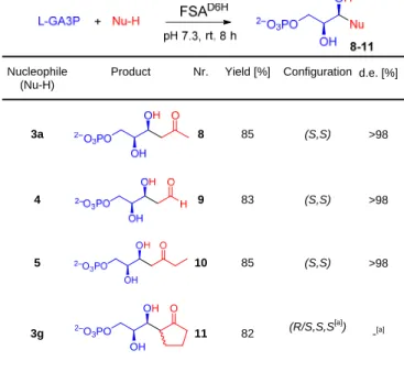
four phosphorylated aldol products (8-11) were isolated in good yields as their Na salts (Table 1).
NMR spectroscopic analysis confirmed the constitution of the expected aldol products and revealed that the ketone products in aqueous solution preferentially existed in their open-chain forms whereas the aldehyde product 9 exists as the furanose forms. NMR spectra of compound 10 clearly indicated that the aldolisation of butanone exclusively occurred on C1. Absolute configuration of the products is given by the chirality of L-GA3P as an internal reference. The relative configuration of the newly created asymmetric centers could be unequivocally assessed by using various strategies depending on the compound considered.
In the case of aldehyde 9, expected to be L
-2-deoxyxylose-5-phosphate by the natural stereoselectivity of FSA, we compared
its 13C NMR spectrum with that of its commercial C3 epimer
D -2-deoxyribose 5-phosphate. Clearly two different spectra were obtained excluding the presence of enantiomers, and thus confirming the configuration for 9a (see SI Figure S2).
Table 1. Small scale synthetic experiments conducted with L-GA3P as electrophile
Nucleophile (Nu-H)
Product Nr. Yield [%] Configuration d.e. [%]
3a 8 85 (S,S) >98 4 9 83 (S,S) >98 5 10 85 (S,S) >98 3g OH 2–O 3PO OH O 11 82 (R/S,S,S [a] ) -[a]
[a] An isotopic exchange was observed in the C- to the carbonyl group (i.e. the tertiary center) of compound 11. Hence, the configuration of this asymmetric carbon is lost under the reaction conditions (pH 7.6-7.8).
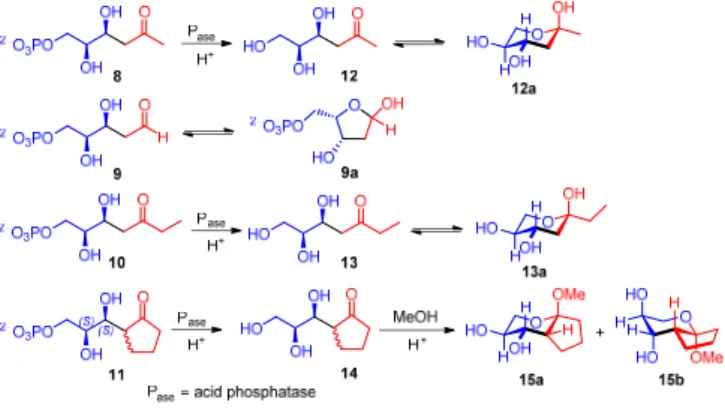
The relative stereochemistry of open chain aldol adducts 8, 10 and 11 was difficult to assess by NMR owing to the free rotation of the bond between chiral carbons. Therefore, mild enzymatic dephosphorylation was used to liberate the terminal hydroxyl group. Compounds 12 and 13 were thus isolated (Scheme 3). This allowed the formation of pyranose isomers in equilibrium with the open chain species, and an unequivocal determination of the relative stereochemical relationships among substituents in the cyclic forms (Scheme 3). In this way compounds 8 and 10 were found to have the same relative configuration as in compound 9. These structures also confirmed that the facial attack at the aldehyde carbonyl group of the electrophilic component during the FSA catalysis was unaffected by the missing hydroxyl function in the nucleophile, fully in line with the
(S)-enantioselectivity observed for the retro-aldol cleavage of
FSA using the fluorogenic substrate analog 1a. In the case of 11, the aldol addition creates two neighboring centers of chirality. However, the tertiary stereocenter was found to epimerize under the reaction conditions, which precluded an unequivocal assessment of the stereopreference. However, considering a similar mechanism to that observed for open-chain hydroxyketols, which form a Z-configured hydroxy-enamine nucleophile at the FSA K85, the rigid cyclopentanone ring would impose an E-configured structure instead for geometrical reasons (Figure 3). As a consequence, in both cases the -proton of the nucleophile component would be abstracted from the same enantiotopic face and as a result, the stereogenic
centers created from 8 and L-GA3P would be formed anti
selectively, whereas the contrary is true for the open chain hydroxyketol nucleophiles. To unambiguously determine the configuration of the second stereocenter, 14 was transformed into a mixture of the methyl glycosides 15a-b by treatment with
methanol under acidic conditions to fix the pyranose ring
structure (Scheme 3).[19] The structural and conformational
characterization of the two epimers 15a-b was performed by a thorough NMR study, including key NOE contacts through-space. Interestingly, 15a-b were recovered in only one configuration (R) at the tertiary stereocenter, possibly directed by the thermodynamic control of the acetalization. The stereochemistry of compounds 15a-b, indicates that FSA D6H was fully (S)-stereoselective for generation of the stereogenic center upon addition to the electrophile.
Scheme 3. Cyclic isomers from phosphorylated and unphosphorylated
derivatives utilized for unambiguous stereochemical analysis.
Figure 3. Schematic representation for FSA K85-enamine nucleophile
structures to demonstrate the origin of the stereochemical outcome of FSA D6H catalysis: A) Z configured FSA K85-hydroxylenamine and B) E configured FSA K85-cyclopentanone enamine nucleophile structures.
In an attempt to investigate the alternative utility of
non-phosphorylated electrophiles, experiments performed using L
-glyceraldehyde revealed that this electrophile reacted only rather sluggishly when exposed to nucleophile 3a when using FSA D6H as catalyst (see SI). The aldol product (12a) could be unequivocally identified by NMR analysis of the resulting product mixture. However, the product could not be isolated in pure form because of difficulties in separating the small amount of aldol adduct from major side products that were arising during the long reaction times required. Under similar reaction conditions, none of the other nucleophiles tested furnished detectable quantities of aldol adducts, using either wt FSA or its D6H variant.
In conclusion, we have discovered that under specific conditions FSA can break the established dogma on the narrow nucleophile specificity of aldolases. Up to now FSA is the first, and so far unique, example of an aldolase with such unprecedented broad nucleophile selectivity. Making use of this
capacity, we succeeded to obtain new aldol products using the FSA D6H variant, by stereoselective addition of aliphatic nucleophiles such as ethanal, propanone, butanone and cyclopentanone to GA3P in good yields. It appears that the presence of the phosphate ester moiety is necessary for the reactivity since only the best known acceptor substrate for FSA, namely GA3P, gave good results. The potential breadth of further applications will be accessible, once more active enzyme variants may be designed and become available. Work in this direction is currently being carried out in our laboratories.
Acknowledgements
This project has received funding from the Auvergne council,the
Bundesministerium für Bildung und Forschung (BMBF grant 0315775B PT-J to W.-D.F.), Ministerio de Economía y Competitividad (MINECO) (grant CTQ2015-63563-R to P.C and CTQ2015-64436-P to T.P.) and COST action CM1303 Systems
Biocatalysis. Anne Samland (Stuttgart University) is warmly
thanked for a gift of plasmid for expressing FSA K85M.
Keywords: fructose-6-phosphate aldolase • aldolisation •
enzyme • phosphorylated compounds • biocatalysis
[1] a) P. Clapés, in Biocatalysis in Organic Synthesis Vol. 2 (Eds.: K. Faber, W.-D. Fessner, N. J. Turner), Georg Thieme Verlag KG, Stuttgart (Germany), 2015, pp. 31-92; b) W.-D. Fessner, in Enzyme Catalysis in Organic Synthesis, Vol. 2, 3rd ed. (Eds.: K. Drauz, H. Groger, O. May), Wiley-VCH, Weinheim, 2011, pp. 857-917; c) P. Clapés, J. Joglar, in Modern Methods in Stereoselective Aldol Reactions (Ed.: R. Mahrwald), John Wiley & Sons Ltd 2013, pp. 475-528; d) C. Guerard-Helaine, M. Debacker, P. Clapes, A. Szekrenyi, V. Helaine, M. Lemaire, Green Chem. 2014, 16, 1109-1113; e) V. Hélaine, R. Mahdi, G. V. S. Babu, V. de Berardinis, R. Wohlgemuth, M. Lemaire, C. Guérard-Hélaine, Adv. Synth. Catal. 2015, 357, 1703-1708; f) A. Szekrenyi, X. Garrabou, T. Parella, J. Joglar, J. Bujons, P. Clapés, Nat. Chem. 2015, 7, 724-729. [2] W.-D. Fessner, C. Walter, Top. Curr. Chem. 1997, 184, 97-194. [3] a) L. Chen, D. P. Dumas, C.-H. Wong, J. Am. Chem. Soc. 1992, 114,
741-748; b) H. L. Arth, W.-D. Fessner, Carbohydr. Res. 1998, 305, 313-321; c) H. A. Chokhawala, H. Cao, H. Yu, Chen.X, J. Am. Chem. Soc.
2007, 129, 10630-10631; d) W.-D. Fessner, G. Sinerius, Angew. Chem.
Int. Ed. 1994, 33, 209-212; e) A. G. Watts, S. G. Withers, Can. J. Chem.
2004, 82, 1581-1588.
[4] V. de Berardinis, C. Guerard-Helaine, E. Darii, K. Bastard, V. Helaine, A. Mariage, J.-L. Petit, N. Poupard, I. Sanchez-Moreno, M. Stam, T. Gefflaut, M. Salanoubat, M. Lemaire, Green Chem. 2017, 19, 519-526. [5] C. Guérard-Hélaine, V. de Berardinis, M. Besnard-Gonnet, E. Darii, M.
Debacker, A. Debard, C. Fernandes, V. Hélaine, A. Mariage, V. Pellouin, A. Perret, J.-L. Petit, M. Sancelme, M. Lemaire, M. Salanoubat, ChemCatChem 2015, 7, 1871-1879.
[6] a) A. L. Concia, C. Lozano, J. A. Castillo, T. Parella, J. Joglar, P. Clapés, Chem. Eur. J. 2009, 15, 3808-3816; b) M. Rale, S. Schneider, G. A. Sprenger, A. K. Samland, W.-D. Fessner, Chem. Eur. J. 2011, 17, 2623-2632.
[7] X. Garrabou, J. A. Castillo, C. Guérard-Hélaine, T. Parella, J. Joglar, M. Lemaire, P. Clapés, Angew. Chem. Int. Ed. 2009, 48, 5521-5525. [8] D. Güclü, A. Szekrenyi, X. Garrabou, M. Kickstein, S. Junker, P. Clapés,
W.-D. Fessner, ACS Catal. 2016, 6, 1848-1852.
[9] a) C.-H. Wong, G. M. Whitesides, J. Org. Chem. 1983, 48, 3199-3205; b) M. D. Bednarski, E. S. Simon, N. Bischofberger, W.-D. Fessner, M. J. Kim, W. Lees, T. Saito, H. Waldmann, G. M. Whitesides, J. Am. Chem.
Soc. 1989, 111, 627-635; c) W.-D. Fessner, G. Sinerius, A. Schneider, M. Dreyer, G. E. Schulz, J. Badia, J. Aguilar, Angew. Chem. 1991, 103, 596-599; d) W.-D. Fessner, O. Eyrisch, Angew. Chem. Int. Ed. 1992, 31, 56-58.
[10] a) B. List, C. F. Barbas, III, R. A. Lerner, Proc. Natl. Acad. Sci. U. S. A.
1998, 95, 15351-15355; b) H.-M. Guo, F. Tanaka, J. Org. Chem. 2009,
74, 2417-2424.
[11] a) N. Jourdain, R. P. Carlon, J.-L. Reymond, Tetrahedron Lett. 1998, 39, 9415-9418; b) F. Tanaka, R. A. Lerner, C. F. Barbas, J Amer Chem Soc 2000, 122, 4835-4836.
[12] a) E. A. Althoff, L. Wang, L. Jiang, L. Giger, J. K. Lassila, Z. Wang, M. Smith, S. Hari, P. Kast, D. Herschlag, D. Hilvert, D. Baker, Protein Science 2012, 21, 717-726; b) S. Bjelic, Y. Kipnis, L. Wang, Z. Pianowski, S. Vorobiev, M. Su, J. Seetharaman, R. Xiao, G. Kornhaber, J. F. Hunt, L. Tong, D. Hilvert, D. Baker, J. Mol. Biol. 2014, 426, 256-271; c) L. Giger, S. Caner, R. Obexer, P. Kast, D. Baker, N. Ban, D. Hilvert, Nat. Chem. Biol. 2013, 9, 494-498; d) L. Jiang, E. A. Althoff, F. R. Clemente, L. Doyle, D. Rothlisberger, A. Zanghellini, J. L. Gallaher, J. L. Betker, F. Tanaka, C. F. Barbas, D. Hilvert, K. N. Houk, B. L. Stoddard, D. Baker, Science 2008, 319, 1387-1391; e) R. Obexer, S. Studer, L. Giger, D. M. Pinkas, M. G. Grutter, D. Baker, D. Hilvert, ChemCatChem 2014, 6, 1043-1050.
[13] E. Gonzalez-Garcia, V. Helaine, G. Klein, M. Schuermann, G. A. Sprenger, W.-D. Fessner, J. L. Reymond, Chem. -Eur. J. 2003, 9, 893-899.
[14] S. Joly, M. S. Nair, J. Mol. Catal. B: Enzym. 2003, 22, 151-160. [15] a) J. A. Castillo, J. Calveras, J. Casas, M. Mitjans, M. P. Vinardell, T.
Parella, T. Inoue, G. A. Sprenger, J. Joglar, P. Clapés, Org. Lett. 2006, 8, 6067-6070; b) M. Sugiyama, Z. Hong, P. H. Liang, S. M. Dean, L. J. Whalen, W. A. Greenberg, C.-H. Wong, J. Am. Chem. Soc. 2007, 129, 14811-14817.
[16] T. Hoffmann, G. F. Zhong, B. List, D. Shabat, J. Anderson, S. Gramatikova, R. A. Lerner, C. F. Barbas, J. Am. Chem. Soc. 1998, 120, 2768-2779.
[17] J. A. Castillo, C. Guérard-Hélaine, M. Gutiérrez, X. Garrabou, M. Sancelme, M. Schürmann, T. Inoue, V. Hélaine, F. Charmantray, T. Gefflaut, L. Hecquet, J. Joglar, P. Clapés, G. A. Sprenger, M. Lemaire, Adv. Synth. Catal. 2010, 352, 1039-1046.
[18] M. Schürmann, G. A. Sprenger, J. Biol. Chem. 2001, 276, 11055-11061. [19] a) H. Zhao, A. Kato, K. Sato, Y.-M. Jia, C.-Y. Yu, J. Org. Chem. 2013, 78, 7896-7902; b) Q. Dang, Z. Zhang, T. Chen, B. Tang, X. He, S. He, Y. Song, S. Bogen, V. Girijavallabhan, D. B. Olsen, P. T. Meinke, Tetrahedron Lett. 2014, 55, 5092-5095.
Entry for the Table of Contents (Please choose one layout)
Layout 1:
COMMUNICATION
Fructose-6-phosphate aldolase (FSA) is famous for being the only aldolase that is able to convert different nucleophiles bearing an alpha-hydroxylated carbonyl group. Here we uncover a hidden activity: FSA can also catalyze aldol additions of aliphatic nucleophiles such as propanone, ethanal, butanone and even cyclopentanone, to glyceraldehyde 3-phosphate thereby providing a stereoselective access to novel aldol adducts.Raquel Roldán, Israel Sanchez-Moreno, Thomas Scheidt, Virgil Hélaine, Marielle Lemaire, Teodor Parella, Pere Clapés*, Wolf-Dieter Fessner* and Christine Guérard-Hélaine*
Page No. – Page No.
Breaking the dogma of aldolase specificity: Simple aliphatic ketones and aldehyde are nucleophiles for fructose-6-phosphate aldolase