HAL Id: hal-01975628
https://hal.archives-ouvertes.fr/hal-01975628
Submitted on 9 Jan 2019
HAL is a multi-disciplinary open access
archive for the deposit and dissemination of
sci-entific research documents, whether they are
pub-lished or not. The documents may come from
teaching and research institutions in France or
abroad, or from public or private research centers.
L’archive ouverte pluridisciplinaire HAL, est
destinée au dépôt et à la diffusion de documents
scientifiques de niveau recherche, publiés ou non,
émanant des établissements d’enseignement et de
recherche français ou étrangers, des laboratoires
publics ou privés.
Structural transformations of the La2–xPrxNiO4+δ
system probed by high-resolution synchrotron and
neutron powder diffraction
Vaibhav Vibhu, Matthew Suchomel, Nicolas Penin, François Weill,
Jean-Claude Grenier, Jean-Marc. Bassat, Aline Rougier
To cite this version:
Vaibhav Vibhu, Matthew Suchomel, Nicolas Penin, François Weill, Jean-Claude Grenier, et al..
Struc-tural transformations of the La2–xPrxNiO4+δ system probed by high-resolution synchrotron and
neu-tron powder diffraction. Dalton Transactions, Royal Society of Chemistry, 2019, 48 (1), pp.266-277.
�hal-01975628�
Structural transformations of the La
2-xPr
xNiO
4+δsystem probed by high-resolution
synchrotron and neutron powder diffraction
Vaibhav Vibhu a,b,Matthew R. Suchomel †a,b, Nicolas Penin a,b, François Weill a,b , Jean-Claude Grenier a,b, Jean-Marc Bassat a,b,
and Aline Rougier a,b
Compositions in the La2-xPrxNiO4+δ series offer an attractive balance of chemical stability and electrochemical performance for use as cathode materials in solid oxide fuel cells (SOFCs). A detailed crystallographic study of this system has been performed, combining both high resolution synchrotron and neutron powder diffraction data, in order to investigate structural details of the series as a function of composition, temperature and oxygen over-stoichiometry. The monoclinic structure (space group F2/m) of ambient temperature Pr-rich compositions for 1.0 < x ≤ 2.0 is discussed in terms of octahedra tilt arrangements and possible long-range structural modulations. In situ synchrotron diffraction experiments and TEM are employed to examine the role of temperature and interstitial oxygen on these structural distortions. With increasing La substitution, a region of mixed monoclinic and tetragonal phases is described for 0.5 ≤ x ≤ 1.0. La-rich compositions are found to be single phase tetragonal (F4/mmm for 0 < x < 0.5) or orthorhombic (Fmmm for x = 0). Possible origins and electrochemical property consequences of the refined structural trends are considered.
1. Introduction
Transition metal oxides exhibiting mixed ionic and electronic conductivity (MIEC) are attractive candidates for cathode materials in solid oxide fuel cells (SOFCs) and oxygen separation membranes.1-3 While SOFCs afford direct and highly efficient
conversion of chemical to electrical energy, the high typical operating temperatures (800-1000 °C) increase device assembly costs and reduce chemical stability of cell components. Consequently, there is considerable interest in improving intermediate temperature (IT-SOFCs) devices operating in a lower temperature range of 600 – 700 °C.4 However, as both electrode
reaction kinetics and electrolyte ion transport mechanisms are temperature dependent, electrochemical performance typically suffers as the temperature is reduced. Problems of high cathode polarization resistance (Rp), induced by slow oxygen reduction
processes, are a particular challenge in developing improved IT-SOFCs. By combining adequate coefficients of oxygen exchange and diffusion with reasonable electronic conductivities, MIEC oxide materials based on perovskite or related derivative structures afford some of best reported performance for IT-SOFC cathode applications.5 This includes the layered Ruddlesden–
Popper (RP) type Ln2MO4 structures in which oxygen ions can be
transported interstitialcy along rock-salt layers separated by slabs of transition metal ions situated in octahedral sites (typical MIEC compositions have Ln = La, Nd, Pr; and M = Cu, Ni, Co).6,7
In particular, the RP-type rare earth nickelates Ln2NiO4+δ have
been shown to exhibit good cathodic performance.8 The general
structure for this first member of the RP series consists of alternate NiO2 square plane layers and Ln2O2 rock salt type-layers.
This type of structure readily accepts interstitial oxygen ions in the Ln2O2 rock-salt interlayer, leading to a mixed Ni2+/3+ valence,
and thus mixed ionic and electronic conductivity (O2-/e-).8 The
interstitial oxygen value δ is strongly dependent on the size of Ln cation radii and can be influenced by annealing conditions.
Regarding previous research on Ln2NiO4+δ (Ln = La, Nd or Pr)
compounds, Pr2NiO4+δ (PNO) showed the lowest polarization
resistance when measured with a Gd-doped ceria (GDC) interface
and a yttria-substituted zirconia (YSZ) electrolyte.9 Despite these
promising electrochemical properties at intermediate temperatures, PNO has rather poor thermodynamic stability in comparison to La2NiO4+δ (LNO). Under oxidizing atmospheres at
elevated temperatures, it has been shown that Pr2NiO4+δ can
decompose to a mixture of Pr4Ni3O10, PrNiO3-δ, and PrOx.10-14 For
this reason, there has been recent interest in mixed PNO-LNO compositions which could offer a compromise of chemical stability and electrochemical performance.15-19 However, despite
investigations spanning three decades, the phase diagram of the La2-xPrxNiO4+δ system; as a function of composition, oxygen
stoichiometry, and temperature, is still unclear. In the current work, a fundamental crystallographic study of the La2-xPrxNiO4+δ
series has been performed using high resolution synchrotron and neutron powder diffraction data. This work aims to provide context of previous work on La2-xPrxNiO4+δ, and to help further
clarify structural details of this series (as a function of composition, temperature and oxygen over-stoichiometry) in order to promote the identification of optimized MIEC compositions.
2. Background
Both of the parent compounds La2NiO4+δ and Pr2NiO4+δ were
examined by Goodenough et al. more than 30 years ago with other Cu2+ and Ni2+ containing K2NiF4-type oxides.20 At high
temperature these compounds adopt a tetragonal structure. On cooling, the strain of increasingly mismatched bond distances between the Ln-O and Ni-O layers can be reduced by tilting of NiO6 octahedra, which results in several structural phase
transitions to lower symmetries. This layer mismatch strain is also partially offset by the insertion of interstitial oxygen ions in the Ln2O2 layer.21
The discovery of superconductivity in related La2CuO4-type
oxides motivated further studies of these compounds in the early 1990’s.22 Jorgensen et al. and Rodriguez-Carvajal et al. used
neutron powder diffraction (NPD) to study La2NiO4+δ as a function
of temperature for different metal oxidation state values.23,24 A
but ambient and low temperature structures of La2NiO4+δ were
found to vary with oxygen content. The stoichiometric (δ ≈ 0.0) composition showed a single Bmab orthorhombic phase at ambient temperature, while oxygen-rich compositions (δ > 0.14) exhibited an F4/mmm orthorhombic structure incorporating an interstitial oxygen ion located near the (¼, ¼, ¼) position in the unit cell. Intermediate oxygen values (0.02 < δ < 0.14) were described by mixed phase coexistence regions of Bmab, P42/ncm,
Pccn, or F4/mmm structures. Subsequent X-ray diffraction and thermogravimetry studies by Buttrey et al. and Tamura et al. broadly supported these findings while adding greater details to the proposed La2NiO4+δ phase diagram.25-28 Demourgues et al.
showed how an electrochemical process could increase interstitial oxygen content in La2NiO4+δ up to δ ≈ 0.25, and used
TEM to determine an expanded C2 monoclinic unit cell accommodating additional oxygen ordering.29,30 More recent
structural studies of non-stoichiometric La2NiO4+δ compositions
have focused on low temperature orthorhombic (LTO) and high temperature tetragonal (HTT) structures under variable oxygen partial pressures, reflecting a shift of research focus on these materials from superconductivity towards the mixed ionic-electronic conductivity properties of layered rare earth nickelates.31-33
Despite early initial conjecture of a monoclinic structural distortion for Pr2NiO4+δ,20,34,35 most subsequent studies reported
orthorhombic or tetragonal structures depending on temperature and oxygen δ content.10,36-41 Sullivan et al. examined
a wide range of non-stoichiometric Pr2NiO4+δ oxygen values (0 ≤
δ < 0.22) and proposed a phase diagram with multiple single and mixed-phase regions distinguished by different canting arrangements of NiO6 octahedra. For higher excess oxygen values
(δ > 0.17) they observed additional weak diffraction peaks which could not be explained nor indexed by the proposed Fmmm orthorhombic structures.10 Pr2NiO4+δ was also extensively probed
by Fernández-Díaz et al. who found magnetic ordering in a tetragonal structure at low temperatures (< 120 K) for near oxygen stoichiometric compositions (δ < 0.02); samples with higher oxygen excess values (δ ≈ 0.20) displayed an “average” orthorhombic structure at ambient temperature (with similar weak superlattice type NPD peaks not indexed by the assigned Bmab cell) which transformed to a tetragonal cell above 690 K.38,39 Similarly, Allançon et al. described a low temperature
tetragonal (LTT) P42/ncm region in Pr2NiO4+δ for low δ values;
whereas more oxidized compositions with 0.09 < δ < 0.22 maintained a LTO Bmab structure with weak unindexed supercell peaks until a HTT phase transition to P42/ncm near 700 K. The
precise HTT transition temperature varied slightly with initial oxygen δ value.40,41 Under low pO2 conditions (≈ 10-4 atm) Flura et
al. report a transition in Pr2NiO4+δ at approximately 675 K from a
LTO over-stoichiometric to a different high temperature orthorhombic structure.33 Later studies have focused primarily
on the stability, oxygen diffusion mechanisms and electrochemical properties of Pr2NiO4+δ.9,11,42-45
Recently, Hernandez et al. used high angular resolution synchrotron powder diffraction to establish that, in fact, oxidized
Pr2NiO4+δ compositions (δ ≈ 0.22) do exhibit a clear but subtle
monoclinic distortion at ambient temperature.46 This so-called
low temperature monoclinic cell (LTM) was indexed with a F2/m space group (non-conventional setting of space group no 12,
C2/m), and is derived from the parent orthorhombic Fmmm cell, but with one non-90° angle of γ ≈ 90.06°. In situ studies showed that the monoclinic distortion persists until the transition to a HTT form above 700 K. As mentioned above for previous Pr2NiO4+δ
works, Hernandez et al. also observe weak non-indexed reflections in diffraction patterns, which they attribute to complex incommensurate modulation, for which the average structure can be described by the proposed LTM cell. Nevertheless, perhaps due to microstructural effects and/or insufficient diffraction data resolution, succeeding reports continue to use orthorhombic structures for Pr2NiO4+δ.19,47
Despite this extensive literature on both LNO and PNO end members, relatively few bulk ceramic structural studies have considered the full La2-xPrxNiO4+δ (LPNO) system. While some
studies did not consider any structural details,15 both Allançon et
al. and Miyake et al. claimed single phase orthorhombic structures across the LPNO system,16,48 indexing Pr-rich (x > 1.0)
and La-rich cell (x < 1.0) compositions with Bmab and Fmmm space groups, respectively. Vibhu et al. used similar structural indexing, but noted the presence of mixed phase region (Bmab and Fmmm) for La rich LPNO compositions with 0.5 < x < 1.0.17
In this current work, compositions of the series La
2-xPrxNiO4+δ are re-examined in light of both the average
monoclinic structural distortion for PNO now clearly established by Hernandez et al., and the recent interest in optimized chemical stability and MIEC properties for mixed LNO-PNO phases. High-resolution data from synchrotron, neutron and TEM probes are used to study the ambient temperature structural characteristics for selected oxidized LPNO compositions. Details of the monoclinic distortion and weak supercell reflections for Pr-rich compositions, as well as the mixed phase region for La-rich samples, are then discussed in the context of understanding the crystallography of La2-xPrxNiO4+δ and informing future
research on this promising SOFC cathode material.
3. Experimental
3.1 Synthesis
La2-xPrxNiO4+δ compositions for the range 0.0 ≤ x ≤ 2.0 were
prepared via a citrate-nitrate route from Pr6O11 (Aldrich, 99.9%),
La2O3 (Strem Chemical, 99.99%) and Ni(NO3)2∙6H2O (Acros
Organics, 99%) precursors. Additional synthesis details were described previously.17 Highly hygroscopic Pr
6O11 and La2O3 were
first pre-fired at 900 °C overnight to remove residual water before weighing targeted precursor mixtures. Final annealing was performed at 1200 °C for 12 h in air, leading to well crystallized phases. The samples were furnace cooled (2 K/min) in static air under identical conditions. As discussed below and shown previously,48 this leads to larger oxygen δ values for Pr-rich
compositions (δ = 0.25 for x = 2.0) than for La-rich compositions (δ = 0.16 for x = 0.0). One sample of Pr2NiO4+δ was also slowly
cooled under an Ar atmosphere (pO2 ≈ 10-4 atm) to independently
study the structural evolution with lower oxygen δ values (δ = 0.19).
3.2 Structural Analyses
The powders were first characterized by laboratory powder X-ray diffraction (PXRD) at room temperature using a PANanalytical X’pert MPD diffractometer with Cu-Kα source and flat-plate reflection geometry. Most LPNO samples were also measured by high-resolution synchrotron powder diffraction (HR-SPD) at beamline 11-BM of the Advanced Photon Source (APS). HR-SPD measurements were performed at room temperature in a transmission capillary mode using an incident beam energy of 30 keV (λ ≈ 0.41 Å) over the range 0.5 to 50° in 2θ. For the x = 2.0 composition of Pr2NiO4+δ, in-situ variable temperature HR-SPD
experiments at 11-BM were also performed from 100 to 450 K to study the structural evolution with temperature. Time of flight neutron powder diffraction (TOF-NPD) data were collected for select LPNO compositions (x = 0.6, 1.2, 1.5 and 2) at 298 K using the POWGEN beamline at the Spallation Neutron Source (SNS). Data obtained via the POWGEN mail-in service were collected on approximately 5 grams of powder loaded into 8.0 mm diameter V cans for approximately 2 hours per sample using the detector banks covering the range 0.4 – 9.0 Å.
Analysis by the Rietveld method for both X-ray and Neutron powder diffraction data (including combined joint refinements of HR-SPD and TOF-NPD data when feasible) were performed using the GSAS/EXPGUI software package.49,50 Peak profiles for X-ray
data were fitted with a pseudo-voigt model incorporating asymmetry due to axial divergence and micro-strain broadening (GSAS Type 4), TOF-NPD profiles have been fit with a convoluted back-to-back exponentials and pseudo-voigt profile (GSAS Type 3). Estimated sample absorption (μR) was considered negligible for both x-ray and neutron datasets. Chebyshev polynomials were used to fit backgrounds. In most cases, lattice parameters, atom positions and anisotropic displacement term values were refined. Profile terms obtained from instrument resolution functions based on refinements of LaB6 SRM data were fixed, while
Lorentzian crystallite size and strain profile terms (including Stephens model type anisotropic microstrain) were refined. Occupancies of interstitial oxygen sites were refined, but with constrained isotropic displacement term values. Other limited refinement constraints, in particular for equivalent Pr and La sites, were employed. Phase weight fractions were refined for the 2-phase region of 0.5 ≤ x ≤ 1.0, but with isotropic atomic displacement terms as required to obtain stable refinements. On close inspection of HR-SPD data for most Pr containing samples (x > 0), extremely weak impurity peaks assigned to NiO (Fm3m, a = 4.176 Å) were observed, which refined to approximately 0.1 wt% when included as a secondary phase in trial Rietveld refinements. NiO was therefore excluded from final structure refinements. Resulting crystal structures and difference Fourier maps used to visualize the interstitial oxygen sites were plotted using the VESTA software package.51
Electron diffraction analysis of PNO sample was performed using a JEOL 2100 electron microscope. Samples were prepared
after grinding under ethanol and crystallites were deposited on a carbon film supported copper grid. Experiments were performed at 100 K, in an attempt to prevent modification of the sample due to beam heating, and also at 300 K. Electron diffraction patterns were obtained by selected area diffraction.
3.3 Additional Characterizations
Thermogravimetric analysis (TGA) was performed to determine oxygen δ values at room temperature. Chemical analyses of La-rich compositions conducted by electron microprobe with approximately 1 μm³ probe size (CAMECA SX 100) confirmed a homogenous distribution of Pr, La and Ni cations in all examined samples. Details of pellet sintering and physical property measurements, including electrical conductivity values determined under air using the four-probe technique, and polarization resistance values obtained by fitting of Nyquist impedance diagrams, have been reported previously.17
4. Results and discussion
Initial examinations of La2-xPrxNiO4+δ samples using low resolution
lab-based Cu Kα source powder X-ray measurements confirmed the phase purity and crystalline nature of the targeted powder compositions and suggested as a first approximation the existence of a continuous series of orthorhombic type structures (e.g. Fmmm or Bmab) across the full composition range. This preliminary result agreed with many previously mentioned ambient temperature literature reports on La2NiO4+δ and
Pr2NiO4+δ.23-28,36-41 However, subsequent inspection using higher
resolution diffraction probes revealed that the true LPNO structural phase diagram is considerably more complex.
4.1 Monoclinic Pr-rich Region (1.0 < x ≤ 2.0)
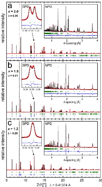
High resolution SPD data collected on Pr-rich samples at 295 K reveal distinct peak splitting clearly visible in certain (hkl) reflections such as {2̅20}/{220}, as seen in Fig. 1a (SPD inset) for the LPNO composition with x = 2.0. The data for the Pr2NiO4+δ
end member with δ ≈ 0.25 are well fit by a monoclinic F2/m space group with lattice parameters of a ≈ 5.39 Å, b ≈ 5.46 Å, c ≈ 12.45 Å, and γ ≈ 90.07° (see Table 1). This unit cell corresponds extremely well with details of the low temperature monoclinic (LTM) structure for Pr2NiO4+δ recently published in a separate
investigation by Broux et al.,46 and provides confirming
experimental evidence for an ambient temperature monoclinic structural distortion of Pr2NiO4+δ first proposed decades
ago.20,34,35 We note that close inspection by monochromatic
lab-based Cu Kα1 powder X-ray measurements of PNO also suggests
a non-orthorhombic distortion, although less distinctly visible than in the HR-SPD datasets.
The non-standard F-centered F2/m unit cell was selected instead of the more common and equivalent C2/m setting (space group no 12) for consistency with previous literature and to
facilitate comparison with other orthorhombic and tetragonal structures in the LPNO series. However, as a monoclinic F-centered unit cell can always be reduced to a smaller C-F-centered monoclinic cell, the smaller and more strictly crystallographic accurate PNO cell is C2/m with a ≈ 5.39 Å, b ≈ 12.45 Å, c ≈ 3.83 Å, and β ≈ 134.62°. Supplemental Fig. S1 illustrates the transformation between the two cells. The F2/m space group is consistent with the parent tetragonal K2NiF4-type HTT structure,
as there is a direct crystallographic group/subgroup path from tetragonal I4/mmm (or its equivalent non-standard F4/mmm setting) space group (no 139) via orthorhombic Fmmm (no 69) to
monoclinic F2/m (no 12) as diagrammed in Supplemental Fig. S2.52
Using nonstandard F centered cells helps to maintain common unit cell axes among all subgroups of the tetragonal parent cell.
Rietveld refinements demonstrate that a single F2/m monoclinic phase provides an excellent fit for Pr-rich LPNO compositions with 1.0 < x ≤ 2.0 at 295 K. This is shown by the refinement difference profiles in Fig. 1 for the compositions x = 2.0, 1.5, and 1.2. The “SPD” plot inset shows a zoomed view of the monoclinic {2̅20}/{220} reflection splitting, and the “NPD” plot inset shows fit profiles for complementary NPD-TOF data collected on the same powder sample as part of the combined Rietveld refinement. The monoclinic distortion is diminished by La substitution as highlighted by the reduced {2̅20}/{220} splitting in Fig 1. As listed in Table 1, for x = 1.1 the refined monoclinic γ angle is essentially 90°. An overall expansion of the lattice and cell volume is observed with La substitution, consistent with replacing Pr3+ by the larger La3+ cation (1.179 Å and 1.216 Å for 9-fold
coordination, respectively).53
For all Pr-rich LPNO compositions, additional weak intensity peaks are seen in both SPD and NPD data which cannot be indexed by a F2/m cell. Except for minor peaks from NiO mentioned earlier, other likely impurity phase sources have been systematically excluded. The small non-indexed peaks for LPNO compositions with x > 1.0 are highlighted in supplemental Figure S3, which plots SPD data sets indexed using a generic orthorhombic F-centered cell with a zoomed vertical scale near the background intensity level. This observation of additional non-indexed peaks is consistent with previous literature on Pr2NiO4+δ referenced earlier.10,38-41,46 Also, as shown in Figure 5
and discussed further below, local TEM electron diffraction analysis similarly revealed additional non-indexed reflections. This confirms that these peaks originate from the main LPNO phase. Indexing attempts to find lower symmetry and/or larger volume cells for PNO that could explain all additional observed peaks were unsuccessful. Moreover, none of the unindexed peak positions can be explained by characteristic {212} or {032} reflections associated with long range order of NiO6 octahedra
in-plane tilting as allowed in the space groups such as Bmab, Pccn, or P42/ncm which were previously proposed.
Although the small monoclinic distortion is not detectable in the lower resolution TOF-NPD data, the complementary scattering sensitivity of neutron data in the joint refinements provides greater detail of atomic site positions and displacement parameters. In particular, for single phase Pr-rich monoclinic compositions with 1.0 < x ≤ 2.0 the combined data allow for stable refinements of not only lattice values and atomic positions, but also anisotropic displacements parameters and occupancy of the interstitial oxygen site. Table 1 lists a summary of select refined structural and lattice values for all examined La2-xPrxNiO4+δ
compositions. Full details of all refined parameters and fit statistics for these compositions are found in supplemental Table S1. Occupancy of the mixed Pr / La sites were fixed to the chemical composition, as no evidence for cation segregation was found during the refinements or by microprobe analysis. Interstitial oxygen ions (refined using an isotropic displacement parameters) partially occupy the (¼,¼,¼) site, and are clearly visible at this position as missing scattering density in Fourier difference maps if excluded from combined refinements.
Fig 1 Combined Rietveld refinement fit profiles for La2-xPrxNiO4+δ
compositions x = 2.0 (a), 1.5 (b), and 1.2 (c) refined with a monoclinic F2/m model at 295 K. Small plot inset highlights splitting of {2̅20}/{220} reflections in SPD data, large inset shows Rietveld refinement fit profiles of NPD datasets.
Estimated oxygen δ values listed in Table S1 are calculated from refined interstitial oxygen O(4) site occupancies and show good agreement when compared to TGA determined δ values measured for select compositions.
A schematic of the ambient temperature refined monoclinic F2/m structure for PNO (x = 2.0) is shown in Figure 2a, where corner sharing NiO6 octahedra are separated along the c direction
of the unit cell by Pr2O2 blocks and in which the oxygen interstitial
sites are partially occupied. The size of atom ellipsoids for Pr (blue), Ni (grey), and O (red) reflect the refined anisotropic displacement parameters (ADP) at 50% probability. Partially filled open circles represent oxygen interstitial O(4) sites. Refined parameters for other LPNO compositions with x > 1.0 show a qualitatively similar structural motif, with decreasing average atomic displacement values for greater La substitution and reduced monoclinic distortion. In general, the refined details for Pr-rich F2/m structures in this study match well with results
recently reported for Pr2NiO4+δ by Broux et al..46 As discussed in
that work, it is not realistic that the large refined anisotropic atomic displacement values for apical O(1) and equatorial O(2/3) oxygen sites imply significant thermal motion of these ions in the structure – which should be relatively small at 300 K - but more likely represent disordered or incommensurate ion displacements, such as octahedra tilting and/or steric repulsions from the interstitial oxygen site. Several previous reports on LNO and PNO have proposed structural models with split apical oxygen sites,31,39,41 however it was recently shown by pair
distribution function (PDF) analysis of neutron and X-ray powder that ambient temperature disorder of apical oxygen atoms in La2NiO4.18 can be well modeled by anisotropic atomic
displacement parameters.54
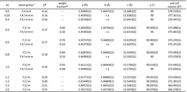
Table 1 Summary of refined structural parameters for select compositions (x) in the La2-xPrxNiO4+δ system, see Supplemental Table S1 for
complete details of all refined parameters and fit statistics.
(x) space group ◊ δ◆ weight
fraction▲ a (Å) b (Å) c (Å) γ (o) unit cell volume (Å3) 0.0 F m m m 0.16 -- 5.45985(1) 5.46471(1) 12.6862(2) 90 378.510(1) 0.25 F 4 / m m m 0.16 -- 5.45992(1) = a 12.6582(1) 90 377.351(1) 0.4 F 4 / m m m 0.16 -- 5.45700(1) = a 12.6414(1) 90 376.447(1) 0.5 F 2 / m 0.17 0.66 5.44209(2) 5.46796(2) 12.6318(3) 90.003(1) 375.888(1) F 4 / m m m 0.34 5.45492(2) = a 12.6315(2) 90 375.867(2) 0.6 F 2 / m 0.17 0.76 5.43737(2) 5.46892(2) 12.6195(2) 90.003(2) 375.259(1) F 4 / m m m 0.24 5.45279(3) = a 12.6207(1) 90 375.251(4) 0.8 F 2 / m 0.18 0.84 5.42874(1) 5.46962(1) 12.5939(1) 90.005(2) 373.994(1) F 4 / m m m 0.16 5.44904(2) = a 12.5952(1) 90 373.976(3) 1.0 F 2 / m 0.19 0.92 5.42111(1) 5.46904(1) 12.5706(2) 90.010(2) 372.695(1) F 4 / m m m 0.08 5.44482(2) = a 12.5702(1) 90 372.656(1) 1.1 F 2 / m 0.20 -- 5.41771(1) 5.46865(1) 12.5572(2) 90.011(2) 372.036(1) 1.2 F 2 / m 0.20 -- 5.41440(1) 5.46809(1) 12.5443(1) 90.026(2) 371.391(1) 1.5 F 2 / m 0.21 -- 5.40572(1) 5.46422(1) 12.5091(1) 90.047(2) 369.495(1) 2.0 F 2 / m 0.25 -- 5.39172(1) 5.45749(1) 12.4478(1) 90.070(2) 366.278(1) ◊ F2/m (full name F112/m) is a non-standard setting of C2/m (no 12); F4/mmm is a non-standard I4/mmm setting (no 139)
◆ Estimated oxygen δ values determined from TGA and/or Rietveld refinements, see Supplemental Table S1 for details. ▲ Refined phase weight fractions for 2-phase samples.
As discussed by Broux et al.,46 the monoclinic F2/m cell for
Pr-rich compositions likely represents the average structure for an incommensurate ordered or a larger supercell which has not yet been determined. In particular, octahedra tilting arrangements along the a, b lattice plane could account for both the large oxygen ellipsoid ADP values and the weak unindexed diffraction peaks. Octahedra tilting arrangements in layered R-P structures have been systematically described previously.35,55,56 For PNO,
tilting may relieve strain from the smaller Pr cation and/or better accommodate interstitial oxygen ions. Figure 2b shows a proposed octahedra tilting arrangement (red arrows) composed of non-equivalent tilt components around the [100] and [1̅10] unit cell direction (black and blue arrows, respectively). Pr and interstitial O(4) positions within the unit cell are marked by blue and open red circles, respectively. A long-range modulation of this average NiO6 octahedra distortion (red arrows) could explain
the average oxygen ADP ellipsoids for the refined Pr2NiO4.25
monoclinic F2/m structure. Using the nomenclature of Aleksandrov,56 this combination of octahedra tilting could be
described as type (Φ1Φ20), indicating alternating non-equivalent
phi (Φx) tilts with axes of rotation parallel to the a and b unit cell
directions. Incommensurate tilting around the [1̅10] direction can account for the subtle monoclinic distortion (γ ≠ 90°) and elongated ADP ellipsoid of the apical oxygen O(1) ions. However, smaller rotations around the [100] direction are only indirectly
related to orthorhombic strain (a ≠ b) observed for the Pr-rich LPNO compositions. Instead, as in other K2NiF4 structures,57 the
orthorhombic distortion here is driven primarily by a contraction of Pr/La-O(1) bonds in the a direction and the concurrent departure from 90° of the in-plane O-Ni-O bond "scissors" angles. Additional aspects of bonding trends and distortions in the LPNO systems are discussed below.
4.2. Intermediate Mixed Phase Region (0.5 ≤ x ≤ 1.0)
Measured HR-SPD and TOF-NPD data for La2-xPrxNiO4+δ samples
with increased La substitution (compositions of 0.5 ≤ x ≤ 1.0) are best fit with a mixed 2-phase refinement model. This is illustrated in Fig. 3a, which shows the combined SPD and NPD Rietveld fit profiles for the x = 0.6 composition employing a 2-phase mixture of tetragonal F4/mmm (upper green tick marks) and monoclinic F2/m (lower brown tick marks) structures. The SPD plot inset in Fig 3a highlights a distinct separation of {113}/{313} and {313} reflections assigned to the monoclinic and tetragonal phases, respectively. In contrast, an equivalent SPD plot inset for the x = 0.4 composition in Fig. 3b clearly reveals a single {313} reflection from the tetragonal F4/mmm unit cell. The evolution of mixed phase La2-xPrxNiO4+δ compositions laying between La-rich single
phase tetragonal F4/mmm (x = 0.4) and Pr-rich monoclinic F2/m (x = 1.1) structures can be visualized by examining the varying intensity of {113}/{313} and {313} reflections in Fig. 3c. Refined
Fig 2 (a) Schematic crystal structure of monoclinic Pr2NiO4.25 at 295 K showing Pr (blue), octahedral Ni (grey), and O (red) sites. Partially
occupied O sites are indicated by open circles. (b) Proposed average octahedra tilting schemes (b) may explain the large average oxygen ADP ellipsoids in the refined F2/m model as viewed along the c axis of the unit cell (c). Distortions in (b) are exaggerated for clarity, where the averaged tilting arrangement (red arrows) is composed of non-equivalent tilt components around the [100] and [1̅10] unit cell directions (black and blue arrows, respectively).
values for this mixed 2-phase region are summarized in Table 1, which includes the refined lattice and weight fraction of both structures for each sample. Full details of refined parameters and fit statistics are found in supplemental Table S1.
Other structural models were considered to fit the diffraction data in this compositional range, such as a lower symmetry single-phase structure, but were ruled out by extensive indexing and refinement analysis. In this compositional range, any peak splitting from a monoclinic distortion is difficult to observe even in the HR-SPD data, but better statistical fits are achieved when the γ angle in the F2/m phase deviates slightly from 90°. The difference in monoclinic {113}/{313} and tetragonal {313} reflection angular positions highlighted in Figure 3a, in fact arises from the non-equivalent in-plane lattice parameters of the F2/m (a ≈ 5.43 Å, b ≈ 5.47 Å) cell vs. the tetragonal F4/mmm (a = b ≈ 5.45 Å) cells. This is seen clearly in the plot of LPNO lattice parameters (Figure 4) which also reveals that c lattice values are essentially identical for the 2 phases and vary linearly across the entire LPNO series.
Therefore, the primary structural difference in this region is the presence of an orthorhombic-type strain (a ≠ b) in F2/m type phases. Due to the overlap of reflections for both phases in the diffraction data, it is impossible to rule out completely a small orthorhombic distortion of the F4/mmm phases. However, trial two-phase refinements with monoclinic and orthorhombic cells for x = 0.6 and 0.8 did not improve overall fit statistics. Moreover,
an orthorhombic cell is inconsistent with refinements of HR-SPD for the La-rich La2-xPrxNiO4+δ compositions (for x = 0.25 and 0.4)
which are well fit by single phase tetragonal F4/mmm cells. Occupancy of the mixed Pr/La sites of each phase were fixed to that of the overall sample composition, as again no evidence for cation segregation was found during the refinements or by microprobe analysis. Moreover, no noticeable differences in refined c lattice values, expected from phases with dissimilar Pr/La composition, were observed. We also noted the absence of any weak non-indexed peaks in the HR-SPD for these LPNO compositions, as demonstrated in supplemental Figure S3.
Intermediate La2-xPrxNiO4+δ compositions (0.5 ≤ x ≤ 1.0)
therefore apparently lack the complex incommensurate ordering of Pr-rich samples, but segregate into two structural motifs distinguished essentially by non-equivalent orthorhombic-type strain in the a-b plane. It is possible that this segregation is connected to oxygen interstitial uptake, with distinct accommodation values and/or configurations more stable than an average intermediate arrangement. Differences in the lattice parameters might be expected for phases with identical Pr/La composition but non-equivalent interstitial oxygen δ content, with a larger δ leading to a unit cell expansion along the c direction of layer stacking, as seen in La2NiO4+δ for δ ≲ 0.16.25
However, for higher oxygen content, reported c lattice values are relatively constant (≈ 12.70 Å) for 0.16 < δ < 0.30.30,32,58 This
suggests oxygen excess at higher concentrations can be accommodated not only by a simple expansion of intra-slab spacing, but instead by different in-plane distortions associated with a-b orthorhombic-type strain. Note that physical analyses by TGA would give only average oxygen δ values for the ensemble mixture, and that it is not statistically feasible to refine separate interstitial oxygen occupancies in the Rietveld analysis. Examples of mixed orthorhombic and tetragonal phase driven by interstitial oxygen ion segregation in K2NiF4-type R-P systems have already
been reported previously. Jorgensen et al. proposed this behavior for La2CuO4+δ, where only one of the phases exhibited
superconductivity.59 In La2NiO4+δ mixed regions of tetragonal
F4/mmm or P42/ncm and orthorhombic Bmab-type structures are
observed for intermediate oxygen values (0.02 < δ < 0.14).23-26
Similar behavior is also reported for the Nd2NiO4+δ system.60
4.3. La-rich Region (0.0 ≤ x < 0.5)
La-rich La2-xPrxNiO4+δ compositions for x < 0.5 are found to be
monophasic, with the HR-SPD data best fit by a tetragonal F4/mmm unit cell for x = 0.25 and x = 0.4 (Figure 3b shows Rietveld refinement difference profile for x = 0.4). No clear orthorhombic type peak distortions are observed on close inspection of the HR-SPD data, and trial refinements using an orthorhombic cell for the x = 0.25 composition did not show meaningful statistical fit improvement (e.g. Rwp of 11% vs. 10%).
Depending on temperature and interstitial oxygen δ values, similar LTT tetragonal (F4/mmm or P42/ncm) structures have
been reported for La2NiO4+δ (in the range 0.06 < δ < 0.15),24,25
Pr2NiO4+δ 40 and Nd2NiO4+δ.60 An orthorhombic Fmmm structure is
proposed for the x = 0.0 LPNO end member with δ = 0.16. The refined orthorhombic distortion is quite small (approximately
Fig 3 Rietveld refinement profiles for La2-xPrxNiO4+δ compositions,
comparing a mixed 2-phase fit for x = 0.6 (a) with a single tetragonal F4/mmm fit for x = 0.4 (b). Evolution of mixed phase region is tracked by following {113}/{313} and {313} reflections in the HR-SPD data for compositions between 0.4 ≤ x ≤ 1.1 (c).
0.1% difference in a and b lattice values) and is consistent with previous literature on the well-studied La2NiO4+δ compound.25,31
4.4 Evolution of non-indexed peaks in Pr-rich compositions
As discussed above in section 4.1, several weak intensity peaks are seen in both SPD and NPD data for Pr-rich La2-xPrxNiO4+δ
compositions (1.0 < x ≤ 2.0) which cannot be indexed by a F2/m cell. Similar observations have been made in previous literature on PNO, 10,38-41,46 and Broux et al. recently discussed how they
could be explained by disordered or incommensurate distortions of the average monoclinic cell.46
In the current study, these non-indexed peaks are examined as a function of La/Pr composition (x), temperature, and interstitial oxygen content (δ). Figures 5a and 5b plot a narrow 2θ range near the indexed {220} and {222} reflections (partially transparent) in the HR-SPD datasets. In Fig. 5a, the intensity of non-indexed peaks clearly diminishes for decreasing x (and δ) and are absent in the mixed phase x = 1.0 composition. Note that all peaks shift together toward smaller 2θ values as the average unit cell expands with La substitution, and that monoclinic splitting observed in {222} peaks is decreasing. In Figure 5b, in-situ variable temperature HR-SPD data measured between 100 and 450 K for
the PNO composition with x = 2.0 are displayed as a stacked contour plot over the same 2θ range. Again, all peaks shift together towards smaller 2θ values as the average unit cell expands with increasing temperature, but no other notable changes are observed. TEM electron diffraction recorded at 300 K (Fig 5c) and 100 K (Fig. 5d) on the x = 2.0 sample similarly show extra non-indexed reflections marked by red arrows for both temperatures. In combination with separate results of Broux et al. collected over a higher temperature range, this suggests that undetermined supercell ordering for over-oxygenated Pr-rich La
2-xPrxNiO4+δ structures may exist from 100 K to nearly 1000 K.46
Monoclinic (F2/m) unit cell parameters refined against the variable temperature data in this study for Pr2NiO4.25 are plotted
in supplemental Figure S4.
Correlations between supercell ordering, Pr/La composition, and interstitial oxygen content, were examined in Pr-rich samples by considering the intensity of non-indexed weak peaks in the HR-SPD data. This qualitative analysis is summarized in Supplemental Figure S5. It assumed that the relative fraction of normalized integrated peak area is proportional to the ordering extent. The integrated peak area of a representative non-indexed peak (at 2θ ≈ 10.88°) was defined as 1.0 for δ = 0.25 and was normalized for each scan as a fraction of the integrated area for the indexed (004) peak at 2θ ≈ 7.60°. The small inset of Fig S5 plots the evolution of this non-indexed peak for air-cooled compositions. In the main plot, red diamonds show air-cooled samples, while the blue diamond represents the Ar-cooled x = 2.0 sample with reduced δ (see Supplemental Table S1 for details of refined parameters). The dashed black curve is a guide for the eye. Note that oxygen over-stoichiometry (δ) values were determined by TGA measurements and/or Rietveld analysis of NPD data.
As discussed previously, with decreasing δ and x, the extent of supercell ordering decreases, and is absent for x ≤ 1.0 and/or δ ≤ 0.19. This is concurrent with the boundary of the single phase F2/m region. However, the estimated ordering fraction for the PNO x = 2.0 Ar cooled sample is much less than that found in the air-cooled x = 2.0 sample, despite very similar refined lattice parameters and monoclinic distortions (γ ≈ 90.06° and 90.07°, respectively). This suggests that supercell ordering in LPNO compositions is more strongly correlated with interstitial oxygen content (δ) than with Pr/La content or average lattice strain. Moreover, it suggests that monoclinic strain in oxygenated Pr-rich compositions of La2-xPrxNiO4+δ is driven primarily by the smaller Pr
cation size. The possibility that a “minimum” oxygen δ is required for supercell ordering in Pr-rich LPNO compositions is consistent with the work of Demourgues et al. on highly oxygenated La2NiO4+δ samples.30 The complete structural model for PNO and
Pr-rich compositions is likely quite complex, as demonstrated for isostructural La2CoO4.14,61 and any full understanding will require
Fig 4 Refined lattice parameters vs. composition (x) for La
2-xPrxNiO4+δ at 295 K, with values for F2/m, F4/mmm, and Fmmm
space groups plotted in red, black and blue, respectively (upper plots). La2-xPrxNiO4+δ compositional dependence at 600 °C of
electrical conductivity (σ) and polarization resistance (Rp) taken
from Vibhu et al. 17 (lower plot) Note dashed lines are a visual
additional single crystal studies similar to those published by Ceretti et al. on Nd2NiO4+δ and Pr2NiO4+δ.45
4.5 Structure and property trends in La2-xPrxNiO4+δ
Refined lattice parameters from the LPNO series are plotted in Figure 4, showing trends across the monoclinic Pr-rich (1.0 < x ≤ 2.0), intermediate mixed phase (0.5 ≤ x ≤ 1.0), and single-phase La-rich (0.0 ≤ x < 0.5) regions. This adds considerable new detail to previous investigations which reported only orthorhombic structures across the La2-xPrxNiO4+δ (LPNO) series.15-19,48 A lower
resolution diffraction study of the related La2-xNdxNiO4+δ system
reports similar trends, with quasi-tetragonal La-rich compositions and smaller orthorhombically strained Nd-rich unit cells.62 It is
possible that future studies of the La2-xNdxNiO4+δ system will
reveal additional complexity analogous to that observed in this current study.
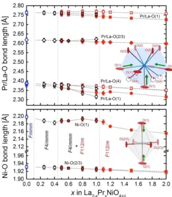
An inspection of cation-oxygen bond distances in the LPNO series helps to give insight into the observed average structural changes. Figure 6 displays selected Pr/La-O and Ni-O bond lengths extracted from structural models refined using the 295 K diffraction data. Schematics of the first shell cation-oxygen bonding environment for Pr/La (top) and Ni (bottom) highlight (green arrows) the most significant bond contractions in the monoclinic Pr2NiO4.25 (x = 2.0) structure, as compared to the
larger and highest symmetry unit cell (F4/mmm) of La-rich
compositions of x < 0.4. Tabulated values of the bond distances are given in Supplemental Table S3.
Strain associated with the replacement of La3+ by the smaller
Pr3+ drives distortions in this system. This approximate 3%
decrease in ionic cation radii gives rise to a roughly equivalent compression of intra-slab spacing along the c direction, which is significantly accommodated by a shrinking of the shortest La/Pr – apical O(1) bond (≈ 2.35 Å) along the same direction. Distinctly non-equivalent contraction of La/Pr-O bonds (≈ 2.75 Å) along the a-b plane produces the orthorhombic strain (a ≠ b) observed for pseudo-monoclinic structures in the mixed phase region of LPNO (and splits the equatorial O(2/3) site). It is speculated that segregation of average structures in the intermediate mixed phase region is connected to oxygen interstitial uptake, with distinct accommodation values and/or configurations more stable than an average intermediate arrangement. The energetically favorable insertion of interstitial oxygen between slab layers reduces overall strain and explains the trend of higher δ values (for equivalent annealing) of Pr-rich compositions. But the effect of excess oxygen insertion on overall unit cell volume is also partially offset by a smaller Ni2+/3+ cation radius with
increased oxidation.
NiO6 octahedra are more rigid and show a slight uniaxial
compression with moderate Pr substitution. In-plane Ni equatorial O(2/3) bonds are relatively constant across the series
Fig 5 Evolution of non-indexed weak intensity peaks observed in HR-SPD data for (a) selected La2-xPrxNiO4+δ compositions at 295 K, and (b) for
Pr2NiO4.25 as a function of temperature from 100 K to 450 K. TEM electron diffraction at (c) 300 K and (d) 100 K showing extra non-indexed
despite the non 90° O-Ni-O bond "scissors" angles associated with orthorhombic strain. In contrast, Ni apical O(1) bonds (≈ 2.18 Å) exhibit a discreet trend of increasing contraction for Pr-rich compositions (x > 1) concurrent with the growth of the average monoclinic distortion (γ ≠ 90°). As discussed previously, the refined F2/m monoclinic structure of Pr-rich compositions likely represents the average of more complex tilting arrangement with short correlation lengths and/or incommensurate ordering. However, the extent of supercell ordering evidenced by weak peaks in LPNO compositions appears to be more strongly correlated with interstitial oxygen content (δ) than with Pr/La content or average lattice distortion.
Promising electrochemical properties of the PNO end member (x = 2.0) are undermined by poor chemical stability at elevated temperatures corresponding to SOFC operating conditions.10-14,18 The monoclinic distortion and supercell
ordering seen at ambient temperature here may therefore reflect a highly strained structure that readily decomposes to more thermodynamically stable compositions. However, good electrochemical performance is maintained with partial La substitutions (x ≤ 0.5). As shown in Figure 4c, the electrical conductivity (σ) for La2-xPrxNiO4+δ compositions increases with x
(data collected at 600 °C, taken from Vibhu et al.17) since the
polarization resistance (Rp) significantly decreases. Based on
thermal stability with ageing, we have shown that the x = 0.5 composition, which is the limit of the La-rich LPNO single-phase domain, is the best compromise between performance and thermal stability for these SOFC cathode materials. However, it is noted that a somewhat different result has been recently reported for materials with the same nominal compositions but
quite different shaping (thin films).19,63 For these thin film studies,
the ideal compromise of La2-xPrxNiO4+δ performance and stability
is reported at x = 1.0. However, in bulk ceramics, this more Pr-rich composition is unstable at operating temperatures vs. time.18
4. Conclusions
A detailed structural study of the La2-xPrxNiO4+δ system, which is
of interest for cathode materials in solid oxide fuel cells, has been performed using high resolution synchrotron and neutron powder diffraction data. This has enabled an investigation of average distortions and cation-oxygen bond bonding as a function of composition, temperature and oxygen over-stoichiometry. Diffraction data for Pr-rich compositions (1.0 < x ≤ 2.0) reveal a monoclinic distortion (F2/m), large ellipsoidal ADP values, and weak supercell peaks which likely represent as-of-yet undetermined octahedra tilt arrangements and/or long-range structural modulations. It is proposed that supercell ordering is more intimately associated with interstitial oxygen content than average unit cell strain. Both features are preserved over the temperature range of 100 - 450 K. In compositions with increasing La substitution (0.5 ≤ x ≤ 1.0), a region of mixed tetragonal and pseudo-monoclinic phases is described, interpreted as local segregation into different oxygen interstitial related structural motifs primarily distinguished by non-equivalent orthorhombic-type strain in the a-b plane. La-rich compositions are found to be single phase tetragonal (F4/mmm for 0 < x < 0.5) or orthorhombic (Fmmm for x = 0) and consistent with previous reports.
A more complete understanding of possible supercell and/or modulations in the Pr-rich La2-xPrxNiO4+δ compositions will
certainly require further detailed studies of single crystal diffraction data. Likewise, details of the mixed phase region might be better resolved with additional examinations of possible compositional segregations varying as a function of average oxygen δ values and annealing conditions. Finally, it is suggested that a high-resolution examination of the analogous La
2-xNdxNiO4+δ system may reveal similar complexity to that observed
in the current study.
Conflicts of interest
There are no conflicts to declare.Acknowledgements
The authors wish to acknowledge gratefully the Agence Nationale de la Recherche (A.N.R., France, PEREN Project; ANR-2011-PREG-016-05) for financial support. This work used resources at the Spallation Neutron Source, a DOE Office of Science User Facility operated by the Oak Ridge National Laboratory. Use of the Advanced Photon Source at Argonne National Laboratory was supported by the U. S. Department of Energy, Office of Science, Office of Basic Energy Sciences, under Contract No. DE-AC02-06CH11357
Fig 6 Selected Pr/La-O (top) and Ni-O (bottom) bond lengths at
295 K vs. composition (x) in La2-xPrxNiO4+δ. Inset graphics illustrate
bonding around Pr/La site (blue ellipsoid at top) and Ni site (grey ellipsoid at bottom). Dashed lines are a guide for the eye.
References
1 A. Tarancón, M. Burriel, J. Santiso, S. J. Skinner and J. A. Kilner, J. Mater. Chem., 2010, 20, 3799–3813.
2 C. Sun, R. Hui and J. Roller, J. Solid State Electrochem., 2010,
14, 1125–1144.
3 A. J. Jacobson, Chem. Mater., 2010, 22, 660–674.
4 D. J. L. Brett, A. Atkinson, N. P. Brandon and S. J. Skinner, Chem. Soc. Rev., 2008, 37, 1568–1578.
5 J.-C. Grenier, J.-M. Bassat and F. Mauvy, in Functional Materials for Sustainable Energy Applications, eds. J. A. Kilner, S. J. Skinner, S. J. C. Irvine and P. P. Edwards, 2014, vol. 1, pp. 402–444.
6 A. Chroneos, D. Parfitt, J. A. Kilner and R. W. Grimes, J. Mater. Chem., 2010, 20, 266–270.
7 Z. Gao, L. V. Mogni, E. C. Miller, J. G. Railsback and S. A. Barnett, Energy Environ. Sci., 2016, 9, 1602–1644. 8 E. Boehm, J.-M. Bassat, P. Dordor, F. Mauvy and J.-C. Grenier,
Solid State Ionics, 2005, 176, 2717–2725.
9 C. Ferchaud, J.-C. Grenier, Y. Zhang-Steenwinkel, M. M. A. van Tuel, F. P. F. van Berkel and J.-M. Bassat, J. Power Sources, 2011, 196, 1872–1879.
10 J. D. Sullivan, D. J. Buttrey, D. E. Cox and J. Hriljac, J. Solid State Chem., 1991, 94, 337–351.
11 P. Odier, C. Allançon and J.-M. Bassat, J. Solid State Chem., 2000, 153, 381–385.
12 A. V. Kovalevsky, V. V. Kharton, A. A. Yaremchenko, Y. V. Pivak, E. V. Tsipis, S. O. Yakovlev, A. A. Markov, E. N. Naumovich and J. R. Frade, J of Electroceramics, 2007, 18, 205–218. 13 V. Vibhu, A. Rougier, C. Nicollet, A. Flura, S. Fourcade, N.
Penin, J.-C. Grenier and J.-M. Bassat, J. Power Sources, 2016, 317, 184–193.
14 J.-M. Bassat, V. Vibhu, C. Nicollet, A. Flura, S. Fourcade, J.-C. Grenier and A. Rougier, ECS Transactions, 2017, 78, 655– 665.
15 G. T. Kim and A. J. Jacobson, Mater. Res. Soc. Symp. Proc., 2006, 972, AA11–04.
16 S. Nishimoto, S. Takahashi, Y. Kameshima, M. Matsuda and M. Miyake, J. Ceram. Soc. Jpn., 2011, 119, 246–250.
17 V. Vibhu, A. Rougier, C. Nicollet, A. Flura, C. Grenier and J.-M. Bassat, Solid State Ionics, 2015, 278, 32–37.
18 V. Vibhu, J.-M. Bassat, A. Flura, C. Nicollet, J.-C. Grenier and A. Rougier, ECS Transactions, 2015, 68, 825–835.
19 R. K. Sharma, S.-K. Cheah, M. Burriel, L. Dessemond, J.-M. Bassat and E. Djurado, Journal of Materials Chemistry A, 2017, 5, 1120–1132.
20 K. K. Singh, P. Ganguly and J. B. Goodenough, J. Solid State Chem., 1984, 52, 254–273.
21 D. J. Buttrey and J. M. Honig, in Chemistry of High Temperature Superconductors, ed. C. N. R. Rao, 1991, pp. 283–305, ISBN: 978-981-02-0805-9.
22 H. Takagi, S.-I. Uchida, K. Kitazawa and S. Tanaka, Jpn. J. Appl. Phys., 1987, 26, L123.
23 J. D. Jorgensen, B. Dabrowski, S. Pei, D. R. Richards and D. G. Hinks, Phys. Rev. B: Condens. Matter, 1989, 40, 2187–2199. 24 J. Rodriguez-Carvajal, M. T. Fernández-Díaz and J. L. Martinez,
J Phys-Condens Mat, 1991, 3, 3215–3234.
25 D. E. Rice and D. J. Buttrey, J. Solid State Chem., 1993, 105, 197–210.
26 J. M. Tranquada, Y. Kong, J. E. Lorenzo, D. J. Buttrey, D. E. Rice and V. Sachan, Phys. Rev. B, 1994, 50, 6340–6351. 27 H. Tamura, A. Hayashi and Y. Ueda, Physica C, 1996, 258, 61–
71.
28 H. Tamura, A. Hayashi and Y. Ueda, Physica C, 1993, 216, 83– 88.
29 A. Demourgues, F. Weill, B. Darriet, A. Wattiaux, J.-C. Grenier, P. Gravereau and M. Pouchard, J. Solid State Chem., 1993,
106, 317–329.
30 A. Demourgues, A. Wattiaux, J.-C. Grenier, M. Pouchard, J. L. Soubeyroux, J. M. Dance and P. Hagenmuller, J. Solid State Chem., 1993, 105, 458–468.
31 S. J. Skinner, Solid State Sciences, 2003, 5, 419–426.
32 A. Aguadero, J. A. Alonso, M. J. Martinez-Lope, M. T. Fernandez-Diaz, M. J. Escudero and L. Daza, J. Mater. Chem., 2006, 16, 3402–3408.
33 A. Flura, S. Dru, C. Nicollet, V. Vibhu, S. Fourcade, E. Lebraud, A. Rougier, J.-M. Bassat and J.-C. Grenier, J. Solid State Chem., 2015, 228, 189–198.
34 B. Willer and M. Daire, C. R. Acad. Sci., Ser. C: Sci. Chim., 1968,
267, 1482–1484.
35 P. Ganguly and C. N. R. Rao, J. Solid State Chem., 1984, 53, 193–216.
36 R. S. Puche, F. Fernández, J. Rodríguez-Carvajal and J. L. Martinez, Solid State Commun., 1989, 72, 273–277. 37 J. L. Martinez, M. T. Fernandez-Diaz, J. Rodriguez-Carvajal and
P. Odier, Phys. Rev. B, 1991, 43, 13766–13769.
38 M. T. Fernandez-Diaz, J. Rodriguez-Carvajal, J. L. Martinez, G. Fillion, F. Fernanadez and R. Saez-Puche, Z. Phys. B: Condens. Matter, 1991, 82, 275–282.
39 M. T. Fernandez-Diaz, J. L. Martinez and J. Rodriguez-Carvajal, Solid State Ionics, 1993, 63-65, 902–906.
40 C. Allançon, A. Gonthier-Vassal, J.-M. Bassat, J. P. Loup and P. Odier, Solid State Ionics, 1994, 74, 1239–1248.
41 C. Allançon, J. Rodríguez-Carvajal, M. T. Fernandez-Diaz, P. Odier, J.-M. Bassat, J. P. Loup and J. L. Martinez, Z. Phys. B: Condens. Matter, 1996, 100, 85–90.
42 M. Yashima, M. Enoki, T. Wakita, R. Ali, Y. Matsushita, F. Izumi and T. Ishihara, J. Am. Chem. Soc., 2008, 130, 2762–2763. 43 D. Parfitt, A. Chroneos, J. A. Kilner and R. W. Grimes, Phys.
Chem. Chem. Phys, 2010, 12, 6834–3.
44 J.-M. Bassat, M. Burriel, O. Wahyudi, R. Castaing, M. Ceretti, P. Veber, I. Weill, A. Villesuzanne, J.-C. Grenier, W. Paulus and J. A. Kilner, J. Phys.Chem C., 2013, 117, 26466–26472. 45 M. Ceretti, O. Wahyudi, A. Cousson, A. Villesuzanne, M.
Meven, B. Pedersen, J.-M. Bassat and W. Paulus, Journal of Materials Chemistry A, 2015, 3, 21140–21148.
46 T. Broux, C. Prestipino, M. Bahout, S. Paofai, E. Elkaïm, V. Vibhu, J.-C. Grenier, A. Rougier, J.-M. Bassat and O. Hernandez, Dalton Transactions, 2016, 45, 3024–3033. 47 E. Dogdibegovic, Q. Cai, N. S. Alabri, W. Guan and X.-D. Zhou,
J. Electrochem. Soc., 2017, 164, F99–F106.
48 C. Allançon, P. Odier, J.-M. Bassat and J. P. Loup, J. Solid State Chem., 1997, 131, 167–172.
49 B. H. Toby, J Appl Crystallogr, 2001, 34, 210–213.
50 A. C. Larson and R. B. Von Dreele, GSAS. Report LAUR 86-748, Los Alamos National Laboratory, New Mexico, USA., 2004. 51 K. Momma and F. Izumi, J Appl Crystallogr, 2011, 44, 1272–
1276.
52 M. I. Aroyo, A. Kirov, C. Capillas, J. M. Perez-Mato and H. Wondratschek, Acta Crystallogr., Sect. A, 2006, 62, 115– 128.
53 R. D. Shannon, Acta Crystallogr., Sect. A, 1976, 32, 751–767. 54 M. Brunelli, M. Coduri, M. Ceretti and W. Paulus, J. Phys. D:
Appl. Phys., 2015, 48, 504009.
55 K. S. Aleksandrov and J. Bartolome, J Phys-Condens Mat, 1994, 6, 8219–8235.
56 K. S. Aleksandrov, B. V. Beznosikov and S. V. Misyul, Phys. Status Solidi A, 1987, 104, 529–543.
57 P. Radaelli, D. Hinks, A. Mitchell, B. Hunter, J. Wagner, B. Dabrowski, K. Vandervoort, H. Viswanathan and J. Jorgensen, Phys. Rev. B, 1994, 49, 4163–4175.
58 A. Aguadero, M. Pérez, J. A. Alonso and L. Daza, J. Power Sources, 2005, 151, 52–56.
59 J. Jorgensen, B. Dabrowski, S. Pei, D. Hinks, L. Soderholm, B. Morosin, J. Schirber, E. Venturini and D. Ginley, Phys. Rev. B: Condens. Matter, 1988, 38, 11337–11345.
60 K. Ishikawa, K. Metoki and H. Miyamoto, J. Solid State Chem., 2009, 182, 2096–2103.
61 L. Le Dréau, C. Prestipino, O. Hernandez, J. Schefer, G. Vaughan, S. Paofai, J. M. Perez-Mato, S. Hosoya and W. Paulus, Inorg. Chem, 2012, 51, 9789–9798.
62 H. Ishikawa, Y. Toyosumi and K. Ishikawa, J Alloy Compd, 2006,
408-412, 1196–1199.
63 R. K. Sharma, O. Celikbilek, M. Burriel, L. Dessemond, J.-M. Bassat and E. Djurado, ECS Transactions, 2016, 72, 1–8.
Supplemental Information
Supplemental Information
Structural transformations of the La
2-x
Pr
x
NiO
4+δ
system probed by
high-resolution synchrotron and neutron powder diffraction
Vaibhav Vibhu
a,b,
Matthew R. Suchomel †
a,b, Nicolas Penin
a,b, François Weill
a,b, Jean-Claude Grenier
a,b,
Jean-Marc Bassat
a,b, and Aline Rougier
a,ba. CNRS, ICMCB, UMR 5026, F-33600 Pessac, France b. Univ. Bordeaux, ICMCB, UMR 5026, F-33600 Pessac, France
† Corresponding author
Supplemental Figure S1: Relationship between expanded F2/m monoclinic unit cell (full name F112/m with unique axis c) used in this work
and its equivalent reduced conventional C2/m cell in its standard crystallographic setting (full name C12/m1 with unique axis b). On the left, the transformation matrix is given, and corresponding lattice values and atomic positions are given for the Pr2NiO4.25 composition. On the
right, a schematic diagram of the relationship between the F2/m cell (in blue) and C2/m cell (in red) is shown. For clarity, only the Ni atom is plotted in the unit cell.
Supplemental Information
Supplemental Figure S2: Selected group/subgroup relationship paths between different space groups commonly referenced in the La2NiO4+δ
and Pr2NiO4+δ literature; from the Bilbao Crystallographic Server 52. Shaded space groups are reported for the LPNO system in the current
study.
Supplemental Figure S3: Near background intensity zoomed view of HR-SPD data (295 K) for La2-xPrxNiO4+δ series. Assigned space groups in
this study are marked on the left near the composition (x). Numerous weak intensity non-indexed peaks are clear for compositions with x > 1.0. Main peaks are labeled with hkl indexing for a generic F-centered orthorhombic cell (hkl condition; all even or all odd). Characteristic positions of (212) and (032) superlattice reflections allowed in Bmab and P42/ncm space groups that arise from ordered in-plane tilting of
Supplemental Information
Supplemental Figure S4: Evolution of refined lattice parameters (a, b, c, γ) as a function of temperature for Pr2NiO4.25. Lattice values were
obtained from refinements of in-situ variable temperature HR-SPD data using the F2/m monoclinic unit cell.
Supplemental Figure S5: Evolution of the relative integrated peak area fraction observed in HR-SPD data at room temperature as a function
of oxygen over-stoichiometry (δ) for Pr-rich La2-xPrxNiO4+δ compositions. The small inset upper figure inset plots the evolution of the
reference non-indexed weak intensity peak (at 2θ ≈ 10.88°) for select air-cooled compositions. The integrated peak area was defined as 1.0 for δ = 0.25, and normalized for each scan as a fraction of the integrated area for a F2/m indexed (004) peak at 2θ ≈ 7.60°. Red plotted diamonds show air-cooled samples; the blue diamond represents the Ar-cooled x = 2.0 sample with reduced δ. The dashed black curve is a guide for the eye. See main text Section 4.4 for details and discussion.
Supplemental Information
Supplemental Table S1: Summary of Rietveld refined structural parameters and fit statistics for compositions (x) in the La2-xPrxNiO4+δ system. Refined values are followed by the uncertainty in
Supplemental Information
Supplemental Table S2: Summary of Rietveld refined anisotropic displacement parameter (ADP) values (Uaniso) for selected Pr-rich compositions (x) in the La2-xPrxNiO4+δ system.
Supplemental Table S3: Summary of selected bond distances determined from Rietveld refined structural models for compositions (x) in the La2-xPrxNiO4+δ system.
Composition (x) 1.1 1.2 1.5 2.0 Space Group F2/m F2/m F2/m F2/m
refined data sets SPD SPD+NPD SPD+NPD SPD+NPD Pr/La: Uaniso ×100 (Å2) U11 0.69(1) 0.77(1) 0.85(1) 0.84(1) U22 0.99(1) 1.19(1) 1.26(1) 1.34(1) U33 0.74(1) 0.76(1) 0.75(1) 0.67(1) U12 -0.18(4) -0.14(1) -0.22(1) -0.24(1) Ni: Uaniso ×100 (Å2) U11 0.23(4) 0.37(2) 0.26(2) 0.22(2) U22 0.16(4) 0.32(3) 0.30(2) 0.25(2) U33 0.92(4) 1.07(2) 1.05(2) 1.17(3) U12 -0.2(1) -0.25(3) 0.18(3) -1.10(3) O(1): Uaniso ×100 (Å2) U11 4.9(3) 5.30(8) 5.23(8) 5.91(8) U22 9.9(4) 9.15(12) 10.06(12) 11.90(12) U33 0.1(2) 0.61(5) 0.59(5) 0.69(5) U12 6.4(4) 6.02(11) 5.30(12) 5.41(13) O(2): Uaniso ×100 (Å2) U11 -- 0.90(11) 0.71(14) 0.82(11) U22 -- 0.31(13) 0.27(14) 0.33(11) U33 -- 3.63(30) 3.36(31) 4.14(29) U12 -- -0.40(8) -0.27(8) -0.25(7) O(3): Uaniso ×100 (Å2) U11 -- 0.39(10) 0.34(13) 0.26(10) U22 -- 0.95(12) 0.57(15) 0.53(11) U33 -- 3.64(33) 4.34(35) 4.38(30) U12 -- 0.18(7) 0.21(8) 0.21(6)