HAL Id: hal-02348606
https://hal.archives-ouvertes.fr/hal-02348606
Submitted on 22 Mar 2021
HAL is a multi-disciplinary open access
archive for the deposit and dissemination of
sci-entific research documents, whether they are
pub-lished or not. The documents may come from
teaching and research institutions in France or
abroad, or from public or private research centers.
L’archive ouverte pluridisciplinaire HAL, est
destinée au dépôt et à la diffusion de documents
scientifiques de niveau recherche, publiés ou non,
émanant des établissements d’enseignement et de
recherche français ou étrangers, des laboratoires
publics ou privés.
Emerging Trends in the Formation and Function of
Tuberculosis Granulomas
Geanncarlo Lugo-Villarino, D. Hudrisier, A. Bénard, Olivier Neyrolles
To cite this version:
Geanncarlo Lugo-Villarino, D. Hudrisier, A. Bénard, Olivier Neyrolles. Emerging Trends in the
Forma-tion and FuncForma-tion of Tuberculosis Granulomas. Frontiers in Immunology, Frontiers, 2013, 3, pp.405.
�10.3389/fimmu.2012.00405�. �hal-02348606�
Emerging trends in the formation and function of
tuberculosis granulomas
Geanncarlo Lugo-Villarino
1,2*
†, D. Hudrisier
1,2†, A. Benard
1,2†and Olivier Neyrolles
1,2*
1
CNRS, Institut de Pharmacologie et de Biologie Structurale, Toulouse, France
2
Institut de Pharmacologie et de Biologie Structurale, Université de Toulouse, Université Paul Sabatier, Toulouse, France
Edited by:
Dov L. Boros, Wayne State University School of Medicine, USA
Reviewed by:
Hans Acha-Orbea, Center for Immunity and Infection Lausanne, Switzerland
Jean-louis Mege, Aix Marseille Université, France
Jennifer A. Philips, NYU School of Medicine, USA
*Correspondence:
Geanncarlo Lugo-Villarino and Olivier Neyrolles, CNRS, Institut de Pharmacologie et de Biologie Structurale, 205 Route de Narbonne, F-31077 Toulouse, France.
e-mail: [email protected]; [email protected]
†
Geanncarlo Lugo-Villarino, D. Hudrisier and A. Benard have contributed equally to this work.
The granuloma is an elaborated aggregate of immune cells found in non-infectious as well
as infectious diseases. It is a hallmark of tuberculosis (TB). Predominantly thought as a
host-driven strategy to constrain the bacilli and prevent dissemination, recent discoveries
indicate the granuloma can also be modulated into an efficient tool to promote microbial
pathogenesis. The aim of future studies will certainly focus on better characterization of the
mechanisms driving the modulation of the granuloma functions. Here, we provide unique
perspectives from both the innate and adaptive immune system in the formation and the
role of the TB granuloma. As macrophages (M
φs) comprise the bulk of granulomas, we
highlight the emerging concept of M
φ polarization and its potential impact in the
micro-bicide response, and other activities, that may ultimately shape the fate of granulomas.
Alternatively, we shed light on the ability of B-cells to influence inflammatory status within
the granuloma.
Keywords: macrophage, B-cells, mycobacteria, tuberculosis, granuloma
INTRODUCTION
“On the basis of my numerous observations I consider it established that, in all tuberculous affections of man and animals, there occur con-stantly those bacilli which I have designated tubercle bacilli and which are distinguishable from all other microorganisms by characteristic properties.”
With those celebrated words in 1882, Koch announced the
discov-ery of the etiological agent of one of the oldest recorded human
afflictions (
Koch, 1982
). The term “tubercle” refers to an
origi-nal description by Sylvius (in 1650) of the apparent lung nodules
characteristic of the “consumption” disease, which became
chris-tened as “tuberculosis (TB)” by Schonlein (in 1839) in recognition
of its intricate correlation with these structures (
Sakula, 1982
).
Today, these tubercles are known as granulomas, defined as
orga-nized immune cell aggregates that form in response to persistent
TB infection (
Ramakrishnan, 2012
). The cellular composition of
TB granulomas includes Mφs, neutrophils, monocytes, dendritic
cells, B- and T-cells, fibroblasts, and epithelial cells (
Russell, 2007
;
Ramakrishnan, 2012
). Moreover, TB granulomas are characterized
by a high-turnover rate of their Mφ population and by specialized
differentiations taking place in mature Mφs such as tightly
inter-digitated cell membranes that make Mφs appear either epithelial
(
Adams, 1974
), fusion into multinucleated giant cells (
Helming
and Gordon, 2007
), or differentiation into foamy cells with a
high lipid content (
Russell et al., 2009
). While granulomas have
been studied for about 200 years, their role in TB etiology remains
unclear. In 1819, Laënnec first proposed granulomas as the cause
of TB (
Sakula, 1982
). Yet, about a century went by before Ghon
correlated the presence of a single caseous granuloma in the
mid-region of the lung with a corresponding nodal involvement (the
Ghon complex) and the pathogen’s dissemination, thus serving
as a marker for latent TB (
Dorhoi et al., 2011
). In spite of this,
subsequent studies and clinical observations established the
gran-uloma as a host-protective structure that“walls off ”Mtb to prevent
its dissemination, a notion that still predominates. Seminal
stud-ies by Ramakrishnan in zebrafish, however, have now evidenced
mycobacteria actually exploit the granuloma into a tool for
patho-genesis, suggesting its function can be modulated depending on
the disease context (
Ramakrishnan, 2012
). Considering TB is still
one of the leading causes of human death due to a single
infec-tious agent, substantial insights into microbe physiology and host
defenses rest in the attempt to better understand the mechanisms
governing TB granulomas.
Here, we will focus exclusively in the role of Mφ polarization in
the formation and function of TB granulomas. Likewise, we will
provide a unique perspective on the significance of B-cells, whose
immune-modulatory function has long been ignored in TB.
MACROPHAGE POLARIZATION IN TB GRANULOMAS
Mφ polarization is broadly classified into M1 and M2 programs
(
Goerdt and Orfanos, 1999
;
Gordon, 2003
;
Mantovani et al., 2004
;
Martinez et al., 2009
). On one hand, the M1 program is a response
to type-1 inflammatory conditions (e.g., IFN-γ), often associated
with intracellular pathogen resistance (
Quintana-Murci et al.,
Lugo-Villarino et al. Emerging trends in tuberculosis granulomas
2007
;
Benoit et al., 2008
). IFN-γ is mainly responsible for the
establishment of the M1 program, granting Mφs the capacity to
kill mycobacteria (
Flynn et al., 1993
;
Ehrt et al., 2001
). The
pro-duction of nitric oxide (NO) in Mφs (characteristic in murine
models) is arguably one of the most important consequences
mediated by IFN-γ, as mice deficient for NO production
suc-cumb to Mtb infection (
Chan et al., 1992
). In fact, the enzyme
iNOS (inducible NO synthase) required for NO production is a
bona fide marker of murine M1 Mφs (
Xie and Nathan, 1993
).
Other marker genes, whose expression is induced in M1, include
ido1, ptgs2, il12b/il23a, socs3, marco, cd86, irf3/irf5, and stat1/stat5,
among others (
Lawrence and Natoli, 2011
;
Murray and Wynn,
2011b
). Collectively, the M1 program is part of the “common host
response” against intracellular bacteria that endows Mφs with a
non-permissible nature (
Ehrt et al., 2001
;
Deretic et al., 2004
;
Mar-tinez et al., 2009
;
Cairo et al., 2011
;
Murray and Wynn, 2011a
). On
the other hand, the M2 program is dictated by type-2
inflam-matory signals (e.g., IL-4, IL-10), enabling Mφs to participate in
the suppression of inflammation, phagocytosis, tissue
remodel-ing, and repair, among others (
Sica et al., 2008
;
Martinez et al.,
2009
;
Murray and Wynn, 2011a
). However, this program also
renders Mφs poorly microbicidal against intracellular pathogens
(
Raju et al., 2008
;
Martinez et al., 2009
). This is best illustrated
by how the arginine metabolism is used in M2 Mφs, which shuts
down NO production in favor of tissue reparation (
Shearer et al.,
1997
). Indeed, M2 polarization is accompanied by ARG1 (type-1
arginase) expression that inhibits NO production by
outcompet-ing iNOS to convert arginine into ornithine and urea (
Munder
et al., 1998
;
El Kasmi et al., 2008
). Along arg1, other M2 marker
genes include fizz1, chi311/chi312/chi313, mrc1, cd36, socs2, il-10,
klf4, jmjd3/irf4, ppar
γ, and stat6, among others (
Lawrence and
Natoli, 2011
;
Murray and Wynn, 2011b
). Altogether, Mtb might
influence the granuloma function by controlling Mφ polarization,
a premise that is presciently in line with the following findings,
which for the purpose of conciseness, are mainly based on the use
of the iNOS/ARG1 polarization axis.
The animal models to study TB granulomas are discussed in
detail elsewhere (
Flynn, 2006
). Here, we highlight recent findings
in mice and zebrafish documenting the TB granuloma
dynam-ics, supported by studies and clinical observations done in TB
patients. It is widely postulated the onset of human pulmonary
TB begins when inhaled Mtb is captured by Mφs and
trans-ported across the alveolar epithelium into the lung tissue. In
zebrafish, the subsequent steps leading to a nascent granuloma
have been captured in real-time imaging (
Davis et al., 2002
).
While infected Mφs undergo apoptosis, they promote the
recruit-ment of phagocytes, which upon arrival, display high motility
conducive for scavenging apoptotic cells. The phagocytosis of
dead Mφs leads to the formation of cell aggregates, fomenting
bacterial growth. Subsequent rounds of this cycle promote the
formation of a stable granuloma in 3 days post-infection (p.i.), a
process that is dependent on the region of difference-1 (RD1)
vir-ulence locus of M. marinum and independent of T-cells (
Davis
et al., 2002
;
Volkman et al., 2004, 2010
;
Davis and Ramakrishnan,
2009
). It is unclear whether zebrafish Mφs undergo polarization.
Yet, since most transcription factors governing T-cell polarization
are highly conserved in zebrafish (
Mitra et al., 2010
), along with
physiological and pathological responses characteristic of type-1
and type-2 immunity (
Aggad et al., 2010
;
Balla et al., 2010
;
Holt
et al., 2011
;
Wittamer et al., 2011
;
Renshaw and Trede, 2012
), it
seems as a matter of time before Mφ polarization is identified
and characterized in this teleost. By contrast, the early stage of
Mtb infection in mice is marked by M1 Mφ polarization,
rem-iniscent of clinical observations in TB patients (
Benoit et al.,
2008
). In fact, transcriptomic analysis of infected murine Mφs
revealed the gene modulation provoked by Mtb overlaps with
that of IFN-γ to establish the M1 program (
Ehrt et al., 2001
).
Type-1 inflammatory signals secreted by infected Mφs induce
cell recruitment and formation of primary granulomas. Unlike
zebrafish, however, granuloma formation in mice takes up to
3 weeks when Mycobacterium reaches a plateau and coincides with
adaptive immunity involvement. For instance, nascent liver
gran-ulomas were visualized by intravital microscopy between 2 and
3 weeks after Mycobacterium bovis Calmette–Guerin (BCG)
chal-lenge (
Egen et al., 2011
). In another study, Mtb infection did not
change the murine Mφ population (iNOS
lowARG1
low) in
bron-choalveolar lavage (BAL) during the first week (
Redente et al.,
2010
). At day 21 p.i., however, M1 Mφs (iNOS
highARG1
low)
dom-inated in BAL and granulomas, coinciding with a peak of IFN-γ
in infected lungs (
Redente et al., 2010
). In humans, although NO
production by monocyte-derived Mφs remains controversial,both
iNOS and NO are detected in granulomas and alleles for NOS2
are associated to TB susceptibility (
Nicholson et al., 1996
;
Facchetti
et al., 1999
;
Choi et al., 2002
;
Schon et al., 2004
;
Moller et al., 2009
).
After 35–60 days p.i., while murine Mφs at the granuloma core
remained iNOS
highARG1
low, there was a dramatic shift toward the
M2 program (iNOS
lowARG1
high) in Mφs surrounding the core,
accompanied by elevated type-2 inflammatory signals (
Redente
et al., 2010
). This is in line with ARG1 detection in human TB
granulomas (
Pessanha et al., 2012
).
The shift toward M2 Mφs during Mtb infection could have
deleterious consequences for the granuloma as a host-protective
structure (Figure 1). First, ARG1 expression in uninfected Mφs
surrounding the granuloma core suggests the development of an
immunosuppressive niche. Indeed, Mtb promotes its survival by
inducing ARG1 expression through MyD88-dependent signaling
pathways (
El Kasmi et al., 2008
;
Qualls et al., 2010
). At the
tran-scriptome level, murine M2 Mφs displayed a diminished
inflam-matory response to Mtb as reflected by a reduced NO production
and increased of iron availability, alluding ARG1 might also be
implicated in nutrient deprivation mechanisms limiting
micro-bial growth (
Forbes and Gros, 2001
;
Kahnert et al., 2006
;
Cairo
et al., 2011
). Furthermore, M1 Mφs possess a “fail-safe” system
sustaining optimum NO production based on citrulline recycling
via argininosuccinate synthase (ASS1), which is absent in M2 Mφs
(
Qualls et al., 2012
). Given the restrictive granuloma
environ-ment where arginine may be limited, the presence of this fail-safe
system may become further accentuated. Second, M2 Mφs may
represent a transitional state into the formation of “foamy” Mφs
that are rich in cholesterol, a carbon source for microbial
intra-cellular survival (
Pandey and Sassetti, 2008
;
Peyron et al., 2008
;
Russell et al., 2009
;
Griffin et al., 2011
). Recently, Mtb lipids were
shown to trigger PPARγ, the master regulator of M2 polarization,
to increase expression of CD36 and induce foam cell formation
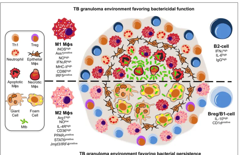
FIGURE 1 | A model illustrating the putative roles of Mφ polarization
and B-cell involvement during the formation and function of TB
granulomas. TB granuloma Mφs undergo various specialized
transformations: they can look like epithelial characterized by tightly interdigitated cell membranes that link adjacent cells; they can fuse into multinucleated giant cells; or they can differentiate into foamy cells with a high content of intracellular lipids. While none of these specialized transformations in the granuloma Mφ population are well understood, we propose they might be reflection of the Mφ polarization status that may render the granuloma structure with a microbicidal capacity (top) or as a tool of pathogenesis (bottom). In the former scenario, the local Mφ population in lung undergoes a M1 polarization early on during Mtb infection and granuloma formation, distinguished by a cell-surface receptor repertoire responsive to pro-inflammatory signaling (e.g., IFN-γRhigh
) and conducive for antigen-presentation (e.g., MHC-IIhigh , CD86high), while acquiring a microbicidal capacity reflected in the NO production (e.g., iNOShigh, ASS1positive), among others. These Mφs have been noted to be most frequently located in the necrotic center of a mature tuberculous granuloma where apoptotic and necrotic Mφs are abundant along with extracellular bacteria. Accompanying the M1 Mφ
polarization is the recruitment of neutrophils and Th1 cells, whose migration and activation status might be influenced by a B-cell involvement likely characterized by a pro-inflammatory phenotype (e.g., IFN-γhigh
IL-6high IgGhigh
). In the latter scenario, we propose a change in the TB granuloma environment during the late stages of Mtb infection, distinguished by the M2 Mφ polarization driven by the high expression of transcription factors (e.g., PPARγhigh, STAT6positive) antagonistic for type-1 inflammation, and characterized by a cell-surface receptor repertoire promoting tissue repair activities (e.g., IL-4Rhigh
) and the formation of foamy cells (e.g., CD36high
), while suppressing the microbicidal functions like NO production (e.g., ARG1high), among others. We envision M2 Mφ polarization might give rise to the formation of foam and multinucleated giant cells, whose presence is noted to be most frequently at the rim and center of mature TB granulomas, and which may favor the intracellular resilience of Mtb. Furthermore, classical M2 Mφs have been noted to be most frequently located surrounding the granuloma center and overwhelmingly in the local lung environment. Along with the M2 Mφ polarization is the inhibition of neutrophil recruitment while enhancing that of Tregs, activities that might be influenced by a B-cell involvement likely characterized by a anti-inflammatory phenotype (e.g., IL-10high
, CD1dpositive ).
(
Mahajan et al., 2012
). Here, we postulate that factors governing
M2 polarization establish additional anti-inflammatory signaling
loops, like that of CD36, to increase microbial fitness within
gran-ulomas (
Kuda et al., 2011
). Third, the shift toward M2 Mφs may
allow Mtb to control the antigen-presentation process to
under-mine adaptive immunity within granulomas (
Benoit et al., 2008
).
Indeed, TB granulomas display a limited antigen-presentation to
evoke significant T-cell responses (
Egen et al., 2011
). While Mφ
polarization was not addressed in this study, M2 Mφs do inhibit
the proliferation of CD4 T-cells while fomenting the activity of
reg-ulatory T-cells (
Schebesch et al., 1997
;
Curiel et al., 2004
;
Biswas
and Mantovani, 2010
). Altogether, the shift toward M2 Mφs might
also occur in human granulomas and contribute to Mtb
pathogen-esis given that TB susceptibility is often accompanied by elevated
Lugo-Villarino et al. Emerging trends in tuberculosis granulomas
type-2 inflammatory and immunosuppressant signals (
Kahnert
et al., 2006
;
Raju et al., 2008
;
Almeida et al., 2009
;
Schreiber et al.,
2009
).
In the near future, we envision the role of Mφ polarization in
the granuloma context will be tested directly in different ways.
First, we expect further advances in real-time imaging in both
zebrafish and mouse models. Highly conserved Mφ polarization
markers are ideal candidates for the development of novel
ani-mal reporter lines expressing different fluorochromes to target the
different Mφ subsets. Second, specific gene inactivation of Mφ
polarization markers with the use of morpholinos (in zebrafish),
siRNA-based technology, or gene-knockout strategy (including
conditional strategies), may be used at different stages of
gran-uloma formation in animal models. The strategies above could be
used in combination with global array-based transcriptomics and
proteomics approaches in order to assess the granuloma and local
lung environment in the presence or absence of Mφ subsets.
Col-lectively, we expect there would be more future efforts to bridge
results obtained in animals into the human context as discussed
in the conclusion section.
A ROLE FOR B-CELLS IN GRANULOMATOUS DISEASES
Alterations in the lung environment by Mtb and/or subsequent
immune responses likely affect the infection outcome. None of
these is more apparent than the type-1 inflammatory storm that
is unleashed in murine lungs at 3 week p.i., when a peak of
IFN-γ/TNF coincides with CD4
+T-cell involvement, an event that
impacts the organization of nascent granuloma structures. Yet,
mice in which CD4
+T-cells are unable to produce IFN-γ/TNF are
still resistant to TB, suggesting a complex scenario for protection
(
Torrado and Cooper, 2011
). In this perspective article, we propose
that, beside T-cells, B-cells modulate the TB granuloma formation
and function through interaction with their cellular components.
Despite extensive evidence for anti-Mtb antibody production
in TB patients (
Kunnath-Velayudhan et al., 2010, 2012
), and a
higher susceptibility of pIgR (IgA receptor)-deficient mice (
Tjarn-lund et al., 2006
), initial studies examining the role of antibodies
in TB indicated a modest impact in protective immunity, with
benefits limited to passive administration of anti-Mtb antibodies
(
Glatman-Freedman and Casadevall, 1998
;
Roy et al., 2005
;
Abebe
and Bjune, 2009
). This contributed to the notion B-cells played a
minor role in TB immunity, if any. Yet, recent studies now provide
compelling reasons to revisit the role of B-cells in TB (
Cooper,
2009
;
Maglione and Chan, 2009
;
Flynn et al., 2011
;
Philips and
Ernst, 2012
). First, B-cells infiltrate the lungs of Mtb-infected mice
and humans (
Tsai et al., 2006
), where they organize in ectopic
B-cell follicles at the periphery of granulomas (
Gonzalez-Juarrero
et al., 2001
;
Ulrichs et al., 2004
;
Kahnert et al., 2007
;
Maglione
et al., 2007
). These foci are the predominant sites of cellular
prolif-eration in the infected lungs attesting to the importance of B-cells
in shaping the local environment during infection (
Ulrichs et al.,
2004
). Moreover, B-cells also infiltrate the granuloma structure, as
shown in non-human primates where activated B-cell clusters are
found in close contact with T-cells (
Phuah et al., 2012
), and in the
lungs from cattle with natural tuberculosis (
Beytut, 2011
).
Mtb-specific B-cells also exist at local sites of infection in pleural fluids,
a strategic place to influence the immunity against Mtb (
Feng et al.,
2011
). Beyond TB, B-cells are well-known cellular components in
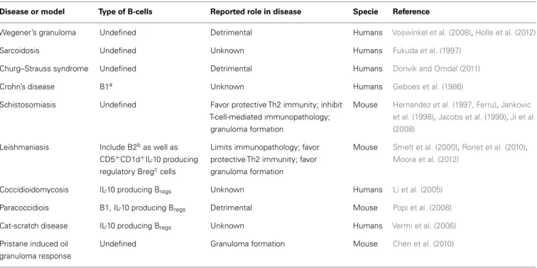
Table 1 | Characteristics of B-cells identified in non-TB granulomatous diseases.
Disease or model Type of B-cells Reported role in disease Specie Reference
Wegener’s granuloma Undefined Detrimental Humans Voswinkel et al. (2008),Holle et al. (2012)
Sarcoidosis Undefined Unknown Humans Fukuda et al. (1997)
Churg–Strauss syndrome Undefined Detrimental Humans Donvik and Omdal (2011)
Crohn’s disease B1a Unknown Humans Geboes et al. (1986)
Schistosomiasis Undefined Favor protective Th2 immunity; inhibit T-cell-mediated immunopathology;
granuloma formation
Mouse Hernandez et al. (1997, Ferru),Jankovic et al. (1998),Jacobs et al. (1999),Ji et al. (2008)
Leishmaniasis Include B2bas well as CD5+
CD1d+
IL-10 producing regulatory Bregccells
Limits immunopathology; favor protective Th2 immunity; favor granuloma formation
Mouse Smelt et al. (2000),Ronet et al. (2010),
Moore et al. (2012)
Coccidioidomycosis IL-10 producing Bregs Unknown Humans Li et al. (2005)
Paracoccidiois B1, IL-10 producing Bregs Detrimental Mouse Popi et al. (2008)
Cat-scratch disease IL-10 producing Bregs Unknown Humans Vermi et al. (2006)
Pristane induced oil granuloma response
Undefined Granuloma formation Mouse Chen et al. (2010)
a
B1 cells: developmentally defined; innate-like B-cells in the mouse; CD5+
or CD5−
subpopulation poorly defined in humans.
b
B2 cells: developmentally defined; include “conventional” follicular B-cells as well as “innate-like” marginal zone B-cells.
cB
several other granulomatous diseases (Table 1). Not only B-cells
are present in granuloma but also they could be important for their
maturation. This is suggested in pristane induced oil granuloma
formation (
Chen et al., 2010
) and during Schistosoma japonicum
infection (
Ji et al., 2008
) where the absence of B-cells results in a
marked delay in granuloma formation. In the context of the TB,
although granulomas form in the absence of B-cells, their
num-bers and size remain lower and they hardly become inflammatory
(
Bosio et al., 2000
;
Maglione et al., 2007
). This could be the result
of the well-known ability of B-cells to contribute to the
organiza-tion of secondary and tertiary lymphoid organs (
Moseman et al.,
2012
).
Second, although this is a rare event, occurrence of
mycobac-terial infections was reported upon rituximab-mediated
deple-tion of B-cells, suggesting a protective role for these lymphocytes
(
Winthrop et al., 2008
;
Gea-Banacloche, 2010
). However, other
granulomatous diseases were successfully treated with rituximab
(
Donvik and Omdal, 2011
;
Holle et al., 2012
), cautioning B-cells
may be detrimental depending on the disease context. Finally,
beyond antibody production, B-cells display diverse roles in the
immunity against multiple pathogens that could operate during
TB. In this regard, Salmonella infection, though not
occasion-ing granuloma formation, represents a paradigm for
antibody-independent roles of B-cells against an intracellular bacterium
with the evidence that B-cells producing IL-10 (B
regs) impairs the
control of natural and vaccine-induced immunity to Salmonella
(
Neves et al., 2010
). Since this role cannot simply be
recapitu-lated in animal models lacking B-cells (
Mastroeni et al., 2000
;
Mittrucker et al., 2000
), this exemplifies how deletion of the B-cell
compartment eclipses specific functions of these cells.
B-cells express adaptive and innate receptors to recognize
pathogens (
Blumenthal et al., 2009
;
Rawlings et al., 2012
). Beyond
antibody production, B-cells secrete various signals including
cytokines, and serve as antigen-presenting cells (
Rawlings et al.,
2012
). These immune-modulatory functions are performed by
different B-cell subsets depending on their differentiation program
(e.g., B1, B2), activation status (e.g., naïve, effector, memory),
tis-sue distribution, the timing of the immune response, or disease
context. From this perspective, the identity of B-cells infiltrating
the lungs of TB patients or animals remains relatively unknown. In
most cases, these cells (likely B2-cells) have undergone class switch
recombination and produce antibodies (
Phuah et al., 2012
).
How-ever, CD5
+CD1d
+B1-cells are also observed predominantly in
TB patients (
Zhang et al., 2012
) and in mouse models of TB and
other granulomatous diseases (
Li et al., 2005
;
Popi et al., 2008
;
Ronet et al., 2010
). Regardless of their identity or individual
con-tribution, we estimate the B-cell compartment influences the TB
granuloma formation and function through interaction with Mφs,
T-cells, and neutrophils (Figure 1).
As B-cells interact with Mφs in TB granulomas (
Tsai et al.,
2006
;
Chakravarty et al., 2008
), they might affect Mφ
polariza-tion within these structures. A case in point, B1-cells differentiate
M2 Mφs via IL-10 in vitro and in a tumor model (
Wong et al.,
2010
). However, mice deficient for B1-cells (xid model) displayed
rather a susceptibility to mycobacterial infection, accompanied
by increased levels of IL-10 (
Junqueira-Kipnis et al., 2005
;
Russo
and Mariano, 2010
). Certainly, there are other B-cell subsets that
could compensate as the in vivo source of IL-10, like B
regs(
O’Garra
et al., 1990
;
Lampropoulou et al., 2008
). Likewise, there exist
alter-native in vivo immunosuppressive mechanisms driven by B-cells
other than the B1-cell subset, as demonstrated for IgG production
favoring FcR-mediated M2 Mφ polarization in a carcinoma model
(
Andreu et al., 2010
). In line with this observation,
FCγRIIB-deficient Mφs displayed a M1 Mφ phenotype upon Mtb infection,
express less IL-10 and better control the infection (
Maglione et al.,
2008
). Since the phenotype manifests after 3 weeks of infection,
IgG-producing B2-cells produced during the course of the
adap-tive immune response might be involved. B1 cells might rather
contribute to M2 polarization through FcγR-independent
IL-10-dependant mechanisms. Whether these events occur within the
granuloma is currently unknown. Collectively, these studies infer
a B-cell contribution to an immunosuppressive niche within TB
granulomas by tilting Mφs toward the M2 program.
If Mφs are the main components in nascent TB granulomas,
then CD4
+T-cells are perhaps the most critical component of
stable TB granulomas as shown by the re-awakening of latent TB
in HIV-1 co-infected patients. In recent years, multiple studies
suggest an immune-modulatory role for B-cells in T-cell
activ-ity at the granuloma level. On one hand, B-cells can co-localize
with T-cells in TB granulomas (
Ulrichs et al., 2004
;
Beytut, 2011
),
and directly interact with them in the granulomas caused by
Leishmania (
Moore et al., 2012
). On the other hand, B-cells
influence T-cell effector functions either through cytokine
pro-duction or antigen-presentation (
Lund and Randall, 2010
). In
TB context, IL-10 derived from B1-cells controls the
homeosta-sis of T-helper-17 (Th17), essential for anti-microbial immunity
at epithelial/mucosal barriers (
Zhang et al., 2012
). Reciprocally,
Th-17-associated cytokines promote the formation of B-cell foci
in Mtb-infected mice, and correlate with B-cell infiltration in TB
patients (
Khader et al., 2011
;
Zhang et al., 2011
). In the mouse
model, IL-17A (
Okamoto Yoshida et al., 2010
) or IL-23-deficient
(
Khader et al., 2011
;
Zhang et al., 2011
) animals have marked
defects in the formation of granulomas and/or B-cell follicles. In
addition 23-deficient mice also have poor levels of 17 and
IL-22. These deficiencies resulted in a marked alteration of CXCL13
production, the chemokine responsible for B-cell recruitment and
follicle formation (
Khader et al., 2011
;
Zhang et al., 2011
). It is
not known if IL-10 production by B-cells is at the initiation or
a secondary consequence of the alterations in IL-17 levels. These
observations might provide an explanation for the links reported
in TB patients between Th17 and formation of B-cell foci and
IL-10 (
Zhang et al., 2011, 2012
).
Evidence obtained in non-TB diseases argue B-cells favor Th1
polarization (involved in TB protective immunity) through
IL-6 and IFN-γ production during Salmonella infection, or
pro-mote Th2 differentiation (thought to be detrimental during TB)
through either IL-2 (
Wojciechowski et al., 2009
) or IL-10 (
Ferru
et al., 1998
;
Popi et al., 2008
;
Ronet et al., 2010
) in the
con-trol of different parasites. Conversely, B-cells also suppress T-cell
activity as best illustrated in mice with a targeted deletion of
MyD88 in B-cells during Salmonella infection (
Neves et al., 2010
).
Finally, evidencing the role of B-cells as antigen-presenting cells,
mice with a targeted deletion of MHC-II in B-cells displayed a
reduction of IL-2 and IFN-γ by CD4
+Lugo-Villarino et al. Emerging trends in tuberculosis granulomas
Salmonella challenge (
Barr et al., 2010
), and low pulmonary Th1
cell counts during Pneumocystis infection (
Lund and Randall,
2010
).
Another cell influencing TB granuloma formation is the
neu-trophil, whose migration can be controlled by B-cells. During
Salmonella infection, mice with a targeted deletion of MyD88 in
B-cells exhibited an accumulation of neutrophils in the spleen,
an effect that likely depends on B
regs-mediated IL-10
produc-tion (
Neves et al., 2010
). In the context of mycobacterial
infec-tions, aberrant neutrophil migration is known to have deleterious
effects in host tissue integrity (
Eruslanov et al., 2005
;
Berry et al.,
2010
). In mice deficient for the B-cell compartment (
Maglione
et al., 2007
), Mtb infection leads to an uncontrolled
accumula-tion of pulmonary neutrophils, an observaaccumula-tion also supported by
the excessive neutrophil migration in the peritoneum after
BCG-vaccination (
Kondratieva et al., 2010
). These examples highlight
the importance of tolerance mechanisms in TB.
Based on the above observation, it is tempting to propose
that B-cell could act at different levels during TB such as
during granuloma progression and by influencing the
effec-tor function of third-party cells like Mφs. To directly
exam-ine this, studying B-cell contribution through comparison of
B-cell-competent vs. B-cell-deficient animals should now be
fur-ther complemented by studies examining the direct response
of B-cells to Mtb infection, and through analyses in animal
models lacking specific pathways in B-cells and biological
consequences.
CONCLUSION
Among trends emerging in TB etiology, the notion that the local
lung environment shifts from a host-protective nature toward one
favorable to microbial resilience is discussed here at the granuloma
level and in the context of Mφ polarization and B-cell function (see
also an illustration in Figure 1). Exploring these issues will likely
bring us closer to uncover the enigma concealed by TB
granulo-mas. One can envisage that studies investigating the role of genes
involved in host tolerance (
Medzhitov et al., 2012
) might be a good
way to explore these aspects of the disease. Although in humans
this could be limited to immunogenetic studies, more
mechanis-tic studies could be conducted in animal models where selective
inactivation of those genes could provide new insights on the
con-sequences on the pathology. These studies could go along with
more sophisticated approaches based on single cell analysis such
as those involving laser microdissection or more global phenotypic
signatures obtained from mass cytometry, in order to further
iden-tify cell subsets involved at different stages of granuloma formation
and TB.
ACKNOWLEDGMENTS
The authors received no specific funding for this work. The
Ney-rolles laboratory is supported by the CNRS, the European Union
(7th Framework Programme and ERA-NET), Agence Nationale de
la Recherche, Fondation pour la Recherche Médicale (FRM), and
Fondation Mérieux. Geanncarlo Lugo-Villarino and A. Benard are
supported by fellowships from FRM.
REFERENCES
Abebe, F., and Bjune, G. (2009). The protective role of antibody responses during Mycobacterium tuberculosis infection. Clin. Exp. Immunol. 157, 235–243.
Adams, D. O. (1974). The struc-ture of mononuclear phagocytes differentiating in vivo. I. Sequen-tial fine and histologic studies of the effect of Bacillus Calmette-Guerin (BCG). Am. J. Pathol. 76, 17–48.
Aggad, D., Stein, C., Sieger, D., Mazel, M., Boudinot, P., Herbomel, P., et al. (2010). In vivo analysis of Ifn-gamma1 and Ifn-gamma2 signal-ing in zebrafish. J. Immunol. 185, 6774–6782.
Almeida, A. S., Lago, P. M., Boechat, N., Huard, R. C., Lazzarini, L. C., Santos, A. R., et al. (2009). Tuberculosis is associated with a down-modulatory lung immune response that impairs Th1-type immunity. J. Immunol. 183, 718–731.
Andreu, P., Johansson, M., Affara, N. I., Pucci, F., Tan, T., Junankar, S., et al. (2010). FcRgamma activation regu-lates inflammation-associated squa-mous carcinogenesis. Cancer Cell 17, 121–134.
Balla, K. M., Lugo-Villarino, G., Spitsbergen, J. M., Stachura, D.
L., Hu, Y., Banuelos, K., et al. (2010). Eosinophils in the zebrafish: prospective isolation, characteriza-tion, and eosinophilia induction by helminth determinants. Blood 116, 3944–3954.
Barr, T. A., Brown, S., Mastroeni, P., and Gray, D. (2010). TLR and B cell receptor signals to B cells dif-ferentially program primary and memory Th1 responses to Salmo-nella enterica. J. Immunol. 185, 2783–2789.
Benoit, M., Desnues, B., and Mege, J. L. (2008). Macrophage polarization in bacterial infections. J. Immunol. 181, 3733–3739.
Berry, M. P., Graham, C. M., McNab, F. W., Xu, Z., Bloch, S. A., Oni, T., et al. (2010). An interferon-inducible neutrophil-driven blood transcriptional signature in human tuberculosis. Nature 466, 973–977.
Beytut, E. (2011). Immunohistochem-ical evaluation of surfactant pro-teins and lymphocyte phenotypes in the lungs of cattle with nat-ural tuberculosis. Res. Vet. Sci. 91, 119–124.
Biswas, S. K., and Mantovani, A. (2010). Macrophage plasticity and interac-tion with lymphocyte subsets: cancer as a paradigm. Nat. Immunol. 11, 889–896.
Blumenthal, A., Kobayashi, T., Pierini, L. M., Banaei, N., Ernst, J. D., Miyake, K., et al. (2009). RP105 facilitates macrophage activation by Mycobac-terium tuberculosis lipoproteins. Cell Host Microbe 5, 35–46. Bosio, C. M., Gardner, D., and Elkins,
K. L. (2000). Infection of B cell-deficient mice with CDC 1551, a clinical isolate of Mycobac-terium tuberculosis: delay in dis-semination and development of lung pathology. J. Immunol. 164, 6417–6425.
Cairo, G., Recalcati, S., Mantovani, A., and Locati, M. (2011). Iron traffick-ing and metabolism in macrophages: contribution to the polarized phe-notype. Trends Immunol. 32, 241–247.
Chakravarty, S. D., Zhu, G., Tsai, M. C., Mohan, V. P., Marino, S., Kirschner, D. E., et al. (2008). Tumor necrosis factor blockade in chronic murine tuberculosis enhances granuloma-tous inflammation and disorganizes granulomas in the lungs. Infect. Immun. 76, 916–926.
Chan, J., Xing, Y., Magliozzo, R. S., and Bloom, B. R. (1992). Killing of virulent Mycobacterium tuberculo-sis by reactive nitrogen intermedi-ates produced by activated murine macrophages. J. Exp. Med. 175, 1111–1122.
Chen, H., Liao, D., Holl, T. M., Snowden, P., Ueda, Y., and Kel-soe, G. (2010). Genetic regulation of pristane-induced oil granuloma responses. Int. J. Exp. Pathol. 91, 472–483.
Choi, H. S., Rai, P. R., Chu, H. W., Cool, C., and Chan, E. D. (2002). Analysis of nitric oxide syn-thase and nitrotyrosine expression in human pulmonary tuberculosis. Am. J. Respir. Crit. Care Med. 166, 178–186.
Cooper, A. M. (2009). Cell-mediated immune responses in tuberculosis. Annu. Rev. Immunol. 27, 393–422. Curiel, T. J., Coukos, G., Zou, L., Alvarez,
X., Cheng, P., Mottram, P., et al. (2004). Specific recruitment of reg-ulatory T cells in ovarian carcinoma fosters immune privilege and pre-dicts reduced survival. Nat. Med. 10, 942–949.
Davis, J. M., Clay, H., Lewis, J. L., Ghori, N., Herbomel, P., and Ramakrish-nan, L. (2002). Real-time visualiza-tion of mycobacterium-macrophage interactions leading to initiation of granuloma formation in zebrafish embryos. Immunity 17, 693–702. Davis, J. M., and Ramakrishnan, L.
(2009). The role of the granuloma in expansion and dissemination of early tuberculous infection. Cell 136, 37–49.
Deretic, V., Vergne, I., Chua, J., Mas-ter, S., Singh, S. B., Fazio, J. A., et al. (2004). Endosomal membrane traffic: convergence point targeted by Mycobacterium tuberculosis and HIV. Cell. Microbiol. 6, 999–1009. Donvik, K. K., and Omdal, R. (2011).
Churg-Strauss syndrome suc-cessfully treated with rituximab. Rheumatol. Int. 31, 89–91. Dorhoi, A., Reece, S. T., and
Kauf-mann, S. H. (2011). For better or for worse: the immune response against Mycobacterium tuberculosis balances pathology and protection. Immunol. Rev. 240, 235–251. Egen, J. G., Rothfuchs, A. G., Feng, C.
G., Horwitz, M. A., Sher, A., and Germain, R. N. (2011). Intravital imaging reveals limited antigen pre-sentation and T cell effector func-tion in mycobacterial granulomas. Immunity 34, 807–819.
Ehrt, S., Schnappinger, D., Bekiranov, S., Drenkow, J., Shi, S., Gingeras, T. R., et al. (2001). Reprogramming of the macrophage transcriptome in response to interferon-gamma and Mycobacterium tuberculosis: signal-ing roles of nitric oxide synthase-2 and phagocyte oxidase. J. Exp. Med. 194, 1123–1140.
El Kasmi, K. C., Qualls, J. E., Pesce, J. T., Smith, A. M., Thompson, R. W., Henao-Tamayo, M., et al. (2008). Toll-like receptor-induced arginase 1 in macrophages thwarts effective immunity against intracel-lular pathogens. Nat. Immunol. 9, 1399–1406.
Eruslanov, E. B., Lyadova, I. V., Kon-dratieva, T. K., Majorov, K. B., Scheglov, I. V., Orlova, M. O., et al. (2005). Neutrophil responses to Mycobacterium tuberculosis infec-tion in genetically susceptible and resistant mice. Infect. Immun. 73, 1744–1753.
Facchetti, F., Vermi, W., Fiorentini, S., Chilosi, M., Caruso, A., Duse, M., et al. (1999). Expression of inducible nitric oxide synthase in human gran-ulomas and histiocytic reactions. Am. J. Pathol. 154, 145–152. Feng, L., Li, L., Liu, Y., Qiao, D., Li,
Q., Fu, X., et al. (2011). B lym-phocytes that migrate to tuberculous pleural fluid via the SDF-1/CXCR4 axis actively respond to antigens spe-cific for Mycobacterium tuberculo-sis. Eur. J. Immunol. 41, 3261–3269. Ferru, I., Roye, O., Delacre, M.,
Auri-ault, C., and Wolowczuk, I. (1998). Infection of B-cell-deficient mice by the parasite Schistosoma mansoni: demonstration of the participation of B cells in granuloma modulation. Scand. J. Immunol. 48, 233–240.
Flynn, J. L. (2006). Lessons from exper-imental Mycobacterium tuberculo-sis infections. Microbes Infect. 8, 1179–1188.
Flynn, J. L., Chan, J., and Lin, P. L. (2011). Macrophages and control of granulomatous inflammation in tuberculosis. Mucosal Immunol. 4, 271–278.
Flynn, J. L., Chan, J., Triebold, K. J., Dal-ton, D. K., Stewart, T. A., and Bloom, B. R. (1993). An essential role for interferon gamma in resistance to Mycobacterium tuberculosis infec-tion. J. Exp. Med. 178, 2249–2254. Forbes, J. R., and Gros, P. (2001).
Divalent-metal transport by NRAMP proteins at the interface of host-pathogen interactions. Trends Microbiol. 9, 397–403.
Fukuda, T., Sato, K., Tachikawa, S., Ohnuki, K., Ohtani, H., and Suzuki, T. (1997). Mucosa-associated lym-phoid tissue lymphoma coexisting with epithelioid granulomas in the stomach of a patient with systemic sarcoidosis. Pathol. Int. 47, 870–875. Gea-Banacloche, J. C. (2010). Rituximab-associated infections. Semin. Hematol. 47, 187–198. Geboes, K., Van Den Oord, J., De
Wolf-Peeters, C., Desmet, V., Rutgeerts, P., Janssens, J., et al. (1986). The cellular composi-tion of granulomas in mesenteric lymph nodes from patients with Crohn’s disease. Virchows Arch. A Pathol. Anat. Histopathol. 409, 679–692.
Glatman-Freedman, A., and Casadevall, A. (1998). Serum therapy for tuber-culosis revisited: reappraisal of the role of antibody-mediated immu-nity against Mycobacterium tuber-culosis. Clin. Microbiol. Rev. 11, 514–532.
Goerdt, S., and Orfanos, C. E. (1999). Other functions, other genes: alternative activation of antigen-presenting cells. Immunity 10, 137–142.
Gonzalez-Juarrero, M., Turner, O. C., Turner, J., Marietta, P., Brooks, J. V., and Orme, I. M. (2001). Tempo-ral and spatial arrangement of lym-phocytes within lung granulomas induced by aerosol infection with Mycobacterium tuberculosis. Infect. Immun. 69, 1722–1728.
Gordon, S. (2003). Alternative acti-vation of macrophages. Nat. Rev. Immunol. 3, 23–35.
Griffin, J. E., Gawronski, J. D., Dejesus, M. A., Ioerger, T. R., Akerley, B. J., and Sassetti, C. M. (2011). High-resolution phenotypic profiling defines genes essential for mycobacterial
growth and cholesterol catabo-lism. PLoS Pathog. 7:e1002251. doi:10.1371/journal.ppat.1002251 Helming, L., and Gordon, S. (2007).
The molecular basis of macrophage fusion. Immunobiology 212, 785–793.
Hernandez, H. J., Wang, Y., and Stadecker, M. J. (1997). In infec-tion with Schistosoma mansoni, B cells are required for T helper type 2 cell responses but not for gran-uloma formation. J. Immunol. 158, 4832–4837.
Holle, J. U., Dubrau, C., Herlyn, K., Heller, M., Ambrosch, P., Noelle, B., et al. (2012). Rituximab for refrac-tory granulomatosis with polyangi-itis (Wegener’s granulomatosis): comparison of efficacy in granulo-matous versus vasculitic manifesta-tions. Ann. Rheum. Dis. 71, 327–333. Holt, A., Mitra, S., Van Der Sar, A. M., Alnabulsi, A., Secombes, C. J., and Bird, S. (2011). Discovery of zebrafish (Danio rerio) interleukin-23 alpha (IL-interleukin-23alpha) chain, a sub-unit important for the formation of IL-23, a cytokine involved in the development of Th17 cells and inflammation. Mol. Immunol. 48, 981–991.
Jacobs, W., Bogers, J., and Van Marck, E. (1999). Distinct B-cell populations are present in hepatic and intesti-nal Schistosoma mansoni granulo-mas. Acta Gastroenterol. Belg. 62, 178–181.
Jankovic, D., Cheever, A. W., Kullberg, M. C., Wynn, T. A., Yap, G., Cas-par, P., et al. (1998). CD4+ T cell-mediated granulomatous pathology in schistosomiasis is downregulated by a B cell-dependent mechanism requiring Fc receptor signaling. J. Exp. Med. 187, 619–629.
Ji, F., Liu, Z., Cao, J., Li, N., Zuo, J., Chen, Y., et al. (2008). B cell response is required for granuloma formation in the early infection of Schistosoma japonicum. PLoS ONE 3:e1724. doi:10.1371/journal.pone.0001724 Junqueira-Kipnis, A. P., Kipnis, A.,
Henao Tamayo, M., Harton, M., Gonzalez Juarrero, M., Basaraba, R. J., et al. (2005). Interleukin-10 production by lung macrophages in CBA xid mutant mice infected with Mycobacterium tuberculosis. Immunology 115, 246–252. Kahnert, A., Hopken, U. E., Stein, M.,
Bandermann, S., Lipp, M., and Kauf-mann, S. H. (2007). Mycobacterium tuberculosis triggers formation of lymphoid structure in murine lungs. J. Infect. Dis. 195, 46–54.
Kahnert, A., Seiler, P., Stein, M., Bander-mann, S., Hahnke, K., Mollenkopf,
H., et al. (2006). Alternative acti-vation deprives macrophages of a coordinated defense program to Mycobacterium tuberculosis. Eur. J. Immunol. 36, 631–647.
Khader, S. A., Guglani, L., Rangel-Moreno, J., Gopal, R., Junecko, B. A., Fountain, J. J., et al. (2011). IL-23 is required for long-term control of Mycobacterium tuberculosis and B cell follicle formation in the infected lung. J. Immunol. 187, 5402–5407. Koch, R. (1982). [The etiology of
tuber-culosis by Dr. Robert Koch. From the Berliner Klinische Wochenschrift, Volume 19 (1882)]. Zentralbl. Bak-teriol. Mikrobiol. Hyg. A 251, 287–296.
Kondratieva, T. K., Rubakova, E. I., Linge, I. A., Evstifeev, V. V., Majorov, K. B., and Apt, A. S. (2010). B cells delay neutrophil migration toward the site of stimulus: tardiness crit-ical for effective bacillus Calmette-Guerin vaccination against tubercu-losis infection in mice. J. Immunol. 184, 1227–1234.
Kuda, O., Jenkins, C. M., Skinner, J. R., Moon, S. H., Su, X., Gross, R. W., et al. (2011). CD36 protein is involved in store-operated calcium flux, phospholipase A2 activation, and production of prostaglandin E2. J. Biol. Chem. 286, 17785–17795. Kunnath-Velayudhan, S., Davidow, A.
L., Wang, H. Y., Molina, D. M., Huynh, V. T., Salamon, H., et al. (2012). Proteome-scale antibody responses and outcome of Mycobac-terium tuberculosis infection in nonhuman primates and in tuber-culosis patients. J. Infect. Dis. 206, 697–705.
Kunnath-Velayudhan, S., Salamon, H., Wang, H. Y., Davidow, A. L., Molina, D. M., Huynh, V. T., et al. (2010). Dynamic antibody responses to the Mycobacterium tuberculosis pro-teome. Proc. Natl. Acad. Sci. U.S.A. 107, 14703–14708.
Lampropoulou, V., Hoehlig, K., Roch, T., Neves, P., Calderon Gomez, E., Sweenie, C. H., et al. (2008). TLR-activated B cells suppress T cell-mediated autoimmunity. J. Immunol. 180, 4763–4773. Lawrence, T., and Natoli, G. (2011).
Transcriptional regulation of macrophage polarization: enabling diversity with identity. Nat. Rev. Immunol. 11, 750–761.
Li, L., Dial, S. M., Schmelz, M., Ren-nels, M. A., and Ampel, N. M. (2005). Cellular immune suppressor activ-ity resides in lymphocyte cell clusters adjacent to granulomata in human coccidioidomycosis. Infect. Immun. 73, 3923–3928.
Lugo-Villarino et al. Emerging trends in tuberculosis granulomas
Lund, F. E., and Randall, T. D. (2010). Effector and regulatory B cells: mod-ulators of CD4(+) T cell immunity. Nat. Rev. Immunol. 10, 236–247. Maglione, P. J., and Chan, J. (2009). How
B cells shape the immune response against Mycobacterium tuberculo-sis. Eur. J. Immunol. 39, 676–686. Maglione, P. J., Xu, J., Casadevall, A., and
Chan, J. (2008). Fc gamma receptors regulate immune activation and sus-ceptibility during Mycobacterium tuberculosis infection. J. Immunol. 180, 3329–3338.
Maglione, P. J., Xu, J., and Chan, J. (2007). B cells moderate inflam-matory progression and enhance bacterial containment upon pul-monary challenge with Mycobac-terium tuberculosis. J. Immunol. 178, 7222–7234.
Mahajan, S., Dkhar, H. K., Chandra, V., Dave, S., Nanduri, R., Janmeja, A. K., et al. (2012). Mycobacterium tuberculosis modulates macrophage lipid-sensing nuclear receptors PPARgamma and TR4 for survival. J. Immunol. 188, 5593–5603. Mantovani, A., Sica, A., Sozzani, S.,
Allavena, P., Vecchi, A., and Locati, M. (2004). The chemokine system in diverse forms of macrophage activation and polarization. Trends Immunol. 25, 677–686.
Martinez, F. O., Helming, L., and Gor-don, S. (2009). Alternative activa-tion of macrophages: an immuno-logic functional perspective. Annu. Rev. Immunol. 27, 451–483. Mastroeni, P., Simmons, C., Fowler, R.,
Hormaeche, C. E., and Dougan, G. (2000). Igh-6(-/-) (B-cell-deficient) mice fail to mount solid acquired resistance to oral challenge with vir-ulent Salmonella enterica serovar typhimurium and show impaired Th1 T-cell responses to Salmonella antigens. Infect. Immun. 68, 46–53. Medzhitov, R., Schneider, D. S., and
Soares, M. P. (2012). Disease toler-ance as a defense strategy. Science 335, 936–941.
Mitra, S., Alnabulsi, A., Secombes, C. J., and Bird, S. (2010). Identification and characterization of the tran-scription factors involved in T-cell development, t-bet, stat6 and foxp3, within the zebrafish, Danio rerio. FEBS J. 277, 128–147.
Mittrucker, H. W., Raupach, B., Kohler, A., and Kaufmann, S. H. (2000). Cutting edge: role of B lymphocytes in protective immunity against Sal-monella typhimurium infection. J. Immunol. 164, 1648–1652. Moller, M., Nebel, A., Valentonyte, R.,
Van Helden, P. D., Schreiber, S., and Hoal, E. G. (2009). Investigation
of chromosome 17 candidate genes in susceptibility to TB in a South African population. Tuberculosis 89, 189–194.
Moore, J. W., Beattie, L., Dalton, J. E., Owens, B. M., Maroof, A., Coles, M. C., et al. (2012). B cell: T cell inter-actions occur within hepatic granu-lomas during experimental visceral leishmaniasis. PLoS ONE 7:e34143. doi:10.1371/journal.pone.0034143 Moseman, E. A., Iannacone, M.,
Bosurgi, L., Tonti, E., Chevrier, N., Tumanov, A., et al. (2012). B cell maintenance of subcapsular sinus macrophages protects against a fatal viral infection independent of adaptive immunity. Immunity 36, 415–426.
Munder, M., Eichmann, K., and Mod-olell, M. (1998). Alternative meta-bolic states in murine macrophages reflected by the nitric oxide syn-thase/arginase balance: competitive regulation by CD4+ T cells corre-lates with Th1/Th2 phenotype. J. Immunol. 160, 5347–5354. Murray, P. J., and Wynn, T. A. (2011a).
Obstacles and opportunities for understanding macrophage polar-ization. J. Leukoc. Biol. 89, 557–563. Murray, P. J., and Wynn, T. A. (2011b). Protective and pathogenic functions of macrophage subsets. Nat. Rev. Immunol. 11, 723–737.
Neves, P., Lampropoulou, V., Calderon-Gomez, E., Roch, T., Stervbo, U., Shen, P., et al. (2010). Signaling via the MyD88 adaptor protein in B cells suppresses protective immunity dur-ing Salmonella typhimurium infec-tion. Immunity 33, 777–790. Nicholson, S., Bonecini-Almeida Mda,
G., Lapa e Silva, J. R., Nathan, C., Xie, Q. W., Mumford, R., et al. (1996). Inducible nitric oxide synthase in pulmonary alveolar macrophages from patients with tuberculosis. J. Exp. Med. 183, 2293–2302. O’Garra, A., Stapleton, G., Dhar, V.,
Pearce, M., Schumacher, J., Rugo, H., et al. (1990). Production of cytokines by mouse B cells: B lym-phomas and normal B cells pro-duce interleukin 10. Int. Immunol. 2, 821–832.
Okamoto Yoshida, Y., Umemura, M., Yahagi, A., O’Brien, R. L., Ikuta, K., Kishihara, K., et al. (2010). Essen-tial role of IL-17A in the forma-tion of a mycobacterial infecforma-tion- infection-induced granuloma in the lung. J. Immunol. 184, 4414–4422. Pandey, A. K., and Sassetti, C. M. (2008).
Mycobacterial persistence requires the utilization of host cholesterol. Proc. Natl. Acad. Sci. U.S.A. 105, 4376–4380.
Pessanha, A. P., Martins, R. A., Mattos-Guaraldi, A. L., Vianna, A., and Mor-eira, L. O. (2012). Arginase-1 expres-sion in granulomas of tuberculo-sis patients. FEMS Immunol. Med. Microbiol. 66, 265–268.
Peyron, P., Vaubourgeix, J., Poquet, Y., Levillain, F., Botanch, C., Bardou, F., et al. (2008). Foamy macrophages from tuberculous patients’ gran-ulomas constitute a nutrient-rich reservoir for M. tuberculosis per-sistence. PLoS Pathog. 4:e1000204. doi:10.1371/journal.ppat.1000204 Philips, J. A., and Ernst, J. D.
(2012). Tuberculosis pathogenesis and immunity. Annu. Rev. Pathol. 7, 353–384.
Phuah, J. Y., Mattila, J. T., Lin, P. L., and Flynn, J. L. (2012). Activated B cells in the granulomas of nonhuman primates infected with Mycobac-terium tuberculosis. Am. J. Pathol. 181, 508–514.
Popi, A. F., Godoy, L. C., Xander, P., Lopes, J. D., and Mariano, M. (2008). B-1 cells facilitate Paracoccidioides brasiliensis infection in mice via IL-10 secretion. Microbes Infect. 10, 817–824.
Qualls, J. E., Neale, G., Smith, A. M., Koo, M. S., Defreitas, A. A., Zhang, H., et al. (2010). Argi-nine usage in mycobacteria-infected macrophages depends on autocrine-paracrine cytokine signaling. Sci. Signal. 3, ra62.
Qualls, J. E., Subramanian, C., Rafi, W., Smith, A. M., Balouzian, L., Defreitas, A. A., et al. (2012). Sus-tained generation of nitric oxide and control of mycobacterial infection requires argininosuccinate synthase 1. Cell Host Microbe 12, 313–323. Quintana-Murci, L., Alcais, A., Abel,
L., and Casanova, J. L. (2007). Immunology in natura: clinical, epidemiological and evolutionary genetics of infectious diseases. Nat. Immunol. 8, 1165–1171.
Raju, B., Hoshino, Y., Belitskaya-Levy, I., Dawson, R., Ress, S., Gold, J. A., et al. (2008). Gene expression pro-files of bronchoalveolar cells in pul-monary TB. Tuberculosis (Edinb.) 88, 39–51.
Ramakrishnan, L. (2012). Revisiting the role of the granuloma in tuberculo-sis. Nat. Rev. Immunol. 12, 352–366. Rawlings, D. J., Schwartz, M. A., Jack-son, S. W., and Meyer-Bahlburg, A. (2012). Integration of B cell responses through Toll-like recep-tors and antigen receprecep-tors. Nat. Rev. Immunol. 12, 282–294.
Redente, E. F., Higgins, D. M., Dwyer-Nield, L. D., Orme, I. M., Gonzalez-Juarrero, M., and Malkinson, A.
M. (2010). Differential polariza-tion of alveolar macrophages and bone marrow-derived monocytes following chemically and pathogen-induced chronic lung inflammation. J. Leukoc. Biol. 88, 159–168. Renshaw, S. A., and Trede, N. S.
(2012). A model 450 million years in the making: zebrafish and verte-brate immunity. Dis. Model Mech. 5, 38–47.
Ronet, C., Hauyon-La Torre, Y., Revaz-Breton, M., Mastelic, B., Tacchini-Cottier, F., Louis, J., et al. (2010). Regulatory B cells shape the devel-opment of Th2 immune responses in BALB/c mice infected with Leish-mania major through IL-10 produc-tion. J. Immunol. 184, 886–894. Roy, E., Stavropoulos, E., Brennan, J.,
Coade, S., Grigorieva, E., Walker, B., et al. (2005). Therapeutic efficacy of high-dose intravenous immunoglobulin in Mycobacterium tuberculosis infection in mice. Infect. Immun. 73, 6101–6109.
Russell, D. G. (2007). Who puts the tubercle in tuberculosis? Nat. Rev. Microbiol. 5, 39–47.
Russell, D. G., Cardona, P. J., Kim, M. J., Allain, S., and Altare, F. (2009). Foamy macrophages and the pro-gression of the human tuberculo-sis granuloma. Nat. Immunol. 10, 943–948.
Russo, R. T., and Mariano, M. (2010). B-1 cell protective role in murine primary Mycobacterium bovis bacillus Calmette-Guerin infection. Immunobiology 215, 1005–1014. Sakula, A. (1982). Robert Koch:
cente-nary of the discovery of the tubercle bacillus, 1882. Thorax 37, 246–251. Schebesch, C., Kodelja, V., Muller, C.,
Hakij, N., Bisson, S., Orfanos, C. E., et al. (1997). Alternatively activated macrophages actively inhibit prolif-eration of peripheral blood lympho-cytes and CD4+ T cells in vitro. Immunology 92, 478–486. Schon, T., Elmberger, G., Negesse, Y.,
Pando, R. H., Sundqvist, T., and Brit-ton, S. (2004). Local production of nitric oxide in patients with tuber-culosis. Int. J. Tuberc. Lung Dis. 8, 1134–1137.
Schreiber, T., Ehlers, S., Heitmann, L., Rausch, A., Mages, J., Murray, P. J., et al. (2009). Autocrine IL-10 induces hallmarks of alterna-tive activation in macrophages and suppresses antituberculosis effector mechanisms without compromising T cell immunity. J. Immunol. 183, 1301–1312.
Shearer, J. D., Richards, J. R., Mills, C. D., and Caldwell, M. D. (1997). Dif-ferential regulation of macrophage
arginine metabolism: a proposed role in wound healing. Am. J. Physiol. 272, E181–E190.
Sica, A., Larghi, P., Mancino, A., Rubino, L., Porta, C., Totaro, M. G., et al. (2008). Macrophage polarization in tumour progression. Semin. Cancer Biol. 18, 349–355.
Smelt, S. C., Cotterell, S. E., Engwerda, C. R., and Kaye, P. M. (2000). B cell-deficient mice are highly resistant to Leishmania donovani infection, but develop neutrophil-mediated tissue pathology. J. Immunol. 164, 3681–3688.
Tjarnlund, A., Rodriguez, A., Cardona, P. J., Guirado, E., Ivanyi, J., Singh, M., et al. (2006). Polymeric IgR knock-out mice are more susceptible to mycobacterial infections in the res-piratory tract than wild-type mice. Int. Immunol. 18, 807–816. Torrado, E., and Cooper, A. M.
(2011). What do we really know about how CD4 T cells control Mycobacterium tubercu-losis? PLoS Pathog. 7:e1002196. doi:10.1371/journal.ppat.1002196 Tsai, M. C., Chakravarty, S., Zhu, G.,
Xu, J., Tanaka, K., Koch, C., et al. (2006). Characterization of the tuberculous granuloma in murine and human lungs: cellular com-position and relative tissue oxy-gen tension. Cell. Microbiol. 8, 218–232.
Ulrichs, T., Kosmiadi, G. A., Trusov, V., Jorg, S., Pradl, L., Titukhina, M., et al.
(2004). Human tuberculous granu-lomas induce peripheral lymphoid follicle-like structures to orchestrate local host defence in the lung. J. Pathol. 204, 217–228.
Vermi, W., Facchetti, F., Riboldi, E., Heine, H., Scutera, S., Stornello, S., et al. (2006). Role of den-dritic cell-derived CXCL13 in the pathogenesis of Bartonella hense-lae B-rich granuloma. Blood 107, 454–462.
Volkman, H. E., Clay, H., Beery, D., Chang, J. C., Sherman, D. R., and Ramakrishnan, L. (2004). Tuberculous granu-loma formation is enhanced by a mycobacterium virulence determinant. PLoS Biol. 2:e367. doi:10.1371/journal.pbio.0020367 Volkman, H. E., Pozos, T. C., Zheng,
J., Davis, J. M., Rawls, J. F., and Ramakrishnan, L. (2010). Tubercu-lous granuloma induction via inter-action of a bacterial secreted protein with host epithelium. Science 327, 466–469.
Voswinkel, J., Assmann, G., Held, G., Pitann, S., Gross, W. L., Holl-Ulrich, K., et al. (2008). Single cell analy-sis of B lymphocytes from Wegener’s granulomatosis: B cell receptors dis-play affinity maturation within the granulomatous lesions. Clin. Exp. Immunol. 154, 339–345.
Winthrop, K. L., Yamashita, S., Beek-mann, S. E., and Polgreen, P. M. (2008). Mycobacterial and other
serious infections in patients receiving anti-tumor necrosis factor and other newly approved biologic therapies: case finding through the Emerging Infections Network. Clin. Infect. Dis. 46, 1738–1740. Wittamer, V., Bertrand, J. Y., Gutschow,
P. W., and Traver, D. (2011). Char-acterization of the mononuclear phagocyte system in zebrafish. Blood 117, 7126–7135.
Wojciechowski, W., Harris, D. P., Sprague, F., Mousseau, B., Makris, M., Kusser, K., et al. (2009). Cytokine-producing effector B cells regulate type 2 immunity to H. polygyrus. Immunity 30, 421–433.
Wong, S. C., Puaux, A. L., Chittezhath, M., Shalova, I., Kajiji, T. S., Wang, X., et al. (2010). Macrophage polar-ization to a unique phenotype dri-ven by B cells. Eur. J. Immunol. 40, 2296–2307.
Xie, Q. W., and Nathan, C. (1993). Pro-moter of the mouse gene encoding calcium-independent nitric oxide synthase confers inducibility by interferon-gamma and bacterial lipopolysaccharide. Trans. Assoc. Am. Physicians 106, 1–12. Zhang, M., Wang, Z., Graner, M. W.,
Yang, L., Liao, M., Yang, Q., et al. (2011). B cell infiltration is associ-ated with the increased 17 and IL-22 expression in the lungs of patients with tuberculosis. Cell. Immunol. 270, 217–223.
Zhang, M., Zheng, X., Zhang, J., Zhu, Y., Zhu, X., Liu, H., et al. (2012). CD19(+)CD1d(+)CD5(+) B cell frequencies are increased in patients with tuberculosis and sup-press Th17 responses. Cell. Immunol. 274, 89–97.
Conflict of Interest Statement: The
authors declare that the research was conducted in the absence of any com-mercial or financial relationships that could be construed as a potential con-flict of interest.
Received: 05 October 2012; paper pending published: 03 November 2012; accepted: 15 December 2012; published online: 07 January 2013.
Citation: Lugo-Villarino G, Hudrisier D, Benard A and Neyrolles O (2013) Emerging trends in the formation and function of tuberculosis gran-ulomas. Front. Immun. 3:405. doi: 10.3389/fimmu.2012.00405
This article was submitted to Frontiers in Inflammation, a specialty of Frontiers in Immunology.
Copyright © 2013 Lugo-Villarino, Hudrisier, Benard and Neyrolles. This is an open-access article distributed under the terms of the Creative Commons Attribution License, which permits use, distribution and reproduction in other forums, provided the original authors and source are credited and subject to any copyright notices concerning any third-party graphics etc.