A Common Neural Code for Perceived and Inferred Emotion
The MIT Faculty has made this article openly available.
Please share
how this access benefits you. Your story matters.
Citation
Skerry, A. E., and R. Saxe. “A Common Neural Code for Perceived
and Inferred Emotion.” Journal of Neuroscience 34, no. 48
(November 26, 2014): 15997–16008.
As Published
http://dx.doi.org/10.1523/jneurosci.1676-14.2014
Publisher
Society for Neuroscience
Version
Final published version
Citable link
http://hdl.handle.net/1721.1/97243
Terms of Use
Article is made available in accordance with the publisher's
policy and may be subject to US copyright law. Please refer to the
publisher's site for terms of use.
Behavioral/Cognitive
A Common Neural Code for Perceived and Inferred Emotion
Amy E. Skerry
1and Rebecca Saxe
21Department of Psychology, Harvard University, Cambridge, Massachusetts 02138, and2Department of Brain and Cognitive Sciences, Massachusetts
Institute of Technology, Cambridge, Massachusetts 02139
Although the emotions of other people can often be perceived from overt reactions (e.g., facial or vocal expressions), they can also be
inferred from situational information in the absence of observable expressions. How does the human brain make use of these diverse
forms of evidence to generate a common representation of a target’s emotional state? In the present research, we identify neural patterns
that correspond to emotions inferred from contextual information and find that these patterns generalize across different cues from
which an emotion can be attributed. Specifically, we use functional neuroimaging to measure neural responses to dynamic facial
expres-sions with positive and negative valence and to short animations in which the valence of a character’s emotion could be identified only
from the situation. Using multivoxel pattern analysis, we test for regions that contain information about the target’s emotional state,
identifying representations specific to a single stimulus type and representations that generalize across stimulus types. In regions of
medial prefrontal cortex (MPFC), a classifier trained to discriminate emotional valence for one stimulus (e.g., animated situations) could
successfully discriminate valence for the remaining stimulus (e.g., facial expressions), indicating a representation of valence that
ab-stracts away from perceptual features and generalizes across different forms of evidence. Moreover, in a subregion of MPFC, this neural
representation generalized to trials involving subjectively experienced emotional events, suggesting partial overlap in neural responses
to attributed and experienced emotions. These data provide a step toward understanding how the brain transforms stimulus-bound
inputs into abstract representations of emotion.
Key words: abstraction; concepts; emotion attribution; multimodal; social cognition; theory of mind
Introduction
To recognize someone’s emotion, we can rely on facial
expres-sion, tone of voice, and even body posture. Perceiving emotions
from these overt expressions poses a version of the “invariance
problem” faced across perceptual domains (
Ullman, 1998
;
Di-Carlo et al., 2012
): we recognize emotions despite variation both
within modality (e.g., sad face across viewpoint and identity) and
across modalities (e.g., sadness from facial and vocal
expres-sions). Emotion recognition may therefore rely on bottom-up
extraction of invariants within a hierarchy of increasingly
com-plex feature-detectors (
Tanaka, 1993
). However, we can also infer
emotions in the absence of overt expressions by reasoning about
the situation a person encounters (
Ortony, 1990
;
Zaki et al., 2008
;
Scherer and Meuleman, 2013
). To do so, we rely on abstract
causal principles (e.g., social rejection causes sadness) rather than
direct perceptual cues. Ultimately, the brain must integrate these
diverse sources of information into a common code that supports
empathic responses and flexible emotion-based inference.
What neural mechanisms underlie these different aspects of
emotion recognition? Previous neuroimaging studies have
re-vealed regions containing information about emotions in overt
expressions: different facial expressions, for example, elicit
dis-tinct patterns of neural activity in the superior temporal sulcus
and fusiform gyrus (
Said et al., 2010a
,
b
;
Harry et al., 2013
; see also
Pitcher, 2014
). In these studies, emotional stimuli were presented
in a single modality, leaving it unclear the precise dimensions
represented in these regions. Given that facial expressions can be
distinguished based on features specific to the visual modality
(e.g., mouth motion, eyebrow deflection, eye aperture;
Ekman
and Rosenberg, 1997
;
Oosterhof and Todorov, 2009
),
face-responsive visual regions could distinguish emotional
expres-sions based on such lower-level features.
To represent what is in common across sad faces and voices,
the brain may also compute multimodal representations. In a
recent study (
Peelen et al., 2010
), subjects were presented with
overt facial, bodily, and vocal expressions: in posterior temporal
cortex (lpSTC) and middle medial prefrontal cortex (MMPFC),
the pattern of response across different modalities was more
sim-ilar for the same emotion than for different emotions. Thus,
emo-tional stimuli sharing no low-level perceptual features seem to be
represented similarly in these regions.
However, we not only recognize emotions from canonical
perceptual cues, but also infer emotions from causal context
alone. We identify emotions in the absence of familiar
expres-sions, even for situations we have never observed or experienced.
In the present study, we test for neural representations of
emo-tional valence that generalize across both overt facial expressions
Received April 25, 2014; revised Sept. 18, 2014; accepted Sept. 24, 2014.
Author contributions: A.E.S. and R.S. designed research; A.E.S. and R.S. performed research; A.E.S. and R.S. analyzed data; A.E.S. and R.S. wrote the paper.
This work was supported by National Science Foundation Graduate Research Fellowship (A.E.S.) and NIH Grant 1R01 MH096914-01A1 (R.S.). We thank Laura Schulz, Nancy Kanwisher, Michael Cohen, Dorit Kliemann, Stefano Anzellotti, and Jorie Koster-Hale for helpful comments and discussion.
The authors declare no competing financial interests.
Correspondence should be addressed to Amy E. Skerry, William James Hall, 33 Kirkland Street, Cambridge, MA 02138. E-mail:[email protected].
DOI:10.1523/JNEUROSCI.1676-14.2014
Copyright © 2014 the authors 0270-6474/14/3315997-12$15.00/0
and emotions inferred from the situation a character is in. We
first identify neural patterns that contain information about
emotional valence for each type of stimulus. We then test whether
these neural patterns generalize across the two stimulus types, the
signature of a common code integrating these very different types
of emotional information. Finally, we investigate whether
attrib-uting emotional experiences to others and experiencing one’s
own emotions recruit a common neural representation by testing
whether these same neural patterns generalize to emotional
events experienced by participants themselves.
Materials and Methods
Summary
In Experiment 1, we used functional magnetic resonance imaging (fMRI) to measure blood oxygen level-dependent (BOLD) responses to emo-tional facial expressions and to animations depicting a character in an emotion-eliciting situation. While emotion-specific representations could, in principle, take the form of a uniform response across voxels in a region (detectable with univariate analyses), prior research has yielded little evidence for consistent and selective associations between discrete brain regions and specific emotions (Fusar-Poli et al., 2009;Lindquist et al., 2012). Thus, the present research uses multivariate analyses that ex-ploit reliable signal across distributed patterns of voxels to uncover neu-ral representations at a spatial scale smaller than that of entire regions (Haxby et al., 2001;Kamitani and Tong, 2005;Kriegeskorte et al., 2006;
Norman et al., 2006). With this approach, we test for representations of emotional valence that are specific to a particular type of stimulus (facial expressions or causal situations) and representations that generalize across the two stimulus types. To identify stimulus-independent repre-sentations, we trained a pattern classification algorithm to discriminate emotional valence for one stimulus type (e.g., dynamic facial expres-sions) and tested its ability to discriminate valence for the remaining type (e.g., animations depicting causal situations). Thus, for each region of interest (ROI), we test whether there is a reliable neural pattern that supports classifying emotions when trained and tested on facial expres-sions, when trained and tested on situations, and when requiring gener-alization across facial expressions and situations.
We then test whether attributing emotions to others engages neural mechanisms involved in the first-person experience of emotion. Previ-ous research has implicated MPFC not only in emotion attribution, but also in subjective experience of emotional or rewarding outcomes (Lin et al., 2012;Clithero and Rangel, 2013;Winecoff et al., 2013;Chikazoe et al., 2014). However, the relationship between experienced reward and emo-tion attribuemo-tion remains poorly understood. In Experiment 2, we mea-sured BOLD responses to positive and negative situations for another individual (replicating Experiment 1) and to trials in which subjects themselves experienced positive and negative outcomes (winning and losing money). Again, we test whether there is a reliable neural pattern that supports classifying the valence of events when trained and tested on third-party situations, when trained and tested on first-person rewards, and when requiring generalization across third-person and first-person experiences.
Regions of interest
Based on prior literature (Peelen et al., 2010), our regions of interest for abstract, conceptual representations of emotion were the pSTC and MMPFC. We localized in individual subjects a middle MPFC ROI com-parable with that ofPeelen et al. (2010), using a standard social versus nonsocial contrast (Saxe and Kanwisher, 2003;Dodell-Feder et al., 2011; see below). Because pSTC could not be identified by standard localizer tasks, we identified bilateral group ROIs based on the peak coordinate fromPeelen et al. (2010). Our primary analyses target these three ROIs, accounting for multiple comparisons with a corrected ␣ ⫽ 0.05/3 (0.017).
In addition to the MMPFC region identified byPeelen et al. (2010), adjacent regions of dorsal and ventral MPFC have been strongly impli-cated in studies of emotion and affective value (Amodio and Frith, 2006;
Hynes et al., 2006;Vo¨llm et al., 2006;Etkin et al. 2011). Moreover, the
MPFC is part of a larger set of regions [the posterior cingulate/precuneus (PC), bilateral temporal parietal junction (rTPJ and lTPJ), and right anterior temporal lobe (rATL)] that are reliably recruited when reason-ing about others’ mental states (Saxe and Kanwisher, 2003;Mitchell, 2009), including emotional states (Zaki et al., 2010;Bruneau et al., 2012;
Spunt and Lieberman, 2012). This set of six regions [dorsal MPFC (DMPFC), ventral MPFC (VMPFC), rTPJ, lTPJ, PC, and rATL, in addi-tion to MMPFC described above) was identified in individual subjects using the social versus nonsocial contrast (described below). We test these remaining regions for representations of both perceived and in-ferred emotions [with␣ ⫽ 0.05/6 (0.008) to correct for comparisons across these six ROIs].
To test for modality-specific representations, we localized regions that might contain information specific to overt facial expressions: the right middle superior temporal sulcus (rmSTS), hypothesized to code for fa-cial motion parameters (Pelphrey et al., 2005;Calder et al., 2007;Carlin et al., 2011), and face-selective patches in right occipitotemporal cortex thought to code for identity-relevant face features [occipital face area (rOFA) and fusiform face area (rFFA);Kanwisher and Yovel, 2006]. For this analysis, we again correct for multiple comparisons using␣ ⫽ 0.017 (0.05/3).
Finally, in Experiment 2, we examined how the mechanisms involved in third-person attribution of emotional states relate to mechanisms involved in processing first-person subjective value. To do so, we identi-fied a region of orbitofrontal cortex (OFC/VMPFC) that has been previ-ously implicated in processing reward/emotional value (Kable and Glimcher, 2007;Plassmann et al., 2007;Chib et al., 2009;Winecoff et al., 2013;Chikazoe et al., 2014). We used a mask derived from two recent meta-analyses (Bartra et al., 2013;Clithero and Rangel, 2013) to investi-gate neural responses in an anatomical region of OFC/VMPFC in which neural responses have been shown to consistently correlate with reward value across reward types and decision contexts (anatomical mask avail-able athttp://www.rnl.caltech.edu/resources/index.html). Note that this mask is only partially overlapping with the search space used to identify VMPFC responses to theory of mind (in Experiment 1).
Participants
Twenty-one right-handed participants (20 – 43 years; Mage⫽ 26.84; 14 male) were recruited for Experiment 1. Sixteen right-handed participants (19 – 40 years; Mage⫽ 27.88; seven male) were recruited for Experiment 2. All participants had normal or corrected-to-normal vision and no history of neurological or psychiatric disorders and gave written, in-formed consent in accordance with the requirements of the MIT institu-tional review board.
fMRI tasks and stimuli
In Experiment 1, each subject participated in several behavioral tasks as well as three fMRI tasks: an Emotion Attribution task and two tasks used to localize regions involved in theory of mind and face perception. Sub-jects in Experiment 2 completed only the Emotion Attribution task and the theory of mind localizer.
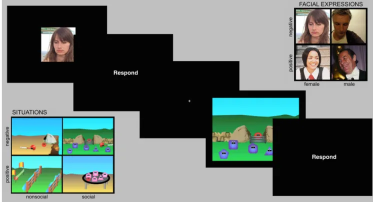
Emotion Attribution task. In the Emotion Attribution task (Fig. 1), subjects viewed brief video clips designed to elicit the attribution of an emotional state to a target (Fig. 1depicts static photos similar to video clips used in the study). The task consisted of video clips of people ex-pressing a positive (happy/smiling) or negative (sad/frowning) emotion (expressions condition) and brief animations in which a simple geomet-ric character experienced an event that would elicit positive or negative emotion (situations condition). In the situations condition, no emotion was expressed, but the character’s emotional state could be inferred based on the character’s goals and the event outcome. To ensure consistent attributions of emotional valence, independent subjects on Amazon Me-chanical Turk (n⫽ 16 per item) rated the stimuli from 1 to 7 (negative to positive valence): M(SEM)pos-faces⫽ 5.597(0.077); M(SEM)neg-faces⫽ 2.694(0.084); M(SEM)pos-situations⫽ 5.401(0.068); M(SEM)neg-situations⫽ 2.695(0.058). Each stimulus type was further divided into two subcatego-ries: “male” and “female” for facial expression clips and “social” and “nonsocial” for situation clips. In the nonsocial condition, the character demonstrated an instrumental goal and achieved or failed to achieve it
(e.g., attempted to climb a hill and succeeded or tumbled to the bottom); in the social condition, there were multiple agents who acted prosocially or antisocially to the target character (e.g., included or excluded the target from their group). This yielded a total of eight stimulus conditions (male positive, male negative, female positive, female negative, social positive, social negative, nonsocial positive, nonsocial negative). Because the face stimuli involved a close-perspective view on a single entity, these stimuli were presented at 7.8⫻ 7.4° visual angle, whereas the context animations were presented at 16.7⫻ 12.5°. We used dynamic, naturalis-tic facial expressions from movies, which are relatively uncontrolled compared with artificial stimuli (e.g., face morphs). However, our main interest is in representations that generalize to animations in the situa-tions condition; low-level visual confounds that generalize across the two perceptually distinct stimulus sets are, therefore, highly unlikely. An ad-vantage of these stimuli in the present design is that they achieve an unusual a balance between external validity (Zaki and Ochsner, 2009;
Spunt and Lieberman, 2012) and experimental control.
The experiment consisted of eight runs (9.43 min/run), each contain-ing 6 stimuli in each of the eight conditions, for a total of 48 stimuli per condition. Each condition contained 24 semantically distinct events, each of which was presented twice over the course of the experiment with superficial transformations (the background scene for context anima-tions and a minor luminance change for facial expressions), and the left–right orientation varied across the two presentations. Each clip was presented at fixation for 4 s, followed by a 1750 ms window during which subjects made a behavioral response and a 250 ms blank screen. Subjects were instructed to press a button to indicate the intensity of the charac-ter’s emotion in each event (1 to 4, neutral to extreme), which focused subjects’ attention on the character’s emotional state but ensured that motor responses (intensity) were orthogonal to the discrimination of interest (valence). The clips were presented in a jittered, event-related design, and a central fixation cross was presented between trials with a variable interstimulus interval of 0 –14 s. Optseq2 (http://surfer.nmr. mgh.harvard.edu/optseq/) was used to create efficient stimulus presen-tation schedules with a first-order counterbalancing constraint such that each condition preceded each other with approximately equal probabil-ity across the experiment. The assignment of conditions to positions
within this sequence was randomized across participants. The order of individual stimulus clips for a given condition was chosen pseudo-randomly for each participant, with the constraint that repetitions of each stimulus occurred in the same even– odd folds as the first presenta-tion (e.g., an event first presented in run 2 would be repeated in run 6, and an event presented in run 3 would be repeated in run 7).
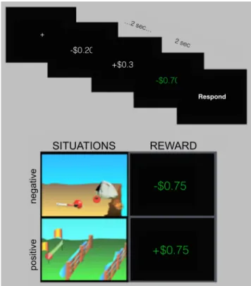
In Experiment 2, subjects completed a modified and abbreviated ver-sion of this task (four runs). On 50% of trials, subjects viewed nonsocial situation stimuli from Experiment 1 (96 total trials); on the remaining trials, subjects were presented with positive and negative events in which they either gained or lost money from a postscan bonus (reward condi-tion;Fig. 2). On each reward trial, subjects viewed a cycle of 20 rapidly presented random monetary values (2 s total), followed by the reward outcome for the trial, shown in green (2 s). Negative values ranged from ⫺$0.20 to ⫺$1.00, and positive values ranged from ⫹$0.20 to ⫹$2.00; this asymmetry allowed subjects to have net gain for their bonus and accounted for the fact that losses are experienced more strongly than comparable gains (Tversky and Kahneman, 1991). The experimental de-sign and behavioral task were identical to Experiment 1, except that subjects were asked to rate the character’s emotional intensity on the situation trials and their own emotional intensity on the reward trials.
Theory of mind localizer. Subjects were presented with short textual
scenarios that required inferences about mental state representations (Belief condition) or physical representations such as a map, photo, or painting (Photo condition;Dodell-Feder et al., 2011; stimuli are available at http://saxelab.mit.edu/superloc.php). These two types of scenarios were similar in their meta-representational demands and logical com-plexity, but only the scenarios in the Belief condition required building a representation of another person’s thoughts and beliefs. Scenarios were displayed for 10 s, followed immediately by a true or false question (4 s) about either the representation (Belief or Photo) or the reality of the situation. Each run (4.53 min) consisted of 10 trials separated by 12 s interstimulus intervals, and 12 s blocks of fixation were included at the beginning and end of each run. One to two runs were presented to each participant. The order of stimulus type (Belief or Photo) and correct answer (True or False) were counterbalanced within and across runs.
Figure 1. Task structure for Emotion Attribution task. Events consisted of a 4 s clip and a 2 s response. Stimuli included two stimulus types (situation stimuli and facial expression stimuli) and two valence categories (positive and negative valence).
Face perception localizer. Subjects viewed two conditions designed to
identify face-selective regions: dynamic faces (video clips of human chil-dren’s faces) and dynamic objects (video clips of objects in motion; from
Pitcher et al., 2011). For each of these conditions, there were a total of 30 clips (3 s each, separated by 333 ms of blank screen), and six clips were presented in each block. This localizer also included two other condi-tions, biological motion and structure from motion, which were not of interest for the present analyses. All conditions were presented as 20 s blocks followed by 2 s of rest, and 12 s blocks of fixation were included at the beginning and end of each run, as well as once in the middle of the run. Each condition was presented twice per run, and subjects received two runs lasting 5 min each, with condition order counterbalanced within and across runs and across participants. To maintain attention, subjects were required to complete a one-back task during viewing. Two of 21 subjects did not complete this localizer because of insufficient scan time.
Behavioral tasks. The Autism-Spectrum Quotient (Baron-Cohen et al., 2001) and the Interpersonal Reactivity Index (Davis, 1983) were com-pleted via on-line Qualtrics surveys. Participants also comcom-pleted an Em-pathic Accuracy task based on the study byZaki et al. (2008)and the verbal reasoning, matrices, and riddles components of the KBIT2 ( Kauf-man, 1990).
Acquisition
Data were acquired on a 3T Siemens Tim Trio scanner in the Athinoula A. Martinos Imaging Center at the McGovern Institute for Brain Re-search at MIT, using a Siemens 32-channel phased array head coil. We collected a high-resolution (1 mm isotropic) T1-weighted MPRAGE an-atomical scan, followed by functional images acquired with a gradient-echo EPI sequence sensitive to BOLD contrast [repetition time (TR), 2 s; echo time, 30 ms; flip angle, 90°; voxel size, 3⫻ 3 ⫻ 3 mm; matrix 64 ⫻ 64; 32 axial slices]. Slices were aligned with the anterior/posterior com-missure and provided whole-brain coverage (excluding the cerebellum).
Analysis
Pilot data. In addition to the 21 subjects reported, 8 independent pilot
subjects were analyzed to fix the parameters of the analyses reported
below (e.g., size of smoothing kernel, type of classifier, method for fea-ture selection). A general concern with fMRI analyses, and with the ap-plication of machine learning techniques to fMRI data in particular, is that the space of possible and reasonable analyses is large and can yield qualitatively different results. Analysis decisions should be made inde-pendent of the comparisons or tests of interest; otherwise, one risks overfitting the analysis to the data (Simmons et al., 2011). One way to optimize an analysis stream without such overfitting is to separate sub-jects into an exploratory or pilot set and a validation or test set. Thus, the analysis stream reported here was selected based on the parameters that appeared to yield the most sensitive analysis of eight pilot subjects.
Preprocessing. MRI data were preprocessed using SPM8 (http://www. fil.ion.ucl.ac.uk/spm/software/spm8/), FreeSurfer (http://surfer.nmr. mgh.harvard.edu/), and in-house code. FreeSurfer’s skull-stripping soft-ware was used for brain extraction. SPM was used to motion correct each subject’s data via rigid rotation and translation about the six orthogonal axes of motion, to register the functional data to the subject’s high-resolution anatomical image, and to normalize the data onto a common brain space (Montreal Neurological Institute). In addition to the smoothing imposed by normalization, functional images were smoothed using a Gaussian filter (FWHM, 5 mm).
Defining regions of interest. To define individual ROIs, we used
hypoth-esis spaces derived from random-effects analyses of previous studies [theory of mind (Dufour et al., 2013): bilateral TPJ, rATL, PC, subregions of MPFC (DMPFC, MMPFC, VMPFC); face perception (Julian et al., 2012): rmSTS, rFFA, rOFA], combined with individual subject activa-tions for the localizer tasks. The theory of mind task was modeled as a 14 s boxcar (the full length of the story and question period, shifted by 1 TR to account for lag in reading, comprehension, and processing of compre-hended text) convolved with a standard hemodynamic response func-tion (HRF). A general linear model was implemented in SPM8 to estimate values for Belief trials and Photo trials. We conducted high-pass filtering at 128 Hz, normalized the global mean signal, and included nuisance covariates to remove effects of run. The face perception task was modeled as a 22 s boxcar, and values were similarly estimated for each of condition (dynamic faces, dynamic objects, biological motion, struc-ture from motion). For each subject, we used a one-sample t test imple-mented in SPM8 to generate a map of t values for the relevant contrast (Belief⬎ Photo for the theory of mind ROIs, faces ⬎ objects for the face perception ROIs), and for each ROI, we identified the peak t value within the hypothesis space. An individual subject’s ROI was defined as the cluster of contiguous suprathreshold voxels (minimum k⫽ 10) within a 9 mm sphere surrounding this peak. If no cluster was found at p⬍ 0.001, we repeated this procedure at p⬍ 0.01 and p ⬍ 0.05. We masked each ROI by its hypothesis space (defined to be mutually exclusive) such that there was no overlap in the voxels contained in each functionally defined ROI. An ROI for a given subject was required to have at least 20 voxels to be included in multivariate analyses. For the pSTC region (Peelen et al., 2010), we generated a group ROI defined as a 9 mm sphere around the peak coordinate from that study, as well as an analogous ROI for the right hemisphere.
Multivariate analyses. Multivoxel pattern analysis (MVPA) was
con-ducted using an in-house code developed in Python using the publicly available PyMVPA toolbox (http://www.pymvpa.org/;Fig. 3). We con-ducted MVPA within ROIs that were functionally defined based on individual subject localizer scans. High-pass filtering (128 Hz) was conducted on each run, and linear detrending was performed across the whole time course. A time point was excluded if it was a global intensity outlier (⬎3 SD above the mean intensity) or corresponded to a large movement (⬎2 mm scan to scan). The data were temporally compressed to generate one voxel-wise summary for each individual trial, and these single trial summaries were used for both training and testing. Individual trial patterns were calculated by averaging the preprocessed bold images for the 6 s duration of the trial, offset by 4 s to account for HRF lag. Rest time points were removed, and the trial summaries were concatenated into one experimental vector in which each value was a trial’s average response. The pattern for each trial was then z-scored relative to the mean across all trial responses in that voxel.
Figure 2. Task structure for Experiment 2. Events consisted of a 4 s trial and 2 s response. Stimuli included two stimulus types (situation stimuli and reward stimuli) and two valence categories (positive and negative valence). Reward trials involved 2 s of rapid cycling through random values, followed by 2 s during which the reward outcome was displayed.
Given the high dimensionality of fMRI data and the relatively small number of training examples available, feature selection is often useful to extract voxels likely to be informative for classification (Mitchell et al., 2004;De Martino et al., 2008;Pereira et al., 2009). Within each ROI, we conducted voxel-wise ANOVAs to identify voxels that were modulated by the task (based on the F statistic for task vs rest contrast). This univar-iate selection procedure tends to eliminate high-variance, noisy voxels (Mitchell et al., 2004). Because this selection procedure is orthogonal to all of the classifications reported here, it could be performed once over
the whole dataset without constituting peeking, meaning that the same voxels could be used as features in each cross-validation fold. The top 80 most active voxels within the ROI were used for classification (selecting a fixed number of voxels also helps to minimize differences in the number of voxels across regions and subjects).
The data were classified using a support vector machine implemented with libSVM (http://www.csie.ntu.edu.tw/~cjlin/libsvm/; Chang and Lin, 2011). This classifier uses condition-labeled training data to learn a weight for each voxel, and subsequent stimuli (validation data not used
Figure 3. MVPA analysis procedure. Top, Valence-labeled voxel patterns (from a single ROI) used to train a linear support vector machine (SVM). Middle, Learned voxel weights used to predict valence of unlabeled test data (voxel patterns not used for training). Bottom, Cross-validation schemes for testing for stimulus-specific and stimulus-independent emotion representations. Skerry and Saxe• A Common Neural Code for Attributed Emotion J. Neurosci., November 26, 2014•34(48):15997–16008 • 16001
for model training) can then be assigned to one of two classes based on a weighted linear com-bination of the response in each voxel. In a support vector machine, the linear decision function can be thought of as a hyperplane di-viding the multidimensional voxel space into two classes, and voxel weights are learned so as to maximize the distance between the hy-perplane and the closest observed example. We conducted binary classification with a linear kernel using a fixed regularization pa-rameter (C⫽ 1) to control the tradeoff be-tween margin size and training error. We restricted ourselves to linearly decodable sig-nal under the assumption that a linear kernel implements a plausible readout mechanism for downstream neurons (Seung and Som-polinsky, 1993;Hung et al., 2005;Shamir and Sompolinsky, 2006). Given that the brain likely implements nonlinear transforma-tions, linear separability within a population can be thought of as a conservative but rea-sonable estimate of the information available for explicit readout (DiCarlo and Cox, 2007).
For each classification, the data were parti-tioned into multiple cross-validation folds where the classifier was trained iteratively on all folds but one and tested on the remaining fold. Classification accuracy was then averaged across folds to yield a single classification accu-racy for each subject in the ROI. A one-sample
t test was then performed over these individual
accuracies, comparing with chance
classifica-tion of 0.50 (all t tests on classificaclassifica-tion accuracies were one-tailed). Whereas parametric tests are not always appropriate for assessing the significance of classification accuracies (Stelzer et al., 2013), the assump-tions of these tests are met in the present case: the accuracy values are independent samples from separate subjects (rather than individual folds trained on overlapping data), and the classification accuracies were found to be normally distributed around the mean accuracy. For within-stimulus analyses (classifying within facial expressions and within situation stimuli), cross-validation was performed across runs (i.e., iteratively train on seven runs, test on the remaining eighth). For cross-stimulus analyses, the folds for cross-validation were based on stimulus type. To ensure complete independence between training and test data, folds for the cross-stimulus analysis were also divided based on even versus odd runs (e.g., train on even run facial expres-sions, test on odd run situations).
Whole-brain searchlight classification. The searchlight procedure was
identical to the ROI-based procedure except that the classifier was ap-plied to voxels within searchlight spheres rather than individually local-ized ROIs. For each voxel in a gray matter mask, we defined a sphere containing all voxels within a three-voxel radius of the center voxel. The searchlight size (123 voxels) was selected to approximately match the size of the regions in which effects were found with the ROI analysis, and we again conducted an ANOVA to select the 80 most active voxels in the sphere. Classification was then performed on each cross-validation fold, and the average classification accuracy for each sphere was assigned to its central voxel, yielding a single accuracy image for each subject for a given discrimination. We then conducted a one-sample t test over subjects’ accuracy maps, comparing accuracy in each voxel to chance (0.5). This yielded a group t-map, which was assessed at a p⬍ 0.05, FWE corrected (based on SPM’s implementation of Gaussian random fields).
Whole-brain random-effects analysis (univariate). We also conducted a
whole-brain random effects analysis to identify voxels in which the uni-variate response differentiated positive and negative valence for faces and for situations. The conjunction of these two contrasts would identify
voxels in which the magnitude of response was related to the valence for both stimulus types.
Results
Experiment 1
Regions of interest
Using the contrast of Belief
⬎ Photo, we identified seven ROIs
(rTPJ, lTPJ, rATL, PC, DMPFC, MMPFC, VMPFC) in each of the
21 subjects, and using the contrast of faces
⬎ objects, we
identi-fied right lateralized face regions OFA, FFA, and mSTS in 18
subjects (of 19 subjects who completed this localizer).
Multivariate results
Multimodal regions (pSTC and MMPFC). For classification of
emotional valence for facial expressions, we replicated the results
of
Peelen et al. (2010)
with above-chance classification in
MMPFC [M(SEM)
⫽ 0.534(0.013), t(18)
⫽ 2.65, p ⫽ 0.008;
Fig.
4
] and lpSTC [M(SEM)
⫽ 0.525(0.010), t(20)
⫽ 2.61, p ⫽ 0.008;
Fig. 5
]. Classification in right posterior superior temporal cortex
(rpSTC) did not reach significance at a corrected (0.05/3)
thresh-old [M(SEM)
⫽ 0.516(0.007), t(20)
⫽ 2.23, p ⫽ 0.019]. Note that
although the magnitude of these effects is small, these results
re-flect classification of single-event trials, which are strongly
influ-enced by measurement noise. Small but significant classification
accuracies are common for single-trial, within-category
distinc-tions (
Anzellotti et al., 2013
;
Harry et al., 2013
).
The key question for the present research is whether these
regions contain neural codes specific to overt expressions or
whether they also represent the valence of inferred emotional
states. When classifying valence for situation stimuli, we again found
above-chance classification accuracy in MMPFC [M(SEM)
⫽
0.553(0.012), t
(18)⫽ 4.31, p ⬎ 0.001]. We then tested for
stimulus-independent representations by training on one kind
Figure 4. DMPFC/MMPFC: Experiment 1. Classification accuracy for facial expressions (green), for situation stimuli (blue), and when training and testing across stimulus types (red). Cross-stimulus accuracies are the average of accuracies for train facial expression/test situation and train situation/test facial expression. Chance equals 0.50.
of stimulus and testing on the other. Consistent with the
exis-tence of an abstract valence code, MMPFC supported
above-chance valence classification across both stimulus types
[M(SEM)
⫽ 0.524(0.007), t(18)
⫽ 3.77, p ⫽ 0.001]. In contrast,
lpSTC did not perform above chance when classifying the valence of
situation stimuli [M(SEM)
⫽ 0.512(0.011), t(20)
⫽ 1.06, p ⫽ 0.152],
nor when requiring generalization across stimulus type [M(SEM)⫽
0.500(0.008), t
(20)⫽ 0.04, p ⫽ 0.486]. To directly compare accuracy
in lpSTC when classifying within facial expression stimuli and when
generalizing across stimulus types, we conducted a paired sample t
test (one-tailed) comparing classification accuracy for faces to
ac-curacy for cross-stimulus classification: classification acac-curacy
was significantly higher for faces compared with cross-stimulus
classification (M
⫽ 0.525, M ⫽ 0.500, t(20)
⫽ 2.00, p ⫽ 0.029).
Theory of mind regions. We performed these same analyses in
six remaining theory of mind regions (at a corrected
␣ ⫽
0.05/6, 0.008). In DMPFC (
Fig. 4
), we observed results very
comparable with those observed in MMPFC: above-chance
classification of facial emotion [M(SEM)
⫽ 0.539(0.016),
t
(18)⫽ 2.39, p ⫽ 0.014], of emotion from situations
[M(SEM)
⫽ 0.570(0.013), t(18)
⫽ 5.38, p ⬍ 0.001], and when
generalizing across stimulus types [M(SEM)
⫽ 0.532(0.008),
t
(18)⫽ 3.95, p ⬍ 0.001]. VMPFC did not perform above chance at
a corrected threshold (p
⬍ 0.008) when classifying facial expressions
[M(SEM)
⫽ 0.525(0.009), t(17)
⫽ 2.62, p ⫽ 0.009] or situation
stim-uli [M(SEM)
⫽ 0.524(0.012), t(17)
⫽ 1.98, p ⫽ 0.032]; however,
cross-stimulus decoding was above chance [M(SEM)
⫽
0.527(0.007), t
(17)⫽ 3.79, p ⫽ 0.001].
None of the other theory of mind regions classified above
threshold when distinguishing positive and negative facial
ex-pressions [rTPJ: M(SEM)
⫽ 0.501(0.010), t(20)
⫽ 0.06, p ⫽ 0.478;
lTPJ: M(SEM)
⫽ 0.521(0.012), t(20)
⫽ 1.85, p ⫽ 0.040; rATL:
M(SEM)
⫽ 0.525(0.012), t(20)
⫽ 2.05, p ⫽ 0.027; PC: M(SEM) ⫽
0.514(0.011), t
(20)⫽ 1.32, p ⫽ 0.102], when distinguishing
posi-tive and negaposi-tive situations [rTPJ: M(SEM)
⫽ 0.528(0.014),
t
(20)⫽ 2.04, p ⫽ 0.027; lTPJ: M(SEM) ⫽ 0.515(0.009), t(20)
⫽
1.57, p
⫽ 0.066; rATL: M(SEM) ⫽ 0.510(0.012), t(20)
⫽ 0.80, p ⫽
0.216; PC: M(SEM)
⫽ 0.523(0.012), t(20)
⫽
1.84, p
⫽ 0.040], or when generalizing
across stimulus types [rTPJ: M(SEM)
⫽
0.503(0.007), t
(20)⫽ 0.45, p ⫽ 0.330; lTPJ:
M(SEM)
⫽ 0.509(0.007), t(20)
⫽ 1.38, p ⫽
0.092; rATL: M(SEM)
⫽ 0.510(0.006),
t
(20)⫽ 1.85, p ⫽ 0.039; PC: M(SEM) ⫽
0.495(0.008), t
(20)⫽⫺0.60, p ⫽ 0.724].
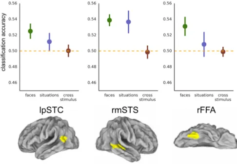
Face-selective cortex. For valence in
fa-cial expressions, we also performed a
sec-ondary analysis in face-selective regions
rOFA, rFFA, and rmSTS (at a corrected
threshold of 0.05/3;
Fig. 5
). We replicated
previous reports (
Said et al., 2010a
,
b
;
Furl et
al., 2012
;
Harry et al., 2013
) with
classifica-tion accuracies significantly above chance in
rmSTS [M(SEM)
⫽ 0.539(0.007), t(14)
⫽
5.20, p
⬍ 0.001] and in rFFA [M(SEM) ⫽
0.531(0.012), t
(14)⫽ 2.59, p ⫽ 0.011];
clas-sification in rOFA did not survive
correc-tion
for
multiple
comparisons
[M(SEM)
⫽ 0.529(0.016), t(13)
⫽ 1.87,
p
⫽ 0.042]. For the situation stimuli, the
rFFA failed to classify valence when it was
inferred from context [rFFA: M(SEM)
⫽
0.508(0.016), t
(14)⫽ 0.54, p ⫽ 0.300]. In
the rmSTS, on the other hand, there was reliable information
about situation stimuli in addition to the face stimuli [M(SEM)
⫽
0.537(0.014), t
(14)⫽ 2.57, p ⫽ 0.011]. However, neither region
sup-ported
above-chance
cross-stimulus
classification
[rFFA:
M(SEM)
⫽ 0.499(0.006), t(14)
⫽ ⫺0.16, p ⫽ 0.563; rmSTS:
M(SEM)
⫽ 0.499(0.008), t(14)
⫽⫺0.17, p ⫽ 0.565], and
classifica-tion accuracy was reliably higher (one-tailed test) when training and
testing on faces compared with when requiring generalization across
stimulus types in rmSTS (M
⫽ 0.539, M ⫽ 0.499, t(14)
⫽ 4.52, p ⬍
0.001) and in rFFA (M
⫽ 0.531, M ⫽ 0.499, t(14)
⫽ 2.26, p ⫽ 0.020).
Follow-up analyses
Given successful valence decoding in dorsal and middle MPFC,
we conducted several follow-up analyses to examine the scope
and generality of these effects. For facial expressions, we
per-formed cross-validation across the orthogonal dimension of face
gender. Both regions of MPFC performed above chance
[DMPFC: M(SEM)
⫽ 0.529(0.015), t(18)
⫽ 1.92, p ⫽ 0.035;
MMPFC: M(SEM)
⫽ 0.532(0.010), t(18)
⫽ 3.20, p ⫽ 0.003],
in-dicating that the valence-specific voxel patterns generalize across
two face sets that differed at the level of exemplars, identity, and
gender. We also tested for generalization across face sets in the
remaining regions that supported decoding of facial expressions
(rmSTS, rFFA, lpSTC). The neural patterns generalized across
the male and female face sets in rmSTS [M(SEM)
⫽ 0.524(0.012),
t
(14)⫽ 2.02, p ⫽ 0.032] but not in rFFA [M(SEM) ⫽
0.512(0.012), t
(14)⫽ 1.00, p ⫽ 0.167] or lpSTC [M(SEM) ⫽
0.509(0.009), t
(20)⫽ 1.05, p ⫽ 0.154].
For situation stimuli, both regions of MPFC were able to
clas-sify valence across the orthogonal dimension: social versus
non-social situations [DMPFC: M(SEM)
⫽ 0.552(0.012), t(18)
⫽ 4.44,
p
⬍ 0.001; MMPFC: M(SEM) ⫽ 0.543(0.011), t(18)
⫽ 3.97, p ⬍
0.001]. Finally, to test for possible asymmetry in the
cross-stimulus classification, we separated the cross-cross-stimulus analysis
into training on faces/testing on situations and training on
situ-ations/testing on faces. We observed above-chance classification
for both train/test partitions in both DMPFC [testing on faces:
Figure 5. Classification accuracy for facial expressions (green), for situation stimuli (blue), and when training and testing across stimulus types (red). Cross-stimulus accuracies are the average of accuracies for train facial expression/test situation and train situation/test facial expression. Chance equals 0.50.
M(SEM)
⫽ 0.523(0.011), t(18)
⫽ 2.18, p ⫽ 0.021; testing on
situ-ations: M(SEM)
⫽ 0.540(0.007), t(18)
⫽ 5.47, p ⬍ 0.001] and
MMPFC [testing on faces: M(SEM)
⫽ 0.525(0.006), t(18)
⫽ 4.13,
p
⬍ 0.001; testing on situations: M(SEM) ⫽ 0.524(0.009), t(18)
⫽
2.64, p
⫽ 0.008].
In summary, it appears that dorsal and middle subregions of
MPFC contain reliable information about the emotional valence
of a stimulus when the emotion must be inferred from the
situa-tion and that the neural code in this region is highly abstract,
generalizing across diverse cues from which an emotion can be
identified. In contrast, although both rFFA and the region of
superior temporal cortex identified by
Peelen et al. (2010)
con-tain information about the valence of facial expressions, the
neu-ral codes in those regions do not appear geneneu-ralized to valence
representations formed on the basis of contextual information.
Interestingly, the rmSTS appears to contain information about
valence in faces and situations but does not form a common code
that integrates across stimulus type.
Whole-brain analyses
To test for any remaining regions that may contain information
about the emotional valence of these stimuli, we conducted a
searchlight procedure, revealing striking consistency with the
ROI analysis (
Table 1
;
Fig. 6
). Only DMPFC and MMPFC
exhib-ited above-chance classification for faces and contexts, and when
generalizing across these two stimulus types. In addition, for
clas-sification of facial expressions alone, we observed clusters in
oc-cipital cortex. Clusters in the other ROIs emerged at a more
liberal threshold (rOFA and rmSTS at p
⬍ 0.001 uncorrected;
rFFA, rpSTC, and lpSTC at p
⬍ 0.01). In contrast, whole-brain
analyses of the univariate response revealed no regions in which
the mean response distinguished between positive and negative
facial expressions or between positive and negative contexts (at
p
⬍ 0.05, FWE correction based on Gaussian random fields).
Experiment 2
The results of Experiment 1 suggest that DMPFC and MMPFC
contain abstract, stimulus-independent information about
emo-tional valence of perceived and inferred emotions. How is this
region related to the regions of MPFC typically implicated in
processing value and/or subjective experience? For Experiment 2,
we first used a group anatomical mask (
Bartra et al., 2013
;
Clithero and Rangel, 2013
) to identify a region of OFC/VMPFC
previously implicated in reward/value processing. Consistent
with previous reports (
Kable and Glimcher, 2007
;
Chib et al.,
2009
), this region showed an overall magnitude effect for
posi-tive
⬎ negative rewards (t(15)
⫽ 3.20, p ⫽ 0.006;
Fig. 7
) and could
classify positive versus negative reward trials reliably above
chance [M(SEM)
⫽ 0.542(0.020), t(15)
⫽ 2.09, p ⫽ 0.027].
Interestingly, this canonical reward region did not reliably
distinguish positive and negative situations for others
[M(SEM)
⫽ 0.521(0.018), t(15)
⫽ 1.15, p ⫽ 0.135], and there was
no evidence for a common valence code generalizing across self
and other [M(SEM)
⫽ 0.512(0.014), t(15)
⫽ 0.80, p ⫽ 0.219].
Classification accuracies were significantly higher when
discrim-inating self-reward values compared with when generalizing
across reward and situation trials (M
⫽ 0.542, M ⫽ 0.512, t(15)
⫽
1.90, p
⫽ 0.038, one-tailed).
What about the regions implicated in abstract valence
rep-resentation in Experiment 1? By decoding valence within the
situation stimuli, we replicate the finding of Experiment 1 that
DMPFC and MMPFC contain information about the emotion
at-tributed to a target even when that emotion must be inferred from
context [DMPFC: M(SEM)
⫽ 0.543(0.021), t(15)
⫽ 2.04, p ⫽ 0.030;
MMPFC: M(SEM)
⫽ 0.536(0.019), t(15)
⫽ 1.95, p ⫽ 0.035;
Fig. 8
]. Do we observe these same neural patterns on trials in
which subjects evaluate their own subjectively experienced
emo-tions? In MMPFC, we observed above-chance valence
classifica-tion for reward trials [M(SEM)
⫽ 0.539(0.018), t(15)
⫽ 2.17, p ⫽
0.023] in addition to situation trials. Moreover, neural patterns
generalized across positive/negative situations and
positive/neg-ative outcomes for the self [M(SEM)
⫽ 0.526(0.010), t(15)
⫽ 2.60,
p
⫽ 0.010]. In dorsal MPFC, in contrast, we observed similar
classifica-tion of the valence of reward outcomes [M(SEM)
⫽ 0.544(0.025), t(15)
⫽ 1.74, p ⫽ 0.051], but this region failed to classify above chance when
generalizingacrossselfandother[M(SEM)
⫽0.514(0.013),t(15)⫽1.07,
p
⫽ 0.150].
Discussion
Are there neural representations of emotions that generalize
across diverse sources of evidence, including overt emotional
expressions and emotions inferred from context alone? In the
present study, we identified regions in which voxel-wise response
patterns contained information about the emotional valence of
facial expressions and a smaller number of regions that
distin-guished the valence of emotion-eliciting situations. Our results,
together with existing literature (
Peelen et al., 2010
), provide
candidate neural substrates for three levels of representation:
modality-specific representations bound to perceptual invariants
in the input, intermediate multimodal representations that
gen-eralize across canonical perceptual schemas, and conceptual
rep-resentations that are fully invariant to the information used to
identify emotions.
Conceptual representations
In DMPFC/MMPFC, we decoded emotional valence from facial
expressions and from animations depicting emotion-eliciting
sit-uations. Like other domains of high-level cognition, emotion
knowledge is theory like (
Carey, 1985
;
Gopnik and Wellman,
1992
), requiring abstract concepts (e.g., of goals, expectations) to
be integrated in a coherent, causal manner. The present results
Table 1. Whole brain, Experiment 1: Searchlight results ( p < 0.05, FWE corrected)
Stimulus Number of voxels Peak t x y z Region Situations 52 11.80 4 46 38 DMPFC 8.24 6 50 28 9 9.49 ⫺8 54 26 DMPFC 28 9.21 4 58 14 MMPFC 9.02 4 56 22 1 7.98 16 60 24 MMPFC 1 7.86 0 50 36 DMPFC 4 7.82 0 54 30 DMPFC 1 7.55 ⫺8 54 18 MMPFC 2 7.43 8 56 20 MMPFC 1 7.40 ⫺2 54 36 DMPFC 1 7.30 ⫺28 ⫺78 32 L OCC/TEMP Faces 8 8.77 ⫺30 ⫺88 ⫺4 L MID OCC GYRUS
2 8.48 38 ⫺92 8 R MID OCC GYRUS
3 8.16 2 52 20 MMPFC 1 7.88 6 52 22 MMPFC 2 7.60 8 56 20 MMPFC 1 7.52 28 ⫺82 32 R SUP OCC Cross-stimulus 42 10.91 ⫺2 50 34 DMPFC 9.28 0 48 24 7.28 8 56 20 1 8.93 8 56 10 MMPFC 1 7.34 12 66 10 MMPFC
L OCC/TEMP, Left occipital/temporal; L MID OCC GYRUS, left middle occipital gyrus, R MID OCC GYRUS, right middle occipital gyrus; R SUP OCC, right superior occipital.
suggest that valence representations in DMPFC/MMPFC are
elicited by such inferential processes. We could classify valence
when training on faces and testing on situations (and vice versa),
replicating the finding that emotion representations in MMPFC
generalize across perceptually dissimilar stimuli (
Peelen et al.,
2010
). Moreover, our results demonstrate an even stronger form
of generalization: perceived emotions and emotions inferred
through generative, theory-like processes activate similar
neu-ral patterns in DMPFC/MMPFC, indicating a mechanism
be-yond mere association of co-occurring perceptual schemas.
Thus, the MPFC may contain a common neural code that
inte-grates diverse perceptual and inferential processes to form
ab-stract representations of emotions.
Previous research leaves open the question of whether activity
in MPFC reflects mechanisms specific to emotion attribution or
mechanisms involved in value or valence
processing more generally. In Experiment
2, we found evidence for both kinds of
representations. First, we found that the
region of OFC/VMPFC implicated in
re-ward processing (
Clithero and Rangel,
2013
; anatomical ROI from
Bartra et al.,
2013
) does not contain information about
the valence of attributed emotions.
Sec-ond, we found no evidence for a shared
representation of experienced and
attrib-uted emotion in dorsal MPFC. Finally, in
MMPFC, we observed neural patterns
that generalized across attributed and
ex-perienced emotional events. One
inter-pretation of this result is that attributing
positive or rewarding experiences to
oth-ers depends on general purpose reward
representations that code value in social
and nonsocial contexts (
Chib et al., 2009
;
Lin et al., 2012
,
Ruff and Fehr, 2014
).
Al-ternatively, neural responses in MMPFC
could reflect the participant’s own
em-pathic
reaction
to
the
depicted
experiences (e.g., witnessing someone
achieve a goal elicits positive emotions in
participants). If so, the participant’s
em-pathic reaction might be causally involved
in the process of attributing emotions to
others (consistent with “simulation
the-ory”;
Goldman and Sripada, 2005
;
Nie-denthal, 2007
) or might be a downstream
consequence of attribution. Previous
re-sults do indicate a causal role for MPFC in
emotion perception and attribution:
damage to MPFC is associated with
defi-cits in emotion recognition (
Shamay-Tsoory et al., 2003
,
2009
), and direct
disruption of MPFC via transcranial
mag-netic stimulation has been shown to
im-pair recognition of facial expressions
(
Harmer et al., 2001
; see also
Mattavelli et
al., 2011
). Moreover, the degree to which
MPFC is recruited during an emotion
at-tribution task predicts individual
differ-ences in the accuracy of emotion
judgments (
Zaki et al., 2009a
,
b
). Future
research should continue to distinguish
the specific contents of attributed emotions from the emotional
response of the participant. For example, can patterns in MPFC
be used to classify the attribution of more specific emotions that
are unlikely to be shared by the observer (e.g., loneliness vs
re-gret)?
Modality-specific representations
In face-selective regions (rFFA and rmSTS), we found that
neural patterns could distinguish positive and negative facial
expressions, replicating previous reports of emotion-specific
neural representations in these regions (
Fox et al., 2009
;
Said
et al., 2010a
,
b
;
Xu and Biederman, 2010
;
Furl et al., 2012
;
Harry et al., 2013
). Neural populations could distinguish facial
expressions by responding to relatively low-level parameters
that differ across expressions, by extracting mid-level
invari-Figure 6. Whole brain: Experiment 1. Classification in whole-brain searchlight (sphere radius, 3 voxels). p⬍ 0.05 (FWE corrected using Gaussian random fields).
Figure 7. OFC/VMPFC. Results from anatomical OFC/VMPFC reward ROI (Bartra et al., 2013;Clithero and Rangel, 2013). Left, Classification accuracy for reward outcomes (purple), for situation stimuli (blue), and when training and testing across stimulus types (red). Chance equals 0.50. Right, Mean values in the ROI for each stimulus condition, asterisk indicates significant differ-ence ( p⬍ 0.05).
ants (e.g., eye motion, mouth
configu-ration) that generalize across
within-modality
transformations
(e.g.,
lighting, position), or by computing
ex-plicit representations of facial emotion
that integrate multiple facial
parame-ters. The present study used naturalistic
stimuli that varied in lighting
condi-tions, face direction, and face position
and found reliable generalization across
male and female face sets in rmSTS.
Thus, it is possible that these neural
pat-terns distinguish facial expressions
based on representations invariant to
certain low-level transformations (
An-zellotti et al., 2013
). Future research
should investigate this possibility by
systematically testing the generalization
properties of neural responses to
emo-tional expressions across variation in
low-level dimensions (e.g., face
direc-tion) and higher-level dimensions (e.g.,
generalization from sad eyes to a sad
mouth). Interestingly, the rmSTS also
contained information about emotional
valence in situation stimuli, but the
neural patterns did not generalize across these distinct sources
of evidence, suggesting two independent valence codes in this
region.
Multimodal representations
We also replicate the finding that pSTC contains information
about the emotional valence of facial expressions (
Peelen et al.,
2010
). However, unlike DMPFC/MMPFC, we find no evidence
for representations of emotions inferred from situations.
Inter-estingly,
Peelen et al. (2010)
found that the pSTC could decode
emotional expressions across modalities (faces, bodies, voices),
suggesting that this region may support an intermediate
repre-sentation that is neither fully conceptual nor tied to specific
per-ceptual parameters. For example, pSTC could be involved in
pooling over associated perceptual schemas, leading to
represen-tations that generalize across diverse sensory inputs but do not
extend to more abstract, inference-based representations. This
interpretation would be consistent with the region’s proposed
role in cross-modal integration (
Kreifelts et al., 2009
;
Stevenson
and James, 2009
). Thus, the present findings reveal a novel
func-tional division within the set of regions (pSTC and MMPFC)
previously implicated in multimodal emotion representation
(
Peelen et al., 2010
).
Open questions
While these data provide important constraints on the levels
of representation associated with different regions, important
questions remain open. First, do the regions identified here
contain information about more fine-grained emotional
dis-tinctions beyond valence? Previous studies have successfully
decoded a larger space of perceived emotions in MMPFC, STS,
and FFA (
Peelen et al., 2010
;
Said et al., 2010a
,
b
;
Harry et al.,
2013
). For emotions inferred from context, the neural
repre-sentation of more fine-grained emotional distinctions (e.g.,
inferring sadness vs fear) will be a key question for future
research.
This study also leaves open the role of other regions (e.g.,
amygdala, insula, inferior frontal gyrus) that have previously
been associated with emotion perception and experience
(
Shamay-Tsoory et al., 2009
;
Singer et al., 2009
;
Pessoa and
Adolphs, 2010
). What is the precise content of emotion
rep-resentations in these regions, and do they contribute to
iden-tifying specific emotional states in others? With the searchlight
procedure, we found little evidence for representations of
emotional valence outside the a priori ROIs. However,
whole-brain analyses are less sensitive than ROI analyses, and
al-though multivariate analyses alleviate some of the spatial
constraints of univariate methods, they still tend to rely on
relatively low-frequency information (
Op de Beeck, 2010
;
Freeman et al., 2011
), meaning that MVPA provides a lower
bound on the information available in a given region (
Krieges-korte and Kievit, 2013
). Neurophysiological studies (
Gothard
et al., 2007
;
Hadj-Bouziane et al., 2012
) may help to elucidate
the full set of regions contributing to emotion attribution.
Relatedly, how does information in these different regions
interact during the process of attribution? A tempting
specula-tion is that the regions described here make up a hierarchy of
information flow (
Adolphs, 2002
;
Ethofer et al., 2006
; e.g.,
modality-specific, face-selective cortex N multimodal pSTC N
conceptual MPFC). However, additional connectivity or causal
information (
Friston et al., 2003
;
Bestmann et al., 2008
) would be
required to confirm such an account and to directly map different
representational content onto discrete stages.
Finally, these findings are complementary to previous
inves-tigations of semantic representations [e.g., object categories (
De-vereux et al., 2013
;
Fairhall and Caramazza, 2013
)], which have
identified modality-specific representations (e.g., in visual
cor-tex) and representations that generalize across modalities (e.g.,
across words and pictures in left middle temporal gyrus). The
present findings highlight a distinction between representations
that are multimodal and those that are based on theory-like
causal inferences. Does this distinction apply to other domains,
Figure 8. MPFC: Experiment 2. Classification accuracy for reward outcomes (purple), for situation stimuli (blue), and when training and testing across stimulus types (red). Cross-stimulus accuracies are the average of accuracies for train reward/test situation and train situation/test reward. Chance equals 0.50.
and can it help to clarify the neural organization of abstract
knowledge more broadly?
General conclusions
The challenge of emotion recognition demands neural processes
for exploiting different sources of evidence for others’ emotions,
as well as a common code for integrating this information to
support emotion-based inference. Here, we demonstrate
success-ful decoding of valence for emotional states that must be inferred
from context as well as emotions directly perceived from overt
expressions. By testing the scope and generality of the responses
in different regions, we provide important constraints on
possi-ble computational roles of these regions and begin to elucidate
the series of representations that make up the processing stream
for emotional perception, attribution, and empathy. Thus, the
present research provides a step toward understanding how the
brain transforms stimulus-bound inputs into abstract
represen-tations of emotions.
References
Adolphs R (2002) Neural systems for recognizing emotion. Curr Opin Neu-robiol 12:169 –177.CrossRef Medline
Amodio DM, Frith CD (2006) Meeting of minds: the medial frontal cortex and social cognition. Nat Rev Neurosci 7:268 –277.CrossRef Medline
Anzellotti S, Fairhall SL, Caramazza A (2013) Decoding representations of face identity that are tolerant to rotation. Cereb Cortex 24:1988 –1995.
CrossRef Medline
Baron-Cohen S, Wheelwright S, Skinner R, Martin J, Clubley E (2001) The Autism-Spectrum Quotient (AQ): evidence from Asperger syndrome/ high-functioning autism, males and females, scientists and mathemati-cians. J Autism Dev Disord 31:5–17.CrossRef Medline
Bartra O, McGuire JT, Kable JW (2013) The valuation system: a coordinate-based meta-analysis of BOLD fMRI experiments examining neural corre-lates of subjective value. Neuroimage 76:412– 427.CrossRef Medline
Bestmann S, Ruff CC, Blankenburg F, Weiskopf N, Driver J, Rothwell JC (2008) Mapping causal interregional influences with concurrent TMS– fMRI. Exp Brain Res 191:383– 402.CrossRef Medline
Bruneau EG, Pluta A, Saxe R (2012) Distinct roles of the “shared pain” and “theory of mind” networks in processing others’ emotional suffering. Neuropsychologia 50:219 –231.CrossRef Medline
Calder AJ, Beaver JD, Winston JS, Dolan RJ, Jenkins R, Eger E, Henson RN (2007) Separate coding of different gaze directions in the superior tem-poral sulcus and inferior parietal lobule. Curr Biol 17:20 –25.CrossRef Medline
Carey S (1985) Conceptual change in childhood. Cambridge, MA: MIT. Carlin JD, Calder AJ, Kriegeskorte N, Nili H, Rowe JB (2011) A head
view-invariant representation of gaze direction in anterior superior temporal sulcus. Curr Biol 21:1817–1821.CrossRef Medline
Chang C, Lin C (2011) LIBSVM: a library for support vector machines. ACM Trans Intellig Sys Tech 2:1–27.
Chib VS, Rangel A, Shimojo S, O’Doherty JP (2009) Evidence for a common representation of decision values for dissimilar goods in human ventro-medial prefrontal cortex. J Neurosci 29:12315–12320.CrossRef Medline
Chikazoe J, Lee DH, Kriegeskorte N, Anderson AK (2014) Population cod-ing of affect across stimuli, modalities and individuals. Nat Neurosci 17: 1114 –1122.CrossRef Medline
Clithero JA, Rangel A (2013) Informatic parcellation of the network in-volved in the computation of subjective value. Soc Cogn Affect Neurosci 9:1289 –1302.CrossRef Medline
Davis MH (1983) Measuring individual differences in empathy: evidence for a multidimensional approach. J Pers Soc Psychol 44:113–126.
CrossRef
De Martino F, Valente G, Staeren N, Ashburner J, Goebel R, Formisano E (2008) Combining multivariate voxel selection and support vector ma-chines for mapping and classification of fMRI spatial patterns. Neuroim-age 43:44 –58.CrossRef Medline
Devereux BJ, Clarke A, Marouchos A, Tyler LK (2013) Representational similarity analysis reveals commonalities and differences in the semantic processing of words and objects. J Neurosci 33:18906 –18916.CrossRef Medline
DiCarlo JJ, Cox DD (2007) Untangling invariant object recognition. Trends Cogn Sci 11:333–341.CrossRef Medline
DiCarlo JJ, Zoccolan D, Rust NC (2012) How does the brain solve visual object recognition? Neuron 73:415– 434.CrossRef Medline
Dodell-Feder D, Koster-Hale J, Bedny M, Saxe R (2011) fMRI item analysis in a theory of mind task. Neuroimage 55:705–712.CrossRef Medline
Dufour N, Redcay E, Young L, Mavros PL, Moran JM, Triantafyllou C, Ga-brieli JD, Saxe R (2013) Similar brain activation during false belief tasks in a large sample of adults with and without autism. PLoS One 8:e75468.
CrossRef Medline
Ekman P, Rosenberg EL (1997) What the face reveals: basic and applied studies of spontaneous expression using the Facial Action Coding System (FACS). Oxford: Oxford UP.
Ethofer T, Anders S, Erb M, Herbert C, Wiethoff S, Kissler J, Grodd W, Wildgruber D (2006) Cerebral pathways in processing of affective pros-ody: a dynamic causal modeling study. Neuroimage 30:580 –587.
CrossRef Medline
Etkin A, Egner T, Kalisch R (2011) Emotional processing in anterior cingu-late and medial prefrontal cortex. Trends Cogn Sci 15:85–93.CrossRef Medline
Fairhall SL, Caramazza A (2013) Brain regions that represent amodal con-ceptual knowledge. J Neurosci 33:10552–10558.CrossRef Medline
Fox CJ, Moon SY, Iaria G, Barton JJ (2009) The correlates of subjective perception of identity and expression in the face network: an fMRI adap-tation study. Neuroimage 44:569 –580.CrossRef Medline
Freeman J, Brouwer GJ, Heeger DJ, Merriam EP (2011) Orientation decod-ing depends on maps, not columns. J Neurosci 31:4792– 4804.CrossRef Medline
Friston KJ, Harrison L, Penny W (2003) Dynamic causal modelling. Neuro-image 19:1273–1302.CrossRef Medline
Furl N, Hadj-Bouziane F, Liu N, Averbeck BB, Ungerleider LG (2012) Dy-namic and static facial expressions decoded from motion-sensitive areas in the macaque monkey. J Neurosci 32:15952–15962.CrossRef Medline
Fusar-Poli P, Placentino A, Carletti F, Landi P, Allen P, Surguladze S, Bene-detti F, Abbamonte M, Gasparotti R, Barale F, Perez J, McGuire P, Politi P (2009) Functional atlas of emotional faces processing: a voxel-based meta-analysis of 105 functional magnetic resonance imaging studies. J Psychiatry Neurosci 34:418 – 432.Medline
Goldman AI, Sripada CS (2005) Simulationist models of face-based emo-tion recogniemo-tion. Cogniemo-tion 94:193–213.CrossRef Medline
Gopnik A, Wellman HM (1992) Why the child’s theory of mind really is a theory. Mind Lang 7:145–171.CrossRef
Gothard KM, Battaglia FP, Erickson CA, Spitler KM, Amaral DG (2007) Neural responses to facial expression and face identity in the monkey amygdala. J Neurophysiol 97:1671–1683.CrossRef Medline
Hadj-Bouziane F, Liu N, Bell AH, Gothard KM, Luh WM, Tootell RB, Murray EA, Ungerleider LG (2012) Amygdala lesions disrupt modulation of functional MRI activity evoked by facial expression in the monkey infe-rior temporal cortex. Proc Natl Acad Sci U S A 109:E3640 –E3648.
CrossRef Medline
Harmer CJ, Thilo KV, Rothwell JC, Goodwin GM (2001) Transcranial mag-netic stimulation of medial–frontal cortex impairs the processing of angry facial expressions. Nat Neurosci 4:17–18.CrossRef Medline
Harry B, Williams MA, Davis C, Kim J (2013) Emotional expressions evoke a differential response in the fusiform face area. Front Hum Neurosci 7:692.CrossRef Medline
Haxby JV, Gobbini MI, Furey ML, Ishai A, Schouten JL, Pietrini P (2001) Distributed and overlapping representations of faces and objects in ven-tral temporal cortex. Science 293:2425–2430.CrossRef Medline
Hung CP, Kreiman G, Poggio T, DiCarlo JJ (2005) Fast readout of object identity from macaque inferior temporal cortex. Science 310:863– 866.
CrossRef Medline
Hynes CA, Baird AA, Grafton ST (2006) Differential role of the orbital fron-tal lobe in emotional versus cognitive perspective-taking. Neuropsycho-logia 44:374 –383.CrossRef Medline
Julian JB, Fedorenko E, Webster J, Kanwisher N (2012) An algorithmic method for functionally defining regions of interest in the ventral visual pathway. Neuroimage 60:2357–2364.CrossRef Medline
Kable JW, Glimcher PW (2007) The neural correlates of subjective value during intertemporal choice. Nat Neurosci 10:1625–1633. CrossRef Medline