HAL Id: hal-03065548
https://hal.archives-ouvertes.fr/hal-03065548
Submitted on 22 Dec 2020HAL is a multi-disciplinary open access archive for the deposit and dissemination of sci-entific research documents, whether they are pub-lished or not. The documents may come from teaching and research institutions in France or abroad, or from public or private research centers.
L’archive ouverte pluridisciplinaire HAL, est destinée au dépôt et à la diffusion de documents scientifiques de niveau recherche, publiés ou non, émanant des établissements d’enseignement et de recherche français ou étrangers, des laboratoires publics ou privés.
N. Doll, S. Royek, S. Fujita, S. Okuda, S. Chamot, A. Stintzi, Thomas
Widiez, M. Hothorn, A. Schaller, N. Geldner, et al.
To cite this version:
N. Doll, S. Royek, S. Fujita, S. Okuda, S. Chamot, et al.. A two-way molecular dialogue between embryo and endosperm is required for seed development. Science, American Association for the Advancement of Science, 2020, 367 (6476), pp.431-435. �10.1126/science.aaz4131�. �hal-03065548�
1
Title: A two-way molecular dialogue between embryo and endosperm
required for seed development
Authors: N. M. Doll1, S. Royek2,†, S. Fujita3,†,‡, S. Okuda4,†, S. Chamot1, A. Stintzi2, T. Widiez1,
M. Hothorn4, A. Schaller2, N. Geldner3, G. Ingram1,*
5
Affiliations:
1Laboratoire Reproduction et Développement des Plantes, University of Lyon, ENS de Lyon,
UCB Lyon 1, CNRS, INRAE, F-69342, Lyon, France.
2Department of Plant Physiology and Biochemistry, University of Hohenheim, Stuttgart,
Germany.
10
3Department of Plant Molecular Biology, University of Lausanne, 1015 Lausanne, Switzerland. 4Structural Plant Biology Laboratory, Department of Botany and Plant Biology, University of
Geneva, 1211 Geneva, Switzerland.
*Correspondence to: [email protected].
†equal contribution
15
‡current address: National Institute of Genetics, 1111 Yata, Mishima, Shizuoka 411-8540, Japan.
Abstract (129 words): The plant embryonic cuticle is a hydrophobic barrier deposited de novo by
the embryo during seed development. At germination it protects the seedling from water loss and
is thus critical for survival. Embryonic cuticle formation is controlled by a signaling pathway
20
involving the ABNORMAL LEAF SHAPE1 subtilase, and the two GASSHO receptor-like
kinases. We show that a sulfated peptide, TWISTED SEED1 (TWS1), acts as a GASSHO ligand.
Cuticle surveillance depends on the action of the subtilase which, unlike the TWS1 precursor and
the GASSHO receptors, is not produced in the embryo but in the neighboring endosperm.
Subtilase-mediated processing of the embryo-derived TWS1 precursor releases the active peptide,
25
triggering GASSHO-dependent cuticle reinforcement in the embryo. A bidirectional molecular
2
One Sentence Summary: Compartmentalized proteolytic activation of a signal peptide provides
spatial cues to ensure an intact embryo cuticle.
Main Text (2047 words): In Angiosperms, seeds comprise three genetically distinct
compartments, the zygotic embryo and the endosperm, and the maternal seed coat. Their
development must be tightly coordinated for seed viability. Here we have elucidated a bidirectional
5
peptide-mediated signaling pathway between the embryo and the endosperm. This pathway
regulates the deposition of the embryonic cuticle which forms an essential hydrophobic barrier
separating the apoplasts of the embryo and endosperm. After germination, the cuticle - one of the
critical innovations underlying the transition of plants from their original, aqueous environment to
dry land - protects the seedling from catastrophic water loss (1, 2).
10
Formation of the embryonic cuticle was previously shown to depend on two Receptor-Like
Kinases (RLKs) GASSHO1/SCHENGEN3 (from here-on named GSO1) and GSO2, and on
ALE1, a protease of the subtilase family (SBTs) (2–5). gso1 gso2 and (to a lesser extent) ale1
mutants produce a patchy, and highly permeable cuticle (2). Mutant embryos also adhere to
surrounding tissues causing a seed-twisting phenotype (6). Since SBTs have been implicated in
15
the processing of peptide hormone precursors (7, 8, 9), we hypothesized that ALE1 may be
required for the biogenesis of the elusive inter-compartmental peptide signal required for
GSO1/2-dependent cuticle deposition.
CASPARIAN STRIP INTEGRITY FACTORs (CIFs), a family of small sulfated signaling
peptides, are ligands for GSO1 and GSO2 (10–12). CIF1 and CIF2 are involved in Casparian strip
20
formation in the root endodermis (10, 11). The function of CIF3 and CIF4 is still unknown. To
assess the role of CIF peptides in cuticle development, the quadruple mutant (cif1 cif2 cif3 cif4)
3
observed in this quadruple mutant (Fig. S1B-E). However, reduction (in the leaky sgn2-1 allele
(10)) or loss (in the tpst-1 mutant (13)) of Tyrosyl-Protein Sulfotransferase (TPST) activity, results
in seed-twisting and cuticle-permeability phenotypes resembling those observed in ale1 mutants
(Fig. 1A-D, Fig. S2 A-D). These data suggest that a sulfated peptide may act as the ligand of
GSO1/2 during seed development.
5
Consistent with the hypothesis that TPST acts in the same pathway as GSO1 and GSO2, no
difference was observed between the phenotype of tpst-1 gso1-1 gso2-1 triple and gso1-1 gso2-1
double mutants (Fig. S2E). In contrast, TPST and ALE1 appear to act synergistically, as a
phenotype resembling that of gso1 gso2 double mutants was observed in tpst-1 ale1-4 double
mutants (Fig. 1E-I) (Fig. S2F-J). This result supports the hypothesis that TPST and ALE1 act in
10
parallel regarding their roles in embryonic cuticle formation, possibly through independent
post-translational modifications contributing to the maturation of the hypothetical peptide signal.
Identification of the peptide signal was facilitated by a study of TWISTED SEED1 (TWS1) (14),
that reported a loss-of-function phenotype strikingly similar to that of gso1 gso2 double mutants.
Because existing alleles of TWS1 are in the WS background, we generated new CRISPR alleles
15
(tws1-3 to tws1-10) in the Col-0 background, and confirmed the phenotype of resulting mutants
(Fig. 1, Fig. S3). No additivity was observed when loss-of-function alleles of TWS1 and of other
pathway components (GSO1, GSO2, TPST and ALE1) were combined, providing genetic evidence
for TWS1 acting in the GSO signaling pathway (Fig. S4). Furthermore, gaps in the cuticle of
embryos and cotyledons similar to those observed in ale1 and gso1 gso2 mutants (2), were detected
20
in both the tws1 mutants and tpst mutants (Fig. 1 J-N, Fig. S5). Inspection of the TWS1 protein
sequence revealed a region with limited similarity to CIF peptides including a DY motif which
4
by TPST (15). Corroborating the functional importance of the putative peptide domain, the
tws1-6 allele (deletion of six codons in the putative peptide-encoding region) and the tws1-5 allele
(substitution of eight amino acids including the DY motif) both showed total loss of function of
the TWS1 protein (Fig. S3).
We tested whether TWS1 is a substrate of ALE1 by co-expression of ALE1:(His)6 and
5
TWS1:GFP-(His)6 fusion proteins in tobacco (N. benthamiana) leaves. A specific TWS1 cleavage
product was observed upon co-expression of ALE1 but not in the empty-vector control suggesting
that TWS1 is processed by ALE1 in planta (Fig. 1P). Likewise, recombinant TWS1 expressed as
GST-fusion in E. coli was cleaved by purified ALE1 in vitro. (Fig. 1Q). Mass spectroscopy
analysis of the TWS1 cleavage product purified from tobacco leaves showed that ALE1 cleaves
10
TWS1 between His54 and Gly55 (Fig. S6). These residues are important for cleavage site selection,
as ALE1-dependent processing was not observed when either His54 or Gly55 was substituted by
site-directed mutagenesis (Fig. 1Q). His54 corresponds to the C-terminal His or Asn of CIF
peptides (Fig. 1O). The data thus suggest that ALE1-mediated processing of the TWS1 precursor
marks the C-terminus of the TWS1 peptide. Because the CIF1 and CIF2 peptides are located at
15
the very end of their respective precursors, C-terminal processing could represent a mechanism of
peptide activation operating in the developing seed but not in the root. A summary of TWS1
modifications is provided in Fig. 1R.
To test the biological activity of TWS1, the predicted peptide encompassing the conserved
N-terminal DY-motif and the C-terminus defined by the ALE1 cleavage site was custom-synthesized
20
in tyrosine-sulfated form. As synthetic TWS1 cannot easily be applied to developing embryos, a
root bioassay for CIF activity was used. In wild-type roots TWS1 induced ectopic endodermal
5
GSO1-dependent, suggesting that processed TWS1 peptide can replace CIF1 and CIF2 as a ligand
for GSO1 during Casparian strip formation (Fig. 2A) (Fig. S7). Supporting this, TWS1 application
complemented the cif1 cif2 mutant, albeit with reduced activity compared to CIF2 (Fig. 2B, Fig.
S8). TWS1 activity in this assay was reduced when sulfation on the DY motif was missing (Fig.
2B). Versions of TWS1 in which Y33 was mutated to either F or T only partially complemented
5
the mutant phenotype of tws1-4 (Fig S9), consistent with a residual but weak activity for
non-sulfated TWS1 in vivo, and with the weak loss-of-function phenotype of the tpst-1 mutant.
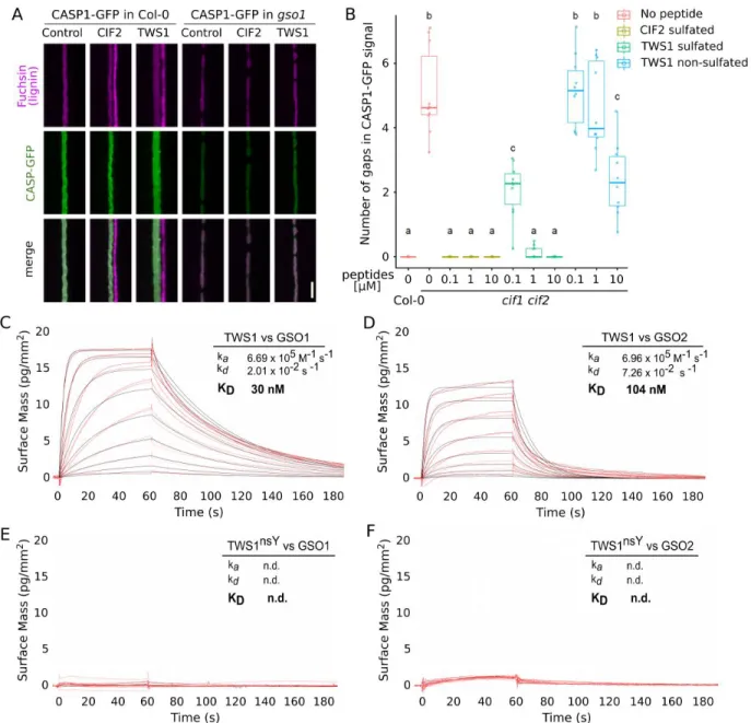
To confirm TWS1 as a ligand of GSO1 and GSO2, the interaction of the synthetic peptide with
the leucine-rich repeat (LRR) ectodomains of the receptors was analyzed in grating – coupled
interferometry binding assays. GSO1 bound sulfated TWS1 with a KD of ~ 30 nM (Fig. 2C). The
10
observed binding affinity is ~10 fold lower compared to the CIF2 peptide (KD = 2.5 nM) (Fig.
S10), which is consistent with the reduced ability of TWS1 to complement the root phenotype of
the cif1 cif2 double mutant (Fig. 2B). Sulfated TWS1 also bound to the LRR domain of GSO2,
albeit with slightly reduced affinity (KD ~ 100 nM) (Fig.2D). As previously shown for other CIF
peptides (11), tyrosine sulfation was critical for the interaction of TWS1 with GSO1 and GSO2 in
15
vitro (Fig. 2E,F). Technical issues at high peptide concentrations may explain discrepancies
between in vitro binding assays and the in vivo activity of non-sulfated TWS1. In vivo activities
for non-sulfated versions of other normally-sulfated peptides, including CIF2, have been reported
(11,16-18). Adding a 3AA C-terminal extension to the sulfated TWS1 peptide reduced binding
affinity to both GSO1 and GSO2 (Fig. S10), consistent with the need for ALE1-mediated
C-20
terminal processing for efficient signaling.
Taken together, our results suggest the sulfated TWS peptide as the missing link in the
6
TPST both contribute to the formation of the bioactive peptide (Fig. 1R) which is perceived by
GSO1 and GSO2 to ensure appropriate cuticle deposition.
To understand how the elements of the signaling pathway cooperate to ensure the formation of a
functional cuticle, we analyzed their spatial organization. In silico data indicate that the TPST gene
is expressed in all seed tissues (Fig. S11) (19, 20). To investigate in which compartment TPST
5
(which acts cell autonomously (13)) is required for TWS1 maturation, reciprocal crosses and
complementation assays using tissue specific promoters were performed. No cuticle permeability
defects were observed when homozygous mutants were pollinated with wild-type pollen,
confirming their zygotic origin. (Fig. 3A-C). Expressing TPST under the ubiquitously active
RPS5A promoter (21), or the PIN1 promoter (which is embryo-specific in seed (Fig. S12))
10
complements tpst-1 cuticle defects. In contrast no complementation was observed using the
endosperm-specific RGP3 promoter (22), indicating that TPST activity is required for TWS1
sulfation specifically in the embryo to ensure cuticle integrity (Fig. 3D) (Fig. S13). Consistent with
this observation, and with a previous report (14), the TWS1 promoter was found to drive expression
specifically in the developing embryo from the early globular stage onwards (Fig. 3E) (Fig S14).
15
The TPST promoter (10) drove expression throughout the embryo proper at the onset of embryo
cuticle establishment (globular stage) before becoming restricted to the root tip (Fig. S11). We
conclude that the TWS1 peptide is both sulfated and secreted specifically in the embryo.
However, production of mature TWS1 requires a C-terminal cleavage event that we have shown
to be mediated by ALE1. ALE1 is expressed only in the endosperm (4, 23), on the opposite side of
20
the nascent cuticle to the GSO1 and GSO2 receptors, which are localized on the membranes of the
epidermal cells that produce the cuticle (Fig. S15-17) (2). Our data thus support a model in which
7
to the endosperm, where it is cleaved and activated by ALE1 before diffusing back to the embryo
to trigger GSO1/2-dependent cuticle deposition. An intact cuticle would separate the subtilase
from its substrate, terminating signaling.
Expressing ALE1 in the embryo, under the control of the TWS1 promoter, provided support for
this model. Multiple transformants were obtained in tws1 mutants, but not in the wild-type
5
background. When tws1 plants from 4 independent plants carrying the pTWS1:ALE1 transgene
were pollinated with wild-type pollen, introducing a functional TWS1 allele into the zygotic
compartments, and thus inducing colocalization of TWS1 precursors, ALE1, GSO1 and GSO2 in
the embryo, premature embryo growth arrest was observed in all seeds. This leads to severe
shriveling of all seeds at maturity (Fig. 3F-M) (Fig. S18, S19). A proportion of seeds could
10
nonetheless germinate to give developmentally normal plants (Fig. S20) indicating that
co-expression of all signaling components in the embryo, although detrimental to embryo
development does not lead to a complete loss of viability. Growth arrest may be due to constitutive
embryonic activation of the GSO1/GSO2 signaling pathway, and indeed stress-responsive genes
shown to require GSO1/GSO2 signaling for expression in the seed (2) were upregulated in seeds
15
co-expressing GSO1, GSO2, TWS1 and ALE1 in the embryo (Fig. S21). We thus postulate that
the spatial separation of the TWS1 precursor and the GSO receptors from the activating protease
by cuticle is required for signaling attenuation.
We next tested if CIF1, CIF2 and TWS1 could complement tws1 and ale1 mutants when expressed
in the endosperm (under the RGP3 promoter). All three peptides complemented tws1 mutants,
20
confirming that retrograde peptide movement from endosperm to embryo is sufficient to allow
integrity monitoring (Fig. 3N and Fig. S22). Lack of full complementation could reflect suboptimal
8
extensions) complemented ale1 mutants much more efficiently than TWS1 (Fig. S23). Weak
complementation of ale1 by TWS1 may reflect the presence of redundantly-acting subtilases in
the endosperm, as suggested by the weak phenotype of ale1 mutants.
The proposed bidirectional signaling model allows efficient embryo cuticle integrity monitoring.
The sulfated TWS1 precursor is produced by the embryo and secreted (probably after N-terminal
5
cleavage of the pro-peptide) to the embryo apoplast. In the absence of an intact cuticular barrier,
it can diffuse to the endosperm and undergo activation by ALE1 (and potentially other subtilases).
Activated TWS1 peptide then leaks back through cuticle gaps to bind the GSO1 and GSO2
receptors and activate local gap repair (Fig. 3O). When the cuticle is intact, proTWS1 peptides are
confined to the embryo where they remain inactive.
10
Our results demonstrate a role for a subtilase in providing spatial specificity to a bidirectional
peptide signaling pathway. In contrast, the related CIF1, CIF2 and GSO1-dependent signaling
pathway controlling Casparian strip integrity is uni-directional, negating the need for C-terminal
cleavage-mediated peptide activation (10, 12). Both pathway components and their spatial
organization differ between the two systems, suggesting an independent recruitment of the GSO
15
receptors to different integrity monitoring functions within the plant.
References and Notes:
1. C. Delude, S. Moussu, J. Joubès, G. Ingram, F. Domergue, Plant Surface Lipids and Epidermis Development. Subcell. Biochem. 86, 287–313 (2016).
2. A. Creff, L. Brocard, J. Joubès, L. Taconnat, N. M. Doll, A.-C. Marsollier, S. Pascal, R. Galletti, S.
20
Boeuf, S. Moussu, T. Widiez, F. Domergue, G. Ingram, A stress-response-related inter-compartmental signalling pathway regulates embryonic cuticle integrity in Arabidopsis. PLoS Genet. 15, e1007847 (2019).
3. R. Tsuwamoto, H. Fukuoka, Y. Takahata, GASSHO1 and GASSHO2 encoding a putative leucine-rich repeat transmembrane-type receptor kinase are essential for the normal development of the
25
9
4. H. Tanaka, H. Onouchi, M. Kondo, I. Hara-Nishimura, M. Nishimura, C. Machida, Y. Machida, A subtilisin-like serine protease is required for epidermal surface formation in Arabidopsis embryos and juvenile plants. Dev. Camb. Engl. 128, 4681–4689 (2001).
5. Q. Xing, A. Creff, A. Waters, H. Tanaka, J. Goodrich, G. C. Ingram, ZHOUPI controls embryonic cuticle formation via a signalling pathway involving the subtilisin protease ABNORMAL
LEAF-5
SHAPE1 and the receptor kinases GASSHO1 and GASSHO2. Dev. Camb. Engl. 140, 770–779 (2013).
6. S. Moussu, N. M. Doll, S. Chamot, L. Brocard, A. Creff, C. Fourquin, T. Widiez, Z. L. Nimchuk, G. Ingram, ZHOUPI and KERBEROS Mediate Embryo/Endosperm Separation by Promoting the Formation of an Extracuticular Sheath at the Embryo Surface. Plant Cell. 29, 1642–1656 (2017).
10
7. A. Schaller, A. Stintzi, S. Rivas, I. Serrano, N. V. Chichkova, A. B. Vartapetian, D. Martínez, J. J. Guiamét, D. J. Sueldo, R. A. L. van der Hoorn, V. Ramírez, P. Vera, From structure to function - a family portrait of plant subtilases. New Phytol. 218, 901–915 (2018).
8. N. Stührwohldt, A. Schaller, Regulation of plant peptide hormones and growth factors by post-translational modification. Plant Biol. (Suppl 1), 49–63 (2019).
15
9. K. Schardon, M. Hohl, L. Graff, J. Pfannstiel, W. Schulze, A. Stintzi, A. Schaller, Precursor processing for plant peptide hormone maturation by subtilisin-like serine proteinases. Science. 354, 1594–1597 (2016).
10. V. G. Doblas, E. Smakowska-Luzan, S. Fujita, J. Alassimone, M. Barberon, M. Madalinski, Y. Belkhadir, N. Geldner, Root diffusion barrier control by a vasculature-derived peptide binding to the
20
SGN3 receptor. Science. 355, 280–284 (2017).
11. S. Okuda, S. Fujita, A. Moretti, U. Hohmann, V. Gonzalez Doblas, Y. Ma, A. Pfister, B. Brandt, N. Geldner, M. Hothorn, Molecular mechanism for the recognition of sequence-divergent CIF peptides by the plant receptor kinases GSO1/SGN3 and GSO2. bioRxiv, 692228.
12. T. Nakayama, H. Shinohara, M. Tanaka, K. Baba, M. Ogawa-Ohnishi, Y. Matsubayashi, A peptide
25
hormone required for Casparian strip diffusion barrier formation in Arabidopsis roots. Science. 355, 284–286 (2017).
13. R. Komori, Y. Amano, M. Ogawa-Ohnishi, Y. Matsubayashi, Identification of tyrosylprotein sulfotransferase in Arabidopsis. Proc. Natl. Acad. Sci. U. S. A. 106, 15067–15072 (2009).
14. E. Fiume, V. Guyon, C. Remoué, E. Magnani, M. Miquel, D. Grain, L. Lepiniec, TWS1, a Novel
30
Small Protein, Regulates Various Aspects of Seed and Plant Development. Plant Physiol. 172, 1732– 1745 (2016).
15. H. Hanai, D. Nakayama, H. Yang, Y. Matsubayashi, Y. Hirota, Y. Sakagami, Existence of a plant tyrosylprotein sulfotransferase: novel plant enzyme catalyzing tyrosine O-sulfation of preprophytosulfokine variants in vitro. FEBS Lett. 470, 97–101 (2000).
35
16. Y. Matsubayashi, Y. Sakagami, Characterization of specific binding sites for a mitogenic sulfated peptide, phytosulfokine-alpha, in the plasma-membrane fraction derived from Oryza sativa L. Eur. J. Biochem. 262, 666–671 (1999).
10
17. A. Kutschmar, G. Rzewuski, N. Stührwohldt, G. T. S. Beemster, D. Inzé, M. Sauter, PSK-α promotes root growth in Arabidopsis. New Phytol. 181, 820–831 (2009).
18. Y. Matsuzaki, M. Ogawa-Ohnishi, A. Mori, Y. Matsubayashi, Secreted peptide signals required for maintenance of root stem cell niche in Arabidopsis. Science. 329, 1065–1067 (2010).
19. B. H. Le, C. Cheng, A. Q. Bui, J. A. Wagmaister, K. F. Henry, J. Pelletier, L. Kwong, M. Belmonte,
5
R. Kirkbride, S. Horvath, G. N. Drews, R. L. Fischer, J. K. Okamuro, J. J. Harada, R. B. Goldberg, Global analysis of gene activity during Arabidopsis seed development and identification of seed-specific transcription factors. Proc. Natl. Acad. Sci. U. S. A. 107, 8063–8070 (2010).
20. A. V. Klepikova, A. S. Kasianov, E. S. Gerasimov, M. D. Logacheva, A. A. Penin, A high resolution map of the Arabidopsis thaliana developmental transcriptome based on RNA-seq profiling. Plant J.
10
Cell Mol. Biol. 88, 1058–1070 (2016).
21. D. Weijers, Franke-van Dijk, M., R. J. Vencken, Quint, A., Hooykaas, P., Offringa, R., An Arabidopsis Minute-like phenotype caused by a semi-dominant mutation in a RIBOSOMAL PROTEIN S5 gene. Dev. Camb. Engl. 128, 4289–4299 (2001).
22. G. Denay, A. Creff, S. Moussu, P. Wagnon, J. Thévenin, M.-F. Gérentes, P. Chambrier, B. Dubreucq,
15
G. Ingram, Endosperm breakdown in Arabidopsis requires heterodimers of the basic helix-loop-helix proteins ZHOUPI and INDUCER OF CBP EXPRESSION 1. Dev. Camb. Engl. 141, 1222–1227 (2014).
23. S. Yang, N. Johnston, E. Talideh, S. Mitchell, C. Jeffree, J. Goodrich, G. Ingram, The endosperm-specific ZHOUPI gene of Arabidopsis thaliana regulates endosperm breakdown and embryonic
20
epidermal development. Dev. Camb. Engl. 135, 3501–3509 (2008).
24. A. Pfister, M. Barberon, J. Alassimone, L. Kalmbach, Y. Lee, J. E. M. Vermeer, M. Yamazaki, G. Li, C. Maurel, J. Takano, T. Kamiya, D. E. Salt, D. Roppolo, N. Geldner, A receptor-like kinase mutant with absent endodermal diffusion barrier displays selective nutrient homeostasis defects. eLife. 3, e03115 (2014).
25
25. D. Roppolo, B. De Rybel, V. D. Tendon, A. Pfister, J. Alassimone, J. E. M. Vermeer, M. Yamazaki, Y.-D. Stierhof, T. Beeckman, N. Geldner, A novel protein family mediates Casparian strip formation in the endodermis. Nature. 473, 380–383 (2011).
26. J. Friml, A. Vieten, M. Sauer, D. Weijers, H. Schwarz, T. Hamann, R. Offringa, G. Jürgens, Efflux-dependent auxin gradients establish the apical–basal axis of Arabidopsis. Nature. 426, 147 (2003).
30
27. Z.-P. Wang, H.-L. Xing, L. Dong, H.-Y. Zhang, C.-Y. Han, X.-C. Wang, Q.-J. Chen, Egg cell-specific promoter-controlled CRISPR/Cas9 efficiently generates homozygous mutants for multiple target genes in Arabidopsis in a single generation. Genome Biol. 16, 144 (2015).
28. F. Fauser, S. Schiml, H. Puchta, Both CRISPR/Cas-based nucleases and nickases can be used efficiently for genome engineering in Arabidopsis thaliana. Plant J. 79, 348–359 (2014).
35
29. E. Logemann, R. P. Birkenbihl, B. Ülker, I. E. Somssich, An improved method for preparing Agrobacterium cells that simplifies the Arabidopsis transformation protocol. Plant Methods. 2, 16 (2006).
11
30. T. Tanaka, H. Tanaka, C. Machida, M. Watanabe, Y. Machida, A new method for rapid visualization of defects in leaf cuticle reveals five intrinsic patterns of surface defects in Arabidopsis. Plant J. 37, 139–146 (2004).
31. U. Roshan, D. R. Livesay, Probalign: multiple sequence alignment using partition function posterior probabilities. Bioinforma. Oxf. Engl. 22, 2715–2721 (2006).
5
32. D. Kurihara, Y. Mizuta, Y. Sato, T. Higashiyama, ClearSee: a rapid optical clearing reagent for whole-plant fluorescence imaging. Development. 142, 4168–4179 (2015).
33. R. Ursache, T. G. Andersen, P. Marhavý, N. Geldner, A protocol for combining fluorescent proteins with histological stains for diverse cell wall components. Plant J. 93, 399–412 (2018).
34. R Core Team, R: A language and environment for statistical computing. R Foundation for Statistical
10
Computing, Vienna, Austria. (2013).
35. Y. Hashimoto, S. Zhang, G. W. Blissard, Ao38, a new cell line from eggs of the black witch moth, Ascalapha odorata (Lepidoptera: Noctuidae), is permissive for AcMNPV infection and produces high levels of recombinant proteins. BMC Biotechnol. 10, 50 (2010).
36. M. Fairhead, M. Howarth, Site-specific biotinylation of purified proteins using BirA. Methods Mol.
15
Biol. Clifton NJ. 1266, 171–184 (2015).
37. A. P. Gleave, A versatile binary vector system with a T-DNA organisational structure conducive to efficient integration of cloned DNA into the plant genome. Plant Mol. Biol. 20, 1203–1207 (1992). 38. A. Shevchenko, M. Wilm, O. Vorm, M. Mann, Mass spectrometric sequencing of proteins
silver-stained polyacrylamide gels. Anal. Chem. 68, 850–858 (1996).
20
39. J. V. Olsen, L. M. F. de Godoy, G. Li, B. Macek, P. Mortensen, R. Pesch, A. Makarov, O. Lange, S. Horning, M. Mann, Parts per million mass accuracy on an Orbitrap mass spectrometer via lock mass injection into a C-trap. Mol. Cell. Proteomics MCP. 4, 2010–2021 (2005).
Acknowledgments: We thank Loïc Lepiniec for providing the tws1-1 and tws1-2 seeds, Carlos
25
Galvan Ampudia, Yvon Jaillais and Laia Armengot for materials and helpful discussions, Audrey
Creff, Ursula Glück-Behrens, Alexis Lacroix, Patrice Bolland, Justin Berger, Isabelle
Desbouchages and Hervé Leyral for technical assistance, Angélique Patole, Brigitte Martin
Sempore, Cindy Vial and Stéphanie Maurin for administrative assistance and Berit Würtz and Jens
Pfannstiel (Core Facility Hohenheim) for mass spectrometric analyses. TEM images were acquired
30
12
Funding: The study was financed by joint funding (project Mind the Gap) from the French Agence
National de Recherche (ANR- 17-CE20-0027) (G.I.) and the Swiss National Science Foundation
(NSF) (N.G., supporting S.F.). N.M.D. was funded by a PhD fellowship from the Ministère de l’Enseignement Supérieur et de la Recherche. Funding was also provided by NSR grant no
5
31003A_176237 (M.H.) and an International Research Scholar grant from the Howard Hughes
Medical Institute (to M.H). S.O. was supported by a long-term postdoctoral fellowship by the
Human Frontier Science Program (HFSP). S.R. was supported by a PhD fellowship from the
Carl-Zeiss Foundation.
Author contributions: G.I. led the study. G.I. and N.G. obtained funding for the study. G.I., N.G.,
10
A.Sc., M.H. T.W and A.St. supervised the work. N.M.D., S.R., S.F., S.O. and S.C. carried out the
experiments. All authors were involved in the analysis of the results. G.I., A.Sc. and N.M.D. wrote
the paper with input from all authors.
Competing interests: The authors declare no competing interests.
Data and materials availability: All lines used in the study will be provided upon signature of
15
an appropriate Material Transfer Agreement. All data is available in the main text or the
supplementary materials.
Supplementary Materials:
Materials and Methods
Figures S1-S23
13
Fig. 1. TPST and ALE1 are required for maturation of the TWS1 peptide. A-C, E-H and
J-K) Toluidine blue tests on etiolated cotyledons. Scale bars = 200µm. D and I) Quantification of toluidine blue uptake by the aerial parts of young seedlings, normalized to chlorophyll content. N=6, ten seedlings per repetition. *** = statistical differences with one-way ANOVA followed by
5
a post-hoc Scheffé multiple comparison test (P < 0,01) in D and I. J and K) Toluidine blue permeability of tws1-4 compared to Col-0. Scale bars = 400µm L-M) Transmission electron micrographs of the embryo/endosperm interface at the heart stage. Scale bars = 200nm. Genotypes are indicated, and gaps in the cuticle are shown by white arrows. O) The predicted TWS1 active peptide sequence and alignment with four other known GSO ligands (CIF1,CIF2, CIF3 and CIF4).
10
The site of predicted sulfation is indicated with a red asterisk. P) Anti-His western blot of protein extracts from N. benthamiana leaves agro-infiltrated to express TWS1::GFP(His)6 (TWS1) or the
14
empty vector (-). Co-expression of ALE1::(His)6 or the empty-vector control are indicated by + and -, respectively. Q) Coomassie-stained SDS-PAGE showing recombinant GST-TWS1 and the indicated site-directed mutants digested in vitro with (+) or without (-) ALE1-(His)6 purified from tobacco leaves. Arrows indicate specific cleavage products. R) The full length TWS1 precursor. Sulfation and ALE1 cleavage sites are indicated.
15
Fig. 2. The TWS1 peptide is a functional GSO1/GSO2 ligand. A) Root over-lignification
following treatment with the active CIF2 or TWS peptide in Col-0 and in the gso1 (sgn3-3) background. Lignin is stained in purple and CASP-GFP fusion protein, marking the Casparian strip domain, in green. Scale bar = 5µm B) Complementation of cif1-2 cif2-2 Casparian strip
5
integrity phenotype by peptide treatments. Number of gaps in CASP1-GFP signal counted after treatment with CIF2 sulfated peptide, TWS1 sulfated peptide, TWS1 non-sulfated peptide. N=10. a, b, and c correspond to a classes statistically supported by one-way ANOVA analysis followed by Tukey tests (P < 0,05). C-F) Grating-coupled interferometry (GCI)-derived binding kinetics. Shown are sensorgrams with raw data in red and their respective fits in black. ka, association rate
16
constant; kd, dissociation rate constant; KD, dissociation constant. C) on the GSO1 extra-cellular
domain in the presence of the sulfated TWS1 peptide. D) on the GSO2 extra-cellular domain in the presence of the sulfated TWS1 peptide. E) on the GSO1 extra-cellular domain in the presence of the non-sulfated TWS1 peptide. F) on the GSO2 extra-cellular domain in the presence of the non-sulfated TWS1 peptide.
17
Fig. 3. Spatial separation of ALE1 and TWS1 expression is critical for pathway function.
A-C) F1 seedlings from reciprocal crosses stained with Toluidine blue. D,N) Toluidine blue quantification as in Fig. 1. a-d = statistical differences with one-way ANOVA followed by a
post-5
hoc Scheffé multiple comparison test (P < 0,01). D) Complementation of tpst-1 mutant with
endosperm-specific expression of TPST (pRGP3::TPST), embryo-specific expression of TPST
(pPIN1:TPST) and ubiquitous expression of TPST (pRPS5a::TPST) compared to tpst-1 and
Col-0. 3 independent lines were analysed. E) Confocal images of pTWS1::mCitrine::NLS-mCitrine reporter lines, signal in yellow, autofluorescence in red. Scale bars = 50µm. F,G) Dry seeds (scale
18
bars = 400 µm) and chloral hydrate cleared seeds (9 DAP) (scale bars = 100 µm) respectively from a line expressing ALE1 in the embryo in the tws1-4 background (pTWS1::ALE1 line#7). H,I) Seeds from crosses of Col-0 pollen onto line#7. J,K) self-fertilized tws1-4 seeds as a control L,M) Seeds from a cross of Col-0 pollen on a tws1-4 pistil as a control. Results for three further independent transgenic lines are shown in Figs. S18 and S19. N) Complementation of tws1-4
5
mutants by expression of TWS1 in the endosperm. Four independent lines were analysed. O) Model for embryonic cuticle integrity monitoring. Panel on left shows the wild-type situation prior to gap-filling (nascent cuticle), illustrating the diffusion and processing of TWS1 across the embryo-endosperm interface. Panel on right shows the wild-type situation when the cuticle is intact, spatially separating signalling components and thus the attenuating signalling.