HAL Id: hal-03039831
https://hal.archives-ouvertes.fr/hal-03039831
Submitted on 4 Dec 2020
HAL is a multi-disciplinary open access
archive for the deposit and dissemination of sci-entific research documents, whether they are pub-lished or not. The documents may come from teaching and research institutions in France or abroad, or from public or private research centers.
L’archive ouverte pluridisciplinaire HAL, est destinée au dépôt et à la diffusion de documents scientifiques de niveau recherche, publiés ou non, émanant des établissements d’enseignement et de recherche français ou étrangers, des laboratoires publics ou privés.
Protein sequence comparison of human and non-human
primate tooth proteomes
Carine Froment, Clément Zanolli, Mathilde Hourset, Emmanuelle
Mouton-Barbosa, Andreia Moreira, Odile Burlet-Schiltz, Catherine Mollereau
To cite this version:
Carine Froment, Clément Zanolli, Mathilde Hourset, Emmanuelle Mouton-Barbosa, Andreia Moreira, et al.. Protein sequence comparison of human and non-human primate tooth proteomes. Journal of Proteomics, Elsevier, 2021, 231, pp.104045. �10.1016/j.jprot.2020.104045�. �hal-03039831�
1
Protein sequence comparison of human and non-human primate tooth proteomes
Carine Froment1, Clément Zanolli2, Mathilde Hourset3,4, Emmanuelle Mouton-Barbosa1, Andreia Moreira3, Odile Burlet-Schiltz1 and Catherine Mollereau3
1 Institut de Pharmacologie et Biologie Structurale (IPBS), Université de Toulouse, CNRS, UPS, Toulouse, France.
2
Laboratoire PACEA, UMR 5199 CNRS, Université de Bordeaux, Pessac, France.
3 Laboratoire d’Anthropobiologie Moléculaire et Imagerie de Synthèse (AMIS), UMR 5288 CNRS, Université de Toulouse, UPS, Toulouse, France
4 Faculté de chirurgie dentaire de Toulouse, Université de Toulouse, UPS, Toulouse, France.
Corresponding authors:
Dr Catherine Mollereau catherine.mollereau-manaute@ipbs.fr Laboratoire AMIS
Faculté de médecine, 37 allées Jules Guesde 31073 Toulouse Cedex 03, France
Tel : 33 561 14 55 13 and
Dr Odile Burlet-Schiltz, odile.schiltz@ibps.fr IPBS
205, Route de Narbonne BP 64182 31077 Toulouse Cedex 04, France Tel: 33 561 17 55 47
Keywords: Palaeoproteomics; Tooth; Primates; nanoLC-MS/MS; Taxonomy
Conflict of interest
The authors declare that they have no conflict of interest.
Acknowledgements
We are sincerely grateful to J. Braga (AMIS, Toulouse France), S. Jiquel (ISEM, Montpellier France) and R. Macchiarelli (HNHP, Poitiers France) for giving us access to their primate collections. We also thank the two colleagues that gave their surgically extracted molars. The work was supported by the French CNRS (PEPS blanc INEE 2016 and DefiXlife 2018-2019), in part by the Région Occitanie, European funds (Fonds Européens de Développement Régional, FEDER), Toulouse Métropole, and by the French Ministry of Research with the
2
Investissement d'Avenir Infrastructures Nationales en Biologie et Santé program (ProFI, Proteomics French Infrastructure project, ANR-10-INBS-08).
3
Abstract
In the context of human evolution, the study of proteins may overcome the limitation of the high degradation of ancient DNA over time to provide biomolecular information useful for the phylogenetic reconstruction of hominid taxa. In this study, we used a shotgun proteomics approach to compare the tooth proteomes of extant human and non-human primates (gorilla, chimpanzee, orangutan and baboon) in order to search for a panel of peptides able to discriminate between taxa and further help reconstructing the evolutionary relationships of fossil primates. Among the 25 proteins shared by the five genera datasets, we found a combination of peptides with sequence variations allowing to differentiate the hominid taxa in the proteins AHSG, AMBN, APOA1, BGN, C9, COL11A2, COL22A1, COL3A1, DSPP, F2, LUM, OMD, PCOLCE and SERPINA1. The phylogenetic tree confirms the placement of the samples in the appropriate genus branches. Altogether, the results provide experimental evidence that a shotgun proteomics approach on dental tissue has the potential to detect taxonomic variation, which is promising for future investigations of uncharacterized and/or fossil hominid/hominin specimens.
4 1. Introduction
1
In the last ten years, major advances have been achieved in the field of human 2
evolution with the increasing recovery of Late Pleistocene human genomes. These advances 3
have unveiled the existence of a new human group, the Denisovan hominins, an extinct 4
relative of Neanderthals [1, 2], in addition to recurrent interbreeding between archaic and 5
modern humans during their period of cohabitation in Eurasia [3, 4]. However, despite the 6
exceptional retrieval of 0.4 million-year (My) old hominin genomic data from Sima de los 7
Huesos in Spain [5, 6], the use of a DNA-based approach for older specimens, beyond the 8
Middle Pleistocene [7], remains limited due to a high degree of fragmentation of ancient DNA 9
over time [8, 9]. Therefore, the next challenging step is the biomolecular characterization of 10
fossils further back in time, in particular for the Early Pleistocene-Late Pliocene key period (3-11
2 My ago) corresponding to the emergence of the genus Homo. Such invaluable missing 12
data are essential for a more accurate reconstruction of the phylogenetic relationships 13
between the different hominin taxa [10]. 14
As an alternative to DNA, proteins appear to be more resistant to post-mortem 15
damage, potentially allowing biomolecular investigation of the deeper time [11, 12]. Even in 16
unfavorable conditions proteins survive longer than DNA, as demonstrated by the recovery of 17
> 3 My old peptides entrapped in ostrich egg shell in the warm African environment [13] and 18
more recently, of dental proteins from a subtropical Early Pleistocene specimen (1.9 My ago) 19
representative of the extinct Asian hominid Gigantopithecus blacki [14]. Given the ever 20
increasing performance of mass spectrometers, in terms of sensitivity, sequencing speed 21
and resolution, it is now possible to study the protein content of ancient samples, using small 22
amount of precious paleontological and paleoanthropological material. This opens the way to 23
a new field of research: Palaeoproteomics. Proteins have been sequenced from Late to Early 24
Pleistocene faunal and human fossils [7, 14-17], providing invaluable functional information 25
on past human (patho)physiology [18-20] and ancient diets [21, 22]. In addition, phylogenetic 26
reconstruction and taxonomic placement based on protein identification have also been 27
possible for extinct species and/or when DNA was no longer available [16, 23-27]. The two 28
main examples below on debated hominin fossils, have recently shed light on the 29
relationships between human lineages during evolution. On the basis of a collagen variant, 30
Chen et al. [24] were able to assign the mandible of Xiahe from the Tibetan plateau to a 31
Denisovan individual. For the first time, the presence of this hominin outside the Siberian 32
Altaï area was therefore demonstrated together with its anatomical relationship with other 33
archaic hominin fossils in China. By analyzing the enamel proteome of a representative of 34
Homo antecessor from Atapuerca (Spain) dated to 0.8-0.9 My ago, Welker et al. [27] have 35
5
suggested a very close affinity between this species and the last common ancestor of 36
modern humans, Neanderthals and Denisovans. 37
Collagen sequences are widely used to identify or characterize specimens [9, 28, 29], 38
but they are sometimes not enough informative because of a high conservation between 39
species limiting accurate phylogenetic reconstruction [30]. It is therefore necessary to identify 40
other proteins with sufficient taxonomic variability. In this context, the tooth proteome is 41
expected to be more informative than the bone proteome [9, 14, 16, 27]. Indeed, in addition 42
to collagens, it also contains a variety of specific non-collagenous proteins with taxonomic 43
interest, including the X and Y forms of amelogenin able to provide information about the sex 44
of ancient individuals [27, 31-34]. 45
In the present study we used a shotgun proteomics approach to compare the tooth 46
proteomes of modern humans from the 21th century and non-human primates (gorilla, 47
chimpanzee, orangutan, baboon) from the 19th-mid 20th century, in order to search for a 48
panel of peptides able to discriminate between taxa. We focused on the analysis of the 49
proteins shared by the five genera datasets to allow for comparison. We found in 14 proteins 50
a combination of peptides with taxonomic variation potentially useful for future studies on 51
uncharacterized and/or older hominid/hominin specimens. 52
53
2. Materials and Methods 54
2.1 Samples 55
Non-human primate teeth (2 individuals per genus) from 19th-mid 20th century 56
collections were obtained from the University of Poitiers with the agreement of Pr R. 57
Macchiarelli (samples T1: Gorilla, T2: Papio, T5: Pan., T6: Pan), from the University of 58
Toulouse with the agreement of Pr J. Braga (samples T3: Papio, T4: Gorilla), and from the 59
University of Montpellier with the agreement of S.Jiquel (samples T7: Pongo, T8: Pongo). 60
Except for the two chimpanzee specimens that are attributed to Pan troglodytes, the species 61
of the other non-human primate samples is not known. Human teeth were obtained from two 62
colleagues who kindly donated their own M3 that had been surgically extracted three (LOS2) 63
and fifteen (LKS2) years ago. Description of the teeth is given in Table S1. Teeth were 64
handled under a laminar flow hood and the operator was equipped with disposable clothes. 65
The surface of the teeth was carefully cleaned with a solution of 1% SDS and abundantly 66
rinsed with sterile pure water. The tooth area to be sampled, (preferably chosen on the 67
cervical part of the crown so that the damaged area is minimally visible after repositioning the 68
tooth on the jaw) was first manually drilled with a micromotor STRONG 207-106 (Pouget-69
Pellerin, France) to abrade the surface. Then a spherical carbide drill (1-1.2 mm in diameter) 70
was used to pierce the enamel through the dentine tissue, over a sterile microtube to collect 71
the powder. Sample amounts ranged from 2 to 26 mg for non-human primates and 60 mg for 72
6
humans, respectively (Table S1). Collected powder was immediately processed for protein 73
extraction. 74
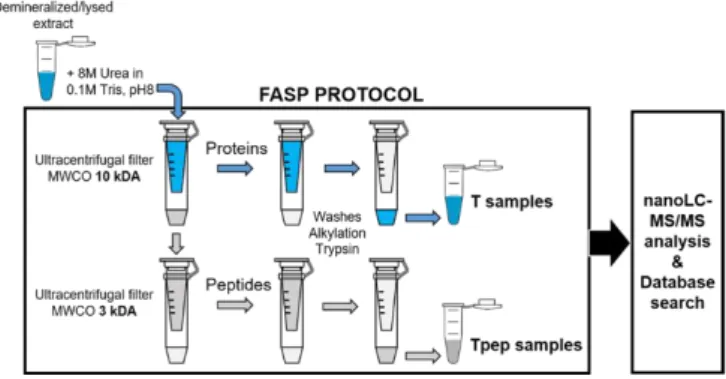
2.2. Protein extraction and Trypsin digestion 75
Samples were prepared as five independent series, each including an extraction blank 76
with no material (Blk) that was processed in the same way as the tooth samples. Protein 77
extraction was performed by using the filter-aided sample preparation (FASP) protocol 78
described in [31]. It was slightly modified to include an additional step for recovering the flow-79
through content of the first Amicon™ Ultra-4 (10kDa) filter unit (Merck Millipore) after 80
centrifugation which may contain material of interest (Fig. 1). After a demineralization step in 81
0.5 M EDTA pH 8 for 18 h at room temperature under rotation, the pellet was extracted in 0.1 82
M Tris pH 8, 0.1 M DTT, 4% SDS for 2h at 60 °C. The supernatants from the 83
demineralization and extraction steps were mixed with 8 M urea in 0.1 M Tris pH 8 and ultra-84
filtered through an Amicon™ Ultra-4 (10kDa) centrifugal filter unit (4000g, swinging rotor, 85
room temperature). The flow-through (except for LOS2) was collected and ultra-filtered 86
through an Amicon™ Ultra-4 (3kDa) centrifugal filter unit to recover smaller protein fragments 87
excluded from the 10kDa filtration. The two filtration units (giving at end samples referred as 88
T and Tpep, respectively) were then similarly processed. After a wash with 2 ml of 8 M urea 89
in 0.1M Tris pH 8, protein alkylation (50 mM 2-Chloroacetamide in 8M urea, 0.1 M Tris pH8) 90
was performed on the filter units for 20-30 min at room temperature in the dark. The units 91
were then washed (2 x 1 ml) with 8M urea in 0.1 M Tris pH 8, followed by 50 mM ammonium 92
bicarbonate washes (1 x 1 ml, 1 x 0.5 ml). Proteins (T samples) and peptides (Tpep 93
samples) retained on the filter were dissolved in 50 mM ammonium bicarbonate and an 94
aliquot was harvested for quantification using the Qubit protein assay kit (Thermo Fisher 95
Scientific). They were digested by overnight incubation at 37 °C with 2 µg sequencing grade 96
modified porcine trypsin (Promega). The digestion was prolonged the next day for 4-6 h with 97
2 µg additional trypsin. The tryptic peptide mixtures were recovered by centrifugation over a 98
new tube. The centrifugates were then transferred to microtubes, dried by using a centrifugal 99
vacuum concentrator and kept at -20°C until mass spectrometry analysis. 100
EDTA, Tris, and SDS were purchased from Invitrogen, urea and ammonium bicarbonate 101
from Acros Organics, chloroacetamide from Sigma-Aldrich. 102
2.3. nanoLC-MS/MS analysis 103
The dried peptides were resuspended with 0.05% trifluoroacetic acid in 2% acetonitrile 104
at an estimated concentration of 1µg/µl based on protein quantification, and then analyzed 105
by online nanoLC using an UltiMate® 3000 RSLCnano LC system (Thermo Scientific, 106
Dionex) coupled to an Orbitrap Fusion Tribrid™ mass spectrometer (Thermo Scientific, 107
Bremen, Germany). 1µl of the samples were loaded on a 300 μm ID x 5 mm PepMap C18 108
7
pre-column (Thermo Scientific, Dionex) at 20 μl/min in 2% acetonitrile, 0.05% trifluoroacetic 109
acid. After 5 minutes of desalting, peptides were on-line separated on a 75 μm ID x 50 cm 110
C18 column (in-house packed with Reprosil C18-AQ Pur 3 μm resin, Dr. Maisch, and 111
equilibrated in 95% of buffer A (0.2% formic acid)) with a gradient of 5 to 25% of buffer B 112
(80% acetonitrile, 0.2% formic acid) for 80 min then 25% to 50% for 30 min at a flow rate of 113
300 nL/min. 114
The instrument was operated in the data-dependent acquisition (DDA) mode using a 115
top-speed approach (cycle time of 3s). The survey scans MS were performed in the Orbitrap 116
over m/z 350-1550 with a resolution of 120,000 (at 200 m/z), an automatic gain control 117
(AGC) target value of 4e5, and a maximum injection time of 50 ms. Most intense ions per 118
survey scan were selected at 1.6 m/z with the quadrupole and fragmented by Higher Energy 119
Collisional Dissociation (HCD). The monoisotopic precursor selection was turned on, the 120
intensity threshold for fragmentation was set to 50,000 and the normalized collision energy 121
was set to 35%. The resulting fragments were analyzed in the Orbitrap with a resolution of 122
30,000 (at 200 m/z), an automatic gain control (AGC) target value of 5e4, and a maximum 123
injection time of 60 ms. The dynamic exclusion duration was set to 30 s with a 10 ppm 124
tolerance around the selected precursor and its isotopes. For internal calibration the 125
445.120025 ion was used as lock mass. 126
Each sample was subjected to two independent LC-MS/MS runs (TR1, TR2) for 127
assessing the identification reproducibility. To control for carry-over contamination, the MS 128
workflow process included a washing step followed by two blank MS runs using gradient 129
conditions similar to those of the samples and performed before and after each sample MS 130
run (including the blank samples). 131
2.4. Bioinformatics analysis of nanoLC-MS/MS data 132
All raw mass spectrometry files were processed in parallel using two different protein 133
identification softwares: Proteome DiscovererTM software 2.3.0.523 (Thermo Fischer 134
Scientific) with Mascot 2.6.2 (Matrix Science, London, UK) combined with the Percolator 135
algorithm (version 2.05) for PSM search optimization, and PEAKS™ Studio 10.0 software 136
(Bioinformatics Solutions Inc., Waterloo, ON, Canada) using the full set of available 137
processes PEAKS de novo > PEAKS DB > PEAKS PTM> PEAKS SPIDER [35, 36]. For both 138
softwares, data obtained from T and Tpep samples were searched against the UniProtKB 139
Swiss-Prot and TrEMBL protein databases (including canonical and isoform sequences, and 140
supplemented with frequently observed contaminants) corresponding to their own taxon: 141
Uniprot_isoF_Human database released 2019_09 with Homo sapiens taxonomy (195349 142
sequences), Uniprot_isoF_Gorilla database released 2019_07 with Gorilla taxonomy (46070 143
sequences), Uniprot_isoF_Pan database released 2019_06 with Pan (chimpanzees) 144
8
taxonomy (154055 sequences), Uniprot_isoF_Pongo released 2019_02 with Pongo 145
(orangutan) taxonomy (96058 sequences), Uniprot_isoF_Papio released 2019_02 with Papio 146
(baboons) taxonomy (46692 sequences). Data obtained from the blank samples were 147
searched against the five databases. 148
For Proteome Discoverer analysis, Mascot database searches were performed 149
individually for each raw file using a processing workflow consisting of the following 150
parameters: mass tolerances in MS and MS/MS were set to 10 ppm and 0.02 Da, 151
respectively. Carbamidomethylation of cysteine was set as a fixed modification. The enzyme 152
specificity was selected as semi-tryptic, with a maximum of three missed cleavages. The 153
main protein modifications commonly observed in damaged and ancient proteins were set as 154
variable modifications: deamidation (N, Q), oxidation (M, P), carbamylation (K, N-terminal 155
protein), and conversion to pyro-glutamic acid (N-terminal Q). The Percolator algorithm was 156
used to validate PSMs and peptides based on Posterior Error Probability (PEP) values at a 157
FDR ≤ 1% [37, 38]. FDR was estimated by a target-decoy approach using the reversed 158
database. Afterwards, the processing workflow results (.msf files) were combined into 159
sample (technical replicates TR1 + TR2), genus (technical replicates TR1 + TR2, T and Tpep 160
samples) or extraction blank multiconsensus reports. Each resulting dataset was then filtered 161
using a consensus workflow consisting of the following parameters: Only PSMs with rank 1 162
and Mascot ion score ≥ 20 were considered. Peptide identifications were grouped into 163
proteins according to the law of parsimony and filtered to 5% FDR. 164
For PEAKS analysis, all the raw files belonging to the same genus (technical replicates 165
TR1 + TR2, T and Tpep samples) or to the extraction blanks were loaded into a single 166
identification workflow per genus or per extraction blank and processed using the following 167
parameters: mass tolerances in MS and MS/MS were set to 10 ppm and 0.02 Da, 168
respectively. Carbamidomethylation of cysteine was set as a fixed modification, deamidation 169
(N, Q) and oxidation (M, P) as variable modifications. A maximum of 3 modifications per 170
peptide was allowed. In the PEAKS PTM module, all the 313 modifications and also a 171
maximum of 3 modifications per peptide were considered, and the validation was based on 172
an average local confidence (ALC) score ≥ 15%. Trypsin with semi-specific digest mode and 173
a maximum of three missed cleavages were selected. Finally, each resulting dataset per 174
genus and per extraction blank obtained from the PEAKS SPIDER module, was filtered and 175
exported using the following threshold values: Peptide score of -10lgP ≥ 20-22 adjusted to 176
obtain a FDR ≤ 1% for PSMs and peptides, Protein score of -10lgP ≥ 25-49 adjusted to 177
obtain a FDR ≤ 5% without taking into account the criterion of unique peptide and only 178
considering significant peptides, and de novo only ALC (%) ≥ 50. FDR was estimated by the 179
PEAKS “decoy-fusion” approach. 180
9
For each analysis, the proteins marked as contaminant or found in the extraction 181
blanks were excluded from the datasets analyzed. All the MS/MS spectra of the taxon-182
specific peptides were inspected manually. The species specificity of peptides with 183
taxonomic interest were checked by a protein Blast (BlastP) search in Uniprot 184
(https://www.uniprot.org/) and NCBI (https://blast.ncbi.nlm.nih.gov/Blast.cgi). 185
All the RAW data files, the output files corresponding to Proteome DiscovererTM (.msf 186
files) and PEAKS (.csv files and HTML reports) analyses, and all the fasta files used for the 187
database searches have been deposited on the ProteomeXchange Consortium [PMID 188
24727771] via the PRIDE partner repository and can be accessed with the dataset identifier 189
PXD018933. 190
2.5. Proteomic data analysis 191
To be able to compare proteomes between genera, the accession numbers of the 192
proteins identified in the different taxa were converted to human gene names by using the 193
Retrieve ID functions of Uniprot (https://www.uniprot.org/), or the db2db conversion tool of 194
bioDBnet (https://biodbnet-abcc.ncifcrf.gov/), and/or by BLAST alignment and manual 195
inspection. Classification of the identified proteins into functional categories according to GO 196
terms was performed by using the GSEA software at https://www.gsea-197
msigdb.org/gsea/msigdb/annotate.jsp [39]. Graphic representations and statistical analysis of 198
the data were performed by using Prism 7 (GraphPad Software Inc., USA). Online tool found 199
at http://bioinformatics.psb.ugent.be/webtools/Venn/ was used to produce the Venn 200
diagrams. 201
2.6 Phylogenetic analysis 202
The phylogenetic tree was based on the alignment of 14 proteins (AHSG, AMBN, 203
APOA1, BGN, C9, COL11A2, COL22A1, COL3A1, DSPP, F2, LUM, OMD, PCOLCE, 204
SERPINA1). The concatenated protein sequences identified in each genus sample were 205
aligned to the concatenated corresponding protein sequences from 8 hominoids (Homo 206
sapiens, Gorilla gorilla, G.gorilla gorilla, Pan troglodytes, Pan paniscus, Pongo abelii, Pongo 207
pygmaeus, Nomascus leucogenys) and 3 Papionini (Papio anubis, Papio hamadryas, 208
Mandrillus leucophaeus) by using the Catfasta2phyml tool available at 209
https://github.com/nylander/catfasta2phyml. The concatenated alignment is given in the 210
supplementary Phylotreealign.fasta file. The best-fit model for generating the phylogenetic 211
tree was selected based on the Akaike’s information criterion corrected (AICc) implemented 212
in jModelTest 2 [40]. The tree was built using RAxML-NG [41] with a bootstrapping procedure 213
(X1000) as statistical test for branch support. 214
10 3. Results
216
3.1. Comparative analysis of human, gorilla, chimpanzee, orangutan and baboon tooth 217
proteomes. 218
For each genus, two types of samples (T and Tpep) were prepared (Fig. 1). The T 219
samples correspond to proteins retained on the filter unit of the FASP protocol and usually 220
analyzed in proteomic experiments. The Tpep samples were recovered from the elution of 221
the filter units that is usually discarded but might also contain material of interest such as 222
peptides or protein fragments present in the demineralized/lysed extracts. The samples from 223
2 individuals per genus (Homo, Gorilla, Pan, Pongo and Papio) were analyzed in duplicate 224
by using a nanoLC-MS/MS data-dependent analysis of the tryptic digestions. The Proteome 225
Discoverer software was used with the Mascot search engine for protein identification in 226
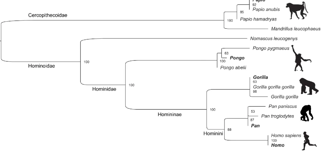
genus-specific protein databases. This led to identification of 32 to 172 proteins per T 227
samples, with only a few additional ones identified in Tpep samples (Fig. S1A and S1B). 228
The total number of proteins identified per taxon by Proteome Discoverer ranged from 229
33 in chimpanzee (Pan) to 228 in human (Fig. S1C and Table S2 for the raw list of proteins). 230
To allow for the comparison between the five proteomes, the protein accessions were 231
converted into the corresponding human gene names. This resulted in a reduced number of 232
identification (Fig.S1C) because of redundant proteins corresponding to multiple accession 233
numbers (isoforms, incomplete sequence, etc.) for a same gene. The analysis of the 234
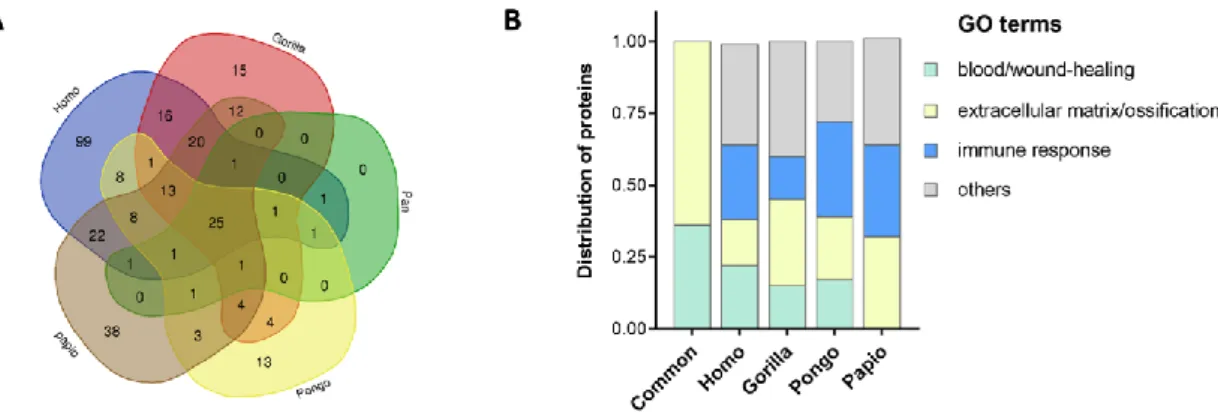
distribution of proteins between the five proteomes (Fig. 2A) indicates that 25 proteins are 235
common to the five genera (Table 1). Less than 15% of proteins are unique to each taxon, 236
with the exception of Homo (45%) and Papio (25%). A gene set overlap analysis according 237
to GO annotation terms indicates that the Top 20 most significant gene sets are different 238
between the common and the exclusive pooled proteins (Fig.2B). While shared proteins are 239
annotated with terms related to extracellular matrix organization/ossification and 240
blood/wound healing, 20% to 46% of the proteins unique in the Homo, Gorilla, Pongo and 241
Papio samples are significantly associated with immunity, in addition to extracellular matrix 242
organization and coagulation (Fig.2B, Table S3). Identifying proteins involved in the immune 243
system in the variable proteome is not unexpected since it may reflect the different sanitary 244
status between taxa and/or individuals, in addition to a possible genus-specific expression of 245
proteins [42]. 246
3.2. Analysis of the proteins of interest for taxonomic discrimination 247
A comparative analysis of the peptide lists from the proteins common to the five 248
primate proteomes was performed to search for the presence of taxon-specific peptides that 249
could be useful markers for specimen identification. Compared to the only use of protein 250
database search engines, de novo peptide sequencing algorithms that do not require a 251
11
protein sequence database [35], or error-tolerant search algorithms that utilize protein 252
sequence databases while allowing sequence deviation [43], represent powerful approaches 253
in palaeoproteomics to allow for the identification of novel amino acid substitutions. 254
Therefore, to potentially improve the identification of taxon-specific peptides, an additional 255
round of bioinformatic data analysis was performed using PEAKS software which uses a de 256
novo-assisted database search algorithm to maximize the peptide identification efficiency 257
[35, 36]. As shown in Fig.S3A, PEAKS yielded slightly more protein identifications than 258
Proteome Discoverer (Fig. S1C), with a 40-72% overlap of the identifications between the 259
two datasets (Fig. S3B) if considering gene name correspondence rather than protein 260
accessions to avoid protein redundancy. 261
The 25 proteins found in the five primate samples, especially the collagens, were 262
mainly identified by peptides covering regions of highly conserved sequences. However, a 263
number of peptides showing amino acid variations between taxa were identified in 14 264
proteins (AHSG, AMBN, APOA1, BGN, C9, COL11A2, COL22A1, COL3A1, DSPP, F2, LUM, 265
OMD, PCOLCE, SERPINA1). The lists of all the peptides identified for each protein in each 266
dataset per genus with Proteome Discoverer or PEAKS softwares are given in Tables S4 to 267
S17. The phylogenetic tree based on the concatenated alignment of the 14 proteins confirms 268
the placement of the five samples in the appropriate genus branches (Fig. 3). Because of 269
missing protein sequences in the database for some species, in particular G. gorilla, P. 270
pygmaeus, P. hamadryas, and as no peptide strictly specific to a species was identified in 271
the dataset, with the exception of H. sapiens (Table 2), the taxonomic discrimination at the 272
level of the species is less guaranteed. The main representative peptides showing amino 273
acid substitutions able to discriminate between the hominids taxa are presented in Table 2, 274
after the validation of their specificity by a BlastP search. The HCD MS/MS spectra are 275
shown in Fig. 4 and 5 and Fig. S4. Peptides specific to the taxa Homo, Gorilla and Pongo 276
were identified. No Pan-specific peptides were detected, probably because of the lower 277
protein coverage in this sample (see discussion). Despite the phylogenetic distance and a 278
high number of amino acid variations with respect to the four other primates, no Papio-279
specific peptide was found (Tables S4-S17). 280
The protein displaying the most diversity is alpha-2-HS-glycoprotein/Fetuin A (AHSG) 281
which is well covered in the five genera (Table 1). A number of discriminating peptides were 282
identified in this protein (Tables 2 and S4). Peptides covering AHSG-[72-99] with a lysine 283
residue at the position 99 are specific to Gorilla (Fig. 4A) while the peptides covering AHSG-284
[104-117] with a lysine residue at the position 117 and AHSG-[328-337] with a leucine 285
residue at the position 329 are specific to Pongo (Fig. 4B and 4C). Interestingly, a particular 286
combination of amino acids in the peptides covering AHSG-[318-337] specifically 287
differentiates Homo (P329/V333) and Pongo (L329/V333) from the two other taxa 288
12
(P329/A333) (Fig. 4C, and Table 2). In addition, the position 45 and 52 in the peptide AHSG-289
[29-57] allows to distinguish Homo and Gorilla (I45/L52) from Pan and Pongo (I45/H52) and 290
from Papio (V45/L52) (Fig.4D and Tables 2 and S4). Among the other proteins, Homo-291
specific peptides are identified in the F2 protein (F2-[199-217]), Gorilla-specific peptides are 292
identified in the proteins COL3A1 (COL3A1-[351-368] and -[902-923]), DSPP (DSPP-[403-293
411]) and SERPINA1 (SERPINA1-[126-149]), and Pongo-specific peptides are identified in 294
the proteins APOA1 (APOA1-[185-195]) and BGN (BGN-[220-239]) (Table 2 and Fig. S4). 295
Beside the taxon-specific peptides, peptides with a variable signature or a combination of 296
variable amino acids among taxa, may be also informative. For example, peptides covering 297
PCOLCE-[305-320] distinguish Homo and Pan (S309) from Gorilla and Pongo (T308) (Table 298
2 and Fig. 5). Peptides C9-[232-242] bearing A238, C9-[546-558] bearing I546/E554/N557 299
and DSPP-[362-370] bearing T365/A367 are specific to the Homininae taxon (Table 2 and 300
Fig. S4). The peptide DSPP-[56-66] with a leucine residue at the position 60 is specific to 301
Homo and Pan. Taken together, the data demonstrate therefore that it is possible by using a 302
MS-based proteomics approach on dental tissue to identify a combination of peptides 303
enabling the distinction between members of the four hominid genera (Homo, Gorilla, Pan 304
and Pongo) in accordance with the phylogenetic tree (Fig. 3). 305
306
4. Discussion 307
The present study is, to our knowledge, the first comparative analysis of tooth 308
proteomes from five living primate genera, including one cercopithecidae (Papio) and the 309
four extant hominids (Homo, Pan, Gorilla, Pongo). It is noteworthy that we had to deal with 310
the incomplete genomic annotation of non-human primates to convert protein accessions 311
from each species into the corresponding canonical (human) gene names, a prerequisite for 312
allowing comparison between taxa. However, even if a few mis-conversions or 313
unrecognitions might still remain, a total of 312 proteins corresponding to the pooled proteins 314
from all samples across all genera after removing of duplicates, were identified (Fig. S5A). 315
67% belong to dentine tissue, compared to the recently reported comprehensive human 316
dentine proteome [44]. The other 30% proteins include the amelogenin protein specific to the 317
enamel tissue, diverse collagens, in addition to constituents of the extracellular matrix and 318
immune system proteins (Fig S5B). The later components may reflect the dynamic and 319
heterogeneous part of dentine, a tissue rich in diverse bioactive peptides involved in host 320
defense, regenerative process, angiogenesis, growth and differentiation [44-47]. These are 321
expected to vary between species or individuals depending on health status and/or traumatic 322
injury [42, 47, 48]. The number of proteins identified was lower in chimpanzee compared to 323
the other specimens, and this was not due to a smaller tooth sampling (same amount as in 324
13
orangutan, Table S1). Without excluding a possible poor preservation of proteins in the 325
chimpanzee specimens, or a less efficient protein extraction, the low number of proteins 326
could also reflect a different level of protein expression in the dental tissue in this taxon [49]. 327
Indeed, the chimpanzee has a particularly thin enamel which is more prone to tooth damage 328
than in the other great apes [50]. Another explanation could be related to the tooth types that 329
were sampled. While for all the other specimens, enamel and dentine powder were collected 330
from permanent teeth, for the two chimpanzees we could only sample their deciduous teeth. 331
The nature and degree of protein expression might differ between permanent and deciduous 332
teeth, although this does not appear to be the case in humans [51] and this will need to be 333
investigated further. 334
All the proteins common to the five proteomes, with the exception of COL10A1 and 335
COL23 A1, have already been identified in human dentine extracts [44, 46, 52], although 336
AMBN may also be derived from the enamel [45, 53]. COL22A1 and F2 have been detected 337
at the enamel-dentine junction (EDJ), an interface between the mineralized tissues involved 338
in mechanical load [54]. The analysis of the peptides in the proteins common to the five 339
proteomes allowed for the identification of sequence variations in AHSG, AMBN, APOA1, 340
BGN, C9, COL11A2, COL22A1, COL3A1, DSPP, F2, LUM, OMD, PCOLCE and SERPINA1 341
enabling a taxonomic placement at the genus level (Fig.3 and Table 2). However, no species 342
marker was detected, excepted for the species H.sapiens. Interestingly, nine of the proteins 343
have been detected in 5000-year-old bovine dentin samples [55]. These include AHSG also 344
identified in the 1.9 My old enamel of the extinct primate Gigantopithecus blacki [14] and 345
COL3A1 identified in the dentine from the Xiahe specimen attributed to Denisova [24]. 346
Therefore, some of the proteins described here and showing a potential taxonomic interest 347
survive in time and to fossilization processes. 348
AHSG has already been reported to be resistant to degradation and to display 349
enough sequence variation to be of interest for phylogenetic studies [9, 12]. Here, although 350
the protein was identified by 3 peptides in only 2 out of 20 blank replicates (Table S2), it was 351
identified by a larger number of peptides in the samples (Table 1) suggesting a probable 352
endogenous origin of the protein in the samples. AHSG was therefore kept in the analysis to 353
make a comparison with the data from the literature on ancient specimens. Among the 354
peptides identified in AHSG, those bearing amino acids K-99 or L-329 are specific to Gorilla 355
and Pongo, respectively. In addition, the combination of P-329 and V-333 is specific to the 356
Homo taxon. In Gigantopithecus blacki [14] only one peptide was identified in a highly 357
conserved region of the protein (AHSG-[133-145]) susceptible to contamination in our 358
analysis (Table S4). AMBN was poorly covered in our samples (Tables 1 and S5) and not 359
detected in the dentine of the Xiahe Denisovan [24], in contrast to the other paleontological 360
specimens sampled from dental enamel [14, 27]. This suggests that using enamel tissue to 361
14
extract this protein is more appropriate than using dentine alone, or both dentine and 362
enamel. Similarly to ancient samples (Homo antecessor, Homo erectus, Gigantopithecus 363
blacki), the N-terminal part of the AMBN including the substitutions S34T and R55G that 364
distinguish Papio from the other hominid taxa were covered in our samples (Table S5). 365
However, no peptides were identified in the region overlapping a combination of amino acid 366
substitutions that differentiates Pongo (V264/G269) and Papio (M265/G170) from the others 367
taxa (V265/E270), and which indeed helps to affiliate Gigantopithecus to the pongine clade. 368
The dentine proteome of the Denisovan specimen from Xiahe exclusively contains collagens. 369
Interestingly, all the peptides identified in the ancient COL3A1 were also identified in our 370
samples (Table S11). In particular, they include the peptides covering the substitution A364V 371
specific to Gorilla, and the substitution S796G that differentiates Homo/Gorilla/Pan (S) from 372
Pongo/Papio (G). 373
A number of taxon-specific peptides were identified in the other dentine proteins 374
(Table 2), with the exception of Pan due to the low protein coverage of the chimpanzee 375
samples, and probably also because of the high sequence homology with Homo (lower 376
bootstrap at the hominin node, Fig.3). Peptides with taxonomic specificity were generally 377
identified together with the corresponding peptides in the other taxa proteins (Fig. 4, Fig. 5, 378
Fig. S4 and Tables S4-S17). As some positions were also covered in ancient samples, the 379
results support the potential of a MS-based proteomics approaches for protein identification 380
and taxonomic discrimination of extant and fossil primates from tooth samples. 381
In conclusion, the present comparison between human and non-human primates 382
tooth proteomes shows that a shotgun proteomics approach on dental tissue has the 383
potential to discriminate between the hominid taxa Homo, Gorilla, Pan and Pongo, despite a 384
high protein sequence homology (Fig.3). The results also suggest that dentine proteins offer 385
informative variability. However the data highlight the limitation of the method to differentiate 386
individual species. A targeted MS-based approach using a combination of the peptides 387
identified in this study, especially in AHSG, APOA1, BGN, COL3A1, DSPP, F2 and 388
PCOLCE, could be applied for further in-depth taxonomic investigations of ancient samples, 389
as previously done with amelogenin peptides for sex estimation [31]. In light of the recent 390
papers on Pleistocene specimens [14, 27] a promising way is open to characterize the 391
taxonomic attribution and phylogenetic relationships of fossil hominid remains, notably for 392
those older than the Middle Pleistocene for which DNA information may not be preserved or 393
not retrievable with the currently available methods. 394
15
References
[1] Meyer M, Kircher M, Gansauge MT, Li H, Racimo F, Mallick S, et al. A high-coverage genome sequence from an archaic Denisovan individual. Science. 2012;338(6104):222-6. Epub 2012/09/01. doi: 10.1126/science.1224344. PubMed PMID: 22936568; PubMed Central PMCID: PMC3617501.
[2] Prufer K, Racimo F, Patterson N, Jay F, Sankararaman S, Sawyer S, et al. The complete genome sequence of a Neanderthal from the Altai Mountains. Nature. 2014;505(7481):43-9. Epub 2013/12/20. doi: 10.1038/nature12886. PubMed PMID: 24352235; PubMed Central PMCID: PMC4031459.
[3] Fu Q, Hajdinjak M, Moldovan OT, Constantin S, Mallick S, Skoglund P, et al. An early modern human from Romania with a recent Neanderthal ancestor. Nature.
2015;524(7564):216-9. doi: 10.1038/nature14558. PubMed PMID: 26098372; PubMed Central PMCID: PMCPMC4537386.
[4] Slon V, Mafessoni F, Vernot B, de Filippo C, Grote S, Viola B, et al. The genome of the offspring of a Neanderthal mother and a Denisovan father. Nature. 2018. doi:
10.1038/s41586-018-0455-x. PubMed PMID: 30135579.
[5] Meyer M, Arsuaga JL, de Filippo C, Nagel S, Aximu-Petri A, Nickel B, et al. Nuclear DNA sequences from the Middle Pleistocene Sima de los Huesos hominins. Nature. 2016;531(7595):504-7. doi: 10.1038/nature17405. PubMed PMID: 26976447.
[6] Meyer M, Fu Q, Aximu-Petri A, Glocke I, Nickel B, Arsuaga JL, et al. A mitochondrial genome sequence of a hominin from Sima de los Huesos. Nature. 2014;505(7483):403-6. doi: 10.1038/nature12788. PubMed PMID: 24305051.
[7] Orlando L, Ginolhac A, Zhang G, Froese D, Albrechtsen A, Stiller M, et al.
Recalibrating Equus evolution using the genome sequence of an early Middle Pleistocene horse. Nature. 2013;499(7456):74-8. Epub 2013/06/28. doi: 10.1038/nature12323. PubMed PMID: 23803765.
[8] Cappellini E, Collins MJ, Gilbert MT. Biochemistry. Unlocking ancient protein palimpsests. Science. 2014;343(6177):1320-2. Epub 2014/03/22. doi:
10.1126/science.1249274. PubMed PMID: 24653025.
[9] Welker F. Palaeoproteomics for human evolution studies. Quaternary Science Reviews. 2018;190:137-47.
[10] Herries AIR, Martin JM, Leece AB, Adams JW, Boschian G, Joannes-Boyau R, et al. Contemporaneity of Australopithecus, Paranthropus, and early Homo erectus in South Africa. Science. 2020;368(6486). doi: 10.1126/science.aaw7293. PubMed PMID: 32241925.
[11] Schweitzer MH, Schroeter ER, Goshe MB. Protein molecular data from ancient (>1 million years old) fossil material: pitfalls, possibilities and grand challenges. Anal Chem.
16
2014;86(14):6731-40. Epub 2014/07/02. doi: 10.1021/ac500803w. PubMed PMID: 24983800.
[12] Wadsworth C, Buckley M. Proteome degradation in fossils: investigating the longevity of protein survival in ancient bone. Rapid communications in mass spectrometry : RCM. 2014;28(6):605-15. Epub 2014/02/13. doi: 10.1002/rcm.6821. PubMed PMID: 24519823. [13] Demarchi B, Hall S, Roncal-Herrero T, Freeman CL, Woolley J, Crisp MK, et al. Protein sequences bound to mineral surfaces persist into deep time. Elife. 2016;5. doi:
10.7554/eLife.17092. PubMed PMID: 27668515; PubMed Central PMCID: PMCPMC5039028.
[14] Welker F, Ramos-Madrigal J, Kuhlwilm M, Liao W, Gutenbrunner P, de Manuel M, et al. Enamel proteome shows that Gigantopithecus was an early diverging pongine. Nature. 2019;576(7786):262-5. doi: 10.1038/s41586-019-1728-8. PubMed PMID: 31723270; PubMed Central PMCID: PMCPMC6908745.
[15] Cappellini E, Jensen LJ, Szklarczyk D, Ginolhac A, da Fonseca RA, Stafford TW, et al. Proteomic analysis of a pleistocene mammoth femur reveals more than one hundred ancient bone proteins. Journal of proteome research. 2012;11(2):917-26. Epub 2011/11/23. doi: 10.1021/pr200721u. PubMed PMID: 22103443.
[16] Cappellini E, Welker F, Pandolfi L, Ramos-Madrigal J, Samodova D, Ruther PL, et al. Early Pleistocene enamel proteome from Dmanisi resolves Stephanorhinus phylogeny. Nature. 2019;574(7776):103-7. doi: 10.1038/s41586-019-1555-y. PubMed PMID: 31511700; PubMed Central PMCID: PMCPMC6894936.
[17] Nielsen-Marsh CM, Richards MP, Hauschka PV, Thomas-Oates JE, Trinkaus E, Pettitt PB, et al. Osteocalcin protein sequences of Neanderthals and modern primates. Proc Natl Acad Sci U S A. 2005;102(12):4409-13. Epub 2005/03/09. doi: 10.1073/pnas.0500450102. PubMed PMID: 15753298; PubMed Central PMCID: PMC555519.
[18] Jersie-Christensen RR, Lanigan LT, Lyon D, Mackie M, Belstrom D, Kelstrup CD, et al. Quantitative metaproteomics of medieval dental calculus reveals individual oral health status. Nature communications. 2018;9(1):4744. doi: 10.1038/s41467-018-07148-3. PubMed PMID: 30459334; PubMed Central PMCID: PMCPMC6246597.
[19] Maixner F, Overath T, Linke D, Janko M, Guerriero G, van den Berg BH, et al. Paleoproteomic study of the Iceman's brain tissue. Cellular and molecular life sciences : CMLS. 2013;70(19):3709-22. Epub 2013/06/07. doi: 10.1007/s00018-013-1360-y. PubMed PMID: 23739949.
[20] Warinner C, Rodrigues JF, Vyas R, Trachsel C, Shved N, Grossmann J, et al. Pathogens and host immunity in the ancient human oral cavity. Nature genetics.
2014;46(4):336-44. Epub 2014/02/25. doi: 10.1038/ng.2906. PubMed PMID: 24562188; PubMed Central PMCID: PMC3969750.
17
[21] Hendy J, Warinner C, Bouwman A, Collins MJ, Fiddyment S, Fischer R, et al. Proteomic evidence of dietary sources in ancient dental calculus. Proc Biol Sci.
2018;285(1883). doi: 10.1098/rspb.2018.0977. PubMed PMID: 30051838; PubMed Central PMCID: PMCPMC6083251.
[22] Warinner C, Hendy J, Speller C, Cappellini E, Fischer R, Trachsel C, et al. Direct evidence of milk consumption from ancient human dental calculus. Scientific reports.
2014;4:7104. Epub 2014/11/28. doi: 10.1038/srep07104. PubMed PMID: 25429530; PubMed Central PMCID: PMC4245811.
[23] Brown S, Higham T, Slon V, Paabo S, Meyer M, Douka K, et al. Identification of a new hominin bone from Denisova Cave, Siberia using collagen fingerprinting and mitochondrial DNA analysis. Scientific reports. 2016;6:23559. Epub 2016/03/30. doi: 10.1038/srep23559. PubMed PMID: 27020421; PubMed Central PMCID: PMC4810434.
[24] Chen F, Welker F, Shen CC, Bailey SE, Bergmann I, Davis S, et al. A late Middle Pleistocene Denisovan mandible from the Tibetan Plateau. Nature. 2019. doi:
10.1038/s41586-019-1139-x. PubMed PMID: 31043746.
[25] Rybczynski N, Gosse JC, Harington CR, Wogelius RA, Hidy AJ, Buckley M. Mid-Pliocene warm-period deposits in the High Arctic yield insight into camel evolution. Nature communications. 2013;4:1550. Epub 2013/03/07. doi: 10.1038/ncomms2516. PubMed PMID: 23462993; PubMed Central PMCID: PMC3615376.
[26] Welker F, Collins MJ, Thomas JA, Wadsley M, Brace S, Cappellini E, et al. Ancient proteins resolve the evolutionary history of Darwin's South American ungulates. Nature. 2015;522(7554):81-4. Epub 2015/03/25. doi: 10.1038/nature14249. PubMed PMID: 25799987.
[27] Welker F, Ramos-Madrigal J, Gutenbrunner P, Mackie M, Tiwary S, Rakownikow Jersie-Christensen R, et al. The dental proteome of Homo antecessor. Nature.
2020;580(7802):235-8. doi: 10.1038/s41586-020-2153-8. PubMed PMID: 32269345.
[28] Buckley M, Collins M, Thomas-Oates J, Wilson JC. Species identification by analysis of bone collagen using matrix-assisted laser desorption/ionisation time-of-flight mass
spectrometry. Rapid communications in mass spectrometry : RCM. 2009;23(23):3843-54. Epub 2009/11/10. doi: 10.1002/rcm.4316. PubMed PMID: 19899187.
[29] Welker F, Hajdinjak M, Talamo S, Jaouen K, Dannemann M, David F, et al.
Palaeoproteomic evidence identifies archaic hominins associated with the Chatelperronian at the Grotte du Renne. Proc Natl Acad Sci U S A. 2016. Epub 2016/09/18. doi:
10.1073/pnas.1605834113. PubMed PMID: 27638212.
[30] Cappellini E, Prohaska A, Racimo F, Welker F, Pedersen MW, Allentoft ME, et al. Ancient Biomolecules and Evolutionary Inference. Annu Rev Biochem. 2018;87:1029-60. doi: 10.1146/annurev-biochem-062917-012002. PubMed PMID: 29709200.
18
[31] Froment C, Hourset M, Saenz-Oyhereguy N, Mouton-Barbosa E, Willmann C, Zanolli C, et al. Analysis of 5000year-old human teeth using optimized large-scale and targeted proteomics approaches for detection of sex-specific peptides. J Proteomics.
2020;211:103548. doi: 10.1016/j.jprot.2019.103548. PubMed PMID: 31626997.
[32] Stewart NA, Gerlach RF, Gowland RL, Gron KJ, Montgomery J. Sex determination of human remains from peptides in tooth enamel. Proc Natl Acad Sci U S A.
2017;114(52):13649-54. doi: 10.1073/pnas.1714926115. PubMed PMID: 29229823; PubMed Central PMCID: PMCPMC5748210.
[33] Wasinger VC, Curnoe D, Bustamante S, Mendoza R, Shoocongdej R, Adler L, et al. Analysis of the Preserved Amino Acid Bias in Peptide Profiles of Iron Age Teeth from a Tropical Environment Enable Sexing of Individuals Using Amelogenin MRM. Proteomics. 2019;19(5):e1800341. doi: 10.1002/pmic.201800341. PubMed PMID: 30650255.
[34] Zanolli C, Hourset M, Esclassan R, Mollereau C. Neanderthal and Denisova tooth protein variants in present-day humans. PLoS One. 2017;12(9):e0183802. doi:
10.1371/journal.pone.0183802. PubMed PMID: 28902892; PubMed Central PMCID: PMCPMC5597096.
[35] Ma B, Zhang K, Hendrie C, Liang C, Li M, Doherty-Kirby A, et al. PEAKS: powerful software for peptide de novo sequencing by tandem mass spectrometry. Rapid
communications in mass spectrometry : RCM. 2003;17(20):2337-42. doi: 10.1002/rcm.1196. PubMed PMID: 14558135.
[36] Zhang J, Xin L, Shan B, Chen W, Xie M, Yuen D, et al. PEAKS DB: de novo
sequencing assisted database search for sensitive and accurate peptide identification. Mol Cell Proteomics. 2012;11(4):M111 010587. doi: 10.1074/mcp.M111.010587. PubMed PMID: 22186715; PubMed Central PMCID: PMCPMC3322562.
[37] Kall L, Storey JD, MacCoss MJ, Noble WS. Posterior error probabilities and false discovery rates: two sides of the same coin. Journal of proteome research. 2008;7(1):40-4. doi: 10.1021/pr700739d. PubMed PMID: 18052118.
[38] Sinitcyn P, Rudolph JD, Cox J. Computational methods for understanding mass spectrometry-based shotgun proteomic data. Annula Review of Biomedical Data Science. 2018;1:28. doi: https://doi.org/10.1146/annurev-biodatasci-080917-013516.
[39] Subramanian A, Tamayo P, Mootha VK, Mukherjee S, Ebert BL, Gillette MA, et al. Gene set enrichment analysis: a knowledge-based approach for interpreting genome-wide expression profiles. Proc Natl Acad Sci U S A. 2005;102(43):15545-50. doi:
10.1073/pnas.0506580102. PubMed PMID: 16199517; PubMed Central PMCID: PMCPMC1239896.
19
[40] Darriba D, Taboada GL, Doallo R, Posada D. jModelTest 2: more models, new heuristics and parallel computing. Nat Methods. 2012;9(8):772. doi: 10.1038/nmeth.2109. PubMed PMID: 22847109; PubMed Central PMCID: PMCPMC4594756.
[41] Kozlov AM, Darriba D, Flouri T, Morel B, Stamatakis A. RAxML-NG: a fast, scalable and user-friendly tool for maximum likelihood phylogenetic inference. Bioinformatics.
2019;35(21):4453-5. doi: 10.1093/bioinformatics/btz305. PubMed PMID: 31070718; PubMed Central PMCID: PMCPMC6821337.
[42] Haley PJ. Species differences in the structure and function of the immune system. Toxicology. 2003;188(1):49-71. doi: 10.1016/s0300-483x(03)00043-x. PubMed PMID: 12748041.
[43] Welker F. Elucidation of cross-species proteomic effects in human and hominin bone proteome identification through a bioinformatics experiment. BMC Evol Biol. 2018;18(1):23. doi: 10.1186/s12862-018-1141-1. PubMed PMID: 29463217; PubMed Central PMCID: PMCPMC5819086.
[44] Widbiller M, Schweikl H, Bruckmann A, Rosendahl A, Hochmuth E, Lindner SR, et al. Shotgun Proteomics of Human Dentin with Different Prefractionation Methods. Scientific reports. 2019;9(1):4457. doi: 10.1038/s41598-019-41144-x. PubMed PMID: 30872775; PubMed Central PMCID: PMCPMC6418255.
[45] Jagr M, Eckhardt A, Pataridis S, Broukal Z, Duskova J, Miksik I. Proteomics of human teeth and saliva. Physiol Res. 2014;63 Suppl 1:S141-54. PubMed PMID: 24564654.
[46] Park ES, Cho HS, Kwon TG, Jang SN, Lee SH, An CH, et al. Proteomics analysis of human dentin reveals distinct protein expression profiles. Journal of proteome research. 2009;8(3):1338-46. doi: 10.1021/pr801065s. PubMed PMID: 19193101.
[47] Smith AJ, Scheven BA, Takahashi Y, Ferracane JL, Shelton RM, Cooper PR. Dentine as a bioactive extracellular matrix. Arch Oral Biol. 2012;57(2):109-21. doi:
10.1016/j.archoralbio.2011.07.008. PubMed PMID: 21855856.
[48] Bahar FG, Ohura K, Ogihara T, Imai T. Species difference of esterase expression and hydrolase activity in plasma. J Pharm Sci. 2012;101(10):3979-88. doi: 10.1002/jps.23258. PubMed PMID: 22833171.
[49] Horvath JE, Ramachandran GL, Fedrigo O, Nielsen WJ, Babbitt CC, St Clair EM, et al. Genetic comparisons yield insight into the evolution of enamel thickness during human evolution. Journal of human evolution. 2014;73:75-87. doi: 10.1016/j.jhevol.2014.01.005. PubMed PMID: 24810709.
[50] Lee JJ, Morris D, Constantino PJ, Lucas PW, Smith TM, Lawn BR. Properties of tooth enamel in great apes. Acta Biomater. 2010;6(12):4560-5. doi: 10.1016/j.actbio.2010.07.023. PubMed PMID: 20656077.
20
[51] Wright JT, Hall K, Yamauchi M. The protein composition of normal and
developmentally defective enamel. Ciba Found Symp. 1997;205:85-99; discussion -106. doi: 10.1002/9780470515303.ch7. PubMed PMID: 9189619.
[52] Jagr M, Eckhardt A, Pataridis S, Miksik I. Comprehensive proteomic analysis of human dentin. Eur J Oral Sci. 2012;120(4):259-68. doi: 10.1111/j.1600-0722.2012.00977.x. PubMed PMID: 22813215.
[53] Castiblanco GA, Rutishauser D, Ilag LL, Martignon S, Castellanos JE, Mejia W. Identification of proteins from human permanent erupted enamel. Eur J Oral Sci. 2015;123(6):390-5. doi: 10.1111/eos.12214. PubMed PMID: 26432388.
[54] Jagr M, Ergang P, Pataridis S, Kolrosova M, Bartos M, Miksik I. Proteomic analysis of dentin-enamel junction and adjacent protein-containing enamel matrix layer of healthy human molar teeth. Eur J Oral Sci. 2019;127(2):112-21. doi: 10.1111/eos.12594. PubMed PMID: 30466169.
[55] Procopio N, Chamberlain AT, Buckley M. Exploring Biological and Geological Age-related Changes through Variations in Intra- and Intertooth Proteomes of Ancient Dentine. Journal of proteome research. 2018;17(3):1000-13. doi: 10.1021/acs.jproteome.7b00648. PubMed PMID: 29356547.
21 Table 1: List of the proteins common to the five proteomes.
The list of proteins was retrieved from the comparison of the protein lists obtained from each dataset per genus with Proteome Discoverer (PD) or PEAKS (PX) softwares. For each protein, Uniprot accession numbers correspond to the master protein and the first protein of the protein group identified by PD and by PX, respectively. The number of total identified peptides (and unique peptides) that allowed for protein identification is indicated. * In the case of proteins identified with only 1 unique peptide, the HCD MS/MS spectra of unique peptides are presented in Fig. S2. nd (not detected)
Homo Gorilla Pan Pongo Papio
Gene name Protein name Ident Software Uniprot Acession # Peptides (unique) Uniprot Acession # Peptides (unique) Uniprot Acession # Peptides (unique) Uniprot Acession # Peptides (unique) Uniprot Acession # Peptides (unique) AHSG Alpha 2-HS PD P02765 27 (27) E1U7Q5 47 (44) A0A2R9ADR9 5 (5) H2PC98E1U7Q6 24 (2) A0A096NPS0 36 (36) glycoprotein PX B7Z8Q2 51 (19) E1U7Q5 89 (85) A0A2R9ADR9 29 (29) H2PC98E1U7Q6 59 (4) B9MSS3 71 (71) AMBN Ameloblastin PD Q9NP70 6 (6) G3RCU1 2 (2) A0A2R9CKL8 2 (2) A0A2J8UTQ2 2 (2) A0A096NFU6 3 (3) PX Q9NP70 8 (8) nd A0A2J8PF83 2 (2) nd A0A096NFU6 1(1) APOA1 Apolipoprotein A1 PD A0A024R3E3 12 (12) G3QY98 7 (7) K7D1U8 5 (5) P0DJG1 9 (9) P68293 12 (12) PX P02647 17 (15) G3QY98 11 (11) P0DJG0 8 (8) A0A2J8X1C8 12 (12) P68293 17 (16) BGN Biglycan PD B4DDQ2 11 (11) A0A2I2YJ91 15 (15) H2R1R5 10 (10) H2PX51 14 (14) A0A096NEE7 27 (27) PX A6NLG9 17 (17) A0A2I2YJ91 38 (37) H2R1R5 20 (19) H2PX51 17 (17) A0A096NEE7 61 (59) C3 Complement C3 PD P01024 6 (6) G3RBJ0 7 (7) A0A2R9B9K1 4 (4) A0A2J8R6I7 3 (3) A0A0A0MUD9 3 (3) PX V9HWA9 18 (17) G3RBJ0 17 (17) K7CUE1 11 (11) A0A2J8R6I7 4 (4) A0A0A0MUD9 13 (13) C9 Complement C9 PD A0A024R035 3 (3) G3RIM1 12 (12) A0A2R9BNI9 2 (2) H2PFE3 4 (4) A0A096N4A4 12 (12) PX P02748 6 (6) G3RIM1 26 (26) A0A2R9BNI9 10 (10) H2PFE3 6 (6) A0A096N4A4 23 (23) CLEC11A C-type lectin PD M0R081 3 (3) G3S6C9 2 (2) A0A2J8J1C9 2 (2) A0A2J8U8Y9 1 (1)* A0A2I3M1B2 5 (5) domain family 11A PX Q5U0B9 4 (2) G3S6C9 4 (4) H2QGY4 3 (0) nd A0A2I3M1B2 8 (8) COL10A1 Collagen alpha-1(X) PD Q5QPC7 1 (1)* G3S3J2 3 (2) A0A2R9A6P1 2 (1)* A0A2J8U152 1 (1)* A0A096NA96 2 (2) chain PX nd A0A2I2YVF5 4 (3) nd A0A2J8U152 1 (1) A0A096NA96 5 (4) COL11A1 Collagen alpha- PD D3DT71 9 (6) A0A2I2Z7V8 20 (18) A0A2I3TI23 6 (5) A0A2J8VCH4 7 (7) A0A2I3LKH0 19 (17) 1(XI) chain PX P12107-4 12 (9) G3QG18 24 (23) A0A2I3RPT1 10 (9) A0A2J8VCG6 8 (8) A0A096MQI3 27 (24) COL11A2 Collagen alpha- PD A0A1U9X7I9 30 (27) G3R2X9 34 (32) H2R4E0 8 (7) A0A2J8Y2T4 7 (7) A0A096NHF5 33 (31) 2(XI) chain PX P13942 34 (30) G3R2X9 52 (50) A0A2J8NYG5 15 (0) A0A2J8Y2T2 11 (11) A0A2I3NDC2 48 (47) COL22A1 Collagen alpha- PD Q8NFW1 7 (7) G3R3K3 19 (19) H2QWR6 5 (5) A0A2J8SAY6 9 (9) A0A096NYZ3 9 (9) 1(XXII) chain PX Q8NFW1-2 10 (1) G3R3K3 27 (27) H2QWR6 9 (2) A0A2J8SAY6 7 (1) A0A2I3NDC7 16 (16) COL23A1 Collagen alpha- PD Q86Y22 1 (1)* G3QVJ2 2 (2) A0A2J8JTY4 4 (3) A0A2J8SD64 1 (1)* A0A096NDQ8 2 (2)
22
1(XXIII) chain PX nd nd A0A2R9C687 2 (1) nd nd COL3A1 Collagen alpha- PD P02461 90 (31) G3RK87 38 (26) K7D7I8 45 (15) A0A2J8WW41 50 (33) A0A096N2I8 59 (41) 1(III) chain PX P02461 92 (69) G3RK87 46 (28) K7D7I8 53 (6) A0A2J8WW41 48 (10) A0A096N2I8 84 (58) COL5A1 Collagen alpha-1(V) PD B2ZZ86 17 (14) G3R760 15 (13) K7CMZ9 11 (10) A0A2J8UIU7 13 (13) A0A096NWJ0 18 (17) chain PX Q59EE7 17 (13) G3R760 25 (24) K7D7I8 22 (3) A0A2J8UIT7 18 (18) A0A096NWJ0 35 (32) COL5A2 Collagen alpha-2(V) PD P05997 39 (37) G3RDT1 37 (33) H2R6B8 18 (14) H2P838 34 (31) A0A096MVP2 42 (41) chain PX P05997 39 (37) G3RDT1 60 (14) A0A2R9AJC2 32 (28) H2P838 32 (29) A0A096MVP2 65 (62) DSPP Dentin PD Q9NZW4 7 (7) G3SE58 11 (11) A0A2J8MA47 2 (2) A0A2J8V572 4 (4) A0A2I3M6W8 4 (4) sialophosphoprotein PX Q9NZW4 11 (11) G3SE58 21 (21) A0A2J8MA47 6 (6) A0A2J8V572 9 (9) A0A2I3M6W8 9 (9) F2 Prothrombin PD P00734 20 (3) G3QVP5 25 (25) A0A2R9C6X1 6 (6) Q5R537 8 (8) A0A096N4Z1 11 (11) PX P00734 31 (4) G3QVP5 35 (7) H2Q3I2 13 (13) Q5R537 16 (16) A0A096N4Z1 24 (24) HSPG2 Heparan sulfate PD P98160 2 (2) A0A2I2YAC3 8 (8) H2PY96 3 (3) H2N8P5 6 (6) A0A096N531 3 (3) proteoglycan PX A0A024RAB6 5 (5) A0A2I2YAC3 10 (10) A0A2R9C5E1 4 (4) H2N8P5 6 (6) A0A096N531 5 (5) LUM Lumican PD P51884 10 (10) G3S376 9 (9) A0A2R8ZXH3 5 (5) Q5RFG1 3 (3) A0A096MQ49 9 (9) PX Q53FV4 10 (10) G3S376 13 (13) H2Q6L3 7 (7) H2NI87 6 (6) A0A096MQ49 15 (15) OMD Osteomodulin PD B2R7N9 7 (7) A0A2I2YQC5 7 (7) H2QXG5 2 (2) H2PSP3 2 (2) A0A096P237 6 (6) PX B2R7N9 11 (11) A0A2I2YQC5 11 (11) H2QXG5 6 (6) H2PSP3 4 (4) A0A096P237 11 (11) PCOLCE Procollagen C- PD A4D2D2 8 (8) G3R5A8 11 (11) A0A2R9C060 3 (3) A0A2J8XUJ5 3 (3) A0A096NNW4 11 (11) endopeptidase
enhancer1
PX Q15113 10 (10) G3R5A8 16 (16) H2QV35 5 (5) A0A2J8XUJ5 4 (4) A0A096NNW4 14 (14) SERPINA1 Alpha-1-antitrypsin PD A0A384MDQ7 9 (9) S4UFD6 4 (4) A0A2J8QMJ5 2 (2) Q5RCW5 5 (5) P01010 3 (3) PX E9KL23 24 (24) G3QXZ8 16 (16) A0A2J8QMJ5 4 (4) Q5RCW5 8 (8) P01010 5 (5) SERPINC1 Antithrombin-III PD P01008 9 (9) G3S9Q7 6 (5) A0A2R9CFX9 5 (5) Q5R5A3 4 (4) A0A096N0R9 7 (7) PX A0A0K0Q2Z1 13 (10) G3S9Q7 9 (8) A0A2R9CFX9 13 (9) Q5R5A3 5 (5) A0A096N0R9 19 (11) SPARC Secreted protein PD P09486 5 (5) G3RJ76 13 (13) A0A2R9BZI6 6 (6) Q5R767 10 (10) A0A096MNJ1 15 (15) acidic rich in
C/Osteonectin
PX D3DQH8 7 (1) G3RJ76 27 (27) H2QRU3 17 (17) Q5R767 11 (11) A0A096MNJ1 48 (48) VTN Vitronectin PD P04004 8 (8) G3R679 7 (7) H2QCH3 2 (2) H2NT31 2 (2) A0A096P388 6 (6) PX D9ZGG2 18 (18) G3R679 11 (11) A0A2R9BDP7 6 (6) A0A2J8TMB3 3 (3) A0A096P388 9 (9)
23
Table 2: Proteins with the main representative peptides showing a taxonomic variation among the hominids.
The list of peptides was retrieved from each dataset obtained per genus with Proteome Discoverer (PD) or PEAKS (PX) softwares (the complete lists of peptides is given in Tables S4-S17, with more detailed information). For each peptide, a Blast search on protein (BlastP) was performed in Uniprot and NCBI to check the species specificity (100% identity and Query cover).
Red bold high size character indicates a position showing an amino acid variation specific to the taxon. *The ancestral/derived amino substitution is provided with respect to the position in the human Uniprot protein accession indicated in brackets. Black bold high size character indicates a discriminative variation between groups of hominids. The spectra of the peptides are shown in Fig. 4, Fig. 5 and Fig. S4.
Homo sample
Gene
name Uniprot accession Position
Ancestral
/derived* BlastP result
AHSG P02765
QPNCDDPETEEAALVAIDYINQNLPWGYK 29-57 H. sapiens, G. gorilla gorilla, G. gorilla
HTFMGVVSLGSPSGEVSHPR 318-337 H. sapiens
LGSPSGEVSHPR 326-337 H. sapiens
DSPP Q9NZW4
ESGVLVHEGDR 56-66 H. sapiens, P. troglodytes, P. paniscus
ESETHAVGK 362-370 H. sapiens, G. gorilla gorilla (na G. gorilla), P. troglodytes, P. paniscus
F2 P00734
SEGSSVNLSPPLEQCVPDR 199-217 S210L H. sapiens
N.LSPPLEQCVPDR.G 206-217 H. sapiens
PCOLCE A4D2D2/Q15113
TEESPSAPDAPTCPK 306-320 H. sapiens, P. troglodytes, P. paniscus
Gorilla sample
Gene
name Uniprot accession Position
Ancestral
/derived* BlastP result
AHSG E1U7Q5/A0A2I2ZQ06 (P02765)
QPNCDDPETEEAALVAIDYINQNLPWGYK 29-57 H. sapiens, G. gorilla gorilla, G. gorilla
QPSGELFEIEIDTLETTCHVLDPTPVAK 72-99 R99K G. gorilla gorilla, G. gorilla
VLDPTPVAK 91-99 G. gorilla gorilla, G. gorilla
HTFMGVVSLGSPSGEASHPR 318-337 G. gorilla gorilla, G. gorilla, P.troglodytes, P. paniscus
LGSPSGEASHPR 326-337 G. gorilla gorilla, G. gorilla, P.troglodytes, P. paniscus
C9 G3RIM1
TSNFNAAISLK 232-242 H. sapiens, G. gorilla gorilla (na G. gorilla), P. troglodytes, P. paniscus
ISEGLPALEFPNE 546-558 H. sapiens, G. gorilla gorilla (na G. gorilla), P. troglodytes, P. paniscus
COL3A1 G3RK87 (P02461)
24
DGPAGPAGNTGAPGSPGVSGPK 902-923 P405A G. gorilla gorilla (na G. gorilla)
DSPP G3SE58 (Q9NZW4)
ESETHAVGK 362-370 H.sapiens, gorilla gorilla, P. troglodytes, P. paniscus
GQHGMILSK 403-411 G410S G. gorilla gorilla (na G. gorilla)
F2 G3QVP5
K.GQPSVLQVVNLPIVER.P 518-533 G.gorilla gorilla (na G. gorilla), H. sapiens, P. troglodytes, P. paniscus
PCOLCE G3R5A8
TEETPSAPDAPTCPK 306-320 G. gorilla gorilla (na G. gorilla), P. abelii (na P. hamadryas)
SERPINA1 S4UFD6/G3QXZ8 (P01009)
TLNQPDSQLQLTTGSGLFLSEGLK 126-149 N140S G. gorilla gorilla, G. gorilla
Pongo sample
Gene
name Uniprot accession Position
Ancestral
/derived* BlastP result
AHSG H2PC98/ E1U7Q6 (P02765)
QPNCDDPETEEAALVAIDYINQNHPWGYK 29-57 P abelii, P. pygmaeus, P. troglodytes, P. paniscus
QLKEHAVEGDCDFK 104-117 Q117K P. abelii, P. pygmaeus
SLSGEVSHPR 328-337 P329L P. abelii, P. pygmaeus
APOA1 P0DJG1/A0A2J8X1C8 (P02647)
THLAPYTDELR 185-195 S191T P. abelli, P.pygmaeus
BGN H2PX51 (P21810)
ELHLDNNKLAGVPSGLPDLK 220-239 R291G P. abelii (na P. pygmaeus)
LAGVPSGLPDLK 228-239 P. abelii (na P. pygmaeus)
PCOLCE A0A2J8XUJ5
25 Legend to figures
Figure 1: Schematic of the FASP protocol describing the steps for the preparation of the T samples (tryptic peptides issued from protein digestion) and the Tpep samples (tryptic peptides issued from the digestion of peptides and/or fragmented proteins already present in the demineralized/lysed extract). See Materials and Methods for details.
Figure 2: Comparison of human (Homo); gorilla (Gorilla) chimpanzee (Pan), orangutan (Pongo) and baboon (Papio) tooth proteomes.
(A) Venn diagram (http://bioinformatics.psb.ugent.be/webtools/Venn/) showing the distribution between genera of the proteins identified in each resulting dataset per genus using Proteome Discoverer, and converted into the corresponding human gene names for allowing comparison. (B) Gene set overlap analysis according to GO annotation terms of the proteins common to the five taxa (Common), or uniquely identified in each genus, by using the GSEA tool at https://www.gsea-msigdb.org/gsea/msigdb/annotate.jspdb. The bar graph shows the pattern of distribution of the proteins sorted with significant FDR q-values <0.05 in the Top 20 GO Gene Sets. The distribution was calculated as the ratio of the protein counts in each category (blood/wound-healing, extracellular matrix/ossification, immune response and others the rest of the less represented GO terms) to the total protein counts in the Top 20 gene sets, and was represented as stacked bars for each sample.
Figure 3: Phylogenetic tree based on the concatenated alignment of 14 proteins (AHSG, AMBN, APOA1, BGN, C9, COL11A2, COL22A1, COL3A1, DSPP, F2, LUM, OMD, PCOLCE, SERPINA1).
The position of the sample datasets is visualized in bold characters.
The tree was built using RAxML-NG. Node values (%) correspond to 1000 bootstraps and branch length indicates the rate of amino acid substitution.
Figure 4: HCD MS/MS spectra of AHSG peptides with taxonomic interest.
(A) AHSG-[91-99] Gorilla-specific peptide VLDPTPVAK (doubly charged precursor ion,
MH2+, at m/z 470.2787; scan 17775;
OFCCF180623_18_SP01_CCF01530_T4_TR1_Gorilla.raw). (B) AHSG-[104-117] Pongo-specific peptide QLKEHAVEGDCDFK (triply charged precursor ion, MH3+, at m/z 559.5901; scan 12157; OFCCF200122_13_SP03_CCF01646_T8_TR1_Pongo.raw). (C) AHSG-[318-337] Homo-specific peptide HTFMGVVSLGSPSGEVSHPR (triply charged precursor ion,