HAL Id: hal-00019406
https://hal.archives-ouvertes.fr/hal-00019406
Submitted on 30 May 2020
HAL is a multi-disciplinary open access
archive for the deposit and dissemination of
sci-entific research documents, whether they are
pub-lished or not. The documents may come from
teaching and research institutions in France or
abroad, or from public or private research centers.
L’archive ouverte pluridisciplinaire HAL, est
destinée au dépôt et à la diffusion de documents
scientifiques de niveau recherche, publiés ou non,
émanant des établissements d’enseignement et de
recherche français ou étrangers, des laboratoires
publics ou privés.
Copyright
ATP-synthesis in Chlamydomonas mitochondria
Ariane Atteia, Robert van Lis, Gabriel Gelius-Dietrich, Annie Adrait, Jérôme
Garin, Jacques Joyard, Norbert Rolland, William Martin
To cite this version:
Ariane Atteia, Robert van Lis, Gabriel Gelius-Dietrich, Annie Adrait, Jérôme Garin, et al.. Pyruvate
formate-lyase and a novel route of eukaryotic ATP-synthesis in Chlamydomonas mitochondria. Journal
of Biological Chemistry, American Society for Biochemistry and Molecular Biology, 2006, 281, pp.9909
- 9918. �10.1074/jbc.M507862200�. �hal-00019406�
Pyruvate Formate-lyase and a Novel Route of Eukaryotic ATP
Synthesis in Chlamydomonas Mitochondria
*
□SReceived for publication, July 19, 2005, and in revised form, February 1, 2006 Published, JBC Papers in Press, February 1, 2006, DOI 10.1074/jbc.M507862200
Ariane Atteia
‡§1, Robert van Lis
‡, Gabriel Gelius-Dietrich
‡, Annie Adrait
¶, Je´roˆme Garin
¶, Jacques Joyard
§,
Norbert Rolland
§, and William Martin
‡From the
‡Institute of Botany, University of Du¨sseldorf, 40225 Du¨sseldorf, Germany, the
§Laboratoire de Physiologie Cellulaire
Ve´ge´tale, UMR5168, CNRS/UJF/INRA/CEA, 38054 Grenoble, France, and the
¶Laboratoire de Chimie des Prote´ines, ERM-0201
INSERM/CEA, 38054 Grenoble, France
Pyruvate formate-lyase (PFL) catalyzes the non-oxidative conver-sion of pyruvate to formate and acetyl-CoA. PFL and its activating enzyme (PFL-AE) are common among strict anaerobic and microaerophilic prokaryotes but are very rare among eukaryotes. In a proteome survey of isolated Chlamydomonas reinhardtii mito-chondria, we found several PFL-specific peptides leading to the identification of cDNAs for PFL and PFL-AE, establishing the exist-ence of a PFL system in this photosynthetic algae. Anaerobiosis and darkness led to increased PFL transcripts but had little effect on protein levels, as determined with antiserum raised against C.
rein-hardtii PFL. Protein blots revealed the occurrence of PFL in both
chloroplast and mitochondria purified from aerobically grown cells. Mass spectrometry sequencing of C. reinhardtii mitochon-drial proteins, furthermore, identified peptides for phosphotrans-acetylase and acetate kinase. The phosphotransphosphotrans-acetylase-acetate kinase pathway is a common route of ATP synthesis or acetate assimilation among prokaryotes but is novel among eukaryotes. In addition to PFL and pyruvate dehydrogenase, the algae also expresses pyruvate:ferredoxin oxidoreductase and bifunctional aldehyde/alcohol dehydrogenase. Among eukaryotes, the oxygen producer C. reinhardtii has the broadest repertoire of pyruvate-, ethanol-, and acetate-metabolizing enzymes described to date, many of which were previously viewed as specific to anaerobic eukaryotic lineages.
Conversion of pyruvate into acetyl-coenzyme A (acetyl-CoA) is a crucial step in carbon energy metabolism. In most eukaryotes studied to date, pyruvate undergoes oxidative decarboxylation in mitochondria via pyruvate dehydrogenase complex (EC 1.2.4.1). In many prokaryotes and in some anaerobic eukaryotes, conversion of pyruvate into acetyl-CoA can occur via pyruvate:ferredoxin oxidoreductase (PFO; EC 1.2.7.1)2or
via pyruvate formate-lyase (PFL; EC 2.3.1.54). PFO, an iron-sulfur
pro-tein, oxidizes pyruvate, yielding 2 mol of reduced ferredoxin, 1 mol of CO2, and 1 mol of acetyl-CoA per mol of pyruvate (1). By contrast, PFL
does not oxidize pyruvate, but uses a radical-based homolytic mecha-nism (2), yielding 1 mol of formate and 1 mol of acetyl-CoA per mol of pyruvate. Both PFO and PFL are oxygen-sensitive enzymes. The evolu-tionary origins of such enzymes for the anaerobic lifestyle in eukaryotes remains heavily debated. Either (i) the eukaryote common ancestor was a strict aerobe and anaerobic biochemistry in eukaryotes was acquired secondarily via lateral gene transfer (3, 4) or (ii) it was a facultative anaerobe, with its descendant lineages having undergone specialization and differential loss (5– 8).
PFL plays a central role in anaerobic glucose fermentation of a num-ber of obligatory or facultative anaerobic bacteria. The Escherichia coli PFL system has been investigated in detail. Following the shift from aerobic to anaerobic conditions, pfl transcription is induced 12–15-fold (9, 10), whereas PFL protein levels increase by about 5–10-fold (11). PFL is converted post-translationally from an inactive to a catalytically active form by a 20-kDa iron-sulfur protein called pyruvate formate lyase-activating enzyme (PFL-AE). PFL-AE introduces a radical on the ulti-mate glycine residue of the PFL, in an S-adenosyl-L-methionine- and
flavodoxin-dependent reaction (12). The radical-containing protein is extremely sensitive to oxygen: exposure of activated E. coli PFL to air results into its cleavage in two polypeptides of⬃82 and 3 kDa (2, 13). Fragmentation of activated PFL by oxygen has also been reported in the ruminal bacterium Streptococcus bovis (14) and in the lactic acid bacte-rium Lactococcus lactis (15). Oxygenolytic cleavage of PFL appears to be a drastic means to inactivate an enzyme that would produce less energy than its aerobic counterpart, pyruvate dehydrogenase. Some bacteria possess a mechanism for the reversible deactivation of PFL. In E. coli, PFL deactivase activity is harbored by the bifunctional enzyme alde-hyde/alcohol dehydrogenase (ADHE), which catalyzes the sequential conversion of acetyl-CoA (a product of PFL activity) into acetaldehyde and ethanol. Quenching of the radical present on activated PFL by ADHE (16, 17) occurs via a yet unknown mechanism. However, ADHE does not always exhibit the PFL deactivase activity, as shown in L. lactis (15).
Reports on eukaryote PFL are limited to a few anaerobic protists and some green algae. PFL activity has hitherto been measured in two ami-tochondriate eukaryotes, the chytridomycetes Neocallimastix sp. L2 and Piromyces sp. E2 (18). In these anaerobic fungi, PFL was localized to the hydrogenosomes (18), which are anaerobic, hydrogen-producing mitochondria (8). Upon a shift to dark anaerobic conditions, a few uni-cellular photosynthetic algae such as Chlamydomonas reinhardtii and
Chlorella fuscaferment their starch into formate, ethanol, and acetate in a molar ratio of 2:1:1, whereas the production of H2and CO2remains
low (19). The pattern of fermentation end products together with the sensitivity of formate production to oxygen were interpreted as the *This work was supported by grants from the Deutsche Forschungsgemeinschaft (to
W. M. and A. Atteia), the CNRS-De´partement des Sciences de la Vie, and the Fondation Rhoˆne-Alpes (to A. A.). The costs of publication of this article were defrayed in part by the payment of page charges. This article must therefore be hereby marked
“adver-tisement” in accordance with 18 U.S.C. Section 1734 solely to indicate this fact.
□SThe on-line version of this article (available at http://www.jbc.org) contains
supple-mental Figs. I–IV.
The nucleotide sequence(s) reported in this paper has been submitted to the GenBankTM/EBI
Data Bank with accession number(s) AJ620190 (ADHE), AJ620191 (PFL), and AJ620192 (PFL-AE).
1To whom correspondence should be addressed: Laboratoire de Physiologie Ve´ge´tale,
UMR5168, CNRS/UJF/INRA/CEA Grenoble, 17 rue des Martyrs, 38054 Grenoble, France. Tel.: 33-4-38-78-56-60; Fax: 33-4-38-78-50-91; E-mail: aatteia@cea.fr.
2The abbreviations used are: PFO, pyruvate:ferredoxin oxidoreductase; ACK, acetate
kinase; ADHE, aldehyde/alcohol dehydrogenase; EST, expressed sequence tag; PFL, pyruvate formate-lyase; PFL-AE, pyruvate formate-lyase activating enzyme; PTA, phosphotransacetylase; TAP, Tris acetate phosphate; MS, mass spectrometry; MOPS, 4-morpholinepropanesulfonic acid; Mes, 4-morpholineethanesulfonic acid.
THE JOURNAL OF BIOLOGICAL CHEMISTRY VOL. 281, NO. 15, pp. 9909 –9918, April 14, 2006 © 2006 by The American Society for Biochemistry and Molecular Biology, Inc. Printed in the U.S.A.
at INRA Institut National de la Recherche Agronomique on June 15, 2018
http://www.jbc.org/
FIGURE 1. A, alignment of C. reinhardtii PFL with known and predicted glycyl radical enzymes. Identical residues are shaded black, conservative replacements are shaded gray. At its N terminus, C. reinhardtii PFL exhibits eight Met residues (italics), of which the third and the fourth (underlined) are the most likely to be the initiation Met based on translation context (47). At its C terminus, C. reinhardtii PFL exhibits a typical glycine radical signature (Prosite PS00850, (STIV)XR(IVT)(CSA)GYX(GACV)); *, glycine residue that is activated into an organic
at INRA Institut National de la Recherche Agronomique on June 15, 2018
http://www.jbc.org/
presence of a PFL-like protein in the algae (19, 20). Neither PFL activity nor PFL genes have been reported in animals or plants.
Here we report the expression and compartmentalization of the PFL system (ADHE, PFL, and PFL-AE) in C. reinhardtii with mass spec-trometry, immunological, and molecular techniques. Furthermore, using mass spectrometry on the soluble fraction of isolated mitochon-dria, backed by the available C. reinhardtii genome sequence, we report the occurrence of a phosphotransacetylase-acetate kinase (PTA-ACK) pathway, which is yet undescribed but, as we show, apparently not unique among eukaryote genomes investigated to date.
MATERIALS AND METHODS
Strains and Culture Conditions—C. reinhardtii wild-type strain 11.32a and cell wall-less strain 83.82 (collection of algae Go¨ttingen) were maintained on Tris acetate phosphate (TAP) medium (21) solidified with 1.5% (w/v) agar. TAP medium and TAP medium supplemented with 34 mMacetate, adjusted to pH 7.2 with KOH (H3 medium), were
used for liquid cultures. For anaerobic cultures, cells were transferred into a Falcon tube that was introduced in a jar containing Anaerocult威A (Merck). Anaerobiosis was typically achieved within 30 min. The jar was
placed on a rotary shaker, under light or dark conditions. Following incubation under either aerobic or anaerobic conditions at 22 °C, the algal cells were harvested by centrifugation at 2,000⫻ g for 5 min and immediately frozen. XL1-Blue MRF’ E. coli strain (Stratagene) was grown anaerobically on LB medium in the presence of 0.4% (w/v) glu-cose and 20 mMMOPS, pH 7.0.
Library Screening for C. reinhardtii cDNAs—Specific probes for C.
rein-hardtiiADHE, PFL, and PFL-AE were obtained by amplification reactions, using as template a cDNA library inZAPII phagemid (Stratagene) made with mRNAs isolated from cells grown in light with 5% CO2. The primers
used were: ADHE, 5⬘-GCCACCCCCCATGCTGAGGTG-3⬘ and 5⬘-GTTGATCTTGGAGAAGAACTC-3⬘; PFL, 5⬘-GACGCGGGC-ATCAACGTCCAG-3⬘ and 5⬘CATGGTGTCGTGGAAGGTGCG-3⬘;
PFL-AE, 5⬘-GTTTTCGGAAACGTGCATTCA-3⬘; and
5⬘-CTCGG-CGCAGATGACGGGAAC-3⬘. The resultant PCR products were cloned in pGEM-T Easy vector (Promega), and sequenced. Specific probes were non-radioactively labeled using a DNA-digoxigenin label-ing kit (Roche), and used to screen the same cDNA library. Isolated cDNAs were excised from the phages and retrieved in pBluescript SK(⫺) (Stratagene).
radical by an AE. F, cysteine residue absolutely conserved in the active site. The first two characters of the abbreviated names refer to the organism: Cb, C. butyricum; Cr, C.
reinhardtii; Nf, N. frontalis; Te, Thermosynecchococcus elongatus. GenBank accession numbers: CbGDH, glycerol dehydratase (AAM54728), CrPFL (AJ620191), EcPFL, E. coli
(P09373); NfPFL, N. frontalis (Q6RFH7), TaBSS, T. aromatica benzylsuccinate synthase (CAA05052); TePFL (Q8DK76). B, alignment of C. reinhardtii PFL-AE with known and predicted activating components of glycyl radical enzymes. Black and gray shadings are as described in A. The position of the radical activating enzyme signature is indicated: (GVPS)X(GKS)X(KRS)X(3)(FL)X(2)GX(0,1)CX(3)CX(2)CX(NLF); F, conserved cysteines involved in the [4Fe-4S] cluster binding (48). Of the four N-terminal Met residues (italics), the second and the third (underlined) are predicted to be the initiation Met. GenBank accession numbers and organisms: CbGDH-AE, AAM54729; CrPFL-AE, AJ620192; EcPFL-AE, NP_752967; NfPFL-AE, Q6RFH6; TePFL-AE, Q8DM95; and TaBSS-AE, CAA05050.
FIGURE 1—continued
Typical Eubacterial Enzymes in C. reinhardtii Mitochondria
at INRA Institut National de la Recherche Agronomique on June 15, 2018
http://www.jbc.org/
Protein Overexpression and Antibody Production—A partial sequence of
C. reinhardtii PFLcDNA (coding for Leu236–Val677; tPFL) was amplified by
PCR using oligonucleotide primers containing the BamHI and HindIII restriction sites (underlined) as follows: 5⬘-GACGGATCCCTGTACAG-CACGGTGCGC-3⬘, and 5⬘-GTCAAGCTTCACCCACTCGGCGA-TCTCGTC-3⬘. The PCR product was cloned in pGEM-T Easy (Promega) and recloned in the BamHI/HindIII sites of the overexpression vector pQE30 (Qiagen). Following the same strategy, the nucleotide sequence cor-responding to the atypical C-terminal extension (Val496–Lys574) of subunit
 of C. reinhardtii mitochondrial ATPase (22) was amplified by PCR and cloned into the expression vector pET15b (Novagen). The primers used were: 5⬘-GAATTCCATATGGTGGAGAAGGCCGACAAGCTG-3⬘, and 5⬘-ATGCTGCTCGAGTTACTTCTTGGGCAGGGGCAC-3⬘. The resultant constructs were introduced into XL1 Blue MRF’ or BL21
E. colistrains to produce the recombinant proteins. His-tagged proteins were purified under denaturing conditions using Ni-NTA matrix (Qiagen), as recommended by the supplier. Antibodies against tPFL were produced at Eurogentec (Leuven, Belgium), and antibodies against the C-terminal extension of subunitF1-ATPase were produced at Charles River
Laboratories.
RNA Blot Analysis—Total RNA from C. reinhardtii cells was isolated with NucleoSpin威RNAII (Macherey-Nagel) and transferred onto Hybond N⫹nylon transfer membrane (Amersham Biosciences). RNA was analyzed by gel blot hybridization in 6⫻ SSC, 5⫻ Denhardt’s solu-tion, 0.5% (w/v) SDS, 100g/ml denatured sheared herring sperm DNA, and 50% (v/v) formamide, at 42 °C. Following hybridization, membranes were washed twice in 2⫻ SSC, 0.5% (w/v) SDS at 48 °C, followed by 2 washes in 0.2⫻ SSC, 0.5% (w/v) SDS, at 48 °C. The PFL probe was a 450-bp amplified PCR fragment corresponding to nucleo-tides 1571–2031 of the isolated PFL cDNA, PFL-AE probe was a 550-bp fragment released after PstI digestion of PFL-AE cDNA, and ADHE probe was a 1.5-kb fragment (corresponding to the 5⬘-end of the open reading frame) released after digestion of ADHE cDNA with SacI. Probes were purified with QIAquick gel extraction columns (Qiagen) and labeled with [32-P]dCTP by random priming.
Isolation of C. reinhardtii Chloroplasts and Mitochondria—Cell wall-less C. reinhardtii cells were grown on TAP medium to late exponential phase, harvested at 2,000⫻ g in a GSA rotor (Sorvall) for 8 min, and resuspended in breaking buffer (0.25Msorbitol, 50 mMTris, 50 mMMes,
10 mMMgCl2, 1 mMMnCl2, 2 mMEDTA, pH 7.2, KOH). Cell
suspen-sion was passed through a cell disruptor (BioNeb, Glas-Col, Terre Haute, IN) with N2gas pressure at 20 p.s.i. Cell lysate was centrifuged
shortly to 5,000 rpm in a GSA rotor (Sorvall). The pellet was used to prepare intact chloroplasts (23). Mitochondria, in the supernatant, were pelleted at 11,000 rpm for 10 min in SS34 rotor (Sorvall) and further purified on Percoll gradient (24).
Protein Analysis—Frozen cells were thawed and resuspended in 50 mMdithiothreitol and 50 mMNa2CO3. For protein concentration
deter-mination, an aliquot of cells was precipitated with 80% (v/v) acetone and resuspended in 0.4% (w/v) SDS. Protein content was determined using the BCA Assay reagent kit (Pierce). For protein gels, cells were solubi-lized in 2% (w/v) SDS and 1 mM-mercaptoethanol and heated at 90 °C,
for 2 min. Proteins were separated by SDS-PAGE using a 10% acrylam-ide (w/v) gel and subsequently transferred onto Hybond C nitrocellu-lose membranes (Amersham Biosciences) for immunodetection. Blots were incubated for 1 h with primary antibodies as follows: 1:5000 for anti-C. reinhardtii PFL (this work), 1:20000 for anti-C. reinhardtii F1-ATPase (this work), and 1:50000 for anti-C. reinhardtii LHC proteins (Dr. O. Vallon, IBPC, France). Immunodetection was carried out using the BM chemiluminescent protein blot kit (Roche Diagnostics)
accord-ing to the manufacturer’s instructions. To reprobe the blots, the Restore Western blot Stripping Buffer (Pierce) was used. TMBZ/H2O2method
was used (25) for in-gel cytochrome detection. Dual color precision plus protein standards (Bio-Rad) and the BenchmarkTMpre-stained protein
ladder (Invitrogen) were used to estimate molecular mass.
Mass Spectrometry and Protein Identification—Mitochondria were resuspended in 50 mMMOPS, pH 7.4, in the presence of 0.1 mM phen-ylmethylsulfonyl fluoride and 1 mM⑀-amino caproic acid to a protein
concentration of 15 mg/ml and sonicated 4 times for 10 s. Soluble and membrane components were fractionated by ultracentrifugation. The soluble fraction was run on a 12% (w/v) acrylamide SDS-PAGE. Gel pieces were excised from the Coomassie Blue-stained gel and subjected to tryptic digestion (26). Samples were injected into a nanoLC system directly coupled to a QTOF Ultima mass spectrometer (Waters). MS and MS/MS data were acquired and processed automatically using Masslynx 4.0 software. Data base searching was performed with Mascot 2.0, using NCBInr, the JGI C. reinhardtii version 2.0 gene models and EST databases. Proteins identified with at least 2 peptides showing scores higher than 40 were validated automatically. Peptides with scores between 18 and 40 were checked manually to confirm or cancel Mascot suggestion.
Sequence Analysis—Expressed sequence tag (EST) clones of C.
rein-hardtiiwere obtained from the EST databases. A draft of the
Chlamy-domonasgenome is available. Sequence alignments were done with ClustalW 1.82 and refined manually. Motif search was done using the Prosite Data base of the protein family and domains. Predictions for intracellular targeting were performed using Predotar version 1.03, Tar-getP version 1.01, and PSORT.
Networks—Homologues were retrieved by BLAST searches against sequenced genomes and the nonredundant protein data base at GenBank威. Sequences were aligned using ClustalW (27). Gapped posi-tions were excluded from phylogenetic analysis. A protein LogDet dis-tance matrix (28) for each alignment was calculated with LDDist (29). Splits were determined by Neighbor-Net (30), a variant of the Neigh-bor-Joining algorithm (31), and plotted as planar graphs with Splitstree (32). For calculating the LogDet distances among ACK sequences, amino acids were recorded as Dayhoff classes, yielding six possible char-acter states instead of 20 (7).
RESULTS
C. reinhardtii Expresses PFL, PFL-AE, and ADHE—A mass spectrom-etry proteomics survey of highly purified Chlamydomonas mitochon-dria revealed numerous peptides matching PFL data base entries (see below). Searching the C. reinhardtii EST databases identified a number of clones for PFL along with homologues for its radical-activating (PFL-AE) and putative deactivating (ADHE) enzymes. These ESTs were used to produce specific DNA probes and to isolate full-length cDNAs for the proteins of interest. The sequence of the longest PFL cDNA clone (3379 bp) contains an open reading frame of 2559 bp coding for a putative protein of 852 amino acids. The inferred PFL amino acid sequence shares extensive similarity with glycyl radical enzymes, an emerging class of anaerobic enzymes. These enzymes use a radical chemistry for carbon-carbon bond formation or cleavage (33–35). Fig. 1A shows sequence alignment of predicted C. reinhardtii PFL with different mem-bers of the larger PFL family (34). The highest sequence identity with prokaryote enzymes was found with the well characterized PFL of E. coli (56%), whereas the identity with other members of the family of PFL-related enzymes was significantly lower: 21% with the glycerol dehydratase from Clostridium butyricum and only 11% with benzylsuc-cinate synthase from Thauera aromatica. C. reinhardtii PFL exhibits
at INRA Institut National de la Recherche Agronomique on June 15, 2018
http://www.jbc.org/
the two adjacent cysteinyl residues (Fig. 1A, Cys513–Cys514), which are
present in all PFLs shown to catalyze homolytic pyruvate cleavage (2). As compared with bacterial PFLs, C. reinhardtii PFL exhibits a long N-terminal extension (Fig. 1A), suggestive of organelle targeting. This extension contains many Met residues, and computer-based subcellular localization predictions vary according to the assumed intiation Met.
The C. reinhardtii PFL-AE cDNA clone (2007 bp) contains an open reading frame of 978 bp coding for a protein of 326 amino acids. The predicted protein exhibits a radical activating enzyme signature (Prosite PS01087) (Fig. 1B), characteristic of all radical S-adenosylmethionine enzymes (36). The highest identity was found with PFL-AE homologues (35% identity with E. coli), whereas the activating enzymes of C.
butyri-cumglycerol dehydratase and T. aromatica benzylsuccinate synthase were more distantly related (23 and 18% identity, respectively). C.
rein-hardtiiPFL-AE exhibits a long N-terminal extension (Fig. 1B) that is predicted to serve as a chloroplast targeting peptide by PSORT, Pre-dotar, and TargetP.
The isolated C. reinhardtii ADHE cDNA (3804 bp) has an open read-ing frame of 2865 bp encodread-ing a precursor protein of 954 amino acids, sharing 62% identity with ADHE from the cyanobacterium
Thermo-synecchococcus elongatus(Q8DM94). Conservation with eukaryote enzymes is to some extent lower with 49% identity with Piromyces sp. ADHE (Q6WJD5) and 53% identity with ADHE of its nonphotosyn-thetic counterpart Polytomella sp. (Q70YJ9) (37). C. reinhardtii ADHE has a long N-terminal extension predicted to target the protein to the mitochondrion.
Effect of Anaerobiosis and Darkness on ADHE, PFL, and PFL-AE mRNA Levels—Kreuzberg (19) reported that formate production in
C. reinhardtiiincreased during the first hours of anaerobiosis, reaching its highest levels after 6 h. Accordingly, transcript levels for ADHE, PFL, and PFL-AE in cells exposed to aerobiosis and to 6 h of anaerobiosis were compared by RNA blot analysis. PFL transcripts were hardly detectable in cells grown in aerated cultures (Fig. 2). Oxygen removal
increased PFL mRNA levels, which were highest in cells kept in darkness (Fig. 2). PFL-AE transcripts were low in cells grown in aerated cultures and were not significantly influenced by anaerobiosis, in contrast to PFL transcripts (Fig. 2). No ADHE mRNAs could be detected in the RNA samples used to follow PFL and PFL-AE transcripts (not shown).
Effect of Anaerobiosis and Darkness on PFL Levels—Truncated
C. reinhardtiiPFL protein (tPFL; Leu236–Val677of the precursor
pro-tein) was expressed in E. coli and used for antibody production. The produced antibody recognized C. reinhardtii PFL as a protein of⬃78 kDa as well as PFL from E. coli and Neocallimastix frontalis (supplemen-tal Fig. I). Protein blots were carried out to determine the effect of 6 h of anaerobiosis on PFL levels. As revealed by immunoblotting, no signifi-cant changes in PFL levels occurred under anaerobiosis in either light or dark conditions (Fig. 3A). The antiserum detected a single band of ⬃78 kDa in protein extracts from light-exposed cells (with or without FIGURE 2. RNA blot analysis of PFL and PFL-AE transcripts in C. reinhardtii. Cells were
grown on TAP medium and transferred to darkness in the presence (⫹O2) or absence
(⫺O2) of oxygen for 6 h. L and D, refer to continuous light and darkness, respectively. Ten
micrograms of total RNA were loaded in each lane. The ribosomal 28 S rRNA band as seen on a nylon membrane stained with methylene blue is shown as a loading control below each RNA blot. Predicted transcript sizes were of 3.3 kb for PFL and 2 kb for PFL-AE, in agreement with the sizes predicted from the isolated cDNAs.
FIGURE 3. Immunoblots to compare PFL levels in C. reinhardtii cells exposed to an
oxygen-depleted environment. A, effect of a short anaerobiosis on PFL levels. A culture
of C. reinhardtii wild-type strain cells grown on TAP medium to 2⫻ 106
cells/ml was divided in three aliquots. Two aliquots were transferred to an anaerobic jar (⫺O2) under
light (L) or dark (D) conditions. The third aliquot, maintained in aerobic conditions (⫹O2),
was used as control. After 6 h of incubation under agitation, the cells from two independ-ent experimindepend-ents (Series I and II) were harvested, and prepared for SDS-PAGE analysis. Proteins (40g) were run on a 10% (w/v) acrylamide gel, transferred to nitrocellulose, and probed with anti-PFL antiserum or anti-light harvesting complex protein antiserum. Note that the ratio between the two PFL bands varied from preparation to preparation.
B, a culture of C. reinhardtii wild-type cells grown on TAP medium (2⫻ 106
cells/ml) was divided into six aliquots. Cells in the aliquots were harvested, and resuspended in fresh culture medium as indicated. Cells were then exposed to either light (L) or darkness (D), in the presence (⫹O2) or absence (⫺O2) of oxygen for 24 h. Cells were then pelleted and
resuspended in gel loading buffer for protein analysis. Proteins, separated on a 10% (w/v) acrylamide SDS-PAGE, were either stained with Coomassie Blue (CBB) or transferred to nitrocellulose for further immunodetection using anti-C. reinhardtii PFL antiserum (anti-PFL).
Typical Eubacterial Enzymes in C. reinhardtii Mitochondria
at INRA Institut National de la Recherche Agronomique on June 15, 2018
http://www.jbc.org/
oxygen), but detected two bands of close molecular mass in protein extracts from cells maintained in anaerobiosis in the dark.
The effects of prolonged darkness or anaerobiosis on PFL accumula-tion were also investigated. TAP-growing cells were harvested in their exponential growth phase, resuspended in fresh culture medium, and incubated for 24 h in the conditions of interest. The medium for resus-pension was either TAP medium (TAP) or a TAP-derived medium that contains 3-fold more acetate (H3). Cells were then harvested and pre-pared for protein analysis. Protein blots showed the presence of PFL in all cells analyzed, although two forms of close Mrwere observed. The
larger form of⬃78 kDa was found in cells from aerated cultures, regard-less of the amount of acetate or the illumination, whereas the smaller form of⬃75 kDa was detected in cells kept under anaerobic conditions, in continuous darkness or continuous light (Fig. 3B). PFL levels appeared to be slightly lower in cells grown on H3 medium relative to the cells grown on TAP medium (Fig. 3B). Further work will determine whether acetate influences PFL steady-state levels in the algae growing under aerobic conditions.
Intracellular Localization of PFL in C. reinhardtii—PFL localization protein was investigated by protein blot analysis. TAP-grown C.
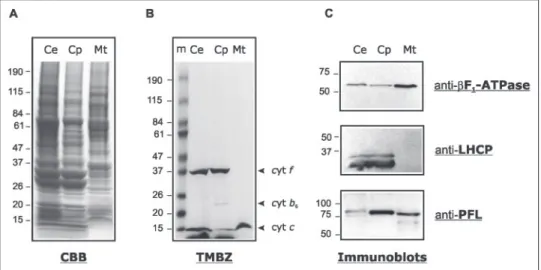
rein-hardtiicells were harvested, and disrupted by nebulization. Chloro-plasts and mitochondria were fractionated by differential centrifugation and further purified on Percoll gradients. To ascertain the purity of the organelle fractions, each was tested for the enrichment of specific marker proteins. Heme staining showed a low contamination of chlo-roplasts by mitochondrial cytochrome c550, whereas chloroplast
cyto-chrome f and cytocyto-chrome b6were not detected in mitochondrial
frac-tions (Fig. 4B). Furthermore, immunoblot analyses revealed that purified mitochondria lacked detectable amounts of light-harvesting complex proteins, whereas chloroplasts contained traces of mitochon-drial F0F1-ATPase (F1-ATPase) (Fig. 4C). These data indicated that
purified mitochondria were devoid of chloroplasts while chloroplasts were slightly contaminated with mitochondria. The same protein blots probed for PFL revealed the occurrence of the protein in both organelles (Fig. 4C). The electrophoretic mobility of the chloroplast and mitochon-drial PFL proteins differs by⬃3 kDa.
Protein Identification by Mass Spectrometry—Protein blot analysis of soluble and membrane fractions of C. reinhardtii mitochondria indi-cated that PFL is a soluble protein (not shown). Therefore, the soluble mitochondrial fraction was run on one-dimensional SDS-PAGE, dis-crete bands were excised from the gel (Fig. 5A) and treated for protein identification by tandem mass spectrometry (26). In the 60 –75-kDa
region, 22 peptides that matched the predicted PFL sequence were iden-tified. These peptides cover a large part of the full-length PFL, although no peptide corresponding to residues Met1–Lys107was identified (Fig.
5B). The coverage of the PFL sequence is comparable with that obtained for the mitochondrial aconitase, indicating that PFL is an abundant protein.
Proteome analysis also uncovered peptides matching proteins that form the PTA-ACK pathway, which in a number of bacteria reversibly interconverts acetyl-CoA and acetate (38). ACK (EC 2.7.2.1) phospho-rylates acetate to acetyl phosphate, which is then converted into acetyl-CoA by phosphotransacetylase (PTA, EC 2.3.1.8). Three tryptic pep-tides that match a putative PTA were identified in the 70 – 85-kDa range (Fig. 5A). All peptides are specific to annotated PTA1 (JGI C_870001), whereas no peptide corresponding to a second putative PTA, annotated PTA2 (C_330071), was obtained. The molecular mass range from PTA1 is 10 –20 kDa higher than the mass of 60 kDa calculated for annotated PTA1, suggesting that the gene model may be incomplete. In the 38 – 42-kDa range, six tryptic peptides that match an ACK sequence were identified. In the C. reinhardtii genome, two genes are annotated as acetate kinase. Whereas one of the six tryptic peptides is common to both ACK1 (C_170112) and ACK2 (C_330070), the other five are spe-cific to ACK2 (Fig. 5C). The molecular mass of ACK2 (Fig. 5A) is in agreement with the mass of 43 kDa calculated from the gene model. No peptide specific to PFL-AE or ADHE were identified in the fraction of soluble proteins, suggesting either that these proteins are present in very low amounts in the sample analyzed or that they are localized to another subcellular fraction.
DISCUSSION
Occurrence of PFL and PFL-AE in a Photosynthetic Eukaryote —Ox-ygen-sensitive formate production in C. reinhardtii cells under dark, anaerobic conditions had suggested the activity of pyruvate formate-lyase (19), an enzyme rare among eukaryotes. The present identification of PFL peptides in C. reinhardtii, along with cDNAs for PFL and PFL-AE, indicate the existence of a PFL system in the algae. Identified
C. reinhardtiiPFL and PFL-AE are well conserved with their counter-parts in bacteria and amitochondriate protists.
In its natural habitats, such as soil and fresh water ponds, C.
rein-hardtiiis exposed to anaerobiosis even more so in darkness. Short anaerobiosis in the dark (6 h) led to a clear increase of PFL transcript levels, but not of protein levels. Ambient levels of PFL in the algae may ensure readiness for immediate adaptation to fermentative conditions, FIGURE 4. Subcellular localization of PFL in C.
reinhardtii. Analysis of purified chloroplast (Cp)
and mitochondria (Mt) from mixotrophically grown cells (Ce). Proteins (60g) were loaded on a 4 –20% SDS-PAGE (A and B) or 10% SDS-PAGE (C).
A, Coomassie Blue (CBB)-stained gel; B, protein gel
stained for hemes using TMBZ: cyt f, cyt b6,
cyto-chromes of the chloroplast b6f complex; cyt c,
mitochondrial cytochrome c550; m, molecular
mass standards; C, immunoblots showing the dis-tribution of light-harvesting complex proteins, of subunit of the mitochondrial F0F1-ATPase, and
of PFL in isolated organelles.
at INRA Institut National de la Recherche Agronomique on June 15, 2018
http://www.jbc.org/
without the need for de novo protein synthesis. More detailed studies will be required to fully describe the regulation of the PFL at RNA, protein, and activity levels.
Compartmentalization of the PFL System—Anaerobic formate production in C. reinhardtii was previously measured in fractions enriched in chloroplasts and mitochondria (20), which suggested the presence of a PFL in both organelles. Here, immunoblots revealed the occurrence of PFL in chloroplasts and mitochondria purified from aerobically grown C. reinhardtii cells. Library screening iden-tified several PFL cDNAs corresponding to a single mRNA, and searching the draft of the C. reinhardtii genome sequence and the EST databases provided no evidence for two PFL genes and only one
PFLtranscript was detected in RNA blots. This may suggest that PFL is dual targeted to chloroplasts and mitochondria. Dual targeting is not uncommon in higher plants (39) but has not been previously reported in the green algae.
The⬃3-kDa difference observed between the chloroplast and the mitochondrial PFL forms might be explained by differential processing of the cytosolic precursor. Alternatively, the 3-kDa difference might be explained by differential activation and radical-induced cleavage at the
C-terminal end. However, it is not currently known whether C.
rein-hardtiiPFL may be subject to oxygenolytic cleavage as its counterparts in bacteria (2, 14, 15). In our mitochondrial proteome analysis, one peptide that matches with the C-terminal sequence of PFL was identi-fied. This peptide stems from (i) a protein of high molecular mass (⬃60–75 kDa) and (ii) a region of the PFL that is C-terminal to the glycine residue to be activated into a glycyl radical. In E. coli, PFL is a homodimer that is post-translationally activated by introduction of a radical on the ultimate Gly residue of only one of the two monomers (2, 15). Whether only one or both PFL subunits are activated in C.
rein-hardtiiis yet unknown.
The rationale behind the differential compartmentalization of PFL in
C. reinhardtiiis not obvious. Indeed, PFL produces formate, a toxic (but freely diffusible) end product. Plants, which do not possess PFL, have a formate dehydrogenase that oxidizes formate into CO2in the presence
of NAD⫹. Plant formate dehydrogenase is localized to the mitochondria (40). No biochemical or molecular evidence for the presence of a for-mate dehydrogenase has yet been reported in the green algae, which would agree with a role of PFL in core energy metabolism and the status of formate as a genuine end product.
FIGURE 5. Identification by mass spectrometry of three atypical eukaryote enzymes in C. reinhardtii mitochondria. A, Coomassie Blue-stained SDS-PAGE used for mass spectrometry analysis. The gel pieces in which peptides for the proteins of interest were identified are indicated. B, PFL sequence that shows the peptides identified are shaded in gray.
C, atypical eukaryotic enzymes identified in C. reinhardtii mitochondria. Tryptic peptides that match gene models for an acetate kinase and a phosphotransacetylase in C. reinhardtii
are listed. The peptides specific to the putative proteins are indicated in bold.
Typical Eubacterial Enzymes in C. reinhardtii Mitochondria
at INRA Institut National de la Recherche Agronomique on June 15, 2018
http://www.jbc.org/
Recycling Coenzyme A in C. reinhardtii Mitochondria—In E. coli, acetyl-CoA resulting from PFL activity is converted into either ethanol or acetate (38). Acetate dissimilation via the PTA-ACK pathway gener-ates one ATP molecule per acetyl-CoA but does not consume reducing equivalents. Ethanol, produced by ADHE, reoxidizes two molecules of NADH per acetyl-CoA without ATP production. In the anaerobe eukaryotes Giardia and Entamoeba, fermentative production of etha-nol is catalyzed by ADHE, whereas acetate is produced by ADP-forming acetyl-CoA synthase (EC 6.2.1.13) (1) (Fig. 6D). The ethanol to acetate ratio depends upon oxygen tension (1). In this sense, the ACK-PTA route, which produces ATP and acetate from acetyl-CoA, Pi, and ADP
in most bacteria would be functionally equivalent to the single enzyme acetyl-CoA synthase in amitochondriate eukaryotes.
Predicted C. reinhardtii ADHE exhibits features typical of ADHE from amitochondriate eukaryotes and bacteria. Under the conditions investi-gated here, i.e. light versus dark and aerobiosis versus anerobiosis, we failed to detect ADHE transcripts and protein (supplemental Fig. II). In this respect, the green algae differs from its colorless counterpart Polytomella sp. where ADHE is a major protein of the mitochondrial matrix in aerobi-cally grown cells (37) (Fig. 6C). The physiological conditions that lead to expression and activity of ADHE in the green algae are not known. Whether
C. reinhardtiiADHE is involved in the anaerobic production of ethanol, and concomitant regeneration of NAD⫹, remains to be determined.
Several peptides from a PTA-ACK pathway were identified in the soluble fraction of mitochondria isolated from aerobically grown algae. These peptides were specific to PTA1 and ACK2 but distinct from PTA2 and ACK1 whose genes are adjacent to HYDA2 (C_330072)
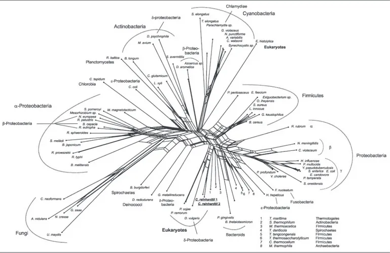
encoding the chloroplast hydrogenase of the algae (41). This gene orga-nization as well as the lack of peptides specific to PTA2 and ACK1 in isolated mitochondria suggest that these proteins are located to the chloroplast (Fig. 6). Phylogenetic networks for C. reinhardtii ACK1 (Fig. 7) and PTA2 (supplemental Fig. III) indicate a common ancestry for both enzymes with the corresponding homologues from the oomycete
Phytophthora sojae, suggesting that the two-step pathway that revers-ibly converts acetyl-CoA into acetate was present in the common ances-tor of oomycetes and plants (42). An ACK but no PTA was found in the amitochondriate eukaryote Entamoeba histolytica genome sequence (Fig. 7). The PTA-ACK system is widespread among prokaryotes (38). Notwithstanding the activity measurements by Kreuzberg et al. (20), ACK and PTA represent novel and typically eubacterial enzymes among eukaryotes. C. reinhardtii is capable of growing heterotrophically on acetate and it is commonly accepted that acetate is assimilated by an acetyl-CoA synthase and metabolized to triose following entry into the glyoxylate cycle (21, 43). The mitochondrial PTA-ACK pathway in C.
reinhardtiimay represent an alternative route for acetate assimilation.
Pyruvate Degrading Systems in the Green Algae C. reinhardtii-PFL Versus PFO—A gene coding for a putative pyruvate:ferredoxin oxi-doreductase (1303 amino acids) was identified in the C. reinhardtii genome (C_140055). Search of EST databases identified only two PFO clones, which correspond to a conserved N-terminal domain (clone BP093588; His241–Ala305) and to the predicted C terminus (BQ821311;
Lys1216–His1303). The PFO sequence inferred from the gene model exhibits four highly conserved PFO motifs including two [4Fe-4S] ferre-doxins, and the iron-sulfur binding region signature (Prosite PS00198) FIGURE 6. Localization and suggested functions of PFL, PFO, PTA, and ACK in C. reinhardtii and for ADHE in its close nonphotosynthetic relative Polytomella sp. Starch is accumulated inside the chloroplast during the light phase and is degraded in the dark. Starch breakdown is greater under anaerobic conditions than in aerobic conditions. In the absence of the functional TCA cycle (anaerobiosis), acetyl-CoA produced either via PFL or PFO enters the acetate-generating ACK-PTA route. The roles for PTA, ACK, and ADHE shown are dissimilatory. Nevertheless, a primarily assimilatory role for PTA-ACK and/or ADHE under some physiological conditions cannot currently be excluded because acetate is a carbon source for C. reinhardtii and ethanol is a source for Polytomella sp. (see text). Chloroplast pyruvate dehydrogenase is not represented. Enzymes for which the localization is not directly demonstrated are underlined. End products are boxed. Fd, ferredoxin; ACS, ADP-forming acetyl CoA synthase; PDH, pyruvate dehydrogenase.
at INRA Institut National de la Recherche Agronomique on June 15, 2018
http://www.jbc.org/
(Cys766–Cys777and Cys822–Cys833), which participate in electron
trans-fer to trans-ferredoxin (44). C. reinhardtii PFO shares 50% identity with PFOs of the strictly anaerobic, sulfate-reducing bacterium Desulfovibrio
vul-garisHildenborough (YP_012236), and the anaerobic parasite E.
histo-lytica (EAL51636) and 49% identity with mitochondrial pyruvate: NADP⫹oxidoreductase (PNO) of Euglena gracilis (Q94IN5). In the phylogenetic network, C. reinhardtii PFO shares common ancestry with other eukaryote PFO homologues (supplemental Fig. IV).
In contrast to Euglena pyruvate:NADP⫹oxidoreductase (44), C.
rein-hardtiiPFO does not exhibit a C-terminal NADPH-generating domain, suggesting that the algal enzyme probably accepts electrons from ferredoxin like its homologues from amitochondriate protists (1, 45). In C. reinhardtii, ferredoxin is present in the stroma where it donates electrons to ferredoxin:NADPH oxidoreductase but also to a hydro-genase (under anaerobic conditions) (46). We thus propose that
C. reinhardtiiPFO is localized to the stroma where under anaerobic
conditions its activity might be coupled to that of the hydrogenase via ferredoxin (Fig. 6A). Under anaerobiosis in the dark, C.
rein-hardtiicells produce large amounts of formate, whereas production of CO2is low (19), which might indicate that under these conditions
PFO is poorly involved in the survival of the algae. The anaerobic enzymes PFL and PFO in the green algae represent an unexpected biochemical and evolutionary link between energy metabolism in amitochondriate and mitochondriate protists.
C. reinhardtiipossesses genes for pyruvate dehydrogenase, PFL, and PFO. This 3-fold pyruvate-metabolizing repertoire is not uncommon in prokaryotes (38) but it is hitherto unique among eukaryotes. Together with the presence of ADHE and HYD, the enzymatic repertoire typical of eukaryote anaerobes (1) is almost completely present in C.
rein-hardtii. The only exception is that ADP-forming acetyl-CoA synthase is lacking, whereby the functionally equivalent PTA-ACK route is present. The large variety of metabolic abilities evidenced in this work are likely FIGURE 7. Neighbor-Net analysis of ACK sequences. Sources of sequences are as given. Phytophthora ramorum (Contig 21 in Scaffold 4 nucleotides 413776 – 415071), P. sojae (Contig 24 in Scaffold 12 reverse complement of nucleotides 795216 –796527), Anabaena variabilis (ZP_00160102), Aspergillus nidulans (EAA60992), Azoarcus sp. (YP_159859), Bacillus
cereus (NP_834343), Bacteroides thetaiotaomicron (AAO78798), Bifidobacterium longum (NP_696143), Borrelia burgdorferi (NP_212756), Bradyrhizobium japonicum (NP_770098), Brucella melitensis (AAL54122), Burkholderia cepacia (ZP_00217734), Campylobacter coli (ZP_00366841), Chlorobium tepidum (NP_662410), Chromobacterium violaceum (AAQ59206), Clostridium thermocellum (ZP_00311922), Corynebacterium glutamicum (BAC00146), Crocosphaera watsonii (ZP_00175966), Cryptococcus neoformans (EAL17325), Dechloromonas aromatica (ZP_00150711), Deinococcus radiodurans (AAF12139), Desulfotalea psychrophila (YP_064295), D. vulgaris (YP_012241), E. histolytica (EAL50605), Enterococcus faecium
(ZP_00287449), Erwinia carotovora (YP_051129), E. coli (NP_754724), Exiguobacterium sp. (ZP_00182864), Fusobacterium nucleatum (AAL95367), Geobacillus kaustophilus (YP_148638), Geobacter metallireducens (ZP_00299464), Gibberella zeae (EAA70606), Gloeobacter violaceus (NP_923946), Haemophilus influenzae (ZP_00154390), Helicobacter
hepati-cus (AAP77905), Leifsonia xyli (YP_061463), Listeria innocua (NP_470952), Magnetospirillum magnetotacticum (ZP_00053788), Mesorhizobium sp. (ZP_00197262), Methanosarcina thermophila (B49338), Moorella thermoacetica (ZP_00331156), Mycobacterium avium (NP_962820), Neisseria meningitidis (AAF41874), Neurospora crassa (CAE76499), Nitrosomonas europaea (NP_842138), Nostoc punctiforme (ZP_00111397), Oceanobacillus iheyensis (NP_693112), Parachlamydia sp. (YP_008361), Pasteurella multocida (NP_245641), Pediococcus pentosaceus (ZP_00323037), Photobacterium profundum (YP_130972), Photorhabdus temperata (AAN08359), Porphyromonas gingivalis (AAQ66195), Ralstonia eutropha
(ZP_00202877), Rhodobacter sphaeroides (ZP_00007961), Rhodopirellula baltica (NP_869003), Rhodopseudomonas palustris (NP_949900), Rhodospirillum rubrum (ZP_00269820),
Rickettsia prowazekii (NP_220502), Rickettsia typhi (YP_066996), Salmonella enterica (NP_804384), Shewanella oneidensis (NP_718485), Silicibacter pomeroyi (AAV93446), Sinorhizo-bium meliloti (AAD24358), Staphylococcus aureus (BAB57873), Streptomyces avermitilis (BAC70535), Symbiobacterium thermophilum (YP_076414), Synechococcus elongatus
(ZP_00165089), Synechocystis sp. (NP_440508), Thermoanaerobacter tengcongensis (NP_623096), Thermoanaerobacterium thermosaccharolyticum (CAA95986), Thermosynechococcus
elongatus (NP_683130), Thermotoga maritima (NP_228087), Treponema denticola (NP_971543), Ustilago maydis (EAK84551), Vibrio cholerae (AAF94257), and Yersinia pseudotubercu-losis (YP_071107).
Typical Eubacterial Enzymes in C. reinhardtii Mitochondria
at INRA Institut National de la Recherche Agronomique on June 15, 2018
http://www.jbc.org/
helping the photosynthetic algae to optimize its metabolism to the ever changing environmental conditions.
Acknowledgments—We thank Dr. J. Davies for providing theC. reinhardtii cDNA library, Dr. S. I. Beale for the use of the BioNeb, and M. Ter Braak for N. frontalis cells. The sequence data were produced by the United States Department of Energy Joint Genome Institute.
REFERENCES
1. Mu¨ller, M. (2003) in Molecular Medical Parasitology (Marr, J., ed) pp. 125–139, Academic Press, London
2. Wagner, A. F., Frey, M., Neugebauer, F. A., Scha¨fer, W., and Knappe, J. (1992) Proc.
Natl Acad. Sci. U. S. A. 89,996 –1000
3. Andersson, S. G., and Kurland, C. G. (1999) Curr. Opin. Microbiol. 2, 535–541 4. Dyall, S. D., Yan, W. H., Delgadillo-Correa, M. G., Lunceford, A., Loo, J. A., Clarke,
C. F., and Johnson, P. J. (2004) Nature 431, 1103–1107 5. Martin, W., and Mu¨ller, M. (1998) Nature 392, 37– 41
6. Tielens, A. G., Rotte, C., van Hellemond, J. J., and Martin, W. (2002) Trends Biochem.
Sci. 27,564 –572
7. Hrdy, I., Hirt, R. P., Dolezal, P., Bardonova, L., Foster, P. G., Tachezy, J., and Embley, T. M. (2004) Nature 432, 618 – 622
8. Boxma, B., de Graaf, R. M., van der Staay, G. W., van Alen, T. A., Ricard, G., Gabaldon, T., van Hoek, A. H., Moon-van der Staay, S. Y., Koopman, W. J., van Hellemond, J. J., Tielens, A. G., Friedrich, T., Veenhuis, M., Huynen, M. A., and Hackstein, J. H. (2005)
Nature 434,74 –79
9. Sawers, G., and Bock, A. (1988) J. Bacteriol. 170, 5330 –5336 10. Knappe, J., and Sawers, G. (1990) FEMS Microbiol. Rev. 6, 383–398
11. Pecher, A., Blaschkowski, H. P., Knappe, K., and Bock, A. (1982) Arch. Microbiol. 132, 365–371
12. Broderick, J. B., Henshaw, T. F., Cheek, J., Wojtuszewski, K., Smith, S. R., Trojan, M. R., McGhan, R. M., Kopf, A., Kibbey, M., and Broderick, W. E. (2000) Biochem.
Biophys. Res. Commun. 269,451– 456
13. Zhang, W., Wong, K. K., Magliozzo, R. S., and Kozarich, J. W. (2001) Biochemistry 40, 4123– 4130
14. Asanuma, N., Iwamoto, M., and Hino, T. (1999) Microbiology 145, 151–157 15. Melchiorsen, C. R., Jokumsen, K. V., Villadsen, J., Johnsen, M. G., Israelsen, H., and
Arnau, J. (2000) 182, 4783– 4788
16. Kessler, D., Herth, W., and Knappe, J. (1992) J. Biol. Chem. 267, 18073–18079 17. Asanuma, N., Yoshii, T., and Hino, T. (2004) Arch. Microbiol. 181, 122–128 18. Akhmanova, A., Voncken, F. G., Hosea, K. M., Harhangi, H., Keltjens, J. T., op den
Camp, H. J., Vogels, G. D., and Hackstein, J. H. (1999) Mol. Microbiol. 32, 1103–1114
19. Kreuzberg, K. (1984) Physiol. Plant. 61, 87–94
20. Kreuzberg, K., Klo¨ch, G., and Großheiser, D. (1987) Physiol. Plant. 69, 481– 488 21. Harris, E. H. (1989) The Chlamydomonas Sourcebook: A Comprehensive Guide to
Biology and Laboratory Use, Academic Press, San Diego, CA 22. Franze´n, L. G., and Falk, G. (1992) Plant Mol. Biol. 19, 771–780 23. Bollivar, D. W., and Beale, S. I. (1996) Plant Physiol. 112, 105–114
24. Eriksson, M., Gadestro¨m, P., and Samuelsson, G. (1995) Plant Physiol. 107, 479 – 483 25. Thomas, P. E., Ryan, D., and Levin, W. (1976) Anal. Biochem. 75, 168 –176 26. Ferro, M., Salvi, D., Rivie`re-Rolland, H., Vermat, T., Seigneurin-Berny, D., Garin, J.,
Joyard, J., and Rolland, N. (2002) Proc. Natl. Acad. Sci. U. S. A. 99, 11487–11492 27. Thompson, J. D., Higgins, D. G., and Gibson, T. J. (1994) Nucleic Acids Res. 22,
4673– 4680
28. Lockhart, P. J., Steel, M. A., Hendy, M. D., and Penny, D. (1994) Mol. Biol. Evol. 11, 605– 612
29. Thollesson, M. (2004) Bioinformatics 20, 416 – 418
30. Bryant, D., and Moulton, V. (2004) Mol. Biol. Evol. 21, 255–265 31. Saitou, N., and Nei, M. (1987) Mol. Biol. Evol. 4, 406 – 425 32. Huson, D. H. (1998) Bioinformatics 14, 68 –73
33. Raynaud, C., Sarcabal, P., Meynial-Salles, I., Croux, C., and Soucaille, P. (2003) Proc.
Natl. Acad. Sci. U. S. A. 100,5010 –5015
34. Lehtio¨, L., and Goldman, A. (2004) Protein Eng. Des. Sel. 17, 545–552 35. Himo, F. (2005) Biochim. Biophys. Acta. 1707, 24 –33
36. Sofia, H. J., Chen, G., Hetzler, B. G., Reyes-Spindola, J. F., and Miller, N. E. (2001)
Nucleic Acids Res. 29,1097–1106
37. Atteia, A., van Lis, R., Mendoza-Herna´ndez, G., Henze, K., Martin, W., Riveros-Rosas, H., and Gonza´lez-Halphen, D. (2003) Plant Mol. Biol. 53, 175–188
38. Wolfe, A. J. (2005) Microbiol. Mol. Biol. Rev. 69, 12–50 39. Karniely, S., and Pines, O. (2005) EMBO Rep. 6, 420 – 425
40. Colas des Francs-Small, C., Ambard-Bretteville, F., Small, I. D., and Remy, R. (1993)
Plant Physiol. 102,1171–1177
41. Forestier, M., King, P., Zhang, L., Posewitz, M., Schwarzer, S., Happe, T., Ghirardi, M. L., and Seibert, M. (2003) Eur. J. Biochem. 270, 2750 –2758
42. Stechmann, A., and Cavalier-Smith, T. (2002) Science 297, 89 –91
43. Heifetz, P. B., Forster, B., Osmond, C. B., Giles, L. J., and Boynton, J. E. (2000) Plant
Physiol. 122,1439 –1445
44. Rotte, C., Stejskal, F., Zhu, G., Keithly, J. S., and Martin, W. (2001) Mol. Biol. Evol. 18, 710 –720
45. Horner, D. S., Hirt, R. P., and Embley, T. M. (1999) Mol. Biol. Evol. 16, 1280 –1291 46. Happe, T., and Kaminski, A (2002) Eur. J. Biochem. 269, 1022–1032
47. Silflow, C. D. (1998) in The Molecular Biology of Chloroplasts and Mitochondria in
Chlamydomonas(Rochaix, J. D., Goldschmidt-Clermont, M., and Merchant, S., eds) pp. 25– 40, Kluwer Academic Publishers, Dordrecht, The Netherlands
48. Ro¨del, W., Plaga, W., Frank, R., and Knappe, J. (1988) Eur. J. Biochem. 177, 153–158
at INRA Institut National de la Recherche Agronomique on June 15, 2018
http://www.jbc.org/
Jacques Joyard, Norbert Rolland and William Martin
Ariane Atteia, Robert van Lis, Gabriel Gelius-Dietrich, Annie Adrait, Jérôme Garin,
Mitochondria
Chlamydomonas
Pyruvate Formate-lyase and a Novel Route of Eukaryotic ATP Synthesis in
doi: 10.1074/jbc.M507862200 originally published online February 1, 2006
2006, 281:9909-9918.
J. Biol. Chem.
10.1074/jbc.M507862200
Access the most updated version of this article at doi:
Alerts:
When a correction for this article is posted
•
When this article is cited
•
to choose from all of JBC's e-mail alerts
Click here
Supplemental material:
http://www.jbc.org/content/suppl/2006/02/02/M507862200.DC1
http://www.jbc.org/content/281/15/9909.full.html#ref-list-1
This article cites 44 references, 12 of which can be accessed free at
at INRA Institut National de la Recherche Agronomique on June 15, 2018
http://www.jbc.org/