HAL Id: tel-00011478
https://tel.archives-ouvertes.fr/tel-00011478
Submitted on 27 Jan 2006
HAL is a multi-disciplinary open access archive for the deposit and dissemination of sci-entific research documents, whether they are pub-lished or not. The documents may come from teaching and research institutions in France or abroad, or from public or private research centers.
L’archive ouverte pluridisciplinaire HAL, est destinée au dépôt et à la diffusion de documents scientifiques de niveau recherche, publiés ou non, émanant des établissements d’enseignement et de recherche français ou étrangers, des laboratoires publics ou privés.
Structural studies on subunits τ 60/τ 91 of yeast
transcription factor IIIC (TFIIIC).
Anastasia Mylona
To cite this version:
Anastasia Mylona. Structural studies on subunits τ 60/τ 91 of yeast transcription factor IIIC (TFIIIC).. Biomolecules [q-bio.BM]. Université Joseph-Fourier - Grenoble I, 2005. English. �tel-00011478�
Université Grenoble I – Joseph Fourier
THÈSE
présentée par
Anastasia MYLONA
Pour obtenir le grade de
Docteur de l’Université Grenoble I – Joseph Fourier
Spécialité : Biologie
Structural studies on subunits τ60/τ91 of yeast
transcription factor IIIC (TFIIIC)
Soutenue publiquement le 18 Juillet 2005 devant le jury composé de:
Dr. Christoph W. Müller Directeur de thèse
Pr. Hans Geiselmann Président du Jury
Dr. Jürg Müller Rapporteur
Pr. Herman Van Tilbeurgh Rapporteur
Dr. Andrea Dessen Examinatrice
Thèse préparée au sein du Laboratoire Européen de Biologie Moléculaire (European Molecular Biology Laboratory, Grenoble Outstation)
Acknowledgements
I am grateful to my supervisor Dr. Christoph Müller for his guidance throughout my thesis.
Many thanks to Dr. Carlos Fernández-Tornero for all the precious help and work that he did in the crystallographic part of my PhD thesis and for being a very good teacher. Ι also thank Martine Moulin and Uli Steuerwald for their help and advice, Dr. Pierre Legrand for helping to solve the ‘phase problem’, Dr. Carlo Petosa for many suggestions and for proofreading the manuscript of my thesis, Dr. Patrice Morand for correcting all the french texts in the manuscript, and all the staff, scientific and non-scientific, at the EMBL for their help.
I would like to thank Dr. Joël Acker for all the help and advice in biochemistry, and Dr. Christian Marck for all the interesting discussions and Dr. André Sentenac for the fruitful collaboration we had.
I would also like to thank the members of my thesis advisory committee Dr. Jürg Müller, Dr. Winfried Weissenhorn and Prof. Hans Geiselmann for all the suggestions and discussions. Σε όλους µου τους φίλους ένα ευχαριστώ γιατί είσαστε πάντα εδώ αυτά τα τέσσερα χρόνια αν και µακριά και γιατί χωρίς εσάς η ζωή δε θα ήταν τόσο ωραία! Στους γονείς µου ένα µεγάλο ευχαριστώ γιατί είναι πάντα δίπλα µου αλλά και γιατί χωρίς αυτούς οι επιλογές του µυαλού και της ψυχής µου µπορεί να ήταν διαφορετικές. Στο Νίκο για την αγάπη και την υποστήριξή του όλα αυτά τα χρόνια αλλά και γιατί µου έµαθε να είµαι αισιόδοξη και να ξεχωρίζω και να εκτιµώ τα σηµαντικά από τα ασήµαντα πράγµατα στη ζωή.
Résumé
TFIIIC est un facteur de transcription de classe III qui se lie spécifiquement et de façon stable sur les boîtes A et B des promoteurs des gènes de l’ARNt. TFIIIC est composé de 6 sous-unités: τ138, τ131, τ95, τ91, τ60 et τ55. Ce travail présente la structure du complexe entre τ60 et la partie C-terminale de τ91 (∆τ91) qui a été résolue à 3.2 Å. La structure comporte trois régions. a) ∆τ91 qui est un β-propeller; b) une partie N-terminale de τ60 qui est aussi un β-propeller et qui se trouve entre ∆τ91 et c) le domaine C-terminal de τ60, qui a un nouveau rempliement. L’interaction entre ∆τ91 et τ60 est formée par les deux β-propellers et la partie C-terminale de τ60 est complètement indépendante. Cette nouvelle interaction β-propeller - β-propeller apparaît importante pour l’assemblage d’un complexe τB stable, capable de se fixer à l’ADN. Nos résultats donnent des informations sur le mécanisme d’assemblage du complexe τB et sa liaison à l’ADN.
Mots de clés : - transcription
- ARN polymérase III - TFIIIC
- structure de cristal - β-propeller
Summary
TFIIIC is a DNA binding complex that serves as an assembly factor in class III genes. It is a multisubunit protein of about 600 kDa organized in two large domains, τA and τB, of similar size and mass. TFIIIC consists of six polypeptides τ138, τ131, τ95, τ91, τ60 and τ55. τ138, τ91 and τ60 make up the τB subcomplex and τ131, τ95 and τ55 belong to the τA subassembly. This work presents the X-ray crystal structure of yeast τ60 complexed with the C-terminal part of τ91 at 3.2 Å resolution. τ60 consists of a N-terminal β-propeller domain and a C-terminal domain that is an overall new fold and very likely is
the part that links τA with τB and contacts the TBP. ∆τ91 is also a β-propeller. The interacting region between the two proteins involves residues in the two β-propellers, whereas the C-terminal domain of τ60 does not contribute at all to the interaction. This novel β-propeller – β-propeller interaction appears to be of great importance for the formation of a very stable τB complex able to bind with high affinity and stability to the B box. Our results provide a starting point for further structural and functional studies aimed at elucidating the mechanism of preinitiation complex formation in the RNA polymerase III transcription machinery.
Keywords:
- transcription
- RNA polymerase III - TFIIIC
- crystal structure - β-propeller
Contents
Part I
...3Introduction
...3Résumé ...4
Eukaryotic transcription
...61.1. RNA polymerase III promoters...7
1.2. RNA polymerase III ...9
1.3. The general factors of transcription system III ...11
1.3.1. TFIIIA ...11
1.3.2. TFIIIC...12
1.3.3. Human TFIIIC ...16
1.3.4. TFIIIB...16
1.4. RNA polymerase III recruitment by TFIIIB ...18
1.5. TFIIIC contacts RNA polymerase III...19
1.6. Termination and recycling...19
1.7. Scope of this thesis ...21
Part II
...22Materials and Methods
...22Résumé ...23
2.1. Molecular Biology and Biochemical Techniques
..242.1.1. Cloning ...24
2.1.2. Protein Expression, Purification and Complex Assembly ...24
2.1.2.1. Production of seleno-methionine substituted protein ...27
2.1.3. Determination of protein concentration ...27
2.1.4. Limited proteolysis ...28
2.1.5. Southern - Western blot ...28
2.2. Crystallogenesis and Crystallographic Methods
...292.2.1. Crystallization...29
2.2.2. Introduction to X-ray crystallography ...33
2.2.3. Data collection and processing ...35
2.2.3.1 Cryogenic protection of crystals...35
2.2.3.2. Data collection...35
2.2.3.3. Data processing ...37
2.2.4. Methods for solving the phase problem...38
2.2.5. Building, refinement and validation of the model...42
2.2.5.1. Model building ...42 2.2.5.2. Refinement ...42 2.2.5.4. Validation ...44
Part III
...46Results
...46 Résumé ...473.1 Expression, proteolytic analysis, reconstitution
and crystallization of the τ60/τ91 complex
...483.1.1. Expression and purification of τ60 and τ91 proteins...48
3.1.3. Crystallization of the τ60/τ91 full-length complex...49
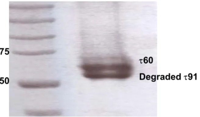
3.1.4. Instability of the τ60/τ91 complex at 20 oC...50
3.1.5. Expression and purification of the protease resistant C-terminal τ91 fragment (∆τ91) followed by τ60/∆τ91 complex formation ...54
3.1.6. Crystallization of the τ60/∆τ91 subcomplex...55
3.1.6.1. Attempts to improve the resolution of the τ60/∆τ91 crystals ...59
3.1.6.2. Co-crystallization with heavy atom derivatives...61
3.2. Data collection and structure determination
...633.2.1. Cryo-crystallography ...63
3.2.2. Data collection and processing ...63
3.2.3. Structure determination...67
3.2.4. Model building and refinement ...73
3.2.5. Crystal packing...77
3.3. Structure of the τ60/∆τ91 complex
...793.3.1. Overall Structure of the τ60/∆τ91 complex...79
3.3.2. Comparison between the two β-propellers ...82
3.3.3. The C-terminal domain of τ60: a new fold...87
3.3.4. τ60/∆τ91 interaction: A β-propeller – β-propeller interaction ...89
3.3.5. DNA binding activity of τ91, τ60 and ∆τ91 ...92
Part IV
...94Discussion and Perspectives
...94Résumé ...95
4.1. The overall architecture of the complex is conserved among different organisms...96
4.2. Importance of the β propeller – β propeller interaction...101
4.3. Possible interactions of the τ60/∆τ91 complex with other TFIIIC subunits...105
4.4. Concluding remarks and perspectives...107
Appendix
...108Part I
Introduction
Résumé
L’expression de l’information génétique est un processus complexe dont la première étape est la transcription de l’ADN en ARN, catalysée par une ARN polymérase. Cette première étape se décompose en trois phases :
• L’initiation consistant en l’assemblage de la machinerie transcriptionnelle sur un site spécifique du gène à transcrire.
• L’élongation correspondant à la synthèse de l’ARN.
• La terminaison permettant la libération du brin d’ARN et de l’ARN polymérase de la matrice d’ADN.
Chez les eucaryotes, on distingue trois formes d’ARN polymérases, définissant trois classes de gènes :
• L’ ARN polymérase I transcrit les gènes de classe I, codant pour l’ARN ribosomique.
• L’ARN polymérase II transcrit les gènes de classe II, codant pour les ARN messagers ainsi que plusieurs petits ARN nucléolaires non traduits.
• L’ARN polymérase III transcrit les gènes de classe III, codant pour l’ARN ribosomal 5S et des petits ARN stables et non traduits tels que les ARN de transfert.
L’ARN polymérase III de la levure S. cerevisiae est un complexe protéique de haut poids moléculaire (~700 kDa) avec 17 sous-unités. Les gènes transcrits par l’ARN polymérase III codent pour une variété des petites molécules d’ARN stables (< 400 pb) et non traduits, comme les ARNt, l’ARNr 5S, 7SL, U6, et VA. Beaucoup de ces petits ARN ont une fonction essentielle dans le métabolisme cellulaire. Les promoteurs des gènes transcrits par l’ARN polymérase III ont la caractéristique de contenir fréquemment des séquences essentielles, localisés dans les régions transcrites des gènes. Ils ont pour la plupart une structure discontinue dénommée IRC (Région Interne de Contrôle). Elles sont constituées de blocs de séquences très conservés entre les différents genres et espèces.
Les facteurs généraux de transcription de classe III sont: TFIIIA, TFIIIC et TFIIIB. TFIIIB et TFIIIC sont nécessaires et suffisants pour transcrire la plupart des gènes de classe III in vitro. En revanche, la transcription du gène de l’ARNr 5S nécessite également TFIIIA. TFIIIB est le facteur d’initiation et ne peut se lier qu’à un complexe TFIIIC-ADN. TFIIIC, encore appelé tau (τ), est un complexe protéique de 600 kDa. Il se lie spécifiquement et de façon stable sur les boîtes A et B des promoteurs des gènes de l’ARNt. La boîte B est responsable de l’ancrage de TFIIIC sur le gène et active la transcription en augmentant la capacité de TFIIIC à fixer la boîte. De plus, l’interaction avec la boîte A est une étape importante pour la détermination du site d’initiation de l’ARN polymérase III. TFIIIC a 6 sous-unités: τ138, τ131, τ95, τ91, τ60, τ55.
Cette thèse présente la structure du complexe entre τ60 et la partie C-terminal de τ91. Nos résultats donnent des informations intéressantes sur le mécanisme de l’assemblage de τB complexe et sur sa liaison à l’ADN.
Eukaryotic transcription
The most important and critical step in the transcription of eukaryotic genes is the recognition of promoter elements by RNA polymerase. In eukaryotes, there are three RNA polymerases which are highly related: RNA polymerase I, II and III. Each of these RNA polymerases transcribes specific genes, and each depends on protein factors, known as transcription factors. The role of the transcription factors is to recognize the promoter sequences of the genes transcribed by the RNA polymerases. RNA polymerase I transcribes the ribosomal RNA genes, which contain a single promoter structure. RNA polymerase II is responsible for the transcription of the mRNA genes as well as of some small nuclear RNA (snRNA) genes. The RNA polymerase II promoters consist of a core region, which is sufficient for directing transcription in vitro, and a regulatory region. The regulatory regions show a high diversity in structure, reflecting the large variety in mechanisms by which cellular proteins are synthesized and the need for complex regulation of these mechanisms. The genes transcribed by RNA polymerase III are generally shorter than 400 base pairs (bp) and encode RNA molecules involved in translation, splicing and tRNA processing, such as tRNA, 5S RNA, U6 spliceosomal RNA, 7SL, and RNase P RNA, as well as RNAs of unknown function (White, 1998; Roberts et al., 2003; Harismendy et al., 2003; Moqtaderi and Struhl, 2004). They display a diversity of promoter organization with internal and/or external elements, but are not as diverse as the RNA polymerase II promoters. RNA polymerase III can be specifically recruited at the transcription start site via a cascade of protein-DNA and protein-protein interactions, which vary each time according to the promoter elements.
1.1. RNA polymerase III promoters
In general, the transcription of RNA polymerase III genes is under the control of intragenic DNA sequences (Ciliberto et al., 1983; Sharp et al., 1981). These internal control regions (ICR) are discontinuous structures, with essential “boxes” separated by nonessential nucleotides, and show a high sequence conservation (Pieler et al., 1985a & 1985b). Sequences located upstream of the transcriptional start site also affect the activities of many RNA polymerase III genes in numerous organisms (White, 1998). The main characteristics of these sequences are that they are not conserved between organisms or between RNA polymerase III genes of the same organism, and that the degree to which they determine activity of the genes varies a lot.
According to the existence of different promoter elements and the participation of different transcription factors, RNA polymerase III genes have been divided into three types (reviewed in Geiduschek and Kassavetis, 2001; Willis, 1993; Schramm and Hernandez, 2002) (Figure 1.1):
• Type 1 genes include the 5S RNA genes whose promoters are characterized by three intragenic sequence elements: a 5’ A box, an intermediate element (IE) and a 3’ C box, which are conserved in the 5S promoters of different species. These elements together span approximately 50 bp, begin at about position +45 and make up the ICR. In the S. cerevisiae 5S genes only the C box is required for transcription.
• Type 2 genes include tRNA genes, adenovirus virus-associated (VA) genes, Alu sequences and other short elements. The promoters of these genes are also internal and contain two highly conserved sequence elements, a proximal A box and a more distal B box, within the transcribed region. The conservation between various species probably is partly due to the fact that the two boxes encode the D and T-loops of tRNAs which are required for the function of tRNAs. The A box is located 10-20 bp from the transcription start site, and this reflects a role in start site selection. The distance between the A and B boxes varies. Distances between ~ 30-60 bp are optimal for transcription, although a distance of 365 bp can be tolerated. Another control element, the
polymerase III terminator, is located 20-25 bp downstream from the B box of tRNA genes.
• Type 3 genes include the vertebrate U6, 7SK, hY4, hY5 and H1 snRNA genes, which contain an upstream TATA element. The well-characterized promoter of the human U6 snRNA gene consists of an upstream TATA box, a proximal sequence element (PSE) and a distal sequence element (DSE). The PSE together with the TATA element are involved in the recruitment of the transcription factors (TFs) SNAPc/PTF and a TFIIIB-like activity (TFIIIB-a). The SNAPc complex is also used by RNA polymerase II as well as TFs for the transcription of U1 (and several other) snRNA genes, although the latter do not contain TATA elements. The transcriptional activator Oct-1 is recruited by the upstream DSE and functions in part by promoting binding of the SNAPc complex to the PSE. Interactions between TFIIIB, SNAPc and Oct-1 bound to their promoter elements lead to a stable initiation complex, which is in part mediated by a nucleosome that is located between the DSE and the PSE.
Figure 1.1 Different types of RNA polymerase III promoters. The type 1 promoter of the Xenopus laevis 5S RNA gene consists of an internal control region (ICR), which can be subdivided into an A
box (+50 to +60), intermediate element (IE, +67 to +72), and C box (+80 to +90). The type 2 promoter of the X. laevis tRNALeu gene consists of an A box (+8 to +19) and a B box (+52 to +62). The type
3 promoter of the Homo sapiens U6 snRNA gene consists of a distal sequence element (DSE, 215 to 240) that enhances transcription and a core promoter composed of a proximal sequence element (PSE, 65 to 48) and a TATA box ( 32 to 25).
1.2. RNA polymerase III
RNA polymerase III is well defined in S. cerevisiae and consists of 17 subunits. All the corresponding genes except for C37 have shown to be essential for cell viability (reviewed in Chedin et al., 1998). Of the 17 subunits, five are unique to RNA polymerase III, and the rest either have paralogues or are identical to RNA polymerase II or I subunits (Table 1.1).
C160 and C128 form the major surface for DNA binding and the active site of the enzyme is located between these two subunits. Subunits AC40, ABC23 and AC19 also belong to the catalytic core. Four additional common polypeptides (ABC27, ABC14.5, ABC10α and ABC10β) are located in the periphery of the complex. The C82, C34, and C31 subunits dissociate from a yeast enzyme carrying a mutation within the zinc finger domain of the largest subunit, and each subunit interacts with the other two in a yeast two-hybrid assay. This suggests that these three subunits form a subcomplex which can be detached from the rest of the enzyme (Werner et al., 1992, 1993). The subunits in the subcomplex are required for initiation (Thuillier et al., 1995; Brun et al., 1997; Wang and Roeder, 1997). C17 and C25 are similar to the RNA polymerase II Rpb4 and Rpb7 subunits (Siaut et al., 2003). C11 is required for the RNA cleavage activity of RNA polymerase III (Chedin et al., 1998) and is thought to interact with subunits C37 and C53.
Table 1.1. Subunit composition of RNA polymerase III.
Module RNAPIII RNAPII Sequence
identity (%) C160 Rpb1 34 C128 Rpb2 39 AC40 Rpb3 21 ABC23 Rpb6 100 Core subunits AC19 Rpb11 18 ABC27 Rpb5 100 ABC14.5 Rpb8 100 ABC10α Rpb12 100 Peripheral subunits ABC10β Rpb10 100 C25 Rpb7 20 Stalk subunits C17 Rpb4 22 C53 - - C37 - - RNAPIII specific complex I C11 Rpb9 26 C82 - - C34 - - RNAPIII specific complex II C31 - -
1.3. The general factors of transcription system III
General transcription factors specific for RNA polymerase III have been best studied in S. cerevisiae and human. Ten essential genes encode the subunits of the S.
cerevisiae RNA polymerase III transcription factors: TFIIIA; TFIIIC, composed of
six subunits; and TFIIIB, consisting of the TATA-binding protein (TBP), the TFIIB-related factor (Brf), and B’’(B double prime, Bdp), which is the central RNA polymerase III recruiting factor. TFIIIC recruits TFIIIB onto the DNA upstream of the transcriptional start. In the case of the 5S rRNA genes TFIIIA binds first to DNA and forms the platform for binding TFIIIC to these genes (Table 1.2).
Table 1.2. RNA polymerase III transcription factors.
TFIIIC
(6 subunits) Assembly factor
A & B box (tRNA & 5S RNA
genes)
assembly of TFIIIB upstream of the transcription start site
TFIIIA
(1 subunit) Assembly factor
C box (5S RNA genes)
assembly of TFIIIB upstream of the transcription start site
TFIIIB
(3 subunits) Initiation factor
Upstream (tRNA & 5S RNA
genes)
responsible for RNA polymerase III
recruitment
1.3.1. TFIIIA
In natural RNA polymerase III promoters, TFIIIB is recruited to the DNA mainly through protein-protein interactions with TFIIIC. The type 1 5S promoters and type 2 promoters both use TFIIIC, but on the 5S promoters TFIIIC is recruited through TFIIIA. TFIIIA belongs to the C2H2 zinc finger family of DNA-binding proteins
(Miller et al., 1985) and contains nine C2H2 zinc fingers. In S. cerevisiae, the only
essential role of TFIIIA is the transcription of the 5S RNA gene, because strains engineered to express 5S rRNA from a tRNA promoter and lacking TFIIIA are viable (Camier et al., 1995). X. laevis TFIIIA was the first eukaryotic transcription factor that was purified (Engelke et al., 1980) and the first for which the cDNA was isolated (Ginsberg et al., 1984). Little is known about how TFIIIA recruits TFIIIC to the
DNA. X. laevis TFIIIA contains a 14-amino acid domain located at the C-terminal end of the ninth zinc-finger that is not necessary for DNA binding but essential for transcription (Mao and Darby, 1993). In S. cerevisiae TFIIIA, a hydrophobic segment in an 84-amino-acid region located between zinc fingers 8 and 9 is also required for cell viability and transcription but not for DNA binding (Rowland and Segall, 1998). These protein domains may play a role in the recruitment of TFIIIC.
1.3.2. TFIIIC
In tRNA genes, the first step during preinitiation complex formation is the binding of TFIIIC to the internal A and B boxes, which is then followed by TFIIIB and RNA polymerase III recruitment, through interactions with TFIIIC. TFIIIB by itself does not bind to RNA polymerase III genes lacking a TATA element but, once it is assembled by TFIIIC, it interacts with DNA, recruits RNA polymerase III and is sufficient for directing accurate initiation by RNA polymerase III during multiple rounds of transcription (reviewed in Geiduschek and Kassavetis, 2001; Schramm and Hernandez, 2002). Hence, TFIIIC acts as an assembly factor (Figure 1.2). However, it has been shown that TFIIIC is a multifunctional protein, involved not only in promoter recognition and TFIIIB recruitment but also in the displacement of nucleosomes to relieve the repression of transcription by chromatin (Burnol et al., 1993). In addition, it is also involved in the termination and reinitiation process, and also very likely in RNA polymerase III recruitment (see 1.5 & 1.6).
Biochemical and genetic studies have led to an extensive characterization of TFIIIC in yeast. S. cerevisiae TFIIIC is a multisubunit protein of about 600 kDa organized in two large subassemblies, τA and τB, of similar mass (300 kDa) and size (10 nm diameter), as shown by scanning transmission electron microscopy (Schultz et al., 1989). τB binds with high affinity to the highly conserved B block of tRNA genes, whereas τA binds to the second well conserved A block. This binding is weaker and mostly B block dependent (Baker et al., 1986; Stillman et al., 1984). The link between the τA and τB subcomplexes shows remarkable flexibility, as it can adapt to different distances separating the A and B blocks in natural tRNA genes (Baker et al., 1987). The chromatographic separation of τA and τB has never been observed, but limited
proteolysis yields a τB subcomplex which is active for B block binding (Marzouki et al., 1986). Affinity-purified yeast TFIIIC consists of six polypeptides: τ138, τ131, τ95, τ91, τ60 and τ55 (Parsons and Weil, 1990; Gabrielsen et al., 1989; Bartholomew et al., 1990) (Table 1.3).
τ138, τ91 and τ60 belong to the τB subcomplex (Arrebola et al., 1998; Deprez et al., 1999). Based on protein-DNA cross-linking experiments, τ138 was mapped over the B block and τ91 at the most 3’ location of TFIIIC-5S RNA gene complexes (Bartholomew et al., 1990; Braun et al., 1992). These two subunits appear to be predominant for the DNA binding activity (Gabrielsen et al., 1989; Arrebola et al., 1998). τ138 is predicted to have two HMG (High Mobility Group) motifs (Lefebvre et al., 1992) which are DNA binding motifs and it cooperates with τ91 in DNA binding, which is predicted to contain an AT-hook (HMG DNA binding motif) domain at the N-terminal region. A temperature sensitive mutation in τ138 (tfc3-G349E) decreases the affinity of TFIIIC for the B box (Lefebvre et al., 1994). A point mutation in τ91 (τ91-E330K) suppresses the thermosensitive phenotype of the tfc3-G349E mutant. This suppressor mutation rescued the DNA binding and transcription defects of mutant TFIIIC in vitro (Arrebola et al., 1998). τ60 is also required together with τ138 and τ91 for DNA binding (Acker J., personal communication), although it is the only TFIIIC polypeptide that could not be cross-linked to DNA. Thus, schematic models for TFIIIC-DNA complexes show τ60 as being unbound to DNA, connecting the τ95 and τ138 subunits and bridging the A and B block. A direct interaction between the TATA Binding Protein (TBP) and τ60 is suggested by coimmunoprecipitation experiments. τ60 very likely participates in TFIIIB assembly via this interaction (Deprez et al., 1999).
τ131, τ95 and τ55 form the τA subassembly (Marck et al., 1993; Rameau et al., 1994; Conesa et al., 1993; Manaud et al., 1998). The main role of τA is transcription activation and start site selection, since it directs the assembly of TFIIIIB (Baker et al., 1987; Joazeiro et al., 1996).
RNA Polymerase III
C17 ABC27 C34 C82 4 ABC10β C37 C53 C11 AC19 ABC 14.5 C160 C31 C128 AC40 ABC23 C25 ABC10α 3 2 1B
A
131 95 60 55 91 138 TBP Bdp1 Brf1TFIIIC
TFIIIB
Figure 1.2 Preinitiation complex formation on a tRNA gene. TFIIIC binds to the internal promoters (A and B block) and then recruits TFIIIB upstream of the transcription start site, which finally (possibly together with TFIIIC) recruits RNA polymerase III at the transcription start site.
This is mediated mainly through its subunit τ131, which can move upstream in the TFIIIB binding region (Bartholomew et al., 1990), possibly due to the flexibility provided by its tetratricopeptide repeats (TPR). τ131 interacts directly with both TFIIIB subunits Brf1 and Bdp1 (Chaussivert et al., 1995; Willis, 2002). The assembly of TFIIIB is a dynamic process that involves significant conformational changes in τ131, the TFIIIB subunits as well as the DNA (see 1.3.4). τ95 was mapped over the A block and is thought to be responsible for A block recognition (Gabrielsen et al., 1989; Bartholomew et al., 1990; Hsieh et al., 1999b; Huang et al., 2000). Coimmunoprecipitation experiments suggest that τ95 bridges τA with τB via its interactions with τ138 and τ91 (Jourdain et al., 2003). Little is known about the smallest polypeptide of 55 kDa. τ55, together with τ95, was located by photo-cross-linking experiments in the vicinity of the A block of tRNA genes, on opposite sides of the DNA helix (Bartholomew et al., 1990), and interact with each other (Manaud et al., 1998).
Table 1.3. Saccharomyces cerevisiae TFIIIC components and orthologues in
Schizosaccharomyces pombe and Homo sapiens.
TFIIIC homologs/orthologs1
DNA-partner Protein partners present Motifs S.
cerevisiae S. pombe H. sapiens
τ95 Sfc1 TFIIIC63 A box
τ55
TFIIIC102, HsBrf1, HsTBP, and HsRPC62
τ55 None None A box τ95
τ131 Sfc4 TFIIIC102 upstream of the start site ScBrf1, ScBdp1, and ABC10α
HsBrf1, HsTBP, TFIIIC63
TPR repeats
τ138 Sfc3 TFIIIC220 B box τ91, τ60, τ95 TFIIIC110 HMG
τ91 Sfc6 TFIIIC110 B box τ60, τ138, τ95 TFIIIC220 WD40 HMG,
repeats τ60 Sfc9 TFIIIC90 τ91, τ138, TBP TFIIIC220, 110, 63, HsBrf1, HsRPC62, and HsRPC39
1.3.3. Human TFIIIC
Human TFIIIC was separated by chromatography into two fractions, TFIIIC1 and TFIIIC2 that were both required for transcription of the Ad2 VAI gene (Yoshinaga et al., 1987). However, recently it was reported that human Bdp is the major component of TFIIIC1 (Weser et al., 2004). Thus, human TFIIIC2 is the protein complex which corresponds to yeast TFIIIC. TFIIIC2 consists of five subunits named TFIIIC220, TFIIIC102, TFIIIC63, TFIIIC110, and TFIIIC90 (Table 1.3). The TFIIIC220 subunit is the homolog of τ138 (Matsutani, 2004), which recognizes the B box. TFIIIC220 does not bind DNA on its own, but it is present in TFIIIC subcomplexes that result from cleavage with proteinase C during poliovirus infection and that are still able to bind DNA (Clark et al., 1991). Some of these subcomplexes only consist of the N-terminal part (83 kDa) of TFIIIC220 together with the TFIIIC110 subunit or a fragment of it. This indicates that the DNA-binding domain is located at the N-terminal region of TFIIIC220 and that TFIIIC220 and TFIIIC110 are sufficient for DNA binding (Shen et al., 1996). The TFIIIC110 subunit corresponds to the yeast τ91 protein, although similarity between these two proteins is only evident when they are both compared with the S. pombe ortholog (Huang et al., 2000). TFIIIC110 has intrinsic HAT (Histone Acetyl-Transferase) activity, and acetylates free and nucleosomal histones H3 and H4 as well as nucleosomal histone H2B (Kundu et al., 1999). TFIIIC90 is thought to be the functional homolog of τ60 because it interacts with TFIIIC220, TFIIIC110, and TFIIIC63, as well as with the TFIIIB subunit Brf1 (Hsieh et al., 1999a). Thus, τ60/TFIIIC90 appears to bridge the TFIIIC τA and τB subcomplexes as well as TFIIIB. However, TFIIIC90 has the additional property of an intrinsic HAT activity for both free and nucleosomal histone H3, and preferentially acetylates histone H3 Lys 14 (Hsieh et al., 1999a). TFIIIC102 and TFIIIC63 are the τ131 and τ95 homologs respectively. TFIIIC102 interacts with TFIIIC63, and both of them interact with TBP and human Brf1 (Hsieh et al., 1999b).
1.3.4. TFIIIB
Three proteins form the budding yeast TFIIIB complex: TBP, Brf1 and Bdp1. Brf1 was named TFIIB related factor 1 due to its similarity with the RNA polymerase II
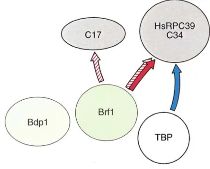
(known as B’), from which Bdp1 can be separated by chromatography (B’’) (Kassavetis et al., 1991). This indicates that the Bdp1 subunit is less tightly incorporated into the TFIIIB complex. TFIIIB is a DNA binding protein and is considered to function as initiation factor because the TFIIIB-DNA complex by itself is able to efficiently recruit RNA polymerase III in vitro (Kassavetis et al., 1990). TFIIIB requires TFIIIC to bind class III genes lacking the TATA box, like most tDNAs, but it can be recruited by itself, in vitro, onto the TATA element of the SNR6 gene (Joazeiro et al., 1994; Margottin et al., 1991; Moenne et al., 1990), although TFIIIC enhances the transcription efficiency (Eschenlauer et al., 1993). In vivo the transcription of the SNR6 snRNA gene requires TFIIIC to direct TFIIIB binding (probably to help TFIIIB finding the correct orientation for binding to DNA) and probably to overcome the repression of chromatin (Burnol et al., 1993; Gerlach et al., 1995) or the competition of TFIIIB with other soluble DNA-binding proteins. Some tRNA genes that contain a TATA-like element can also be transcribed in vitro without TFIIIC (Dieci et al., 2000). The mechanism by which TFIIIC recruits TFIIIB onto class III genes, which do not contain a TATA element, involves different steps of protein-protein interactions and conformational changes. The TFIIIC-DNA complex is thought to interact first with Brf1 via τ131 (Chaussivert et al., 1995; Khoo et al, 1994). Incorporation of TBP in the complex, mediated by Brf1 (Kassavetis et al., 1992; Librizzi et al., 1996) and by τ60 (Deprez et al., 1999), stabilizes the complex via minor groove interactions similar to those seen in RNA polymerase II transcription complexes (Persinger et al., 1999). A masked DNA-binding domain at the C-terminal of Brf1 (covered by domain interactions within Brf1, but unmasked by conformational changes during transcription complex assembly) may contribute to the stabilization of the complex (Huet et al., 1997). Addition of Bdp1 further stabilizes the TFIIIC-TFIIIB-DNA complex (Grove et al., 1999) and leads to more drastic changes in the conformation and stability of the complex: (i) the TFIIIB-DNA complex becomes resistant to high salt or polyanion concentrations (Kassavetis et al., 1989) and (ii) the pattern of cross-linking of τ131, Brf1 and TBP changes (Kassavetis et al., 1992; Persinger et al., 1999; Kumar et al., 1997).
1.4. RNA polymerase III recruitment by TFIIIB
Eight RNA polymerase III subunits can be cross-linked to DNA in a transcription initiation complex (Bartholomew et al., 1993). Of these, C34 is required for transcription initiation (Werner et al., 1993; Wang and Roeder, 1997) and has been localized the furthest upstream from the transcription start site between positions –17 and +6, close to TFIIIB (Bartholomew et al., 1993). Yeast Brf1 interacts in vivo and
in vitro with C34, while human Brf1 interacts with the human C34 homolog, RPC39, in vitro (Werner et al., 1993; Khoo et al., 1994; Wang and Roeder, 1997). Yeast Brf1
also interacts with C17 (Ferri et al., 2000). Human TBP interacts with RPC39 in vitro (Wang and Roeder, 1997). Interactions between Bdp1 and RNA polymerase III subunits have not been described (Figure 1.3). TBP and Brf1 alone are sufficient to recruit RNA polymerase III and direct multiple rounds of transcription, although the efficiency is only 5% to 10% of that observed with the full TFIIIB complex. Thus, Bdp1 plays an essential role in the opening of promoters (Kassavetis et al., 1998).
Figure 1.3 Protein-protein interactions between TFIIIB and RNA polymerase III subunits. The solid
arrows represent contacts with human subunits, the stippled arrows show contacts with Saccharomyces
1.5. TFIIIC contacts RNA polymerase III
TFIIIC τ131 subunit has been shown to interact directly with RNA polymerase III subunits. τ131 interacts with the RNA polymerase III C53 subunit in a two-hybrid assay (Flores et al., 1999), and with the conserved C-terminal domain of the universal RNA polymerase III subunit ABC10α in a yeast two-hybrid assay as well as in vitro. This interaction is likely to be functionally significant because a thermosensitive mutation within the C-terminal domain of ABC10α that makes the interaction weaker can be suppressed by overexpression of a variant form of τ131 that makes the interaction stronger (Dumay et al., 1999). These interactions between TFIIIC and RNA polymerase III show that TFIIIC not only recruits TFIIIB, but may also participate in the recruitment of RNA polymerase III, and/or the cycle of elongation, termination and reinitiation.
1.6. Termination and recycling
RNA polymerase III is unique among the eukaryotic RNA polymerases in recognizing a simple run of T residues as a termination signal. S. cerevisiae RNA polymerase III requires a minimum of six or seven dT residues for efficient termination, while vertebrate RNA polymerase III can terminate very efficiently with four dT residues (Geiduschek and Tcchini-Valentini, 1988; Allison and Hall, 1985; Bogenhagen and Brown, 1981). Mutation or deletion of the termination signal yields incomplete transcripts. In addition, mutation of T residues in the VAI gene decreased the efficiency of single- and multiple- round transcription in a HeLa cell extract (Wang and Roeder, 1996; Wang et al., 2000). Similarly, in an S. cerevisiae system in which RNA polymerase III was limited, deletion of the terminator affected multiple-round transcription, although single-multiple-round transcription was not affected (Dieci and Sentenac, 1996). These results suggest that the T residues can contribute to the efficiency of initiation and reinitiation, and therefore that they may play a role in RNA polymerase recycling. However, the reinitiation process seems to be more complicated, since not only RNA polymerase III, but also TFIIIC, and TFIIIB have been shown to participate in this process. (Ferrari et al., 2004). In higher eukaryotes, a
number of factors seem to be involved in termination and RNA polymerase III recycling, including the La protein, NF1 polypeptides, DNA topoisomerase I, and PC4.
1.7. Scope of this thesis
A thorough understanding of the molecular details underlying preinitiation complex formation on class III genes has been hampered by a lack of structural information. In tRNA genes, which occupy more than 80% of the total number of class III genes, the most critical step during the formation of the preinitiation complex is binding of the τB subcomplex to the B block. This appears to be the most important step since it determines all the following steps until the recruitment of RNA polymerase III, and thus the binding to the B block has to be done with very high affinity and ‘fidelity’. Therefore, the scope of this thesis was to shed some light into this mechanism from a structural point of view. The initial goal of this project was the structure determination of the τB complex bound to DNA. However, due to problems with the expression of the τ138 subunit, we decided to work only with the other two subunits, τ60 and τ91, which interestingly we found out that they form a very stable binary complex. In this thesis, the crystal structure at 3.2 Å resolution of the yeast full-length τ60 in complex with a C-terminal domain of τ91 (∆τ91) is presented. Our findings provide insights into the recruitment of the τB complex and its binding to the very well conserved B block.
Part II
Résumé
Cette partie est consacrée aux techniques et méthodes utilisées pour la résolution de la structure de la protéine τ60 complexée au domaine C-terminal de la protéine τ91 (∆τ91). Elle comprend l’expression des protéines recombinantes, les étapes de la purification et les méthodes de caractérisation biochimique (protéolyse limitée) ainsi que les principes de la cristallographie et les méthodes utilisées pour le traitement des données de la diffraction, la résolution du problème de phases, et le raffinement du model tridimensionnel.
2.1. Molecular Biology and Biochemical Techniques
2.1.1. Cloning
The cDNA of full-length τ91 was kindly provided by J. Acker. For subcloning τ91, oligos of 25-30 bp length were designed to contain NcoI-XhoI restriction sites. Oligos for PCR were ordered from MWG. For PCR the annealing temperature Tm was
chosen to be approximately 5 oC below the theoretical Tm of the oligos and the
elongation was performed at 72 oC. 30 cycles were carried out. The PCR products were purified with the PCR purification kit (Qiagen), digested with restriction enzymes and ligated to the target vector, previously digested with NcoI-XhoI. The ligation was carried out with the Rapid Ligation kit (La Roche) according to the manufacturer’s instructions. The ligation product was directly transformed into chemically competent XL1-blue cells (Stratagene) according to the manufacturer’s instructions and plated on LB-agarose plates supplemented with the appropriate antibiotics. Plasmids were purified from colonies by using the Wizard mini-prep kit (Promega) and analysed on agarose gels after double-digestions. The sequence of the ∆τ91 (residues 159-672) clone, as well as the sequences of all the other τ91 clones, were confirmed by sequencing.
2.1.2. Protein Expression, Purification and Complex Assembly
Recombinant baculoviruses containing full-length τ60 and τ91 genes were kindly provided by J. Acker. The virus was used to infect High Five insect cells at a multiplicity of infection of 1 for τ60 and 2 for τ91. Infection was carried out in TC100 medium (Life Technologies) complemented with 10% fetal bovine serum (FBS). Insect cells were harvested after 3 days by centrifuging at 2800 g for 10 minutes.
The pellet was resuspended in lysis buffer containing 50 mM HEPES [pH 7.4], 30 mM NaCl for τ60 (and 150 mM NaCl for τ91), 5 mM β-mercaptoethanol, 20% glycerol and a Complete protease inhibitor cocktail (Roche Diagnostics), and cells were lysed by three steps of freeze-thawing and sonication at 4 oC (in the case of τ91, the protein was treated with DNase I before sonication). The cell debris was spun down by centrifugation at 39000 g for 40 min.
For the purification of τ60, the τ60 supernatant was loaded onto a Heparin Hi Trap column (Pharmacia) and eluted with a linear gradient 30-1000 mM NaCl in 50 mM HEPES [pH 7.4], 20% glycerol, 5 mM β-mercaptoethanol, 1 mM PMSF. τ60 was then dialyzed against buffer 1 (50 mM Tris [pH 7.4], 10% glycerol, 5 mM β-mercaptoethanol) and further purified on a MonoQ anion-exchange column (Pharmacia) using a 0-500 mM NaCl gradient in buffer 1.
The crude extract containing τ91 was loaded onto a Heparin Hi Trap column (Pharmacia) and eluted with a linear 200-800 mM NaCl gradient in 50 mM HEPES [pH 7.4], 5 mM β-mercaptoethanol, 1 mM PMSF. τ91 was then dialyzed against 50 mM Tris [pH 7.4], 50 mM NaCl, 5 mM β-mercaptoethanol and further purified on a MonoS cation-exchange column (Pharmacia) using a 200-800 mM NaCl gradient. Finally, both proteins were concentrated and additionally purified over a Superdex 200 HR10/30 gel filtration column (Pharmacia) in buffer 2 (50 mM Tris [pH 7.4], 150 mM NaCl, 5 mM β-mercaptoethanol, 1 mM PMSF).
A fragment encoding ∆τ91 residues 159-672 with a C-terminal non-cleavable His tag was expressed from a pET28a vector (Novagen) in E. coli strain BL21(DE3)pLysS (Stratagene). This plasmid contains a kanamycin resistance gene for selection of transformed cells and a pBR322 replication origin. The target gene is placed under the control of the T7 RNA polymerase promoter. Bacteria were grown in LB medium at 37 oC to an optical density of 0.6 at 600 nm, cooled to 30 oC, induced with 0.3 mM isopropyl-thiogalactoside (IPTG), and grown for an additional 5 hr before harvesting. The bacteria were harvested by centrifugation at 4300 g for 15 minutes. The pellet was resuspended in 50 mM Tris [pH 7.4], 300 mM NaCl, 5 mM β-mercaptoethanol, 3 mM imidazole and a Complete EDTA-free protease inhibitor cocktail (Roche Diagnostics). Cells were lysed by sonication at 4 oC.
The cell debris was spun down by centrifugation at 48300 g for 30 minutes and the supernatant was incubated with Co-TALON affinity resin (Clontech) previously equilibrated with lysis buffer but without β-mercaptoethanol. Incubation was done at 4 oC for 1 h under gentle agitation. The resin was then transferred to a column and washed extensively with 50 mM Tris [pH 7.4], 300 mM NaCl, 1 mM β-mercaptoethanol, 8 mM Imidazole, 1 mM PMSF. The protein was eluted with 50 mM Tris [pH 7.4], 300 mM NaCl, 1 mM β-mercaptoethanol, 250 mM imidazole, 1 mM PMSF. Finally, the protein was concentrated and further purified on a Superdex 75 HR10/30 gel filtration column (Pharmacia) in buffer 2.
All the other N-terminal τ91 constructs (residues 168-672, 175-672, 181-672) were cloned in the same way as ∆τ91 using the same restriction sites, but were inserted into the pETM11 vector (provided by the Protein Expression and Purification Unit in EMBL/Heidelberg), which contains an N-terminal TEV protease cleavable 6xHis-tag. These constructs were expressed and purified in the same way as ∆τ91 except that after the Co-TALON affinity resin, they were dialyzed overnight against 50 mM Tris [pH 7.4], 300 mM NaCl, 1 mM DDT, 1 mM PMSF to remove imidazole. The next day they were cleaved with TEV protease (1:50 w/w) for 7 h at room temperature in the presence of 0.5 mM EDTA. After cleavage, they were loaded again onto the Co-resin, in order to remove the TEV protease and then further purified on a Superdex 75 gel filtration column in the same conditions as described above for τ60, τ91 and ∆τ91. For the purification of digested ∆τ91-159 and 168 constructs, after the Co-resin they were digested with 1:500 (w/w) elastase at room temperature for 4 h. The reaction was stopped with PMSF (10 mM final concentration) and the proteins were further purified on a Superdex 75 gel filtration column as already described.
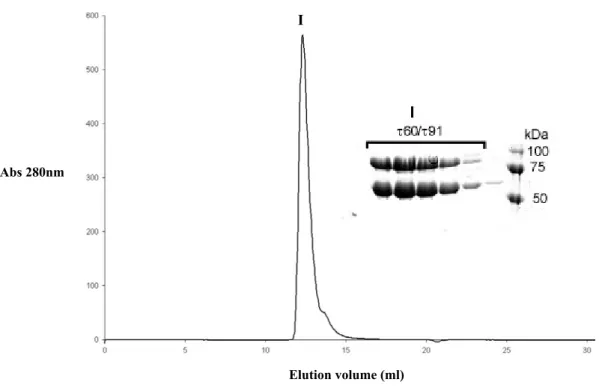
The complex was formed at 4 oC by mixing pure τ60 with τ91 in a ratio 1:1 and was purified over a Superdex 200 HR10/30 column (Pharmacia) in 50 mM Tris [pH 7.4], 75 mM NaCl, 5 mM DTT, 1 mM PMSF. The final concentration was achieved by centrifugation using an amicon concentrator. The interaction between τ60 and τ91 constructs was tested as in the case of the full-length complex.
2.1.2.1. Production of seleno-methionine substituted protein
Seleno-methionine substituted ∆τ91 was expressed in the same BL21(DE3)pLysS E.
coli strain as used for the native protein. Cells were grown at 37 oC, in minimal medium M9 (supplemented with 20% glucose as unique carbon source) until an OD600 of 0.6. The following amino acids and seleno-methionine were added to the
culture at the given final concentrations: • Lys, Phe, Thr: 100 mg/ml
• Ile, Leu, Val: 50 mg/ml • Se-Met : 60 mg/ml
with an additional growth of 15 minutes at 37 oC. Then the culture was cooled to 30
oC and induced with 0.3 mM IPTG and grown for an additional 8 hr. Purification of
seleno-methionine labeled ∆τ91 was performed as already described for the non-labeled protein, except that 10 mM β-mercaptoethanol was added to all purification buffers to prevent seleno-methionine oxidation.
2.1.3. Determination of protein concentration
Protein concentration was determined in all cases by photometric measurement at λ = 280 nm according to the Beer – Lambert Law:
A = ε I C where A is the measured absorbance.
ε is the wavelength-dependent molar extinction coefficient with units of L mol-1 cm-1. I is the path length of the cuvette in which the sample is contained.
C is the concentration of the protein in solution, expressed in mol L-1.
The molar extinction coefficient ε was calculated for each protein by program ProtParam (www.expasy.org). The ε for the complex was calculated by the equation:
2.1.4. Limited proteolysis
Digestion reactions were performed at room temperature with a protease:protein ratio of 1:500 (w/w). Reactions were started by the addition of freshly dissolved protease, and aliquots of the reaction mixture were withdrawn at regular intervals (5, 10, 20, 45 and 120 minutes). Reactions were stopped by the addition of PMSF to a final concentration of 10 mM and 4X sodium dodecyl sulfate (SDS) protein sample buffer. Samples were boiled for 4 min and analysed on a 10% SDS-polyacrylamide gel electrophoresis (PAGE) followed by coomassie blue staining.
2.1.5. Southern - Western blot
For the preparation of DNA probes, specific or non-specific DNA fragments were PCR amplified and purified on agarose gel. 100 ng of this purified fragment was phosphorylated with PNK T4 kinase (Biolabs) and ATP γ32P, during 1 h at 37 oC. The DNA was then purified with a PCR purification kit (Qiagen). To obtain a single strand probe, the probe was boiled for 5 minutes and then incubated on ice for 10 minutes, just before hybridization.
10-20 µg of protein sample was loaded on a 8% SDS PAGE. Protein samples were then transferred to a nitrocellulose membrane previously incubated in transfer buffer (25 mM Tris, 40 mM glycine, 10% [v/v] methanol). The membrane containing the protein samples was washed with 20 mM Hepes pH 7.5, 0.1 mM EDTA, 5 mM MgCl2, 100 mM KCl for 30 minutes at 20 oC and then incubated in blocking buffer
(2.5% (v/v) NP40, 1% (w/v) gelatin, 20 mM PBS 1X pH 7.2, 40 mM NaCl, 0.5 mM EDTA, 10% (v/v) glycerol) for 30 minutes at 20 oC. For the hybridization, the
membrane was first washed with hybridization buffer (20 mM PBS 1X pH 7.2, 40 mM NaCl, 0.05% (w/v) gelatin) for 10 minutes at 4 oC. Then the membrane was incubated with 10 ml of hybridization buffer containing the probe (107 cpm) for 1 h at 4 oC. Finally, the membrane was washed three times for 5 minutes with hybridization buffer and autoradiographed.
2.2. Crystallogenesis and Crystallographic Methods
2.2.1. Crystallization
The most critical step in determining the three-dimensional structure of a macromolecule by X-ray crystallography is obtaining well-diffracting single crystals. Crystallization of macromolecules involves three main steps: nucleation, growth and cessation of growth. It is usually achieved by varying the physical parameters that affect the solubility of the macromolecule in order to achieve a supersaturation state, which will force the macromolecules into the solid state, i.e. the crystal (McPherson, 1992). There are a number of devices, procedures and methods to achieve supersaturation in a protein solution, generally by a slow increase in the concentration of some precipitant such as salt or PEG. Among them, the method of vapor diffusion is undoubtedly the most widely used approach at present. It is ideal for screening a lot of crystallization conditions, and it can be scaled up to obtain large crystals suitable for X-ray diffraction analysis. Furthermore, it is not expensive, it is convenient, and takes advantage of a number of different physical arrangements. The main principle of the method is that a small volume of protein solution is brought into a closed system with a large reservoir solution. A concentration difference between the reservoir and the sample causes vapor diffusion between the two solutions until the vapor pressure in the system is at equilibrium. Hence, the change of conditions in the protein solution can result in the precipitation of the protein. Under the right conditions this happens by the formation of crystals; in most cases, however, by amorphous aggregation. In practice, 0.1-10 µl of the purified protein is mixed with the reservoir solution, which contains a precipitant at a certain concentration. If the ratio is 1:1 both protein and precipitant concentration are halved upon mixing and will return to the initial concentration at equilibrium. However, due to the presence of the precipitant the solubility of the protein can now be lower so that it precipitates.
The two most common vapor diffusion techniques are the sitting drop and the hanging drop method. In the sitting drop method the protein-precipitant mixture is placed onto a small depression or bridge on top of the reservoir. In the hanging drop method, the protein sample is prepared on a cover slip that is turned upside-down before sealing the well (Figure 2.1).
Optimal conditions of crystal growth for biological macromolecules in general are very difficult to determine a priori, since the number of parameters affecting crystallization and the total number of possible solution conditions to be tested is very large (McPherson, 1992). Jancarik and Kim developed a set of 50 screening conditions for initial experiments in protein crystallization, chosen from known or published crystallization conditions of various proteins (Jancarik and Kim, 1991). This and other commercially available crystallization screens (Hampton Research) were automatically dispersed with the Cartesian robot (available at the EMBL/Grenoble) and were used in the first trials to grow the crystals described in this thesis. In all cases, the parameters that were tested were: nature and concentration of the precipitant, protein concentration, composition of protein and reservoir solutions (pH, ionic strength etc.), temperature.
A B
Crystallization trials were carried out as hanging drops with complex concentrations between 17-20 mg/ml. The mixing ratio was usually 1:1 with 2 µl total drop volume and 0.5 ml reservoir solution. For the full-length complex all the available screens from Hampton research were tested. For the truncated complex, the initial screening was performed by the Cartesian robot (which in our case prepared drops with 0.4 µl total volume) and the conditions that gave the best crystals were repeated manually in 2 µl total drop volume. Conditions where crystals were appearing were further refined by varying the precipitant concentration, protein concentration and pH. The effect of temperature (20 and 4 oC) was also tested.
Once initial crystallization conditions had been determined, additive screens (Hampton Research) were used according to the manufacturer’s instructions (the additive was mixed with the precipitant in a ratio 1:5 and this mixture was subsequently mixed with the protein solution in a 1:1 ratio) with a reservoir solution of 0.8 M ammonium sulfate, 0.1 M Hepes pH 7.0.
For micro-seeding the protein complex (10-20 mg/ml) in 50 mM Tris pH 7.4, 75 mM NaCl, 5 mM DTT was prepared as a hanging drop mixed with 0.8 M ammonium sulfate, 0.1 M Hepes pH 7.0. After the drops were equilibrated, each drop was streak-seeded with a cat whisker. A crystal from a previous preparation was touched with the whisker, the whisker was washed in reservoir solution and immersed into the fresh drop. For micro-seeding of drops containing the τ60/∆τ91 complex, where subunit ∆τ91 was SeMet labeled, native crystals were used as seeds.
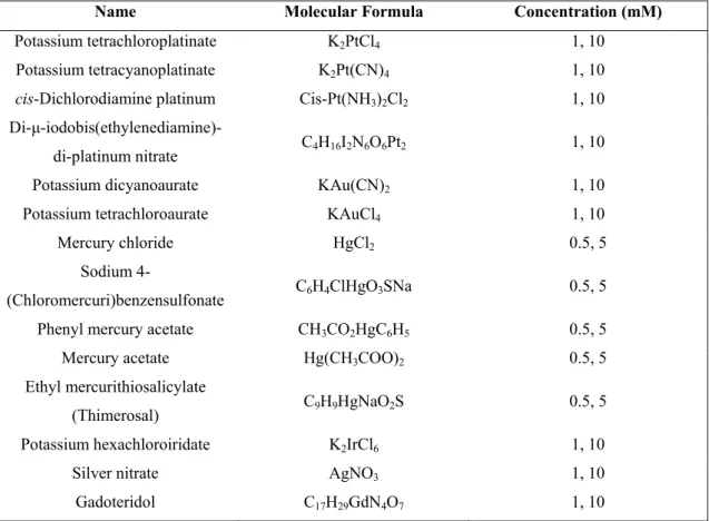
For co-crystallization with heavy atom derivatives, each heavy atom compound was dissolved in harvesting buffer (0.9 M ammonium sulfate, 0.1 M Hepes pH 7.0) to a final concentration of 1 and 10 mM for all derivatives, apart from the Hg derivatives, where the final concentrations were 0.5 and 5 mM. Protein solution (18 mg/ml) was mixed with each heavy atom solution in a ratio 1:1, to a total 2 µl drop volume. The heavy atom compounds that were tested are listed in Table 2.1.
Table 2.1. Heavy atom derivatives used for co-crystallization with the τ60/∆τ91 complex.
Name Molecular Formula Concentration (mM)
Potassium tetrachloroplatinate K2PtCl4 1, 10
Potassium tetracyanoplatinate K2Pt(CN)4 1, 10
cis-Dichlorodiamine platinum Cis-Pt(NH3)2Cl2 1, 10
Di-µ-iodobis(ethylenediamine)-di-platinum nitrate C4H16I2N6O6Pt2 1, 10 Potassium dicyanoaurate KAu(CN)2 1, 10
Potassium tetrachloroaurate KAuCl4 1, 10
Mercury chloride HgCl2 0.5, 5
Sodium
4-(Chloromercuri)benzensulfonate C6H4ClHgO3SNa 0.5, 5 Phenyl mercury acetate CH3CO2HgC6H5 0.5, 5
Mercury acetate Hg(CH3COO)2 0.5, 5
Ethyl mercurithiosalicylate
(Thimerosal) C9H9HgNaO2S 0.5, 5 Potassium hexachloroiridate K2IrCl6 1, 10
Silver nitrate AgNO3 1, 10
2.2.2. Introduction to X-ray crystallography
X-ray crystallography is one of the techniques that allow to obtain structural information on biological macromolecules at the atomic or near-atomic level. The most important limitation of the technique is the requirement for diffracting crystals. A crystal is an ordered three-dimensional array of atoms or molecules. The simplest portion in the crystal which is repeated and shows its fully symmetry is defined as the unit cell. The location of an atom in the unit cell (real space) is usually given by a set of three-dimensional Cartesian coordinates, x, y, and z. One of the vertices (a lattice point or any other convenient point) is used as the origin of the unit cell’s coordinate system, and is assigned the coordinates x=0, y=0, z=0 (0,0,0).
According to Bragg’s definition of diffraction, during X-ray diffraction of a crystal the diffracted waves from the crystal are treated as reflections from an imaginary set of planes through the crystal. The angle of reflection must be in accordance with the following equation:
2d sinθ = nλ
where d is the distance between the planes, θ the diffraction angle, λ the wavelength, and n an integer. Each reflection can be assigned three coordinates or indices (hkl), called Miller indices. The diffraction pattern produced by a crystal constitutes the reciprocal space and its dimensions are the inverse of the real space dimensions. The central reflection in the diffraction pattern is taken as the origin and assigned the coordinates h=0, k=0, l=0, (0,0,0).
The X-ray scattering of an atom is proportional to the electron density of each atom. Every atom in the unit cell contributes to every reflection in the diffraction pattern. The structure factor Fhkl for a reflection (hkl) is the sum of the scattering from all the
atoms in the unit cell and is a vector: atoms
Fhkl = Σj=1 fj exp [2π*i(hxj + kyj + lzj)]
The fj is the scattering factor of atom j and depends on the kind of atom and the
diffraction angle of the corresponding reflection (hkl). The exponent is complex with x, y, z the fractional coordinates of each atom in the summation, and (hkl) the Miller indices of the corresponding reflection. This complex exponential function is periodic, and with the above parameters it is limited between –1 and 1 for its real part, and –i and i for the imaginary part. In such cases of periodic functions a Fourier transformation can be applied. The Fourier transformation with a set of structure factors as its Fourier coefficient is the electron density:
ρxyz = (1/V) Σhkl |Fhkl| exp [-2π * i(hxj + kyj + lzj - αhkl)]
where V is the volume of the unit cell.
The minus sign preceding the exponent means that the reciprocal space (h,k,l) is transformed into real space (the electron density at point x,y,z). If the structure factors (their amplitudes |Fhkl| and their phases αhkl) are known, the actual real structure (the
density of the electrons in real space) can be calculated. However, during a diffraction experiment, only the intensities and the positions of reflections are measured. From the position of these reflections, Miller indices (hkl) can be determined and the appropriate intensity assigned. This intensity is proportional to the square of the structure factor amplitude, |Fhkl|2. Thus, from the diffraction data we have no
information about the phase αhkl of each structure factor. This loss of phase
information during data collection is known as the “Phase Problem”, which can be solved experimentally by a number of methods, such as multiple isomorphous replacement (MIR), single anomalous dispersion (SAD), multiwavelength anomalous dispersion (MAD), or molecular replacement. Some of these methods will be discussed below (see 2.2.4).
2.2.3. Data collection and processing
2.2.3.1 Cryogenic protection of crystals
Diffraction data collection at cryogenic temperatures from flashed-cooled protein crystals has become routine in macromolecular crystallography. Reduced radiation damage of the crystals, the requirement of a smaller number of crystals to collect complete data sets and solve the crystal structure, the use of intense beamlines at synchrotron radiation facilities for collecting data on weakly diffracting crystals, the ability to transport crystals at the temperature of liquid nitrogen, the possibility to mount and store crystals for extended periods, and the sometimes higher diffraction resolution limit have made flash-cooling of crystals an extremely important technique in macromolecular crystallography (Garman and Schneider, 1997).
For flash-cooling protein crystals prior to data collection, a crystal is mounted on a small loop of non-diffracting material, which is then plunged into a cryogen (such as liquid nitrogen or propane) or directly mounted in a stream of cryogenic gas (Teng, 1990). During data collection crystals are maintained at approximately 100 K in the stream of a cryogenic gas. When a crystal is cooled to cryogenic temperatures the formation of ice must be avoided, because it could lead to the disruption of the crystal lattice, due to the increase of the specific volume during transition from water to ice. In order to avoid ice formation the addition of cryoprotectants to the solution is required (Rubinson et al., 2000).
2.2.3.2. Data collection
Two main types of X-ray sources exist in protein crystallography: X-ray tubes (i.e. rotating anode) and synchrotrons. The main principle of X-ray tubes is that heat applied to a metal cathode produces free electrons. The X-rays are produced when the rapidly moving electrons are suddenly stopped as they strike the metal target of the tube anode. A rotating anode produces more powerful X-rays than fixed tubes. In this case the target is a rapidly rotating metal disk, which prevents overheating of the anode material.
Synchrotrons are enormous rings, where electrons circulate at velocities near the speed of light and are maintained in circular motion by powerful magnets. Because the electrons are forced on their circular orbit, photons are emitted in tangential direction. All the energy is concentrated in a narrow beam yielding X-rays of very high brilliance. Thus, the most important advantage of synchrotron radiation for X-ray diffraction is its high brilliance. This is very advantageous when collecting data on weakly diffracting crystals. Furthermore, in synchrotrons X-rays are produced over a certain range of wavelengths and the optimal wavelength can be selected with the monochromator. This property of synchrotron radiation is for example employed for MAD experiments.
During data collection the intensities of all reflections within a given resolution range are measured. For measuring the reflection intensities in our experiments we used the ADSC Q4 CCD detector. These detectors contain a thin screen of phosphor which emits photons when hit by X-rays. The photons are collected through an optical taper and detected on the CCD array. Phosphoimage detectors are very sensitive to X-rays and are characterized by fast readout times. The 4-module Quantum 4 detector is the latest and most advanced of the commercial CCD detectors from ADSC.
In all our experiments, data were collected using the oscillation technique, where the diffraction image is taken while the crystal is rotated by a rotation angle around an axis (the oscillation axis) that is perpendicular to the X-ray beam. For a complete data set collection, rotation images are recorded until the crystal has been rotated through enough reciprocal space to collect all unique data. The choice of the rotation angle per image depends on the unit cell size (the larger the unit cell the smaller the rotation angle), spot size or mosaic spread, and resolution (the higher the resolution the smaller the rotation angle).
2.2.3.3. Data processing
Dataprocessing involves the following steps:
1. Indexing an image, which means assigning correct Miller indices to each reflection of the image. The positions of the theoretical diffraction spots can be estimated if the accurate cell dimensions, crystal orientation, space group, wavelength and detector distance are known.
2. Refinement of the crystal and detector parameters using least-squares minimization. The differences between the experimental reflection centers are minimized with respect to the theoretical positions.
3. Study of the crystal symmetry and determination of the space group. The crystal symmetry can be deduced from the experimental diffraction pattern and the systematic absence of certain specific reflections.
4. Integration of the diffraction spots and evaluation of each reflection. 5. Crystal parameters post-refinement using all acquired data.
6. Calculation of the relative scale factors among different images in the data set. 7. Statistical analysis of the collected reflection intensities and errors.
The CCP4 suite (CCP4, 1994) is a collection of disparate programs covering most of the computation required for macromolecular crystallography. MOSFLM, SCALA, and TRUNCATE are included in this suite of programs. MOSFLM is a program used to process diffraction images developed by A. G. W. Leslie (Leslie, 1991). This software provides all the tools necessary to carry out the integration of each reflection observed in the experimental X-ray diffraction image. It generates the reflection list, reads the image, integrates the spots and writes the intensities and standard deviations into the mtz file. SCALA (Evans, 1993) is a program that calculates the scale factors among the collected images, adds the partially recorded reflections, rejects the wrong measurements and averages them. In addition, this program performs a statistical analysis of the scaled data. To determine the space group, statistic parameters of symmetry correlation are essential.
The Rsym value is an estimation of the degree of accordance among all the reflections
related by symmetry operations:
Rsym = Σhkl Σj( | Ij - <I> | ) / Σhkl Σj Ij
where <I> is the average intensity of equivalent reflections and Ij is a general
reflection contributing to the average intensity <I>.
TRUNCATE (French and Wilson, 1978) transforms the reflection intensities to structure factor amplitudes and determines the overall temperature factor of the data set from the fall-off in intensity as a function of the resolution.
2.2.4. Methods for solving the phase problem
As already discussed there are several methods to solve “the phase problem” in macromolecular crystallography. Only multiple isomorphous replacement (MIR), single and multiwavelength anomalous dispersion (SAD/MAD) will be discussed here.
1. MIR
This method involves collection of data from crystals of the protein alone, and crystals soaked in various heavy atom compounds. To form a useful derivative, heavy atoms have to bind specifically and with sufficient occupancy to the protein without introducing other changes in the crystal. In this case their locations can be identified, and the phase problem can be solved from the differences in the structure factors between the protein and its heavy-atom derivatives. These differences are referred to as isomorphous differences. Thus, this method requires production of heavy atom derivatives isomorphous to the native crystals.
The exact positions of the heavy atoms can be determined by difference Patterson or difference Fourier techniques. The Patterson function is a Fourier series described as:
P(uvw) = (1/V) Σhkl |F(h,k,l) |2 exp [-2πi (hu + kv + lw)]
where u, v, w are the Patterson fraction coordinates. Peaks in the Patterson function correspond to interatomic vectors. Peaks corresponding to vectors between heavy atoms can be calculated by a difference Patterson function using the difference between the structure factor amplitude Fph and the native Fp: (|Fph| - |Fp|)2. Once the
positions of the heavy atoms are known the structure factor of the heavy atom Fh can
be calculated and subsequently the phases for the native structure factors can be determined.
2. SAD and MAD
These methods take advantage of the heavy atom’s capacity to absorb X-rays in function of the energy of the incident X-ray beam. The strongest changes are observed near the characteristic X-ray absorption edges of the heavy atoms. The scattering factor of an atom j can be described as:
fanom j = fj + Dfj’ + iDfj’’
The values for Dfj’ and Dfj’’ vary with wavelength and type of atom.
Friedel's law states that Friedel pair reflections have equal amplitudes and opposite phases. If there is an anomalous scatterer in the crystal then Friedel’s law does no longer hold, and the reflections hkl and –h-k-l are not equal in intensity. This inequality of symmetry-related reflections is caused by the anomalous scattering (or anomalous dispersion). The structure factors of Friedel's pairs will be different in amplitude and phase and by measuring accurately the differences in their structure factor amplitudes we can determine the phase of the native structure factor.
In this approach during a MAD or SAD experiment, the wavelength is varied around the absorption edge of the heavy atom in the crystal. At wavelengths near the absorption edge, there is significant variation in the real and imaginary components f' and f" of the anomalous scattering of the heavy atoms.