HAL Id: pasteur-01980391
https://hal-pasteur.archives-ouvertes.fr/pasteur-01980391v2
Submitted on 4 Feb 2019
HAL is a multi-disciplinary open access archive for the deposit and dissemination of sci-entific research documents, whether they are pub-lished or not. The documents may come from teaching and research institutions in France or abroad, or from public or private research centers.
L’archive ouverte pluridisciplinaire HAL, est destinée au dépôt et à la diffusion de documents scientifiques de niveau recherche, publiés ou non, émanant des établissements d’enseignement et de recherche français ou étrangers, des laboratoires publics ou privés.
Resolving the apparent transmission paradox of African
sleeping sickness
Paul Capewell, Katie Atkins, William Weir, Vincent Jamonneau, Mamadou
Camara, Caroline Clucas, Nono-Raymond K Swar, Dieudonne Ngoyi, Brice
Rotureau, Paul Garside, et al.
To cite this version:
Paul Capewell, Katie Atkins, William Weir, Vincent Jamonneau, Mamadou Camara, et al.. Resolving the apparent transmission paradox of African sleeping sickness. PLoS Biology, Public Library of Science, 2019, 17 (1), pp.e3000105. �10.1371/journal.pbio.3000105�. �pasteur-01980391v2�
ESSAY
Resolving the apparent transmission paradox
of African sleeping sickness
Paul CapewellID1, Katie Atkins2,3,4, William Weir5, Vincent Jamonneau6,
Mamadou Camara7, Caroline Clucas1, Nono-Raymond K. SwarID8, Dieudonne M. Ngoyi8, Brice RotureauID9, Paul Garside1, Alison P. Galvani10, Bruno BuchetonID6,7‡,
Annette MacLeod1‡*
1 Wellcome Centre for Molecular Parasitology, College of Medical, Veterinary and Life Sciences, Glasgow
Biomedical Research Centre, University of Glasgow, Glasgow, United Kingdom, 2 Department of Infectious Disease Epidemiology, Faculty of Epidemiology and Population Health, London School of Hygiene and Tropical Medicine, London, United Kingdom, 3 Centre for Mathematical Modelling of Infectious Diseases, London School of Hygiene and Tropical Medicine, London, United Kingdom, 4 Centre for Global Health, Usher Institute for Population Health Sciences and Bioinformatics, University of Edinburgh, Edinburgh, United Kingdom, 5 School of Veterinary Medicine, University of Glasgow, Glasgow, United Kingdom, 6 Unite´ Mixte de Recherche IRD-CIRAD 177, INTERTRYP, Institut de Recherche pour le De´ veloppement (IRD), Montpellier, France, 7 Programme National de Lutte contre la Trypanosomiase Humaine Africaine, Conakry, Guinea, 8 University of Kinshasa, Kinshasa, Democratic Republic of the Congo, 9 Trypanosome Transmission Group, Trypanosome Cell Biology Unit, INSERM U1201 and Department of Parasites and Insect Vectors, Institut Pasteur, Paris, France, 10 Center for Infectious Disease Modeling and Analysis, Yale School of Public Health, New Haven, Connecticut, United States of America
‡ These authors are joint last authors on this work.
Abstract
Human African trypanosomiasis (HAT), or African sleeping sickness, is a fatal disease found throughout sub-Saharan Africa. The disease is close to elimination in many areas, although it was similarly close to elimination once before and subsequently reemerged, despite seemingly low rates of transmission. Determining how these foci persisted and over-came an apparent transmission paradox is key to finally eliminating HAT. By assessing clini-cal, laboratory, and mathematical data, we propose that asymptomatic infections contribute to transmission through the presence of an overlooked reservoir of skin-dwelling parasites. Our assessment suggests that a combination of asymptomatic and parasitaemic cases is sufficient to maintain transmission at foci without animal reservoirs, and we argue that the current policy not to treat asymptomatic HAT should be reconsidered.
Introduction
Human African trypanosomiasis (HAT) is a deadly disease of sub-Saharan Africa caused by two morphologically identical subspecies of the vector-borne parasiteTrypanosoma brucei, T. b. rhodesiense and T. b. gambiense. T. b. rhodesiense causes a more acute form of HAT and is
found predominantly in eastern Africa.T. b. gambiense is prevalent in western and central
Africa and leads to a more chronic disease. Although there has been a recent resurgence in both forms of HAT,T. b. gambiense is by far the most common and represents over 97% of a1111111111 a1111111111 a1111111111 a1111111111 a1111111111 OPEN ACCESS
Citation: Capewell P, Atkins K, Weir W, Jamonneau
V, Camara M, Clucas C, et al. (2019) Resolving the apparent transmission paradox of African sleeping sickness. PLoS Biol 17(1): e3000105.https://doi. org/10.1371/journal.pbio.3000105
Academic Editor: Steven Riley, Imperial College
London, UNITED KINGDOM
Published: January 11, 2019
Copyright:© 2019 Capewell et al. This is an open access article distributed under the terms of the
Creative Commons Attribution License, which permits unrestricted use, distribution, and reproduction in any medium, provided the original author and source are credited.
Funding: The authors received no specific funding
for this work.
Competing interests: The authors have declared
that no competing interests exist.
Abbreviations: ATF, adipose tissue form; HAT,
human African trypanosomiasis; NGO, nongovernment organisation; TL, trypanolysis.
Provenance: Not commissioned; externally peer
reported cases [1]. Both human infective forms are transmitted by tsetse flies (Glossina spp.)
and contribute to a cycle of poverty in some of the poorest regions of sub-Saharan Africa, pre-dominantly inT. b. gambiense endemic areas. HAT is one of the diseases targeted by the
WHO 2020 and 2030 elimination goals and, as a result of concerted control measures, the number of annual cases dropped from 22,800 reported (300,000 estimated) cases in 1995 to less than 3,000 reported cases in 2015 and is now approaching elimination in many countries [2]. The disease was similarly close to elimination during the 1960s when intensive control efforts reduced the number of reported cases to just 5,000. Thereafter, political turmoil, cou-pled with a decline in screening and control programmes, led to a rapid rise and reemergence of several high-prevalence foci, with reported cases reaching a new peak by the end of the 20th century [2]. Renewed HAT control and surveillance efforts through bilateral cooperation between WHO, various nongovernment organisations (NGOs), and African administrations over the last two decades have again presented an opportunity to finally eliminate this debili-tating and deadly disease [2]. However, success depends on understanding the mechanisms that led to the persistence and reemergence of HAT in low-transmission settings.
The transmission paradox
A key issue is the apparent transmission paradox that surrounds the disease as it approaches elimination, particularly with regardsT. b. gambiense. Although T. b. rhodesiense often
pres-ents with a high parasitaemia, there are relatively few parasites observed in a typicalT. b. gam-biense infection. Combined with the small volume of a tsetse blood meal (approximately 10 to
30μL) that limits the opportunity to ingest a parasite, the difficulty in establishing an infection in the tsetse and the low prevalence of infected vectors at many foci [3], it would seemT. b. gambiense transmission should have ceased. These issues lead to the obvious questions: how
doesT. b. gambiense HAT persist, and how did it return from apparent elimination? Several
hypotheses to explain the persistence and reemergence ofT. b. gambiense HAT foci have been
proposed, including animal reservoirs, the use of suboptimal diagnostic methods, underre-porting, and the possible involvement of infectious but persistently asymptomatic individuals [4]. Although trypanosome-infected animals have been detected in many HAT foci, and it is generally accepted that such reservoirs contribute to the maintenance and transmission ofT. b. rhodesiense, this is not universally the case for T. b. gambiense. The role that animal
reser-voirs may play inT. b. gambiense HAT foci persistence has recently been reviewed, suggesting
that they contribute to transmission maintenance in some areas [5]. However, there areT. b. gambiense HAT foci that have few or no infected domestic animals despite intensive sampling,
indicating that the disease could be maintained by predominantly human infections (or an as yet unidentified wild animal reservoir) [3]. Recently, evidence has also emerged to suggest that aparasitaemic asymptomatic human infections may constitute an overlooked reservoir of dis-ease that contributes to the maintenance of disdis-ease foci [6].
Resolving the paradox
Counter to the long-standing belief that HAT is always fatal in the absence of treatment, recent studies suggest that some individuals infected withT. b. gambiense are able to tolerate the
para-site with few specific symptoms [7]. This has not yet been demonstrated inT. b. rhodesiense.
These asymptomatic individuals infected withT. b. gambiense are serologically positive but
microscopy negative when their blood is examined, even when assayed via the trypanolysis (TL) test that eliminates false positives [8]. In some individuals, lymph node aspirate was also examined and was similarly negative. Current diagnostic protocols for definitive diagnosis of bothT. b. rhodesiense and T. b. gambiense HAT rely on the visible identification of parasites in
the blood or lymph node aspirate. As asymptomatic infected individuals inT. b. gambiense
areas do not present with disease symptoms and have undetectable parasitaemia, they are not diagnosed via passive surveillance, and their potential impact on transmission is overlooked. Even under active surveillance schemes, serologically positive but aparasitaemic and asymp-tomatic individuals are not treated due to the toxicity of available drugs (pentamidine and eflornithine), potentially meaning they continue to contribute to transmission.
This asymptomatic infection period can be extremely protracted, if not indefinite, with a recent case persisting for 29 years before the patient developed symptoms [9]. The ability to occasionally detect parasite genetic material using PCR from these individuals supports the assertion that they harbour active infections, and they have consequently received increased attention in recent years. For example, over a two-year follow-up in the Fore´cariah focus in Guinea, approximately 13% of serologically positive individuals without visible parasites in an initial screen developed clinical symptoms and parasitaemia [10], suggesting that these indi-viduals were early-stage clinical cases or had been subsequently reinfected by a tsetse fly. Of the remaining positive cases, 39% became serologically negative (indicating clearance of infec-tion or an initial false positive serology result), whereas 48% remained seropositive for at least two years [10], consistent with harbouring latentT. b. gambiense infections. As previously
mentioned, few infected animals have been identified in similar settings in Guinea, suggesting that transmission is primarily human to human [3]. Although it may be possible that asymp-tomatic animal reservoirs harbouring parasites exist, molecular and serological techniques used to identify human asymptomatic cases would also have detected asymptomatic animals, suggesting that there are few such infected animals in theseT. b. gambiense foci. Although
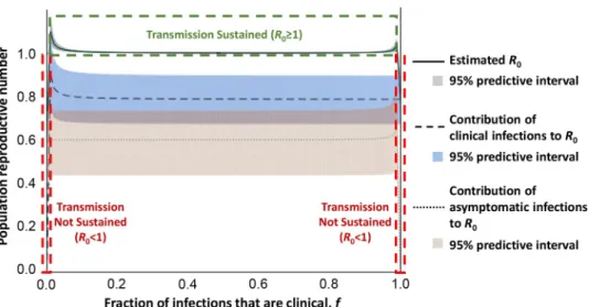
transmission in this focus appears to be largely human to human, it is unclear what contribu-tion asymptomatic human infeccontribu-tions may make to transmission. To address this, we adapted a previously published mathematical model [11] and parameterised it using data from the Fore´-cariah focus [10] to simulate a polymorphic human population (Fig 1). This analysis suggests that both asymptomatic and clinical cases are required to maintain HAT in the absence of ani-mal reservoirs and indicates that long-term asymptomatic human infections may indeed rep-resent an important but overlooked source of transmission. In order to detect both
symptomatic and asymptomatic infected individuals, active rather than passive surveillance is required for HAT monitoring. As the number of symptomatic cases falls, the proportion of infected individuals that harbour asymptomatic infections will consequently rise, representing a new challenge to elimination.
The skin as a reservoir
There are several lines of evidence supporting the suggestion that asymptomatic individuals harbour trypanosomes in their skin and thus form an anatomical reservoir forT. b. gambiense,
including xenodiagnosis assays showing that asymptomatic and symptomatic infected humans and animals are able to infect tsetse flies [12,13]. These experiments provide independent validation that tsetse flies can become infected despite feeding on hosts with undetectable parasitaemia, indicating that the parasites are derived from an extravascular location. We hypothesise that the skin is a likely source of these infective trypanosomes due to experimental work in mice, indicating that animals with no visible parasitaemia have extravascular parasites in their dermis and are infectious to the tsetse fly vector [6,14]. The presence ofT. brucei
para-sites outside the vasculature of the host was recognised in the original characterisation of the disease, although this understanding has been gradually eclipsed by descriptions ofT. brucei as
largely a blood parasite. This is in part due to a reliance on the detection of blood parasites for diagnosis.
Recent developments have begun to refocus attention on the existence and significance of extravascular skin-dwelling parasites. These include the description of aT. brucei adipose
tis-sue form (ATF) in mice, a distinct life-cycle stage of the parasite that exploits fatty acid oxida-tion to survive in the lipid-rich environment of host adipose tissue [15]. The high density of adipose tissue in the skin would make this a likely site for these forms, although there is not yet any direct evidence of ATF parasites in the skin. Independent experiments have also shown that trypanosomes recently inoculated into mouse skin by tsetse flies reside close to dermal adipocytes and can be retransmitted [14]. Immunohistological analysis of archived human skin punches has found trypanosomes in the skin of individuals not previously diagnosed with HAT, consistent with the hypothesis that these are asymptomatic infections with transmissible extravascular parasites [6]. The hypothesis that asymptomatic individuals harbour skin-dwell-ing trypanosomes is currently beskin-dwell-ing formally tested in an immunohistological survey of skin biopsies from infected individuals in Guinea.
Given the mean unstressed intravascular volume of the blood (3.8 L) and the average vol-ume of the skin (3.6 L), skin-dwelling parasites may represent a significant population of
Fig 1. Modelling of the relative contributions of asymptomatic and clinical cases toR0in a polymorphic human HAT focus without animal reservoirs. We estimated the contribution of asymptomatic and clinical infections toR0
(the basic reproductive number) under equilibrium prevalence by adapting a previously published trypanosomiasis transmission model [11]. Consistent with empirical data showing no domestic or wildlife reservoir, we removed the nonhuman-animal contribution toR0and instead allowed for a polymorphic human population in which a fraction (f)
of the population develop clinical infections when infected (population I), with the remainder (1–f) developing asymptomatic infections (population II). This leads toR0¼
ffiffiffiffiffiffiffiffiffiffiffiffiffiffiffiffiffiffiffiffiffiffiffiffiffiffiffiffiffiffiffiffiffiffiffiffiffiffiffiffiffiffiffiffiffiffiffiffi 1 ð1 i� vÞðI�IþI � IIÞ I� I 1I�I fN þ I�II 1 I�II ð1f ÞN " # v u u t , wherei�
vis the infected tsetse
prevalence,II�is the number of clinical infections present at equilibrium,III�is the number of asymptomatic infections
at equilibrium, andN is the total human population size. We simulated this equation with epidemiologic and demographic surveys from the Fore´cariah focus in Guinea. Specifically, there were 13 clinical infections and 16 suspected asymptomatic infections identified during the survey. Of the suspected asymptomatic individuals, one-third tested negative on follow-up using the TL test and one-third developed symptoms [10]. Therefore, we set the number of asymptomatic infections asIII�= 16X (where X~Uniform(1/3,1)) and the number of clinical infections as II�= 29–
III�. The total population size was set asN = 10,837, based on 7,586 surveyed individuals and a survey completeness of
70%. These results suggest that transmission is not sustainable (R0< 1) when nearly all infections are either clinical
(f > 0.98) or asymptomatic infections (f < 0.02). The shaded areas represent 95% predictive intervals when the number of asymptomatic and clinical infections were sampled 1,000 times. HAT, human African trypanosomiasis; TL, trypanolysis.
dividing parasites in a chronic HAT infection that is similar in size to the population of blood parasites. The strategy of residing in the skin may have evolved to maximise passage by telmo-phagus (slash and suck) insects, like tsetse flies, that rupture the skin rather than feeding on blood directly from active vessels. Indeed, other parasites that exist in the skin of the host, such asLeishmania and Onchocerca, rely on telmophagus vectors for transmission.
Future perspectives
There is, therefore, clear evidence from both human and animal studies that skin invasion occurs duringT. brucei infection and likely represents an anatomical reservoir that could
resolve the transmission paradox ofT. b. gambiense transmission and maintenance at foci
without animal reservoirs. The hypothesis thatT. b. gambiense infection can lead to the
asymp-tomatic carriage of transmissible parasites has important implications for the WHO 2020 and 2030 HAT elimination goals [2]. Current policy does not advise the treatment of individuals without confirmation of parasites in bodily fluids due to the toxic nature of currently available drugs. However, identification and treatment of asymptomatic infections could improve the effectiveness of disease control approaches and increase the likelihood of HAT elimination at persistent foci. Such efforts could be bolstered by vector-control measures aimed at decreasing human and tsetse contact, with prior studies demonstrating that such efforts [16] reducedT. b. gambiense transmission [17]. Although it would not be advisable for asymptomatic individuals to be treated with currently available drugs due to their toxicity, the development of less harm-ful alternatives and improving the identification of asymptomatic individuals may remove this barrier. For example, oral fexinidazole and acoziborole have both been shown to be almost as effective as current treatments but with much less toxicity [18,19]. However, it is essential that the capacities of these new drugs to treat skin-dwelling parasites are also ascertained, although only in-hospital and under strictly controlled administration. If efficacy is demonstrated, we argue that there should be a reassessment of the policy not to treat asymptomatic individuals, in addition to concerted surveillance efforts to improve the diagnosis and identification of such individuals. This would potentially remove an important reservoir forT. b. gambiense
HAT in many areas, reducing transmission and contributing to the elimination of this devast-ing disease.
References
1. WHO. Control and surveillance of human African trypanosomiasis: report of a WHO Expert Committee [on Control and Surveillance of Human African Trypanosomiasis, Geneva, 22–26 April 2013]. Geneva: World Health Organization; 2013.
2. Franco JR, Cecchi G, Priotto G, Paone M, Diarra A, Grout L, et al. Monitoring the elimination of human African trypanosomiasis: Update to 2014. Boelaert M, editor. PLoS Negl Trop Dis. 2017; 11: e0005585. https://doi.org/10.1371/journal.pntd.0005585PMID:28531222
3. Kagbadouno MS, Camara M, Rouamba J, Rayaisse J-B, Traore´ IS, Camara O, et al. Epidemiology of Sleeping Sickness in Boffa (Guinea): Where Are the Trypanosomes? Matovu E, editor. PLoS Negl Trop Dis. 2012; 6: e1949.https://doi.org/10.1371/journal.pntd.0001949PMID:23272259
4. Koffi M, Solano P, Denizot M, Courtin D, Garcia A, Lejon V, et al. Aparasitemic serological suspects in Trypanosoma brucei gambiense human African trypanosomiasis: A potential human reservoir of para-sites? Acta Trop. 2006; 98: 183–188.https://doi.org/10.1016/j.actatropica.2006.04.001PMID: 16723098
5. Bu¨scher P, Bart J-M, Boelaert M, Bucheton B, Cecchi G, Chitnis N, et al. Do Cryptic Reservoirs Threaten Gambiense-Sleeping Sickness Elimination? Trends Parasitol. 2018; 34: 197–207.https://doi. org/10.1016/j.pt.2017.11.008PMID:29396200
6. Capewell P, Cren-Travaille´ C, Marchesi F, Johnston P, Clucas C, Benson RA, et al. The skin is a signifi-cant but overlooked anatomical reservoir for vector-borne African trypanosomes. eLife. 2016; 5.https:// doi.org/10.7554/eLife.17716PMID:27653219
7. Jamonneau V, Ilboudo H, Kabore´ J, Kaba D, Koffi M, Solano P, et al. Untreated Human Infections by Trypanosoma brucei gambiense Are Not 100% Fatal. Ndung’u JM, editor. PLoS Negl Trop Dis. 2012; 6: e1691.https://doi.org/10.1371/journal.pntd.0001691PMID:22720107
8. Jamonneau V, Bucheton B, Kabore´ J, Ilboudo H, Camara O, Courtin F, et al. Revisiting the Immune Try-panolysis Test to Optimise Epidemiological Surveillance and Control of Sleeping Sickness in West Africa. Masiga DK, editor. PLoS Negl Trop Dis. 2010; 4: e917.https://doi.org/10.1371/journal.pntd. 0000917PMID:21200417
9. Sudarshi D, Lawrence S, Pickrell WO, Eligar V, Walters R, Quaderi S, et al. Human African Trypanoso-miasis Presenting at Least 29 Years after Infection—What Can This Teach Us about the Pathogenesis and Control of This Neglected Tropical Disease? Franco-Paredes C, editor. PLoS Negl Trop Dis. 2014; 8: e3349.https://doi.org/10.1371/journal.pntd.0003349PMID:25522322
10. Ilboudo H, Jamonneau V, Camara M, Camara O, Dama E, Le´no M, et al. Diversity of response to Trypa-nosoma brucei gambiense infections in the Forecariah mangrove focus (Guinea): perspectives for a better control of sleeping sickness. Microbes Infect. 2011; 13: 943–952.https://doi.org/10.1016/j.micinf. 2011.05.007PMID:21658462
11. Funk S, Nishiura H, Heesterbeek H, Edmunds WJ, Checchi F. Identifying Transmission Cycles at the Human-Animal Interface: The Role of Animal Reservoirs in Maintaining Gambiense Human African Try-panosomiasis. Pascual M, editor. PLoS Comput Biol. 2013; 9: e1002855.https://doi.org/10.1371/ journal.pcbi.1002855PMID:23341760
12. Frezil JL. [Application of xenodiagnosis in the detection of T. gambiense trypanosomiasis in immunolog-ically suspect patients]. Bull Soc Pathol Exot Filiales. 1971; 64: 871–878. PMID:5172722
13. Wombou Toukam CM, Solano P, Bengaly Z, Jamonneau V, Bucheton B. Experimental evaluation of xenodiagnosis to detect trypanosomes at low parasitaemia levels in infected hosts. Parasite. 2011; 18: 295–302.https://doi.org/10.1051/parasite/2011184295PMID:22091459
14. Caljon G, Van Reet N, De Trez C, Vermeersch M, Pe´rez-Morga D, Van Den Abbeele J. The Dermis as a Delivery Site of Trypanosoma brucei for Tsetse Flies. Peters NC, editor. PLoS Pathog. 2016; 12: e1005744.https://doi.org/10.1371/journal.ppat.1005744PMID:27441553
15. Trindade S, Rijo-Ferreira F, Carvalho T, Pinto-Neves D, Guegan F, Aresta-Branco F, et al. Trypano-soma brucei Parasites Occupy and Functionally Adapt to the Adipose Tissue in Mice. Cell Host Microbe. 2016; 19: 837–848.https://doi.org/10.1016/j.chom.2016.05.002PMID:27237364
16. Courtin F, Camara M, Rayaisse J-B, Kagbadouno M, Dama E, Camara O, et al. Reducing Human-Tsetse Contact Significantly Enhances the Efficacy of Sleeping Sickness Active Screening Campaigns: A Promising Result in the Context of Elimination. Aksoy S, editor. PLoS Negl Trop Dis. 2015; 9: e0003727.https://doi.org/10.1371/journal.pntd.0003727PMID:26267667
17. Kagbadouno MS, Camara O, Camara M, Ilboudo H, Camara ML, Rayaisse J-B, et al. Ebola outbreak brings to light an unforeseen impact of tsetse control on sleeping sickness transmission in Guinea. bioR-xiv, 2018;https://doi.org/10.1101/202762
18. Pollastri MP. Fexinidazole: A New Drug for African Sleeping Sickness on the Horizon. Trends Parasitol. 2018; 34: 178–179.https://doi.org/10.1016/j.pt.2017.12.002PMID:29275007
19. Baker CH, Welburn SC. The Long Wait for a New Drug for Human African Trypanosomiasis. Trends Parasitol. 2018; 34: 818–827.https://doi.org/10.1016/j.pt.2018.08.006PMID:30181071