HAL Id: inserm-00630240
https://www.hal.inserm.fr/inserm-00630240
Submitted on 7 Oct 2011
HAL is a multi-disciplinary open access
archive for the deposit and dissemination of
sci-entific research documents, whether they are
pub-lished or not. The documents may come from
teaching and research institutions in France or
abroad, or from public or private research centers.
L’archive ouverte pluridisciplinaire HAL, est
destinée au dépôt et à la diffusion de documents
scientifiques de niveau recherche, publiés ou non,
émanant des établissements d’enseignement et de
recherche français ou étrangers, des laboratoires
publics ou privés.
ColVI-myopathies: where do we stand, where do we go?
Valérie Allamand, Laura Briñas, Pascale Richard, Tanya Stojkovic, Susana
Quijano-Roy, Gisèle Bonne
To cite this version:
Valérie Allamand, Laura Briñas, Pascale Richard, Tanya Stojkovic, Susana Quijano-Roy, et al..
ColVI-myopathies: where do we stand, where do we go?. Skeletal Muscle, BioMed Central, 2011, 1 (1), pp.30.
�10.1186/2044-5040-1-30�. �inserm-00630240�
Skeletal Muscle
d IノW
;y
Fラノェキ
1j]ヶ#ヱ 1j]ヶ#ヲ 1j]ヶ#ンW ;マW
}Э}"ノキミニ
cラミラマW
7キマW
}Э}"ノキミニ
0;
;ノ
"ノ;マ
キミ
;
Nミ
W
キキ
;ノ
マ
;
キ
EキH キノノ; Iラノノ;ェWミ
Nミ Wェ キミ
α7F
β7F
1ラノN
1ラノ N
];マキミキミどヲヱヱ
0キェノ I;ミ
}; Iラェノ I;ミ
α
γ δ
β
}; Iラ ;ミ
α7 β1
dキSラェWミ
vW ノWI;ミ
7WIラ キミ
}WI W キラミ
αヱふ Nぶ
αヲふ Nぶ
αンふ Nぶ
1 ラ ノ; マ;
ColVI myopathies: where do we stand, where do
we go?
Allamand et al.
Allamand et al. Skeletal Muscle 2011, 1:30 http://www.skeletalmusclejournal.com/content/1/1/30 (23 September 2011)
R E V I E W
Open Access
ColVI myopathies: where do we stand,
where do we go?
Valérie Allamand
1,2,3,4*, Laura Briñas
1,2,3,4, Pascale Richard
5, Tanya Stojkovic
1,2,3,4,6, Susana Quijano-Roy
7and
Gisèle Bonne
1,2,3,4,5Abstract
Collagen VI myopathies, caused by mutations in the genes encoding collagen type VI (ColVI), represent a clinical
continuum with Ullrich congenital muscular dystrophy (UCMD) and Bethlem myopathy (BM) at each end of the
spectrum, and less well-defined intermediate phenotypes in between. ColVI myopathies also share common
features with other disorders associated with prominent muscle contractures, making differential diagnosis difficult.
This group of disorders, under-recognized for a long time, has aroused much interest over the past decade, with
important advances made in understanding its molecular pathogenesis. Indeed, numerous mutations have now
been reported in the COL6A1, COL6A2 and COL6A3 genes, a large proportion of which are de novo and exert
dominant-negative effects. Genotype-phenotype correlations have also started to emerge, which reflect the various
pathogenic mechanisms at play in these disorders: dominant de novo exon splicing that enables the synthesis and
secretion of mutant tetramers and homozygous nonsense mutations that lead to premature termination of
translation and complete loss of function are associated with early-onset, severe phenotypes. In this review, we
present the current state of diagnosis and research in the field of ColVI myopathies. The past decade has provided
significant advances, with the identification of altered cellular functions in animal models of ColVI myopathies and
in patient samples. In particular, mitochondrial dysfunction and a defect in the autophagic clearance system of
skeletal muscle have recently been reported, thereby opening potential therapeutic avenues.
Review
Collagen VI: an important component of connective
tissues
Collagens are major constituents of the extracellular
matrix (ECM), and are found in most connective tissues.
They provide structural and mechanical stability to
tis-sues, but they also play crucial roles in cell-ECM
inter-actions through various receptors [1]. In particular,
collagen type VI (ColVI), an important component of
skeletal muscle ECM, is involved in maintaining tissue
integrity by providing a structural link between different
constituents of connective-tissue basement membranes
(for example, collagen types I and IV, biglycan, and
dec-orin) and cells [2-15] (Figure 1). In addition to its
struc-tural role, ColVI supports adhesion, spreading and
migration of cells, and cell survival, as discussed later in
this review.
ColVI is a heterotrimeric molecule composed of three
individual a(VI) chains that display a similar structure,
with a triple helical domain characterized by the
repeti-tion of the Gly-X-Y amino acid sequence, flanked by
globular domains homologous to von Willebrand factor
A domains [16,17]. In addition to the well-known a1
(VI), a2(VI) and a3(VI) chains encoded in human by
the COL6A1, COL6A2 (located head-to-tail on
chromo-some 21q22.3), and COL6A3 (on chromochromo-some 2q37)
genes [18], three novel chains, a4(VI), a5(VI) and a6
(VI), have recently been identified [19,20]. These chains
have high structural homology to the a3(VI) chain. In
humans, the COL6A4, COL6A5 and COL6A6 genes are
all located on chromosome 3q22.1, with the COL6A4
gene being split by a chromosome break and thus not
coding for a protein [19-21]. The murine orthologs of
these genes are organized in tandem on chromosome 9
(Col6a4, Col6a5 and Col6a6) and encode the a4(VI), a5
(VI) and a6(VI) chains. The expression pattern of the
three novel chains differs between mice and humans,
and also between fetal and adult tissues [19,20].
* Correspondence: [email protected]
1Inserm, U974, Paris, France
Full list of author information is available at the end of the article Allamand et al. Skeletal Muscle 2011, 1:30
http://www.skeletalmusclejournal.com/content/1/1/30
Skeletal Muscle
© 2011 Allamand et al; licensee BioMed Central Ltd. This is an Open Access article distributed under the terms of the Creative Commons Attribution License (http://creativecommons.org/licenses/by/2.0), which permits unrestricted use, distribution, and reproduction in any medium, provided the original work is properly cited.
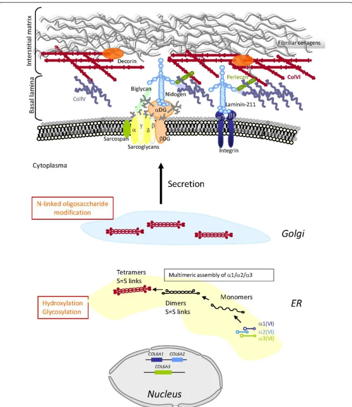
Figure 1Schematic representation of the collagen type VI (ColVI) intracellular assembly process, and interactions with skeletal muscle extracellular matrix (ECM) components. Individual a(VI) chains fold through their triple helical domains to form monomers (1:1:1 ratio) in the endoplasmic reticulum (ER), which further align in an anti-parallel manner as dimers and tetramers that are stabilized by disulfide bonds between cysteine residues (S = S links). Post-translational modifications (indicated in orange) take place in the ER and Golgi, followed by secretion of tetramers that align covalently end to end, to form beaded microfibrils in the ECM. ColVI interacts with collagenous and non-collagenous components of the basal lamina and interstitial matrix surrounding muscle fibers.
Allamand et al. Skeletal Muscle 2011, 1:30
http://www.skeletalmusclejournal.com/content/1/1/30
Importantly, in the context of ColVI myopathies, the a6
(VI) chain is the only one expressed at high levels in
human skeletal muscle, at higher levels in fetal than
adult tissue [19]. In skin, a detailed analysis of the
expression of the human a5(VI) and a6(VI) chains
revealed that both chains are expressed, albeit
differ-ently, and that they are variably altered in tissues from
patients with mutations in the COL6A1, COL6A2 and
COL6A3
genes [22]. Interestingly, the COL6A5 gene had
previously been reported as associated with atopic
der-matitis under the name COL29A1 [23], but this
associa-tion has recently been quesassocia-tioned [24,25]. The knee
osteoarthritis susceptibility locus DVWA was shown to
correspond to the 5’ part of the split COL6A4 gene [21].
Although largely ubiquitous, the expression of ColVI
seems to be finely regulated in different cell types and
tissues, as shown for the murine Col6a1 gene. The
iden-tification of a transcriptional enhancer located in the
5’-flanking sequence of the gene points to a collaborative
crosstalk between myogenic and
mesenchymal/endomy-sial cells, enabling transcription of ColV in muscle
con-nective tissue [26-28].
The a1(VI), a2(VI) and a3(VI) chains assemble
intra-cellularly as monomers (1:1:1 ratio), from their
C-term-inal ends, and subsequently form dimers (two
anti-parallel, overlapping monomers) and tetramers (four
monomers) that are stabilized by disulfide bonds
between cysteine residues of the three chains [29-34].
ColVI chains are subjected to extensive
post-transla-tional modifications such as hydroxylation of lysine and
proline residues [35], and glycosylation of
hydroxyly-sines, which have been shown to be essential for the
tet-ramerization and further secretion of ColVI [36,37].
Upon secretion, tetramers are further aligned end to
end as microfibrils in the extracellular space, with a
characteristic beaded appearance [33] (Figure 1). To
date, somewhat contradictory results have been obtained
regarding the possible assembly of the newly
character-ized a(VI) polypeptides with the a1(VI), a2(VI) chains.
In transfection experiments, only a4(VI) appeared to
have this ability [19], whereas in mouse muscle, all three
were reported to do so [20]. Whether and how these
additional chains may fit in the pathogenesis of ColVI
myopathies remains unresolved to date, and needs to be
addressed more comprehensively. To date, in our cohort
of patients, no pathogenic mutations have been found
by sequencing of the COL6A5 and COL6A6 genes in
patients without mutations in the COL6A1-3 genes (V.
Allamand, data not shown).
Clinical phenotypes of collagen VI myopathies
The etiological definition of ColVI myopathies as a
spe-cific condition has evolved over the years with the
blur-ring of boundaries between two disorders, initially
described separately but now recognized as the extreme
ends of a continuous clinical spectrum [38,39] (Figure
2). The severe endpoint of this spectrum corresponds to
Ullrich congenital muscular dystrophy (UCMD, OMIM
254090; http://www.ncbi.nlm.nih.gov/omim), described
in 1930 as ‘congenital atonic-sclerotic muscular
dystro-phy’, emphasizing its early onset and the presence of
proximal joint contractures associated with a striking
distal hyperlaxity [40,41]. Orthopedic deformities (joint
contractures, scoliosis) and respiratory impairment with
diaphragmatic failure generally develop within the first
decade of life, and may be life-threatening. Arrest of
motor milestones with no acquisition of walking ability
is seen in a subset of patients, but most children are
able to walk, and show later progression of muscle
weakness with loss of ambulation around 10 years of
age, and a requirement for mechanical ventilation in late
childhood or young adulthood [42,43].
At the other end of the spectrum is the milder form
Bethlem myopathy (BM, OMIM 158810), described in
1976, which begins in the first or second decade,
although a neonatal history may be recognized,
charac-terized by early contractures of finger flexors, wrist,
elbows and ankles [44,45]. Respiratory failure and distal
hyperlaxity are usually absent or are milder than in
UCMD, although the latter may not be so uncommon
in very young children with BM. The course is usually
slow, with most of the patients remaining ambulatory.
However, progression of muscle weakness occurs often
in the fifth decade, resulting in about 50% of patients
requiring walking aids or a wheelchair [46]. Intermediate
phenotypes have been described, and named ‘mild
UCMD’ or ‘severe BM’, thereby reinforcing the notion
of clinical overlap between Ullrich and Bethlem
pheno-types [38,39].
Skin features such as follicular hyperkeratosis and
hypertrophic scars or keloid formation are common
[38,39,42,43,47-49]. Other common findings include
normal cognitive abilities, normal or only slightly raised
serum creatine kinase (CK) levels, and absence of
car-diac phenotype. Two other conditions that fall within
the spectrum of ColVI myopathies have been
documen-ted: autosomal dominant limb-girdle muscular
dystro-phy (LGMD) (in three families) and, more recently,
autosomal recessive myosclerosis myopathy (OMIM
255600) (in one family) [50,51].
The prevalences of UCMD and BM in northern
Eng-land has recently been reported as 0.13 and 0.77 per
100,000, respectively, amounting collectively to 0.9 per
100,000 [52]. UCMD seems to be the second most
com-mon type of congenital muscular dystrophy (CMD) in
Europe (behind laminin a2 chain deficiency; OMIM
607855) and also in Japan (behind Fukuyama congenital
muscular dystrophy; OMIM 253800, [53]) and Australia
Allamand et al. Skeletal Muscle 2011, 1:30
http://www.skeletalmusclejournal.com/content/1/1/30
(behind a-dystroglycan glycosylation defects; [54]). In
the cohort from northern England, BM emerges as the
fourth most common myopathy behind myotonic
dys-trophy (OMIM 160900), facio-scapulo-humeral
muscu-lar dystrophy (OMIM 158900) and Duchenne/Becker
muscular dystrophy (OMIM 310200 and 300376) [52].
Differential diagnosis of ColVI-related myopathies
With the most prominent clinical presentation of
ColVI myopathies being muscle weakness and
contrac-tures, associated with variable degrees of hyperlaxity,
an important difficulty lies in defining boundaries and
contiguities, with the possible differential diagnosis
including congenital myopathies, Emery-Dreifuss
mus-cular dystrophy (EDMD; OMIM 181350), LGMD, rigid
spine muscular dystrophies, and other diseases of
con-nective tissues such as Ehlers-Danlos syndrome
[55-57]. Imaging techniques, such as computed
tomography or magnetic resonance imaging (MRI) of
muscle, are now recognized as very helpful in the
diag-nostic approach of muscle disease, because there are
specific patterns of muscle involvement in each of
these contractile myopathies as reported for EDMD
with LMNA mutations [58], muscular dystrophies with
rigidity of the spine [59], and ColVI myopathies
[58,60,61]. From these studies, the typical pattern of
muscle involvement in ColVI myopathies is now
con-sidered to be constituted by a diffuse, concentric
hypo-density of the thigh muscles with relative sparing of
the sartorius, gracilis and adductor longus muscles.
The vasti muscles are the most affected muscles. In
addition, a peculiar central area of abnormal signal is
seen within the rectus femoris, initially referred to as a
‘central shadow’ [62].
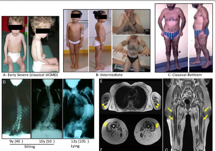
Figure 2Clinical spectrum, associated spine deformation and muscle MRI in collagen type VI (ColVI) myopathies. (A), Early severe phenotypes, corresponding to classic Ullrich congenital muscular dystrophy (UCMD), (B) intermediate forms seen in children or adults and (C) less severe, classic Bethlem myopathy (BM) forms constitute the overlapping clinical presentations of ColVI myopathies. (D) Radiography showing the evolution of spine deformation in a patient presenting with a classic early-onset UCMD phenotype. T1 transverse section of Bethlem myopathy upper limb girdle (E) and (F) thighs. Note the fatty infiltration, which appears as hyperintense area on T1-weighted images, located around the triceps brachialis muscles in (E) and along the fascia of vastus lateralis and vastus medialis muscles in (F) (yellow arrows). (G) The concentric fatty involvement of the thigh muscles is also seen on whole body MRI. (Images courtesy of Drs Susana Quijano-Roy and Tanya Stojkovic).
Allamand et al. Skeletal Muscle 2011, 1:30
http://www.skeletalmusclejournal.com/content/1/1/30
In the context of the differential diagnosis, the absence
of raised CK levels, the lack of a cardiac phenotype, and
the presence of a specific MRI pattern are strongly
sug-gestive of a ColVI myopathy.
Molecular diagnosis and genetics
In light of the clinical variability and the overlapping
presentation with other muscular disorders, a definite
diagnosis can only be made after the identification of
pathogenic mutations in one of the COL6A genes,
which to date are restricted to COL6A1, COL6A2 and
COL6A3
. However, the large size (106 coding exons in
total corresponding to 150 kb of genomic DNA) of
these genes makes routine molecular diagnostics costly
and time-consuming. The road to this ‘holy grail’ of
diagnosis is thus often lengthy and full of pitfalls, and
relies on a combination of clinical, biochemical and
molecular findings.
Historically, muscle biopsies were the routine and
pri-mary step undertaken for diagnostic purposes, and
dou-ble immunostaining with a basement-membrane marker
enabled recognition of ColVI deficiency in patients with
UCMD [63], but not in patients with BM. The current
diagnostic method of determining ColVI involvement is
primarily based on immunocytochemistry of cultured
skin fibroblasts, but this analysis is only available in a
limited number of laboratories to date. A number of
antibodies recognizing human ColVI are now
commer-cially available and may be used for such techniques; in
particular, the refined protocol proposed by Hicks et al.
[64], using a polyclonal antibody raised against mature
ColVI from human placenta, has better sensitivity,
espe-cially in fibroblast cultures from patients with BM
(Figure 3). The absence or alteration of ColVI secretion
in cultured fibroblasts, associated with clinical symptoms
compatible with a diagnosis of ColVI myopathy,
cer-tainly warrants further genetic analysis.
Over the past decade, the development of genetic
stu-dies has demonstrated the heterogeneity and complexity
of the molecular mechanisms at play in ColVI
myopa-thies. An autosomal recessive pattern of inheritance was
initially thought to be involved in UCMD, and linkage
analysis led to the identification of mutations in the
COL6A2
and COL6A3 genes [65-67]. However,
numer-ous dominant de novo mutations have now been shown
to be involved, accounting for more than 50% of the
mutations causing UCMD [38,39,42,68]. Similarly,
auto-somal dominant mutations were first identified in the
COL6A1
and COL6A2 genes in families with BM,
sug-gesting that BM was mostly familial and inherited as an
autosomal dominant disease [69], although rare de novo
mutations and autosomal recessive mutations have now
been reported [70-73]. To date, over 200 mutations
have been identified in these genes, mostly distributed
in the COL6A1 and COL6A2 genes. The most common
types of mutations are point mutations, and mutations
leading to premature termination codons (PTCs) and
exon skipping (Figure 4). Among the former, missense
changes affecting glycine residues in the triple helical
domains of the corresponding proteins are the most
common, and are often dominant de novo. Because
these changes affect crucial amino acids within the
col-lagenous domains, they hamper triple-helix formation
[74-77]. Splice mutations resulting in in-frame exon
skipping are generally dominant de novo mutations, and
exons 16 of COL6A3 and 14 of COL6A1 seem to be
preferentially affected, leading to UCMD or BM
pheno-types, respectively [53,75,78-83]. Nonsense mutations
and small deletions or insertions inducing PTCs within
the coding frame are mostly inherited as recessive
muta-tions, and lead to loss of function of the protein
[42,53,68,75,76,79-92]. These mutations are responsible
for most UCMD phenotypes. Nevertheless, it should be
noted that genotype-phenotype correlations are very
dif-ficult to identify.
It has recently been shown that all types of mutations
alter transcript levels, and that in the case of
PTC-bear-ing transcripts, which are specifically degraded via the
nonsense-mediated mRNA decay (NMD [93,94])
path-way, quantification of the three COL6A mRNAs is a
helpful tool to pinpoint the mutated gene, thereby
facili-tating these cumbersome molecular analyses [42]. The
NMD-induced degradation of PTC-bearing transcripts
may also, at least in part, explain why the parents of
patients with UCMD who themselves harbor recessive
mutations are asymptomatic; their heterozygous status
sustains the expression of 50% of the ‘normal’ protein,
thereby leading to a ‘functional loss of heterozygosity’.
The study by Briñas and collaborators also provided
some genotype-phenotype correlations in a cohort of
patients with early-onset ColVI myopathy, showing that
recessive mutations leading to PTC were associated with
severe phenotypes [42]. Genetic studies are further
com-plicated by a possibly variable penetrance as reported by
Peat et al. [89].
Finally, the highly polymorphic nature of the COL6A
genes makes it difficult to definitely assign pathogenicity
to some variants, especially missense ones that do not
affect glycine residues within the triple-helix domains of
the proteins. In addition, these ‘polymorphisms’ may
very well play a role in the extreme clinical variability of
these conditions, particularly in patients carrying
identi-cal mutations but presenting with variable severity.
The types of mutations identified also reflect the
methods used in laboratories performing these analyses
(for example, sequencing of genomic DNA or of the
coding sequences on cDNA), but the emergence of
high-throughput methods (arrays) is likely to allow the
Allamand et al. Skeletal Muscle 2011, 1:30
http://www.skeletalmusclejournal.com/content/1/1/30
identification of as yet unknown or under-recognized
pathogenic mechanisms, such as large gene
rearrange-ments, or promoter or deep intronic mutations, as
recently illustrated in two reports [95,96].
Animal models and pathophysiology
Limited access to muscle biopsies hinders extensive
investigations of the specific cellular mechanisms leading
to the development of the muscle pathology, and in
vitro/ex vivo
cellular systems only partially reproduce
the complexity of the tissue. The development of the
first animal model of ColVI deficiency in 1998,
engi-neered by invalidating the Col6a1 gene in mice, has
proven central to understanding the cellular pathways
involved in these diseases. Homozygous animals were
reported to develop a mild myopathic phenotype, and
were initially described as a model of BM [97].
Interest-ingly, the diaphragm was the most affected muscle, with
signs of necrosis evidenced by uptake of Evan’s blue dye
[97]. Subsequently, a latent mitochondrial dysfunction
accompanied by ultrastructural alterations of
mitochon-dria and the sarcoplasmic reticulum, resulting in
sponta-neous apoptosis, was found in about one-third of muscle
fibers [98]. Reduced contractile strength of the
dia-phragm and other muscle groups was also reported in
Col6a1
-/-mice in this initial study [98]. The maximal
Figure 3Collagen type VI (ColVI) expression study in cultured skin fibroblasts. (A) Representative images obtained using the protocol from [67] in which ColVI (red) is labeled with monoclonal antibody MAB1944 (Chemicon (now Millipore), Billerica, MA, USA) and perlecan (green) with monoclonal antibody MAB1948 (Chemicon). Note that ColVI expression appeared clearly altered in a patient with an early severe (ES) form and less so in patients with intermediate (Int) or Bethlem myopathy (BM) forms, compared with control fibroblasts (Cont). (B) Using the protocol of Hicks et al. [64], which detects ColVI (red) with polyclonal antibody Ab6588 (Abcam, Cambridge, UK) and fibronectin (green) with monoclonal antibody F15 (Sigma Chemical Co., St Louis, MO, USA), the sensitivity of the method is increased, and defective ColVI secretion could be detected in all patients’ samples. Insets indicate nuclei, labeled using DAPI. Bars are 50 μm. (Images courtesy of Corine Gartioux and Valérie Allamand).
Allamand et al. Skeletal Muscle 2011, 1:30
http://www.skeletalmusclejournal.com/content/1/1/30
isometric tension generated by ColVI-deficient skinned
fibers from gastrocnemius was found to be reduced in a
recent report; however, using a protocol of eccentric
contractions in vivo, no muscle force drop was found,
indicating that the lack of ColVI does not impair
myofi-brillar function [99]. Importantly, mitochondrial
dys-function was also reported in cultured muscle cells from
patients and could be reversed by cyclosporin (Cs)A, an
immunosuppressive drug that prevents the opening of
the mitochondrial permeability transition pore through
binding to cyclophilin D, and also inhibits the
phospha-tase calcineurin [100,101]. Another in vitro study
showed that patients-derived skin fibroblasts behave
dif-ferently from myoblasts in that respect, and also
ques-tioned the specificity of this mitochondrial dysfunction
[102], warranting further studies on the matter.
A role for cell survival had previously been proposed
for ColVI because it was shown to prevent anti-a1
integrin-mediated apoptosis and trigger the
downregula-tion of bax, a pro-apoptotic molecule [103,104].
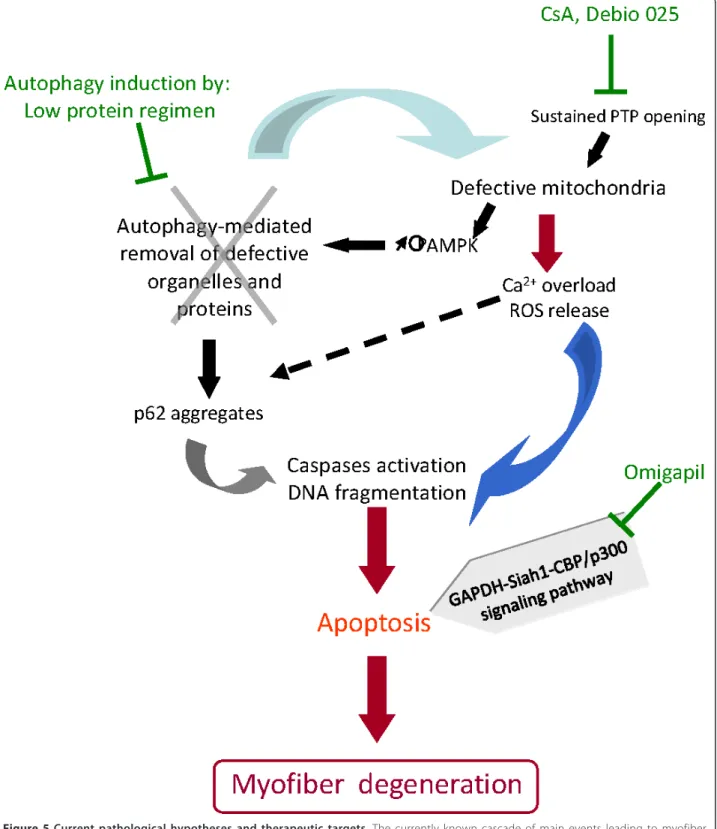
Recently, a study of the autophagic process in muscles
of Col6a1 knockout mice revealed that autophagy was
not induced efficiently [105]. The ensuing defective
autophagy provides the link between the previously
Figure 4Repartition of the various types of mutations identified in the COL6A1, COL6A2 and COL6A3 genes. This schematic reflects, to the best of our ability, the distribution of 258 allelic mutations (98 on COL6A1, 113 on COL6A2 and 47 on COL6A3). Dominant de novo mutations represent 67% of the missense mutations, affecting glycine residues in the TH domains (Gly in TH), 57% of small deletions (< 5 amino acids) and 44% of splice-site mutations leading to in-frame exon skipping, whereas 97% of mutations leading to premature termination codons (PTCs) are familial (recessive or dominant).
Allamand et al. Skeletal Muscle 2011, 1:30
http://www.skeletalmusclejournal.com/content/1/1/30
Figure 5Current pathological hypotheses and therapeutic targets. The currently known cascade of main events leading to myofiber degeneration in ColVI-deficient skeletal muscle is shown. Mitochondrial dysfunction (due in part to the defective permeability transition pore (PTP) opening) triggers an energetic imbalance with the increased levels of phosphorylated adenosine monophosphate-activated protein kinase (p-AMPK), Ca2+overload and the production of reactive oxygen species (ROS). Lack of autophagy induction exacerbates the cellular dysfunction
because defective mitochondria and proteins (such as p62 aggregates) are not cleared from the cytoplasm. Together, these defects lead to increased apoptosis. Potential therapeutic interventions are indicated in green.
Allamand et al. Skeletal Muscle 2011, 1:30
http://www.skeletalmusclejournal.com/content/1/1/30
described mitochondrial dysfunction and myofiber
degeneration, as abnormal organelles and molecules
cannot be efficiently cleared from the cell. This study
further showed that the forced induction of autophagy,
either by dietary restriction or by treatment with
rapa-mycin or CsA, ameliorated the phenotype of the
Col6a1
-/-mice (Figure 5). A similar alteration of
autop-hagy was also detected in muscle biopsies derived from
nine patients with UCMD or BM [105]. These data thus
provide a basis for novel therapeutic targets to promote
the elimination of defective organelles in ColVI-deficient
skeletal muscle.
Morpholino-mediated knock-down of the col6a1 and
col6a3
genes in zebrafish embryos showed that collagen
VI deficiency significantly impairs muscle development
and function [106]. Increased apoptosis, partially
pre-vented by CsA treatment, was also described in the
zeb-rafish morphants [106]. As in other instances,
perturbation of muscle components leads to a more
severe phenotype in zebrafish than in mouse models,
which may in part be due to intrinsic differences in
muscle development in these species, especially in terms
of timing. Zebrafish models have emerged as major in
vivo
models of neuromuscular disorders, and seem to be
particularly well suited for whole-organism screens for
potential pharmacological treatments, as recently
illu-strated in zebrafish models of Duchenne muscular
dys-trophy [107].
Therapeutic intervention
To date, no curative treatment exists for these disorders,
and most patients rely on supportive treatment of
symp-toms, usually involving orthopedic (spinal deformations,
contractures) and respiratory complications [108].
The unveiling of mitochondrial dysfunction led to an
open pilot trial in five patients with UCMD or BM
trea-ted orally with CsA for 1 month [109]. This study
reported normalization of the mitochondrial dysfunction
and decrease of apoptosis of muscle cells following this
short-term treatment [110,111]. Longer treatment (up to
2 years) had some beneficial effect on muscle function
in these patients but did not prevent progression of the
disease in the children [38]. Debio-025
(D-MeAla3Et-Val4-cyclosporin; DebioPharm) prevents the
inappropri-ate opening of the mitochondrial permeability transition
pore (PTP) without interfering with calcineurin [112],
and was shown to restore mitochondrial function in
cul-tured muscle cells of patients [100] and in Col6a1
-/-mice [113]. Debio-025 is currently being tested in a
phase II clinical trial in patients with chronic hepatitis
C. Another anti-apoptotic pharmacological agent that is
being investigated in the context of ColVI myopathies is
Omigapil (N-(dibenz(b,
f)oxepin-10-ylmethyl)-N-methyl-N-prop-2-ynylamine maleate; Santhera Pharmaceuticals),
a chemical derivative of (-)-deprenyl, which was shown
to reduce GAPDH-Siah1-mediated apoptosis in a mouse
model of laminin a2 chain deficiency [114].
It should be noted that translating some of this
research from animal models to patients represents a
challenging task, particularly because, to date, these
drugs have not been approved for use in children, the
patient population with the most severe forms of ColVI
myopathies. In addition, there is concern about these
therapeutic approaches because of the pleiotropic, and
potentially harmful, consequences of anti-apoptotic and/
or pro-autophagy treatments. Furthermore, such
approaches aiming at modulating downstream pathways
would not address the primary defect in these disorders,
that is, lack of ColVI in the connective tissue, and
would thus need to be continually administered. For the
sake of discussion, several alternative, and not
necessa-rily exclusive, therapeutic avenues that would sustain
re-expression of ColVI may be envisioned. These
approaches may consist of gene-based therapies, such as
vector delivery of ColVI-coding sequence, and antisense
inhibition of mutant transcripts exerting
dominant-negative effects [96]. Additionally, as nonsense
muta-tions leading to PTCs are often associated with
early-onset, severe phenotypes [42], pharmacological
approaches aiming to ‘force’ translation of PTCs (a
phe-nomenon known as ‘translational readthrough’ [115])
may prove beneficial for a subset of patients carrying
these types of mutations. However, the complex
assem-bly process and regulation of ColVI may prove
challen-ging and may limit the realistic options to be
investigated.
Conclusions
The past decade of research on neuromuscular disorders
has proven very exciting, and has seen ColVI
myopa-thies emerge as an important set of disorders, rather
under-recognized until recently. Many challenges remain
despite the tremendous advances in the understanding
of their genetic, biochemical and pathophysiological
bases. It is hoped that the decade(s) to come will see the
development of safe and efficient therapies for these
dis-orders. Consequently, as for other rare diseases, the
scientific community, and patient organizations, and
patients and their families have become increasingly
aware of the need for databases, both clinical and
genetic, to facilitate recruitment of patients for
upcom-ing clinical trials.
List of abbreviations used
BM: Bethlem myopathy; ColVI: collagen type VI; COL6A: gene(s) encoding the alpha chain(s) of collagen VI; CsA: cyclosporin A; DAPI: 4’,6-diamidino-2-phenylindole; EDMD: Emery-Dreifuss muscular dystrophy; EDS: Ehlers-Danlos syndrome; LMNA: gene encoding lamin A/C; MDC1A: congenital muscular Allamand et al. Skeletal Muscle 2011, 1:30
http://www.skeletalmusclejournal.com/content/1/1/30
dystrophy with laminin α2 chain deficiency; PTP: permeability transition pore; MRI: magnetic resonance imaging; PTC: premature termination codon; ROS: reactive oxygen species; SEPN1: gene encoding selenoprotein N; TNXB: gene encoding tenascin-X; UCMD: Ullrich congenital muscular dystrophy Acknowledgements
We thank Corine Gartioux and Céline Ledeuil for excellent technical skills. This study was funded by the Institut National de la Santé et de la Recherche Médicale (Inserm), Association Française contre les Myopathies (AFM), UPMC Université Paris 06, Centre National de la Recherche Scientifique (CNRS), Assistance Publique-Hôpitaux de Paris (AP-HP). We apologize to all colleagues who might have not been cited in this review due to space limitations.
Author details
1
Inserm, U974, Paris, France.2CNRS, UMR7215, Paris, France.3UPMC Univ Paris 06 UM76, IFR14, Paris, France.4Institut de Myologie, Paris, France.5
AP-HP, Groupe Hospitalier Pitié-Salpêtrière, UF Cardiogénétique et
Myogénétique, Service de Biochimie Métabolique, Paris, France.6Centre de
Référence Neuromusculaire Paris-Est, Institut de Myologie, Groupe Hospitalier Pitié-Salpêtrière, Paris, France.7AP-HP, Service de Pédiatrie, Centre de
Référence Maladies Neuromusculaires (GNMH) Hôpital Raymond Poincaré, Garches, France.
Authors’ contributions
All authors contributed to the writing of this manuscript. All authors read and approved the final manuscript.
Authors’ information
Valérie Allamand holds a PhD in Human Genetics, and currently leads a group focusing on ColVI myopathies in the research unit directed by Thomas Voit. In 2009, she co-organized, with Drs Kate Bushby and Luciano Merlini, the 166th ENMC International Workshop on Collagen Type VI-Related Myopathies (22-24 May 2009, Naarden, The Netherlands). She is the genetic curator of the UMD-COL6 databases, developed in the context of the Treat-NMD European network of excellence.
Laura Briñas obtained her PhD in Molecular Biology. She joined the Institut de Myologie in September 2006 as a post-doctoral fellow, and has been involved in the molecular analysis of mutations in the genes encoding collagen VI and the dissection of their cellular consequences.
Susana Quijano-Roy is a child neurologist with previous medical training in La Paz Hospital (Madrid, Spain) and Boston Children’s Hospital (USA). She has held a clinical practice since 2001 at Garches Neuromuscular Reference Center (GNMH), and leads the pediatric EMG laboratory at Necker Enfants Hospital, Paris. She obtained her PhD in 2004 with a study on congenital muscular dystrophies (CMDs). She is part or the international expert group that is currently defining diagnosis, standards of care and natural history of CMDs and establishing outcome measures for future therapeutic trials. Tanya Stojkovic is a medical doctor, specialized in neurophysiology and neuromuscular disorders. She has worked in the neuromuscular clinical unit directed by Professor Eymard since 2006 (Pitié-Salpêtière, Institut de Myologie, Paris, France). She is involved, as a clinician, in the diagnosis of neuromuscular disorders. She has a special interest in ColVI-related myopathies.
Pascale Richard holds a PharmD, Graduation from Medical Biologist and a PhD in molecular genetics. She is head of the ‘Functional Unit of Molecular Cardiogenetics and Myogenetics’ at Pitié-Salpêtrière Hospital in Paris, where she developed a functional unit focused on the molecular diagnosis of cardiomyopathies and congenital and progressive myopathies in close collaboration with the research units. This unit constitutes the only laboratory in France where the genetic diagnosis of ColVI myopathies is proposed.
Gisèle Bonne holds a PhD in developmental physiology, followed by a post-doctoral training in human genetics. She currently leads the team ‘Genetics and Pathophysiology of Neuromuscular Disorders’ in the research unit directed by Thomas Voit at the Institut de Myologie in Paris. Her research group focuses on neuromuscular disorders caused by mutations in the lamin A/C gene.
Competing interests
The authors declare that they have no competing interests.
Received: 24 March 2011 Accepted: 23 September 2011 Published: 23 September 2011
References
1. Leitinger B, Hohenester E: Mammalian collagen receptors. Matrix Biol 2007, 26:146-155.
2. Bidanset D, Guidry C, Rosenberg L, Choi H, Timpl R, Hook M: Binding of the proteoglycan decorin to collagen type VI. J Biol Chem 1992, 267:5250-5256.
3. Bonaldo P, Russo V, Bucciotti F, Doliana R, Colombatti A: Structural and functional features of the alpha 3 chain indicate a bridging role for chicken collagen VI in connective tissues. Biochemistry 1990, 29:1245-1254. 4. Burg MA, Tillet E, Timpl R, Stallcup WB: Binding of the NG2 proteoglycan
to type VI collagen and other extracellular matrix molecules. J Biol Chem 1996, 271:26110-26116.
5. Doane KJ, Yang G, Birk DE: Corneal cell-matrix interactions: type VI collagen promotes adhesion and spreading of corneal fibroblasts. Exp Cell Res 1992, 200:490-499.
6. Heino J: The collagen family members as cell adhesion proteins. BioEssays 2007, 29:1001-1010.
7. Keene D, Engvall E, Glanville R: Ultrastructure of type VI collagen in human skin and cartilage suggests an anchoring function for this filamentous network. J Cell Biol 1988, 107:1995-2006.
8. Kielty CM, Whittaker SP, Grant ME, Shuttleworth CA: Type VI collagen microfibrils: evidence for a structural association with hyaluronan. J Cell Biol 1992, 118:979-990.
9. Kuo H-J, Maslen CL, Keene DR, Glanville RW: Type VI collagen anchors endothelial basement membranes by interacting with Type IV collagen. J Biol Chem 1997, 272:26522-26529.
10. McDevitt CA, Marcelino J, Tucker L: Interaction of intact type VI collagen with hyaluronan. FEBS Letters 1991, 294:167-170.
11. Pfaff M, Aumailley M, Specks U, Knolle J, Zerwes HG, Timpl R: Integrin and Arg-Gly-Asp dependence of cell adhesion to the native and unfolded triple helix of collagen type VI. Exp Cell Res 1993, 206:167-176. 12. Specks U, Mayer U, Nischt R, Spissinger T, Mann K, Timpl R, Engel J,
Chu ML: Structure of recombinant N-terminal globule of type VI collagen alpha 3 chain and its binding to heparin and hyaluronan. Embo J 1992, 11:4281-4290.
13. Takahashi T, Cho HI, Kublin CL, Cintron C: Keratan sulfate and dermatan sulfate proteoglycans associate with type VI collagen in fetal rabbit cornea. J Histochem Cytochem 1993, 41:1447-1457.
14. Tillet E, Ruggiero F, Nishiyama A, Stallcup WB: The membrane-spanning proteoglycan NG2 binds to collagens V and VI through the central nonglobular domain of its core protein. J Biol Chem 1997, 272:10769-10776.
15. Wiberg C, Klatt AR, Wagener R, Paulsson M, Bateman JF, Heinegard D, Morgelin M: Complexes of matrilin-1 and biglycan or decorin connect collagen VI microfibrils to both collagen II and aggrecan. J Biol Chem 2003, 278:37698-37704.
16. Chu ML, Pan TC, Conway D, Kuo HJ, Glanville RW, Timpl R, Mann K, Deutzmann R: Sequence analysis of alpha 1(VI) and alpha 2(VI) chains of human type VI collagen reveals internal triplication of globular domains similar to the A domains of von Willebrand factor and two alpha 2(VI) chain variants that differ in the carboxy terminus. EMBO J 1989, 8:1939-1946.
17. Chu ML, Zhang RZ, Pan TC, Stokes D, Conway D, Kuo HJ, Glanville R, Mayer U, Mann K, Deutzmann R, Timpl R: Mosaic structure of globular domains in the human type VI collagen alpha 3 chain: similarity to von Willebrand factor, fibronectin, actin, salivary proteins and aprotinin type protease inhibitors. EMBO J 1990, 9:385-393.
18. Weil D, Mattei MG, Passage E, N’Guyen VC, Pribula-Conway D, Mann K, Deutzmann R, Timpl R, Chu ML: Cloning and chromosomal localization of human genes encoding the three chains of type VI collagen. Am J Hum Genet 1988, 42:435-445.
19. Fitzgerald J, Rich C, Zhou FH, Hansen U: Three novel collagen VI Chains, {alpha}4(VI), {alpha}5(VI), and {alpha}6(VI). J Biol Chem 2008,
283:20170-20180.
20. Gara SK, Grumati P, Urciuolo A, Bonaldo P, Kobbe B, Koch M, Paulsson M, Wagener R: Three novel collagen vi chains with high homology to the {alpha}3 chain. J Biol Chem 2008, 283:10658-10670.
Allamand et al. Skeletal Muscle 2011, 1:30
http://www.skeletalmusclejournal.com/content/1/1/30
21. Wagener R, Gara SK, Kobbe B, Paulsson M, Zaucke F: The knee
osteoarthritis susceptibility locus DVWA on chromosome 3p24.3 is the 5’ part of the split COL6A4 gene. Matrix Biol 2009, 28:307-310.
22. Sabatelli P, Gara SK, Grumati P, Urciuolo A, Gualandi F, Curci R, Squarzoni S, Zamparelli A, Martoni E, Merlini L, Paulsson M, Bonaldo P, Wagener R: Expression of the collagen VI [alpha]5 and [alpha]6 chains in normal human skin and in skin of patients with collagen VI-related myopathies. J Invest Dermatol 2010, 131:99-107.
23. Söderhäll C, Marenholz I, Kerscher T, Rüschendorf F, Esparza-Gordillo J, Worm M, Gruber C, Mayr G, Albrecht M, Rohde K, Schulz H, Wahn U, Hubner N, Lee YA: Variants in a novel epidermal collagen gene (col29a1) are associated with atopic dermatitis. PLoS Biology 2007, 5:e242. 24. Castro-Giner F, Bustamante M, Ramon Gonzalez J, Kogevinas M, Jarvis D,
Heinrich J, Anto J-M, Wjst M, Estivill X, de Cid R: A pooling-based genome-wide analysis identifies new potential candidate genes for atopy in the European Community Respiratory Health Survey (ECRHS). BMC Medical Genetics 2009, 10:128.
25. Esparza-Gordillo J, Weidinger S, Folster-Holst R, Bauerfeind A, Ruschendorf F, Patone G, Rohde K, Marenholz I, Schulz F, Kerscher T, Hubner U, Wahn S, Schreiber A, Franke R, Vogler S, Heath H, Baurecht N, Novak E, Rodriguez T, Illig M-A, Lee-Kirsch A, Ciechanowicz M, Kurek T, Piskackova M, Macek Y-A, Lee Ruether A: A common variant on chromosome 11q13 is associated with atopic dermatitis. Nat Genet 2009, 41:596-601.
26. Braghetta P, Fabbro C, Piccolo S, Marvulli D, Bonaldo P, Volpin D, Bressan GM: Distinct regions control transcriptional activation of the alpha1(VI) collagen promoter in different tissues of transgenic mice. J Cell Biol 1996, 135:1163-1177.
27. Braghetta P, Ferrari A, Fabbro C, Bizzotto D, Volpin D, Bonaldo P, Bressan GM: An enhancer required for transcription of the Col6a1 gene in muscle connective tissue is induced by signals released from muscle cells. Exp Cell Res 2008, 314:3508-3518.
28. Girotto D, Fabbro C, Braghetta P, Vitale P, Volpin D, Bressan GM: Analysis of transcription of the Col6a1 gene in a specific set of tissues suggests a new variant of enhancer region. J Biol Chem 2000, 275:17381-17390. 29. Baldock C, Sherratt MJ, Shuttleworth CA, Kielty CM: The supramolecular
organization of collagen VI microfibrils. J Mol Biol 2003, 330:297-307. 30. Ball S, Bella J, Kielty C, Shuttleworth A: Structural basis of type VI collagen
dimer formation. J Biol Chem 2003, 278:15326-15332.
31. Chu M, Conway D, Pan T, Baldwin C, Mann K, Deutzmann R, Timpl R: Amino acid sequence of the triple-helical domain of human collagen type VI. J Biol Chem 1988, 263:18601-18606.
32. Engel J, Furthmayr H, Odermatt E, Von Der Mark H, Aumailley M, Fleischmajer R, Timpl R: Structure and macromolecular organization of type VI collagen. Annals of the New York Academy of Sciences 1985, 460:25-37.
33. Engvall E, Hessle H, Klier G: Molecular assembly, secretion, and matrix deposition of type VI collagen. J Cell Biol 1986, 102:703-710. 34. Furthmayr H, Wiedemann H, Timpl R, Odermatt E, Engel J:
Electron-microscopical approach to a structural model of intima collagen. Biochem J 1983, 211:303-311.
35. Myllyharju J, Kivirikko KI: Collagens, modifying enzymes and their mutations in humans, flies and worms. Trends in Genet 2004, 20:33-43. 36. Risteli M, Ruotsalainen H, Salo AM, Sormunen R, Sipilä L, Baker NL,
Lamande SR, Vimpari-Kauppinen L, Myllylä R: Reduction of Lysyl hydroxylase 3 causes deleterious changes in the deposition and organization of extracellular Matrix. J Biol Chem 2009, 284:28204-28211. 37. Sipilä L, Ruotsalainen H, Sormunen R, Baker NL, Lamande SR, Vapola M,
Wang C, Sado Y, Aszodi A, Myllylä R: Secretion and assembly of type IV and VI collagens depend on glycosylation of hydroxylysines. J Biol Chem 2007, 282:33381-33388.
38. Allamand V, Merlini L, Bushby K: 166th ENMC International Workshop on Collagen type VI-related Myopathies, 22-24 May 2009, Naarden, The Netherlands. Neuromusc Disord 2010, 20:346-354.
39. Lampe AK, Bushby KMD: Collagen VI related muscle disorders. J Med Genet 2005, 42:673-685.
40. Nonaka I, Une Y, Ishihara T, Miyoshino S, Nakashima T, Sugita H: A clinical and histological study of Ullrich’s disease (congenital atonic-sclerotic muscular dystrophy). Neuroped 1981, 12:197-208.
41. Ullrich O: Kongenitale, atonisch-sklerotische Muskeldystrophie, ein weiteres Typus der heredodegenerativen Erkrankungen des neuromuskulären Systems. Z Gesamte Neurol Psychiat 1930, 126:171-201.
42. Briñas L, Richard P, Quijano-Roy S, Gartioux C, Ledeuil C, Makri S, Ferreiro A, Maugenre S, Topaloglu H, Haliloglu G, Pénisson-Besnier I, Jeannet P-Y, Merlini L, Navarro C, Toutain A, Chaigne D, Desguerre I, de Die-Smulders C, Dunand M, Echenne B, Eymard B, Kuntzer T, Maincent K, Mayer M, Plessis G, Rivier F, Roelens F, Stojkovic T, Taratuto A, Lubieniecki F, et al: Early onset collagen VI myopathies: genetic and clinical correlations. Annals of Neurol 2010, 68:511-520.
43. Nadeau A, Kinali M, Main M, Jimenez-Mallebrera C, Aloysius A, Clement E, North B, Manzur AY, Robb SA, Mercuri E, Muntoni F: Natural history of Ullrich congenital muscular dystrophy. Neurology 2009, 73:25-31. 44. Bethlem J, Van Wijngaarden GK: Benign myopathy with autosomal
dominant inheritance: a report of three pedigrees. Brain 1976, 99:91-100. 45. De Visser M, van der Kooi AJ, Jobsis GJ: Bethlem myopathy. In Myology.
Edited by: Franzini-Amstrong AGEaC. New York: McGraw-Hill; 2004:1135-1146.
46. Jobsis GJ, Boers JM, Barth PG, de Visser M: Bethlem myopathy: a slowly progressive congenital muscular dystrophy with contractures. Brain 1999, 122:649-655.
47. Kirschner J, Hausser I, Zou Y, Schreiber G, Christen HJ, Brown SC, Anton-Lamprecht I, Muntoni F, Hanefeld F, Bonnemann CG: Ullrich congenital muscular dystrophy: connective tissue abnormalities in the skin support overlap with Ehlers-Danlos syndromes. Am J Med Genet A 2005, 132:296-301.
48. Nalini A, Gayathri N: Bethlem myopathy: A study of two families. Neurol India 2010, 58:665-666.
49. Pepe G, Bertini E, Bonaldo P, Bushby K, Giusti B, de Visser M, Guicheney P, Lattanzi G, Merlini L, Muntoni F, Nishino I, Nonaka I, Ben Yaou R, Sabatelli P, Sewry C, Topaloglu H, van der Kooi A: Bethlem myopathy (BETHLEM) and Ullrich scleroatonic muscular dystrophy: 100th ENMC International Workshop, 23-24 November 2001, Naarden, The Netherlands. Neuromusc Disord 2002, 12:984-993.
50. Merlini L, Martoni E, Grumati P, Sabatelli P, Squarzoni S, Urciuolo A, Ferlini A, Gualandi F, Bonaldo P: Autosomal recessive myosclerosis myopathy is a collagen VI disorder. Neurology 2008, 71:1245-1253.
51. Scacheri PC, Gillanders EM, Subramony SH, Vedanarayanan V, Crowe CA, Thakore N, Bingler M, Hoffman EP: Novel mutations in collagen VI genes: expansion of the Bethlem myopathy phenotype. Neurology 2002, 58:593-602.
52. Norwood FLM, Harling C, Chinnery PF, Eagle M, Bushby K, Straub V: Prevalence of genetic muscle disease in northern England: in-depth analysis of a muscle clinic population. Brain 2009, 132:3175-3186. 53. Okada M, Kawahara G, Noguchi S, Sugie K, Murayama K, Nonaka I,
Hayashi YK, Nishino I: Primary collagen VI deficiency is the second most common congenital muscular dystrophy in Japan. Neurology 2007, 69:1035-1042.
54. Peat RA, Smith JM, Compton AG, Baker NL, Pace RA, Burkin DJ, Kaufman SJ, Lamande SR, North KN: Diagnosis and etiology of congenital muscular dystrophy. Neurology 2008, 71:312-321.
55. Bateman JF, Boot-Handford RP, Lamande SR: Genetic diseases of connective tissues: cellular and extracellular effects of ECM mutations. Nat Rev Genet 2009, 10:173-183.
56. Voermans NC, Bönnemann CG, Huijing PA, Hamel BC, van Kuppevelt TH, de Haan A, Schalkwijk J, van Engelen BG, Jenniskens GJ: Clinical and molecular overlap between myopathies and inherited connective tissue diseases. Neuromusc Disord 2008, 18:843-856.
57. Voermans NC, van Alfen N, Pillen S, Lammens M, Schalkwijk J, Zwarts MJ, van Rooij IA, Hamel BC, van Engelen BG: Neuromuscular involvement in various types of Ehlers-Danlos syndrome. Annals of Neurol 2009, 65:687-697.
58. Deconinck N, Dion E, Ben Yaou R, Ferreiro A, Eymard B, Briñas L, Payan C, Voit T, Guicheney P, Richard P, Allamand V, Bonne G, Stojkovic T: Differentiating Emery-Dreifuss muscular dystrophy and collagen VI-related myopathies using a specific CT scanner pattern. Neuromusc Disord 2010, 20:517-523.
59. Mercuri E, Clements E, Offiah A, Pichiecchio A, Vasco G, Bianco F, Berardinelli A, Manzur A, Pane M, Messina S, Gualandi F, Ricci E, Rutherford M, Muntoni F: Muscle magnetic resonance imaging involvement in muscular dystrophies with rigidity of the spine. Annals of Neurol 2010, 67:201-208.
60. Mercuri E, Cini C, Pichiecchio A, Allsop J, Counsell S, Zolkipli Z, Messina S, Kinali M, Brown SC, Jimenez C: Muscle magnetic resonance imaging in Allamand et al. Skeletal Muscle 2011, 1:30
http://www.skeletalmusclejournal.com/content/1/1/30
patients with congenital muscular dystrophy and Ullrich phenotype. Neuromusc Disord 2003, 13:554-558.
61. Mercuri E, Lampe A, Allsop J, Knight R, Pane M, Kinali M, Bonnemann C, Flanigan K, Lapini I, Bushby K, Pepe G, Muntoni F: Muscle MRI in Ullrich congenital muscular dystrophy and Bethlem myopathy. Neuromusc Disord 2005, 15:303-310.
62. Bönnemann C, Brockmann K, Hanefeld F: Muscle ultrasound in Bethlem myopathy. Neuroped 2003, 34:335-336.
63. Kawahara G, Okada M, Morone N, Ibarra CA, Nonaka I, Noguchi S, Hayashi YK, Nishino I: Reduced cell anchorage may cause sarcolemma-specific collagen VI deficiency in Ullrich disease. Neurology 2007, 69:1043-1049.
64. Hicks D, Lampe AK, Barresi R, Charlton R, Fiorillo C, Bonnemann CG, Hudson J, Sutton R, Lochmuller H, Straub V, Bushby K: A refined diagnostic algorithm for Bethlem myopathy. Neurology 2008, 70:1192-1199. 65. Camacho Vanegas O, Bertini E, Zhang R-Z, Petrini S, Minosse C, Sabatelli P,
Giusti B, Chu M-F, Pepe G: Ullrich scleroatonic muscular dystrophy is caused by recessive mutations in collagen type VI. Proc Natl Acad Sci USA 2001, 98:7516-7521.
66. Demir E, Ferreiro A, Sabatelli P, Allamand V, Makri S, Echenne B, Maraldi NM, Merlini L, Topaloglu H, Guicheney P: Collagen VI status and clinical severity in Ullrich congenital muscular dystrophy: phenotype analysis of 11 families linked to the COL6 loci. Neuroped 2004, 35:103-112. 67. Demir E, Sabatelli P, Allamand V, Ferreiro A, Moghadaszadeh B,
Makrelouf M, Topaloglu H, Echenne B, Merlini L, Guicheney P: Mutations in COL6A3 cause severe and mild phenotypes of Ullrich congenital muscular dystrophy. Am J Hum Genet 2002, 70:1446-1458. 68. Baker NL, Morgelin M, Peat R, Goemans N, North KN, Bateman JF,
Lamande SR: Dominant collagen VI mutations are a common cause of Ullrich congenital muscular dystrophy. Hum Mol Genet 2005, 14:279-293. 69. Jobsis GJ, Keizers H, Vreijling JP, de Visser M, Speer MC, Wolterman RA,
Baas F, Bolhuis PA: Type VI collagen mutations in Bethlem myopathy, an autosomal dominant myopathy with contractures. Nat Genet 1996, 14:113-115.
70. Foley AR, Hu Y, Zou Y, Columbus A, Shoffner J, Dunn DM, Weiss RB, Bonnemann CG: Autosomal recessive inheritance of classic Bethlem myopathy. Neuromusc Disord 2009, 19:813-817.
71. Gualandi F, Urciuolo A, Martoni E, Sabatelli P, Squarzoni S, Bovolenta M, Messina S, Mercuri E, Franchella A, Ferlini A, Bonaldo P, Merlini L: Autosomal recessive Bethlem myopathy. Neurology 2009, 73:1883-1891. 72. Pepe G, Bertini E, Giusti B, Brunelli T, Comeglio P, Saitta B, Merlini L,
Chu ML, Federici G, Abbate R: A novel de novo mutation in the triple helix of the COL6A3 gene in a two-generation Italian family affected by Bethlem myopathy. A diagnostic approach in the mutations’ screening of type VI collagen. Neuromusc Disord 1999, 9:264-271.
73. Pepe G, de Visser M, Bertini E, Bushby K, Vanegas OC, Chu ML, Lattanzi G, Merlini L, Muntoni F, Urtizberea A: Bethlem myopathy (BETHLEM) 86th ENMC international workshop, 10-11 November 2000, Naarden, The Netherlands. Neuromusc Disord 2002, 12:296-305.
74. Lamandé SR, Morgelin M, Selan C, Jobsis GJ, Baas F, Bateman JF: Kinked collagen VI tetramers and reduced microfibril formation as a result of Bethlem myopathy and introduced triple helical glycine mutations. J Biol Chem 2002, 277:1949-1956.
75. Lamandé SR, Shields KA, Kornberg AJ, Shield LK, Bateman JF: Bethlem myopathy and engineered collagen VI triple helical deletions prevent intracellular multimer assembly and protein secretion. J Biol Chem 1999, 274:21817-21822.
76. Pace R, Peat R, Baker N, Zamurs L, Mörgelin M, Irving M, Adams N, Bateman J, Mowat D, Smith N, Lamont P, Moore S, Mathews K, North K, Lamandé S: Collagen VI glycine mutations: perturbed assembly and a spectrum of clinical severity. Annals of Neurol 2008, 64:294-303. 77. Sasaki T, Hohenester E, Zhang RZ, Gotta S, Speer MC, Tandan R, Timpl R,
Chu ML: A Bethlem myopathy Gly to Glu mutation in the von Willebrand factor A domain N2 of the collagen alpha3(VI) chain interferes with protein folding. Faseb J 2000, 14:761-768.
78. Baker NL, Mörgelin M, Pace RA, Peat RA, Adams NE, Gardner RJM, Rowland LP, Miller G, De Jonghe P, Ceulemans B, Hannibal MC, Edwards M, Thompson EM, Jacobson R, Quinlivan RCM, Aftimos S, Kornberg AJ, North KN, Bateman JF, Lamandé SR: Molecular consequences of dominant Bethlem myopathy collagen VI mutations. Annals of Neurol 2007, 9999:NA.
79. Lampe AK, Dunn DM, von Niederhausern AC, Hamil C, Aoyagi A, Laval SH, Marie SK, Chu M-L, Swoboda K, Muntoni F, Bönnemann CG, Flanigan KM, Bushby KMD, Weiss RB: Automated genomic sequence analysis of the three collagen VI genes: applications to Ullrich congenital muscular dystrophy and Bethlem myopathy. J Med Genet 2005, 42:108-120. 80. Lampe AK, Zou Y, Sudano D, O’Brien KK, Hicks D, Laval SH, Charlton R,
Jimenez-Mallebrera C, Zhang RZ, Finkel RS, Tennekoon G, Schreiber G, van der Knaap MS, Marks H, Straub V, Flanigan KM, Chu ML, Muntoni F, Bushby KMD, Bönnemann CG: Exon skipping mutations in collagen VI are common and are predictive for severity and inheritance. Human Mut 2008, 29:809-822.
81. Lucioli S, Giusti B, Mercuri E, Camacho Vanegas O, Lucarini L, Pietroni V, Urtizberea J-A, Ben Yaou R, de Visser M, van der Kooi AJ, Bönnemann CG, Iannaccone ST, Merlini L, Bushby K, Muntoni F, Bertini E, Chu ML, Pepe G: Detection of common and private mutations in the COL6A1 gene of patients with Bethlem myopathy. Neurology 2005, 64:1931-1937. 82. Pan T, Zhang R, Sudano D, Marie S, Bönnemann C, Chu M: New molecular
mechanism for Ullrich congenital muscular dystrophy: a heterozygous in-frame deletion in the COL6A1 gene causes a severe phenotype. Am J Hum Genet 2003, 73:355-369.
83. Pepe G, Giusti B, Bertini E, Brunelli T, Saitta B, Comeglio P, Bolognese A, Merlini L, Federici G, Abbate R, Chu M-L: A Heterozygous splice site mutation in COL6A1 leading to an in-frame deletion of the [alpha]1(VI) collagen chain in an Italian family affected by Bethlem myopathy. Biochem Biophys Res Com 1999, 258:802-807.
84. Giusti B, Lucarini L, Pietroni V, Lucioli S, Bandinelli B, Petrini S, Gartioux C, Talim B, Roelens F, Merlini L, Topaloglu H, Bertini E, Guicheney P, Pepe G: Dominant and recessive COL6A1 mutations in Ullrich scleroatonic muscular dystrophy. Ann Neurol 2005, 58:400-410.
85. Jimenez-Mallebrera C, Maioli MA, Kim J, Brown SC, Feng L, Lampe AK, Bushby K, Hicks D, Flanigan KM, Bonnemann C, Sewry CA, Muntoni F: A comparative analysis of collagen VI production in muscle, skin and fibroblasts from 14 Ullrich congenital muscular dystrophy patients with dominant and recessive COL6A mutations. Neuromusc Disorders 2006, 16:571-582.
86. Lamandé SR, Sigalas E, Pan TC, Chu ML, Dziadek M, Timpl R, Bateman JF: The role of the alpha3(VI) chain in collagen VI assembly. Expression of an alpha3(VI) chain lacking N-terminal modules N10-N7 restores collagen VI assembly, secretion, and matrix deposition in an alpha3(VI)-deficient cell line. J Biol Chem 1998, 273:7423-7430.
87. Lucarini L, Giusti B, Zhang R-Z, Pan TC, Jimenez-Mallebrera C, Mercuri E, Muntoni F, Pepe G, Chu M-L: A homozygous COL6A2 intron mutation causes in-frame triple-helical deletion and nonsense-mediated mRNA decay in a patient with Ullrich congenital muscular dystrophy. Hum Genet 2005, 117:460-466.
88. Martoni E, Urciuolo A, Sabatelli P, Fabris M, Bovolenta M, Neri M, Grumati P, D’Amico A, Pane M, Mercuri E, Bertini E, Merlini L, Bonaldo P, Ferlini A, Gualandi F: Identification and characterization of novel collagen VI non-canonical splicing mutations causing ullrich congenital muscular dystrophy. Human Mut 2009, 30:E662-E672.
89. Peat RA, Baker NL, Jones KJ, North KN, Lamande SR: Variable penetrance of COL6A1 null mutations: Implications for prenatal diagnosis and genetic counselling in Ullrich congenital muscular dystrophy families. Neuromusc Disorders 2007, 17:547-557.
90. Pepe G, Lucarini L, Zhang RZ, Pan T, Giusti B, Quijano-Roy S, Gartioux C, Bushby KM, Guicheney P, Chu ML: COL6A1 genomic deletions in Bethlem myopathy and Ullrich muscular dystrophy. Ann Neurol 2006, 59:190-195. 91. Tooley LD, Zamurs LK, Beecher N, Baker NL, Peat RA, Adams NE,
Bateman JF, North KN, Baldock C, Lamande SR: Collagen VI microfibril formation is abolished by an alpha(VI) von Willebrand factor A-domain mutation in a patient with Ullrich congenital muscular dystrophy. J Biol Chem 2010.
92. Zhang R-Z, Zou Y, Pan T-C, Markova D, Fertala A, Hu Y, Squarzoni S, Reed UC, Marie SKN, Bonnemann CG, Chu M-L: Recessive COL6A2 C-globular missense mutations in Ullrich congenital muscular dystrophy. J Biol Chem 2010, 285:10005-10015.
93. Chang YF, Imam JS, Wilkinson MF: The nonsense-mediated decay RNA surveillance pathway. Annual Review of Biochemistry 2007, 76:51-74. 94. Maquat LE, Carmichael GG: Quality control of mRNA function. Cell 2001,
104:173-176. Allamand et al. Skeletal Muscle 2011, 1:30
http://www.skeletalmusclejournal.com/content/1/1/30
95. Bovolenta M, Neri M, Martoni E, Urciuolo A, Sabatelli P, Fabris M, Grumati P, Mercuri E, Bertini E, Merlini L, Bonaldo P, Ferlini A, Gualandi F: Identification of a deep intronic mutation in the COL6A2 gene by a novel custom oligonucleotide CGH array designed to explore allelic and genetic heterogeneity in collagen VI-related myopathies. BMC Medical Genetics 2010, 11:44.
96. Foley AR, Hu Y, Zou Y, Yang M, Medne L, Leach M, Conlin LK, Spinner N, Shaikh TH, Falk M, Neumeyer AM, Bliss L, Tseng BS, Winder TL, Bönnemann CG: Large genomic deletions: A novel cause of Ullrich congenital muscular dystrophy. Annals of Neurol 2011, 69:206-211. 97. Bonaldo P, Braghetta P, Zanetti M, Piccolo S, Volpin D, Bressan GM:
Collagen VI deficiency induces early onset myopathy in the mouse: an animal model for Bethlem myopathy. Hum Mol Genet 1998, 7:2135-2140. 98. Irwin W, Bergamin N, Sabatelli P, Reggiani C, Megighian A, Merlini L,
Braghetta P, Columbaro M, Volpin D, Bressan G, Bernardi P, Bonaldo P: Mitochondrial dysfunction and apoptosis in myopathic mice with collagen VI deficiency. Nat Genet 2003, 35:367-371.
99. Blaauw B, Agatea L, Toniolo L, Canato M, Quarta M, Dyar KA, Danieli-Betto D, Danieli-Betto R, Schiaffino S, Reggiani C: Eccentric contractions lead to myofibrillar dysfunction in muscular dystrophy. J Applied Physiology 2010, 108:105-111.
100. Angelin A, Tiepolo T, Sabatelli P, Grumati P, Bergamin N, Golfieri C, Mattioli E, Gualandi F, Ferlini A, Merlini L, Maraldi NM, Bonaldo P, Bernardi P: Mitochondrial dysfunction in the pathogenesis of Ullrich congenital muscular dystrophy and prospective therapy with cyclosporins. Proc Natl Acad Sci USA 2007, 104:991-996.
101. Bernardi P, Bonaldo P: Dysfunction of mitochondria and sarcoplasmic reticulum in the pathogenesis of collagen VI muscular dystrophies. Annals of the New York Academy of Sciences 2008, 1147:303-311. 102. Hicks D, Lampe AK, Laval SH, Allamand V, Jimenez-Mallebrera C, Walter MC,
Muntoni F, Quijano-Roy S, Richard P, Straub V, Lochmuller H, Bushby KMD: Cyclosporine. A treatment for Ullrich congenital muscular dystrophy: a cellular study of mitochondrial dysfunction and its rescue. Brain 2009, 132:147-155.
103. Howell SJ, Doane KJ: Type VI collagen increases cell survival and prevents anti-[beta]1integrin-mediated apoptosis. Exp Cell Res 1998, 241:230-241.
104. Ruhl M, Sahin E, Johannsen M, Somasundaram R, Manski D, Riecken EO, Schuppan D: Soluble collagen VI drives serum-starved fibroblasts through S Phase and prevents apoptosis via down-regulation of Bax. J Biol Chem 1999, 274:34361-34368.
105. Grumati P, Coletto L, Sabatelli P, Cescon M, Angelin A, Bertaggia E, Blaauw B, Urciuolo A, Tiepolo T, Merlini L, Maraldi NM, Bernardi P, Sandri M, Bonaldo P: Autophagy is defective in collagen VI muscular dystrophies, and its reactivation rescues myofiber degeneration. Nat Med 2010, 16:1313-1320.
106. Telfer WR, Busta AS, Bonnemann CG, Feldman EL, Dowling JJ: Zebrafish models of collagen VI-related myopathies. Hum Mol Genet 2010, 19:2433-2444.
107. Kawahara G, Karpf JA, Myers JA, Alexander MS, Guyon JR, Kunkel LM: Drug screening in a zebrafish model of Duchenne muscular dystrophy. Proc Natl Acad Sci USA 2011, 108:5331-5336.
108. Wang CH, Bonnemann CG, Rutkowski A, Sejersen T, Bellini J, Battista V, Florence JM, Schara U, Schuler PM, Wahbi K, Aloysius A, Bash RO, Béroud C, Bertini E, Bushby K, Cohn RD, Connolly AM, Deconinck N, Desguerre I, Eagle M, Estournet-Mathiaud B, Ferreiro A, Fujak A, Goemans N,
Iannaccone ST, Jouinot P, Main M, Melacini P, Mueller-Felber W, Muntoni F, et al: Consensus statement on standard of care for congenital muscular dystrophies. J Child Neurol 2010, 25:1559-1581.
109. Merlini L, Angelin A, Tiepolo T, Braghetta P, Sabatelli P, Zamparelli A, Ferlini A, Maraldi NM, Bonaldo P, Bernardi P: Cyclosporin A corrects mitochondrial dysfunction and muscle apoptosis in patients with collagen VI myopathies. Proc Natl Acad Sci USA 2008, 105:5225-5229. 110. Maraldi NM, Sabatelli P, Columbaro M, Zamparelli A, Manzoli FA, Bernardi P,
Bonaldo P, Merlini L: Collagen VI myopathies: From the animal model to the clinical trial. Advances in Enzyme Regulation 2009, 49:197-211. 111. Merlini L, Bernardi P: Therapy of collagen VI-related myopathies (Bethlem
and Ullrich). Neurotherapeutics 2008, 5:613-618.
112. Hansson MJ, Mattiasson G, Månsson R, Karlsson J, Keep MF, Waldmeier P, Ruegg UT, Dumont J-M, Besseghir K, Elmér E: The Nonimmunosuppressive cyclosporin analogs NIM811 and UNIL025 display nanomolar potencies
on permeability transition in brain-derived mitochondria. Journal of Bioenergetics and Biomembranes 2004, 36:407-413.
113. Tiepolo T, Angelin A, Palma E, Sabatelli P, Merlini L, Nicolosi L, Finetti F, Braghetta P, Vuagniaux G, Dumont JM, Baldari CT, Bonaldo P, Bernardi P: The cyclophilin inhibitor Debio 025 normalizes mitochondrial function, muscle apoptosis and ultrastructural defects in Col6a1-/- myopathic mice. British Journal of Pharmacology 2009, 157:1045-1052.
114. Erb M, Meinen S, Barzaghi P, Sumanovski LT, Courdier-Fruh I, Ruegg MA, Meier T: Omigapil ameliorates the pathology of muscle dystrophy caused by laminin-{alpha}2 deficiency. J Pharmacol Exp Ther 2009, 331:787-795.
115. Zingman LV, Park S, Olson TM, Alekseev AE, Terzic A: Aminoglycoside-induced translational read-through in disease: overcoming nonsense mutations by pharmacogenetic therapy. Clin Pharmacol Ther 2007, 81:99-103.
doi:10.1186/2044-5040-1-30
Cite this article as: Allamand et al.: ColVI myopathies: where do we stand, where do we go? Skeletal Muscle 2011 1:30.
Submit your next manuscript to BioMed Central
and take full advantage of:
• Convenient online submission • Thorough peer review
• No space constraints or color figure charges • Immediate publication on acceptance
• Inclusion in PubMed, CAS, Scopus and Google Scholar • Research which is freely available for redistribution
Submit your manuscript at www.biomedcentral.com/submit
Allamand et al. Skeletal Muscle 2011, 1:30
http://www.skeletalmusclejournal.com/content/1/1/30
![Figure 3 Collagen type VI (ColVI) expression study in cultured skin fibroblasts. (A) Representative images obtained using the protocol from [67] in which ColVI (red) is labeled with monoclonal antibody MAB1944 (Chemicon (now Millipore), Billerica, MA, USA)](https://thumb-eu.123doks.com/thumbv2/123doknet/12903032.371645/8.892.88.808.134.695/collagen-expression-fibroblasts-representative-monoclonal-chemicon-millipore-billerica.webp)