HAL Id: hal-02655713
https://hal.inrae.fr/hal-02655713
Submitted on 29 May 2020
HAL is a multi-disciplinary open access
archive for the deposit and dissemination of
sci-entific research documents, whether they are
pub-lished or not. The documents may come from
teaching and research institutions in France or
abroad, or from public or private research centers.
L’archive ouverte pluridisciplinaire HAL, est
destinée au dépôt et à la diffusion de documents
scientifiques de niveau recherche, publiés ou non,
émanant des établissements d’enseignement et de
recherche français ou étrangers, des laboratoires
publics ou privés.
Gene expression profiling of Spodoptera frugiperda
hemocytes and fat body using cDNA microarray reveals
polydnavirus-associated variations in lepidopteran host
genes transcript levels
Mouna Barat-Houari, Frédérique Hilliou, Fran?oise-Xavi?re Jousset, Luc
Sofer, Emeline Deleury, Janick Rocher, Marc Ravallec, Lionel Galibert, Pierre
Delobel, René Feyereisen, et al.
To cite this version:
Mouna Barat-Houari, Frédérique Hilliou, Fran?oise-Xavi?re Jousset, Luc Sofer, Emeline Deleury, et
al.. Gene expression profiling of Spodoptera frugiperda hemocytes and fat body using cDNA
mi-croarray reveals polydnavirus-associated variations in lepidopteran host genes transcript levels. BMC
Genomics, BioMed Central, 2006, 7 (160), n. p. �10.1186/1471-2164-7-160�. �hal-02655713�
BioMed Central
Page 1 of 20 (page number not for citation purposes)
BMC Genomics
Open Access
Research article
Gene expression profiling of Spodoptera frugiperda hemocytes and
fat body using cDNA microarray reveals polydnavirus-associated
variations in lepidopteran host genes transcript levels
M Barat-Houari
1, F Hilliou
2, F-X Jousset
1, L Sofer
2, E Deleury
2, J Rocher
1,
M Ravallec
1, L Galibert
1, P Delobel
3, R Feyereisen
2, P Fournier
1and
A-N Volkoff*
1Address: 1UMR 1231 Biologie Intégrative et Virologie des Insectes. INRA – Université de Montpellier II. Place Eugène Bataillon, Case Courrier 101, 34 095 Montpellier Cedex, France, 2UMR 1112 R.O.S.E. INRA – Université de Nice-Sophia Antipolis, Laboratoire de Génomique Fonctionnelle des Insectes, 400 route des Chappes, BP 167, 06 903 Sophia Antipolis Cedex, France and 3INRA U.M.R. Sciences pour l'Oenologie, Equipe Microbiologie – Bât 28, 2, place Viala, 34 060 Montpellier Cedex 01, France
Email: M Barat-Houari - mouna.barat@chu-nimes.fr; F Hilliou - hilliou@antibes.inra.fr; F-X Jousset - jousset@ensam.inra.fr;
L Sofer - sofer@antibes.inra.fr; E Deleury - deleury@antibes.inra.fr; J Rocher - Janick.Rocher@ema.fr; M Ravallec - ravallec@ensam.inra.fr; L Galibert - galibert@ensam.inra.fr; P Delobel - delobelp@ensam.inra.fr; R Feyereisen - rfeyer@antibes.inra.fr;
P Fournier - fourniep@ensam.inra.fr; A-N Volkoff* - volkoff@ensam.inra.fr * Corresponding author
Abstract
Background: Genomic approaches provide unique opportunities to study interactions of insects with their pathogens. We
developed a cDNA microarray to analyze the gene transcription profile of the lepidopteran pest Spodoptera frugiperda in response to injection of the polydnavirus HdIV associated with the ichneumonid wasp Hyposoter didymator. Polydnaviruses are associated with parasitic ichneumonoid wasps and are required for their development within the lepidopteran host, in which they act as potent immunosuppressive pathogens. In this study, we analyzed transcriptional variations in the two main effectors of the insect immune response, the hemocytes and the fat body, after injection of filter-purified HdIV.
Results: Results show that 24 hours post-injection, about 4% of the 1750 arrayed host genes display changes in their transcript
levels with a large proportion (76%) showing a decrease. As a comparison, in S. frugiperda fat body, after injection of the pathogenic JcDNV densovirus, 8 genes display significant changes in their transcript level. They differ from the 7 affected by HdIV and, as opposed to HdIV injection, are all up-regulated. Interestingly, several of the genes that are modulated by HdIV injection have been shown to be involved in lepidopteran innate immunity. Levels of transcripts related to calreticulin, prophenoloxidase-activating enzyme, immulectin-2 and a novel lepidopteran scavenger receptor are decreased in hemocytes of HdIV-injected caterpillars. This was confirmed by quantitative RT-PCR analysis but not observed after injection of heat-inactivated HdIV. Conversely, an increased level of transcripts was found for a galactose-binding lectin and, surprisingly, for the prophenoloxidase subunits. The results obtained suggest that HdIV injection affects transcript levels of genes encoding different components of the host immune response (non-self recognition, humoral and cellular responses).
Conclusion: This analysis of the host-polydnavirus interactions by a microarray approach indicates that the presence of HdIV
induces, directly or indirectly, variations in transcript levels of specific host genes, changes that could be responsible in part for the alterations observed in the parasitized host physiology. Development of such global approaches will allow a better understanding of the strategies employed by parasites to manipulate their host physiology, and will permit the identification of potential targets of the immunosuppressive polydnaviruses.
Published: 21 June 2006
BMC Genomics 2006, 7:160 doi:10.1186/1471-2164-7-160
Received: 01 February 2006 Accepted: 21 June 2006
This article is available from: http://www.biomedcentral.com/1471-2164/7/160 © 2006 Barat-Houari et al; licensee BioMed Central Ltd.
This is an Open Access article distributed under the terms of the Creative Commons Attribution License (http://creativecommons.org/licenses/by/2.0), which permits unrestricted use, distribution, and reproduction in any medium, provided the original work is properly cited.
BMC Genomics 2006, 7:160 http://www.biomedcentral.com/1471-2164/7/160
Page 2 of 20 (page number not for citation purposes)
Background
Unlike mammals, the defense against microorganisms and foreign organisms in insects relies exclusively on the innate immune response composed of complex and inter-connected humoral and cellular mechanisms [1,2]. The humoral response consists of the synthesis of a large vari-ety of antimicrobial peptides (AMPs) mainly by the fat body cells (the equivalent of the liver in mammals) and proteolytic cascades which, upon activation, lead to blood coagulation or melanization [3-6]. Cellular responses include phagocytosis of invading bacteria, apoptotic bod-ies or small abiotic targets, and the formation of capsules around larger invading intruders such as parasitic wasps' eggs [7]. Lastly, insect antiviral response is still poorly understood but recent studies demonstrate the increasing interest raised by this response [8-10]. The immune response is well known for dipteran insects such as flies and mosquitoes and a large amount of data is also availa-ble for lepidopteran insects. In the latter, several AMPs have been characterized. Signal transduction pathways leading to their transcription are probably similar to those of Drosophila, since regulatory motifs such as the kappaB-like and GATA sequences have been identified [11-13] and transcriptional induction by immune challenge has been reported [14]. Pattern recognition proteins such as hemolin, peptidoglycan recognition protein, beta-1,3-glucan recognition proteins and immulectins have also been described in lepidopteran insects [15]. Regarding antiviral response within the insect hemocoel in lepidop-teran insects, recent studies suggest involvement of humoral effectors such as prophenoloxydase [16] or hemolin [17] and of a cell-mediated response [18]. An increasing number of studies focus on the lepidopteran cellular response and several effectors, including a cytokine-like, receptors or cellular adhesion molecules, have been identified [2,5,19-22]. Encapsulation is a rapid event that results from the activity of hemocytes capable of adhering to invading foreign organisms, the granulo-cytes and plasmatogranulo-cytes [2,22,23]. In S. frugiperda, half an hour after their injection into last instar larvae, hemocytes are already binding to the latex beads (Figure 1A, a). The subsequently recruited hemocytes adhere to the one already spread on the bead in successive layers (Figure 1A, b and 1c) and the capsule, with abundant desmosome-like structures, is complete around most of the beads 8 hours after injection (Figure 1A, d and 1d').
In parasitic wasps, various mechanisms have evolved to circumvent the host immune response and allow success-ful pre-imaginal development [24-28]. Some species, such as the endoparasitoid wasp Hyposoter didymator from the
Ichneumonidae family, utilize endosymbiotic viruses of the
Polydnavirus family, characterized by circular segmented DNA genomes, which protect the developing wasp against host defenses. The Hyposoter didymator ichnovirus (HdIV)
particles are injected along with the egg into the hemol-ymph of the lepidopteran host Spodoptera frugiperda lar-vae. Viral replication occurs exclusively in the calyx of the wasp ovary and high concentrations of viral particles are produced [29]. When introduced into the lepidopteran host hemocoel, HdIV infects several host tissues and the HdIV genes that have been studied to date are rapidly and consistently transcribed [30-33]. Expression of viral genes is associated with phenotypic variations resulting from physiological and developmental alterations in the para-sitized larvae, which are required for successful parasitoid development. Among these physiological alterations, HdIV inhibits host humoral and cellular immunity. Indeed, when HdIV is injected 5 hours before or simulta-neously with latex beads, the latter are no longer encapsu-lated (Figure 1B). However, S. frugiperda hemocytes conserve the ability to adhere to the bead and to each other (Figure 1B). HdIV-induced impaired encapsulation is due in part to disruption of the actin cytoskeleton (Volkoff, pers. data), as observed in several other systems [34,35]. Another effect of parasitism by polydnavirus-associated wasps is a reduction of prophenoloxidase (proPO) activity and of plasma melanization [36-41]. PO plays a role in cuticle sclerotization, wound healing, and sequestering/killing of invading pathogens (reviewed in [42]). The question as to why it is advantageous for the parasitic wasp or the virus to inhibit proPO activation remains unresolved.
Thus, polydnaviruses are responsible for the disruption of various insect immune components (reviewed in [24]). However, little is known about the precise molecular mechanisms involved. Recent polydnavirus sequencing projects allowed the discovery of a large panel of candi-dates as virulence or immune suppressive factors, which are encoded by the segmented DNA genomes [43-45]. These polydnavirus genomes, including that of HdIV, encode a family of genes with homology with the
Dro-sophila ankyrin-repeat Cactus protein, the inhibitor of
NF-κB in the Toll pathway [43,46]. Some of these viral inhib-itor kappaB-like proteins have been recently shown to be potent inhibitors of the insect immune system by target-ing the NF-κB pathway [47]. With the exception of the viral ankyrins and the viral innexins [48], none of the genes identified until now in the HdIV genome have sig-nificant similarities with known genes. Therefore, despite increasing available information on polydnavirus gene sequences, little is known to date about the host cellular targets of the viral gene products. Parasitism often dramat-ically affects levels of host hemolymph proteins or enzymes (growth-associated proteins such as arylphorin, riboflavin binding hexamer or juvenile hormone esterase, or antibacterial molecules such as lysozyme and cecropin) [49-55]. These changes can be explained by consumption of hemolymph by the parasitoid larva or can arise from
BMC Genomics 2006, 7:160 http://www.biomedcentral.com/1471-2164/7/160
Page 3 of 20 (page number not for citation purposes)
Encapsulation of beads in Spodoptera frugiperda last instar larvae Figure 1
Encapsulation of beads in Spodoptera frugiperda last instar larvae. A. Time course of encapsulation, by transmission electronic and photonic observations. a) Bead 30 minutes post-injection (p.i.). Beads are covered by an uncharacterized granular material (as described in Galleria mellonella by Schmit and Ratcliffe [115]). b) Bead 1 hour p.i. c) Bead 2 hours p.i. d) Bead 4 hours p.i. d') Detail showing desmosomes between two cells involved in the capsule. e) Bead 24 hours p.i. (phase contrast observation). B. Effect of injection of HdIV on cellular response. In a) and b) HdIV have been injected 5 hours prior to the beads, in c) HdIV and beads were injected simultaneously. a) Bead 15 hours p.i. b) Bead 6 hours p.i. c) Beads 24 hours p.i. (phase contrast observa-tion). LB: latex bead; C: capsule; gm: granular material; des: desmosome-like junction; H: hemocyte; J: cellular junction; p: pseu-dopode-like extension; SB: sephadex bead; arrowheads indicate cells already partially attached to each other. The scale bar corresponds to 1 μm in all views, with exception of Figure 1d'), where the scale bar corresponds to 0.5 μm.
BMC Genomics 2006, 7:160 http://www.biomedcentral.com/1471-2164/7/160
Page 4 of 20 (page number not for citation purposes) alterations in levels of gene transcription [51], protein
synthesis or other post-transcriptional modifications [49,52-55]. However, reports indicate that gene transcript levels are not changed by polydnavirus infection [53-55] or by calyx fluid injection [51]. Thus, there is no data today that demonstrate regulation of host transcripts by polydnaviruses or calyx fluid.
In insects, microarray approaches have previously been used for analysis of host-pathogen interactions (for exam-ple, for Drosophila: [8,56,57]; for Anopheles: [58]) or host-parasite interactions (for Drosophila: [9,59]; for Anopheles: [60]). These studies generally pertain to model insects for which genomic tools are available. However, none of these well-known models are hosts for parasitic wasps associated with polydnaviruses, since this family of viruses is reported only from larval endoparasitoids that develop on lepidopteran hosts, and to our knowledge no such parasitoid attacks the silkworm Bombyx mori, a model lepidopteran with a fully sequenced genome. Thus, because of lack of available tools, such global analyses have not yet been undertaken with lepidopteran species hosting polydnavirus-associated parasitic wasps.
The present study was aimed at investigating by a micro-array approach whether HdIV injection induces modifica-tions in the levels of S. frugiperda gene transcript levels,
resulting either directly from viral gene products or from upstream regulations or feed-back mechanisms. Analysis of the nature of the host genes displaying modified tran-script levels, if any, may thus provide clues on the path-ways targeted by polydnaviruses and associated factors during parasitism. Moreover, since we are dealing with an immunosuppressive agent, some of those genes may rep-resent new candidate molecules involved in the lepidop-teran immune response.
In this study we aimed to establish the gene expression profile of the two main effectors of the S. frugiperda immune response, the fat body and the hemocytes, 24 hours after injection of the polydnavirus HdIV. We report in this paper that transcript levels for several host genes are indeed modified in the two immune tissues following HdIV injection. As discussed below, a number of these genes correspond to genes previously shown to be involved in lepidopteran innate immunity.
Results and discussion
A preliminary construction of four EST libraries for the noctuid pest S. frugiperda (EST sequences deposited in the database Spodobase: [61]) allowed us to design a micro-array comprising triplicates of 1750 cDNA PCR products printed on glass slides. This cDNA microarray was used to analyze the transcriptional changes in S. frugiperda tissues 24 hours after injection of filter-purified HdIV. At this chosen time point, the HdIV genes studied so far are all transcribed in the lepidopteran host [30-33]. Ten viral cDNAs were spotted on the microarray. Among these genes, four (D8, K29, M24 and P30) displayed significant increases in transcript levels in S. frugiperda hemocytes and two (M24 and P30) in the fat body (Additional file 1A). These results were consistent with previous North-ern-blot analyses showing high levels of transcripts for these viral genes 24 hours post injection (pi) [30,32]. After microarray analyses, a subset of responsive S.
fru-giperda genes was selected and their transcript levels were
analyzed by quantitative RT-PCR 24 hours after injection of heat-inactivated HdIV. Heat inactivation resulted in the absence of viral gene transcription, which was verified by RT-PCR for the viral innexin-1 gene, but did not impair entrance of heat-treated viral particles in host hemocytes (HdIV particles were found in the cytoplasm as well as in vacuoles, similarly to non-heated HdIV; Figure 2). We expected this assay to provide clues whether the observed variations in gene transcript levels were related to HdIV gene transcription or were only the result of the presence of virus particles (recognition).
Moreover, to ascertain that microarray results were rele-vant to HdIV, and did not merely reflect a global response of the lepidopteran host to the presence of an invader of
Infection of Spodoptera frugiperda hemocytes after infection of untreated HdIV (left panels, a and b) and heat-inactivated HdIV (right panels, c and d) by transmission electronic obser-vations
Figure 2
Infection of Spodoptera frugiperda hemocytes after infection of untreated HdIV (left panels, a and b) and heat-inactivated HdIV (right panels, c and d) by transmission electronic obser-vations. For both control and heat treatment, HdIV particles can be observed within the cytoplasm of hemocytes (black arrows, a and c) and in vacuoles (white arrows, b and d). C: cytoplasm; N: nucleus; V: vacuole.
BMC Genomics 2006, 7:160 http://www.biomedcentral.com/1471-2164/7/160
Page 5 of 20 (page number not for citation purposes) small size such as a virus, we also analyzed the
transcrip-tional profile of S. frugiperda fat body 24 hours after injec-tion of the Junonia coenia densovirus (JcDNV). JcDNV is a highly pathogenic non-enveloped virus for S. frugiperda larvae that presents a wide range of tissue tropism, includ-ing hemocytes and fat body, the latter beinclud-ing the main tar-get organ [62].
General overview of transcriptional profiles in S. frugiperda after injection of HdIV
As shown in Table 1, 18 genes are significantly up-regu-lated and 54 genes are significantly down-reguup-regu-lated in S.
frugiperda hemocytes, 24 hours after injection of
filter-purified HdIV. This represents 1.4 % and 3.7 % of the arrayed cDNAs, respectively (q-value = 0.0056; they corre-spond to a total of 24 and 65 spots, respectively; see Addi-tional file 1B). The S. frugiperda transcripts increase up to 5.9-fold for the up-regulated ones (56% range from 1.6 to 2.7 and 44% from 3.0 to 5.9), and decrease up to 3.8-fold for the down-regulated ones (61% from -1.5 to -1.9 and 39% from -2.0 to -3.8). In the HdIV-infected fat body, using both SAM and GeneAnova analyses, we detected only 7 transcripts (0.40 % from the overall arrayed cDNAs) that were significantly modulated, all negatively (q-value = 0.077). Five genes were down-regulated in both hemocytes and fat body (alpha and beta tubulins, eno-lase, deoxyribose-phosphate aldolase-like and calreticu-lin; Table 1). Fold changes in fat body are similar to those observed in the hemocytes (up to -2.6). A quantitative RT-PCR analysis corroborated the microarray results in HdIV-infected hemocytes and fat body for a subset of 8 genes putatively implicated in insect immunity (Table 2). After injection of heat-inactivated HdIV, 5 out of the 8 analyzed genes did not display any significant changes in their tran-script level and 2 varied in the opposite direction when compared to the results of injection of non-inactivated HdIV (galectin and lysozyme; Table 2). These results sug-gest that for these genes, the variations in transcript level probably resulted from the transcription of HdIV genes. The transcript levels of 8 genes (0.51 % of the arrayed cDNAs) displayed a significant increase (up to 5.8-fold) in
S. frugiperda fat body 24 hours after injection of the
dens-ovirus JcDNV. Genes responsive after JcDNV injection dif-fered from those modified after HdIV injection (Table 1). As opposed to HdIV injection, no gene was significantly down-regulated. This result strongly suggests that the tran-scriptional variations observed in HdIV-injected samples are associated with HdIV rather than merely reflect a response of the lepidopteran host to injection of small foreign invaders such as viruses. However, when the tran-script levels of 8 selected genes were analyzed in the hemocytes, we found 2 genes, galectin and immulectin-2 that were similarly increased and decreased, respectively, by HdIV and JcDNV injection (Table 2). Injection of
heat-inactivated HdIV led to a decrease of galectin transcript level and did not affect significantly immulectin (Table 2). This suggests that variations do not reflect S. frugiperda response to injection of viral particles but rather that these two genes are targeted by both HdIV and JcDNV.
Thus, both microarray and quantitative RT-PCR results indicate that HdIV injection affects S. frugiperda gene tran-script levels. Whether these effects are direct or not remains to be further investigated. The expression of the gene encoding the storage protein arylphorin was not affected in our experiments, in agreement with previous reports [49,54]. Considering the hemocyte and fat body tissues, 4% of the arrayed genes displayed modified tran-script levels following HdIV injection, the majority (76%) of them being down-regulated (Table 1). Whereas classi-cal immune challenges cause the majority of the genes to be induced or up-regulated, our finding that genes are essentially down-regulated by HdIV injection was expected, since HdIV represses immune response and development.
Several of the S. frugiperda down-regulated genes 24 hours after injection of HdIV encode proteins related to the immune response
In the hemocytes, 24 genes (44% of the down-regulated genes) have either no significant similarity with sequences deposited in databases or similarity with hypothetical proteins of unknown function (classes EII and EIII, as defined in Additional file 2). Based on the hypothesis that host targets for polydnaviruses and associated factors may represent key molecules in lepidopteran physiology, these novel molecules should be further investigated. Of partic-ular interest are those that are prevalent in either one of the hemocyte or fat body libraries. For example two cDNAs that displayed the highest fold changes belong to large clusters in the hemocyte library (Sf1H00035-3-1 and Sf1H02709-3-1, with 19 and 38 clones, respectively, decreased 3.2 and 2.2 fold, respectively).
Among the 56 genes down-regulated in S. frugiperda hemocytes and fat body after HdIV injection, 32 have sim-ilarity with known proteins. At least 37% (12 genes belonging to classes AI, AII, AV, AVI and AIX; Table 1) have presumed ubiquitous functions in cell metabolism, but most of the others are potentially involved in different steps of insect immune responses, as discussed below.
Genes encoding proteins previously described as involved in the insect humoral response
Antimicrobial molecules
Two potential antimicrobial molecules, the cobatoxin-like (decrease 1.98-fold) and a c-type lysozyme (decrease 2.2-fold) have lower transcript levels in hemocytes 24 hours after injection of HdIV compared to injection of
BMC Geno mi cs 20 06, 7:1 60 http://ww w.b iomed central.com/1 471 -2 164 /7 /16 0 Pa ge 6 of 2 0 (page nu mber not for cit a tion pur poses)
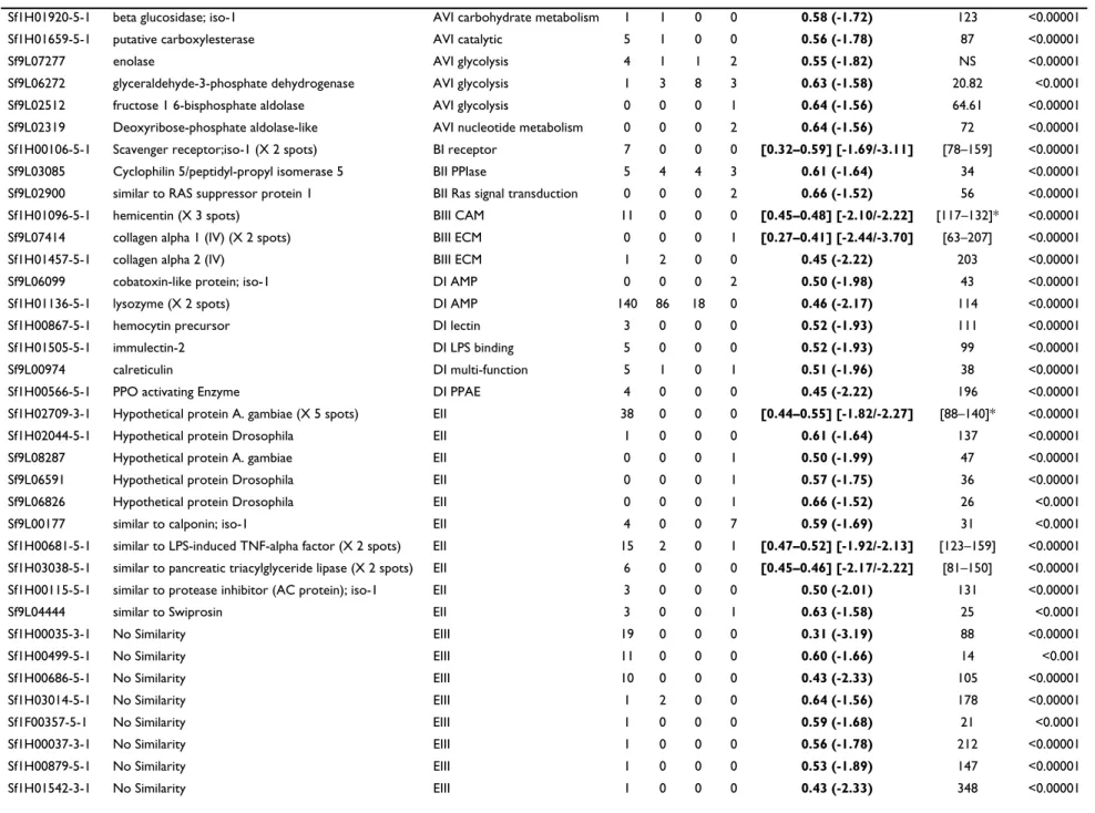
Table 1: List of Spodoptera frugiperda genes transcriptionally modified by HdIV infection.
Spodobase ID Gene Name Functional class Number of clones SAM Fold GeneANOVA
H F M L F gene-cond° p-value
Hemocytes_HdIV : 72 significant genes, 18 up-regulated and 54 down-regulated q-value 0.0056
Sf1H03393-5-1 Fumarate hydratase AVI tricarboxylic acid cycle 1 3 0 0 2.24 74 <0.00001
Sf1H00151-5-1 retinol dehydratase; iso-1 AVI catalytic 7 0 0 0 2.11 152 <0.00001
Sf9L04641 90-kDa heat shock protein HSP83 AVII stress. hsp 0 0 2 5 2.19 33 <0.0001
Sf1H03026-5-1 heat shock cognate 70 protein AVII stress. hsp 1 0 0 0 1.58 62 <0.00001
Sf1H00903-5-1 prefoldin subunit 2 AVIII processing 2 0 1 0 1.88 91 <0.00001
Sf1H00522-5-1 Galectin; iso-1 BIII lectin 3 0 0 0 5.66 161 <0.00001
Sf1H01648-5-1 Galectin; iso-2 BIII lectin 2 0 0 0 5.50 110 <0.00001
Sf1H01841-5-1 lectin (CrV) BIII lectin 11 0 0 0 1.66 44 <0.00001
Sf1H03263-5-1 MBF2 C regulator 10 3 0 0 2.18 NS <0.00001
Sf1H00908-5-1 PPO-1 (X 4 spots) DI PPO 75 0 0 0 [1.9-4.89] [27–144] <0.00001
Sf1H00508-5-1 PPO-2 (X 3 spots) DI PPO 60 0 0 0 [4.02–5.91] [67–104] <0.00001
Sf1H01637-5-1 Hypothetical protein A.gambiae EII 1 0 0 0 2.73 250 <0.00001
Sf9L08591 Hypothetical protein Drosophila EII 0 0 1 1 2.52 96 <0.00001
Sf1H00837-3-1 No Similarity EIII 1 0 0 0 2.74 134 <0.00001
Sf1H00871-5-1 No Similarity EIII 1 0 0 0 2.98 261 <0.00001
Sf1H01434-3-1 No Similarity EIII 1 0 0 0 3.03 102 <0.00001
Sf1H02673-5-1 No Similarity EIII 1 0 0 0 1.96 173 <0.00001
Sf1H00201-3-1 NS. similar to Hypothetical protein (5' seq) EIII 2 0 0 0 2.73 93 <0.00001
Sf1H02439-5-1 Annexin IX-B AI Ca binding 7 3 0 0 0.62 (-1.61) 66 <0.00001
Sf9L07393 Calmodulin AI Ca binding 5 2 11 6 0.59 (-1.69) 31 <0.0001
Sf9L07135 Cytochrome C oxydase I AI electron transport 162 117 232 146 0.62 (-1.61) 30 <0.0001
Sf1H02665-5-1 splicing factor 3a AII splicing 1 0 0 0 0.48 (-2.08) 196 <0.00001
Sf9L06869 cofilin AIV actin 16 1 10 2 0.54 (-1.85) 62 <0.00001
Sf1H03446-5-1 profiling AIV actin 30 2 16 0 0.54 (-1.85) 142 <0.00001
Sf9L03392 Thymosin beta AIV actin 7 1 3 13 0.45 (-2.22) 33 <0.0001
Sf9L06055 alpha tubulin AIV tubulin 5 5 7 7 0.56 (-1.80) 56 <0.00001
Sf9L01568 beta-1 tubulin AIV tubulin 1 2 2 1 0.56 (-1.80) NS <0.00001
Sf1H02495-5-1 synaptotagmin-like; granuphilin AIX exocytosis 2 0 0 0 0.65 (-1.54) 27 <0.0001
Sf9L01210 Ribosomal protein S24 AV ribosomal 1 0 4 21 0.58 (-1.71) 84 <0.00001
BMC Geno mi cs 20 06, 7:1 60 http://ww w.b iomed central.com/1 471 -2 164 /7 /16 0 Pa ge 7 of 2 0 (page nu mber not for cit a tion pur poses)
Sf1H01920-5-1 beta glucosidase; iso-1 AVI carbohydrate metabolism 1 1 0 0 0.58 (-1.72) 123 <0.00001
Sf1H01659-5-1 putative carboxylesterase AVI catalytic 5 1 0 0 0.56 (-1.78) 87 <0.00001
Sf9L07277 enolase AVI glycolysis 4 1 1 2 0.55 (-1.82) NS <0.00001
Sf9L06272 glyceraldehyde-3-phosphate dehydrogenase AVI glycolysis 1 3 8 3 0.63 (-1.58) 20.82 <0.0001
Sf9L02512 fructose 1 6-bisphosphate aldolase AVI glycolysis 0 0 0 1 0.64 (-1.56) 64.61 <0.00001
Sf9L02319 Deoxyribose-phosphate aldolase-like AVI nucleotide metabolism 0 0 0 2 0.64 (-1.56) 72 <0.00001
Sf1H00106-5-1 Scavenger receptor;iso-1 (X 2 spots) BI receptor 7 0 0 0 [0.32–0.59] [-1.69/-3.11] [78–159] <0.00001
Sf9L03085 Cyclophilin 5/peptidyl-propyl isomerase 5 BII PPIase 5 4 4 3 0.61 (-1.64) 34 <0.00001
Sf9L02900 similar to RAS suppressor protein 1 BII Ras signal transduction 0 0 0 2 0.66 (-1.52) 56 <0.00001
Sf1H01096-5-1 hemicentin (X 3 spots) BIII CAM 11 0 0 0 [0.45–0.48] [-2.10/-2.22] [117–132]* <0.00001
Sf9L07414 collagen alpha 1 (IV) (X 2 spots) BIII ECM 0 0 0 1 [0.27–0.41] [-2.44/-3.70] [63–207] <0.00001
Sf1H01457-5-1 collagen alpha 2 (IV) BIII ECM 1 2 0 0 0.45 (-2.22) 203 <0.00001
Sf9L06099 cobatoxin-like protein; iso-1 DI AMP 0 0 0 2 0.50 (-1.98) 43 <0.00001
Sf1H01136-5-1 lysozyme (X 2 spots) DI AMP 140 86 18 0 0.46 (-2.17) 114 <0.00001
Sf1H00867-5-1 hemocytin precursor DI lectin 3 0 0 0 0.52 (-1.93) 111 <0.00001
Sf1H01505-5-1 immulectin-2 DI LPS binding 5 0 0 0 0.52 (-1.93) 99 <0.00001
Sf9L00974 calreticulin DI multi-function 5 1 0 1 0.51 (-1.96) 38 <0.00001
Sf1H00566-5-1 PPO activating Enzyme DI PPAE 4 0 0 0 0.45 (-2.22) 196 <0.00001
Sf1H02709-3-1 Hypothetical protein A. gambiae (X 5 spots) EII 38 0 0 0 [0.44–0.55] [-1.82/-2.27] [88–140]* <0.00001
Sf1H02044-5-1 Hypothetical protein Drosophila EII 1 0 0 0 0.61 (-1.64) 137 <0.00001
Sf9L08287 Hypothetical protein A. gambiae EII 0 0 0 1 0.50 (-1.99) 47 <0.00001
Sf9L06591 Hypothetical protein Drosophila EII 0 0 0 1 0.57 (-1.75) 36 <0.00001
Sf9L06826 Hypothetical protein Drosophila EII 0 0 0 1 0.66 (-1.52) 26 <0.0001
Sf9L00177 similar to calponin; iso-1 EII 4 0 0 7 0.59 (-1.69) 31 <0.0001
Sf1H00681-5-1 similar to LPS-induced TNF-alpha factor (X 2 spots) EII 15 2 0 1 [0.47–0.52] [-1.92/-2.13] [123–159] <0.00001 Sf1H03038-5-1 similar to pancreatic triacylglyceride lipase (X 2 spots) EII 6 0 0 0 [0.45–0.46] [-2.17/-2.22] [81–150] <0.00001
Sf1H00115-5-1 similar to protease inhibitor (AC protein); iso-1 EII 3 0 0 0 0.50 (-2.01) 131 <0.00001
Sf9L04444 similar to Swiprosin EII 3 0 0 1 0.63 (-1.58) 25 <0.0001
Sf1H00035-3-1 No Similarity EIII 19 0 0 0 0.31 (-3.19) 88 <0.00001 Sf1H00499-5-1 No Similarity EIII 11 0 0 0 0.60 (-1.66) 14 <0.001 Sf1H00686-5-1 No Similarity EIII 10 0 0 0 0.43 (-2.33) 105 <0.00001 Sf1H03014-5-1 No Similarity EIII 1 2 0 0 0.64 (-1.56) 178 <0.00001 Sf1F00357-5-1 No Similarity EIII 1 0 0 0 0.59 (-1.68) 21 <0.0001 Sf1H00037-3-1 No Similarity EIII 1 0 0 0 0.56 (-1.78) 212 <0.00001 Sf1H00879-5-1 No Similarity EIII 1 0 0 0 0.53 (-1.89) 147 <0.00001 Sf1H01542-3-1 No Similarity EIII 1 0 0 0 0.43 (-2.33) 348 <0.00001
BMC Geno mi cs 20 06, 7:1 60 http://ww w.b iomed central.com/1 471 -2 164 /7 /16 0 Pa ge 8 of 2 0 (page nu mber not for cit a tion pur poses) Sf1H01712-5-1 No Similarity EIII 1 0 0 0 0.43 (-2.33) 106 <0.00001 Sf1H02105-5-1 No Similarity EIII 1 0 0 0 0.41 (-2.44) 163 <0.00001 Sf9L00090 No Similarity EIII 1 0 0 4 0.62 (-1.61) 35 <0.00001 Sf9L01158 No Similarity EIII 0 0 1 2 0.45 (-2.22) 73 <0.00001 Sf9L06863 No Similarity EIII 0 0 0 1 0.53 (-1.89) 43 <0.00001 Sf9L08779 No Similarity EIII 0 0 0 1 0.52 (-1.91) NS <0.00001
Fat Body_HdIV : 7 significant genes, down-regulated q-value 0.077
Sf9L07198 cytoplasmic actin A3 AIV actin 27 6 47 2 0.59 (-1.69) 27 0.00001
Sf9L06055 alpha tubulin AIV tubulin 5 5 7 7 0.45 (-2.22) 135 <0.00001
Sf9L01568 beta-1 tubulin AIV tubulin 1 2 2 1 0.65 (-1.54) 80 <0.00001
Sf9L07277 enolase AVI glycolysis 4 1 1 2 0.54 (-1.85) 39 <0.00001
Sf9L02319 Deoxyribose-phosphate aldolase-like AVI nucleotide metabolism 0 0 0 2 0.51 (-1.96) 82 <0.00001
Sf9L00974 calreticulin DI multi-function 5 1 0 1 0.38 (-2.63) 85 <0.00001
Sf1F00299-5-1 Hemolymph glycoprotein precursor DIII unknown 1 17 1 0 0.51 (-1.96) 33 <0.00001
Fat Body_JcDNV : 8 significant genes, up-regulated q-value 0.056
Sf9L00253 mRNA export factor binding AII transcription coactivator 0 0 0 2 1.87 35 <0.000001
Sf9L07586 Ribosomal protein S27A AV ribosomal 5 3 8 11 3.82 136 <0.000001
Sf1H03263-5-1 MBF2 C regulator 10 3 0 0 1.54 22 0.00006
Sf9L06099 cobatoxin-like protein; iso-1 DI AMP 0 0 0 2 2.12 70 <0.000001
Sf1H00171-5-1 lysozyme (X 2 spots) DI AMP 140 86 18 0 [1.59–1.66] [10–20] 0.0001
Sf9L06812 immunolectin-A precursor DI LPS binding 0 1 0 2 5.88 137 <0.000001
Sf9L02793 No Similarity EIII 0 0 0 1 5.46 212 <0.000001
Sf9L03186 No Similarity EIII 2 1 7 21 5.42 149 <0.000001
List of Spodoptera frugiperda genes for which transcript levels are significantly modified in the hemocytes and in the fat body 24 hours after injection of HdIV, and in the fat body 24 hours after injection of JcDNV, as detected by a modified t-test from the SAM package and an ANOVA based microarray analysis, GeneANOVA. Only the genes with a fold change superior or equal to 1.5 with both a FDR median and 90th percentiles of 0% were kept. For each assay, the SAM q-value (the lowest FDR at which the gene is called significant) is given. For each gene, the F value for "gene-condition" and the p-value calculated by GeneANOVA are given. Single genes for which more than one cDNA was spotted on the glass slide are indicated (x spots), in those cases, only the spot with the best score was kept in the list. Genes are organized according to their functional annotation (see Additional file 2 for legends). Spodobase ID: identity of the arrayed cDNA in the database Spodobase ([61]); Gene Name: annotation of the sequence after BlastX search against the GenBank nr database; Functional class: classes and subclasses to which the sequences belong to, based on the classification described in Shida et al. [114] for the ascidian Ciona intestinalis (details in Additional file 2); Number of clones: distribution of the cDNA clones corresponding to the given gene with available sequences in the different S. frugiperda cDNA libraries (H:hemocytes; F: fat body; M:midgut; L:Sf9 cell line). For genes represented by at least two cDNAs, we reported the interval [min, max] for the SAM fold change and for the F gene-condition from GeneANOVA analysis. Asterisk (*) indicate that one of the spots was non significant for GeneANOVA (clusters Sf1H01096-5-1 and Sf1H02709-3-1). The list of additional clones corresponding to each gene is provided in Additional file 1B.
BMC Geno mi cs 20 06, 7:1 60 http://ww w.b iomed central.com/1 471 -2 164 /7 /16 0 Pa ge 9 of 2 0 (page nu mber not for cit a tion pur poses)
Table 2: Variation in gene transcript levels in response to virus infection measured by quantitative RT-PCR.
Micro-array RT-qPCR
Fold (V/C) mean N0 Fold (V/C) mean N0 Fold (V/C)
H/F HdIV DNV control PBS (n = 24) HdIV (n = 18) HdIV95° (n = 18) HdIV HdIV95 control Sf (n = 24) DNV (n = 18) DNV
fold fold fold min mean max min mean max min mean max fold fold min mean max min mean max fold
HEMOCYTES PPO-1 NS +4.9 ND 0,73 0,9 1,1 11.7 30.1 48.5 3.8 7.1 10.5 +32.0 +7.6 3.5 4.7 5.9 4.5 4.7 4.9 NS PPO-2 10.7 +5.9 ND 5.3 6.1 7.0 8.7 43.8 78.9 7.1 7.9 8.7 +9.8 NS 20.5 24.3 28.1 23.7 27.0 30.3 NS Galectin NS +5.6 ND 0.7 0.9 1.1 3.8 8.9 14.1 0.3 0.5 0.7 +10.0 -1.7 0.6 0.8 0.9 4.0 4.8 5.6 +6.5 Scavenger R 26,0 -3.1 ND 10,6 11,8 13,0 1,8 4,3 7,0 8,4 9,5 10,6 -2.8 NS 22.0 25.0 27.9 21.5 24.2 26.9 NS PPAE 26.7 -2.2 ND 7,0 9,1 11,2 1,7 4,3 7,0 6.5 7.2 7.8 -2.11 NS 7.3 7.9 8.5 9.2 10.5 11.8 NS Immulectin-2 66,0 -1.9 ND 22.1 26.9 31.8 2.7 6.0 9.3 20.9 25.0 29.0 -4.5 NS 20.0 25.9 31.7 3.2 7.6 12.0 -3.4 Lysozyme 4,0 -2.2 ND 36.0 46.8 57.6 8.2 18.7 29.1 55.4 79.9 104.3 -2.3 +1.7 22.7 31.8 40.8 32.4 44.7 57.0 NS Calreticulin NS -2.0 ND 5,5 7,4 9,2 1,1 2,4 3,8 3.5 5.5 7.5 -3,0 NS 3.5 4.3 5.1 3.7 4.6 5.4 NS FAT BODY Lysozyme -- NS +1.7 9.9 11.8 13.8 10.9 11.7 12.5 39.6 86.7 133.8 NS +7.3 33.8 35.7 37.6 45.5 56.3 67.1 +1.6 Calreticulin -- -2.6 NS 6.5 7.7 8.9 2.0 2.5 2.9 6.5 7.6 8.6 -2.9 NS 4.5 5.8 7.2 4.8 5.8 6.8 NS Variations in gene transcript levels measured by quantitative RT-PCR in response to virus infection. Calculated mean of the relative cDNA starting quantity (N0) and consequential fold changes (V/C: virus/control) for selected Spodoptera frugiperda genes in control and virus-infected hemocytes and fat body 24 hours after injection. Minimum and maximum values obtained from the 18 or 24 replicates are given in addition to the mean N0 value. For HdIV assays, the control consisted in injection of PBS, for JcDNV (DNV), in injection of healthy S. frugiperda larvae crushed in PBS; HdIV95 corresponds to injections of heat-inactivated HdIV. H/F: ratio of the relative cDNA starting quantity (N0) between hemocytes and fat body in control PBS samples. ND: not done; NS: not significant; fold change threshold at 1.5 for both microarray and quantitative RT-PCR analyses, with FDR median and 90th percentiles at 0%.
BMC Genomics 2006, 7:160 http://www.biomedcentral.com/1471-2164/7/160
Page 10 of 20 (page number not for citation purposes) saline buffer. Conversely, lysozyme mRNA levels are
higher 24 hours after injection of heat-inactivated HdIV, as measured by quantitative RT-PCR (Table 2), with higher fold changes in the fat body (+7.3) than in the hemocytes (+1.7). JcDNV injection also results in increased transcript levels in the fat body (+2.1 for coba-toxin and +1.7 for lysozyme). Cobacoba-toxin is a cysteine-rich peptide that may be involved in antimicrobial defense [12] whereas lysozyme is a widely distributed ubiquitous enzyme involved in self-defense from bacterial infection [63]. In Drosophila adults, lysozyme genes are up-regu-lated 24 hrs after protozoan invasion and down-reguup-regu-lated after fungal invasion [9], but transcript levels remain unchanged after microbial infection [56], or infection with a picorna-like virus [9]. In lepidopteran insects, lys-ozyme genes are induced in both eggs and pre-imaginal instars in response to bacterial injection [14,37,64,65]. In
S. frugiperda, our results indicate that the c-lysozyme
mRNA is more abundant in fat body in response to inac-tivated-HdIV or to JcDNV compared to control. This sug-gests that transcript levels of this gene increased in response to viral presence. In host-parasitoid models involving polydnaviruses, the activity of host lysozyme is reported to be inhibited by parasitism [37]. In Heliothis
virescens parasitized with Campoletis sonorensis, the
observed reduced plasma lysozyme activity involves inhi-bition at a post-transcriptional step [53]. Our study is the first report of a transcriptional regulation of this antimi-crobial gene due to polydnavirus presence and expression. Down-regulation could reflect an active way of protecting both parasitoid wasp and virus from host defense.
The pro-PO cascade
Our results indicate that HdIV down-regulates the proPO-activation system by directly interfering with transcript levels of genes encoding proteins involved in the enzy-matic cascade. Indeed, in this study, we found that a S.
fru-giperda sequence (named Sf_PPAE), similar to the Manduca sexta proPO-activating enzyme PAP-1 gene [66],
is specifically down-regulated in the hemocytes 24 hours after injection of HdIV particles (2.2 in microarrays and -2.1 in quantitative RT-PCR). Similarly to the proPO-acti-vating enzymes (PPAE), Sf_PPAE contains a carboxyl-ter-minal proteinase domain typical of the chymotrypsin family and has an amino-terminal regulatory "clip" domain (reviewed in [67]). Insect PO is synthesized as an active zymogen (proPO), and is activated by proteolytic cleavage, mediated by a proteinase cascade plus addi-tional factors, including immulectins. The PPAEs are the terminal serine proteinases that carry out the proteolysis of the proPO precursor. In M. sexta, transcription of the proPO-activating enzyme PAP-1 gene is up-regulated in hemocytes and fat body in response to a bacterial chal-lenge and down-regulated by treatment with 20-hydroxy-ecdysone, suggesting that this gene is under the dual
control of immune and hormonal signals [68]. Our results show that HdIV infection down-regulates the tran-script levels of the gene encoding this proteinase. Con-versely, injection of heat-inactivated HdIV or of JcDNV did not affect transcript levels of Sf_PPAE. Down regula-tion of the Sf_PPAE, as well as of the immulectin gene (see below), should thus reduce the efficiency of PO pro-teolysis, which could be linked to the inhibition of hemo-lymph melanization observed in HdIV-infected S.
frugiperda caterpillars.
Genes encoding proteins putatively involved in the insect cellular immune response and/or non-self recognition
Immulectin
Immulectins are C-type lectins specific to Lepidoptera with a unique structure consisting of tandem carbohy-drate recognition domains (CRDs). They function as path-ogen recognition receptors in the innate immune system by activating proPO in hemolymph [69], and by partici-pating in hemocyte nodule formation [70] and encapsu-lation [71,72]. HdIV infection down-regulates (-1.9 in microarrays and -4.5 in quantitative RT-PCR) a S.
fru-giperda gene with high similarity with the M. sexta
immulectin-2 (AAF91316; Blast E value = 6e-81), which is induced in M. sexta fat body after bacterial challenge [73]. This S. frugiperda immulectin gene (Sf1H01505-5-1) seems to be preferentially transcribed in hemocytes (Table 2, H/F column) thus differing from other immulectin genes generally reported as synthesized in the fat body and secreted in the hemolymph [72]. Four immulectins are known in M. sexta [72], and we identified 3 different cDNAs in the S. frugiperda libraries. One of the other immulectins spotted in the microarray (Sf9L06812) is up-regulated by JcDNV in the fat body (+5.9), but is not mod-ified by HdIV, suggesting that different immulectin genes may be differentially affected by HdIV. Down-regulation by HdIV of the S. frugiperda immulectin-2 homologue could be linked to inhibition of encapsulation observed in parasitized caterpillars. Interestingly, this gene is also down-regulated by JcDNV in the hemocytes (-3.4 in quan-titative RT-PCR), suggesting an important immune func-tion of this protein.
Calreticulin
In fat body, the highest fold change (-2.6 in microarrays and -3.0 in quantitative RT-PCR) was found for the tran-scripts related to the multifunctional Ca-binding protein calreticulin (CRT). Transcript levels for CRT also decrease in the hemocytes (-2.0 in microarrays and -2.9 in quanti-tative RT-PCR). Conversely, no variation in the number of transcripts was noted after injection of either JcDNV or heat-inactivated HdIV, suggesting that this variation is related to HdIV gene transcription. CRT is a conserved multifunctional Ca2+-binding protein, detected in a large variety of cellular compartments (reviewed in [74]).
Intra-BMC Genomics 2006, 7:160 http://www.biomedcentral.com/1471-2164/7/160
Page 11 of 20 (page number not for citation purposes) cellular CRT functions as a molecular chaperone and also
regulates Ca2+ homeostasis [75]. CRT is also found on the cell surface where it may play a role in cellular adhesion and migration [76] or clearance of apoptotic cells [77]. In insects, CRT has several functions, including functions associated with immunity. In Galleria mellonella, CRT pro-tein was detected in the soluble fraction of hemocyte lysate and surrounding DEAE beads thus suggesting that CRT participates in the non-self recognition of early-stage encapsulation response [78]. The role of CRT in lepidop-teran cellular response is also suggested by its presence on the surface of Pieris rapae hemocytes during phagocytosis of yeast cells [79]. Recently, a CRT was identified in the venom fluid of the parasitoid wasp Cotesia rubecula and shown to inhibit host hemocytes in vitro spreading [80]. The authors suggest that the wasp CRT functions as an antagonist of host CRT. In the Hyposoter didymator model, the host CRT gene is down-regulated in HdIV-infected lar-vae, suggesting a different strategy than C. rubecula. The down-regulation of CRT in HdIV-infected S. frugiperda lar-vae occurs in the same range in both hemocytes and fat body and may thus be responsible for the decrease in non-self recognition and encapsulation efficiency.
Scavenger receptor
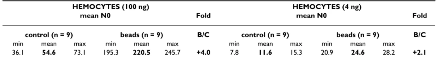
One S. frugiperda gene that is down-regulated in response to HdIV injection (1.7/3.1-fold decrease for the two spots, Table 1) encodes a protein with significant similarities with Drosophila class C scavenger receptors (SR-C) (blast E value = 5e-46 with GenBank:AAW79423). The S.
fru-giperda gene encodes a predicted transmembrane protein
that contains a MAM domain and two tandem comple-ment control protein (CCP) domains. In Drosophila, the functionally characterized SR-C, dSR-CI, is implicated in phagocytosis and MAM and CCP domains are sufficient for binding of bacteria [81]. Scavenger receptors (SRs) are probably also involved in phagocytosis in Lepidoptera. Indeed, adhesion of E. coli on Spodoptera littoralis granular hemocytes is inhibited with polyinosinic acid, a specific ligand of SRs [82]. Since the dSR-CI recognizes a broad range of polyanionic ligands, similarly to the mammalian class A scavenger receptors [83], SR-C may be the SR responsible for the attachment of bacteria to lepidopteran hemocytes. To test the involvement of S. frugiperda SR-C
in cellular responses other than phagocytosis, we ana-lyzed the transcript levels of this gene after injection of large Sephadex beads. Beads are fully encapsulated 8 hours after injection and a preliminary macroarray analy-sis has indicated up-regulation of the SR-C gene 6 hours after injection (not detected 1 hour after injection, Volkoff, unpub.). So SR-C transcript levels were analyzed 6–7 hours after injection by quantitative RT-PCR in 2 dif-ferent technical and biological conditions. Results indi-cate that SR-C is up-regulated (2.0/4.0-fold increase) in hemocytes after injection of Sephadex beads (Table 3), strongly suggesting that this novel SR-C plays a role in the
S. frugiperda cellular response against parasites. The SR-C
gene is down-regulated only when HdIV genes are tran-scribed (non significant with heat-inactivated virus and with JcDNV; Table 2). We thus found two opposite effects on SR-C gene transcript levels for the beads and the virus, suggesting that HdIV acts directly on SR-C transcript levels to impair the host cellular response.
Others
In addition to the three genes discussed above, HdIV injection down-regulates genes encoding proteins poten-tially involved in hemocytes adhesion and migration. For example, HdIV injection alters transcript levels of the annexin IX-B (decrease 1.6-fold), a calcium binding pro-tein that plays a role in biological important functions such as membrane fusion and control of cell proliferation and differentiation [84]. In Drosophila, annexin IX is an immune responsive gene that is up-regulated in response to bacterial immune challenge [56,57]. Interestingly, a new discovered hemicentin-like gene (Volkoff et al., unpub.), a putative adhesion molecule of unknown func-tion, is down-regulated in infected hemocytes (fold decrease -2.1/-2.2) as well as a transcript encoding a pro-tein with significant similarity with the C-terminal region of the silkworm humoral lectin (hemocytin, -1.9), an adhesive protein related to encapsulation of foreign sub-stances [85].
Polydnavirus, as other immunosuppressive virulence fac-tors, commonly results in disruption of the hemocytes actin cytoskeleton [24]. Microarrays indicate that, besides down-regulating the two tubulin subunits in the
hemo-Table 3: Variation in SR-C gene transcript levels measured by quantitative RT-PCR.
HEMOCYTES (100 ng) HEMOCYTES (4 ng)
mean N0 Fold mean N0 Fold
control (n = 9) beads (n = 9) B/C control (n = 9) beads (n = 9) B/C min mean max min mean max min mean max min mean max
36.1 54.6 73.1 195.3 220.5 245.7 +4.0 7.8 11.6 15.3 20.9 24.6 28.2 +2.1 Variation in SR-C gene transcript levels measured by quantitative RT-PCR in response to injection of Sephadex beads in the hemocyte population collected 6 to 7 hours after injection. Calculated mean of the relative cDNA starting quantity (N0) and consequential fold changes (B/C: beads/ control). Fold change threshold at 1.5 for quantitative RT-PCR analyses, with FDR median and 90th percentiles at 0%.
BMC Genomics 2006, 7:160 http://www.biomedcentral.com/1471-2164/7/160
Page 12 of 20 (page number not for citation purposes) cytes and in the fat body, injection of HdIV affects
tran-scripts of several genes encoding proteins involved in actin-based motility processes, such as thymosin (-2.2), profilin (-1.85) and cofilin (-1.85). The reason for the down-regulation of structural cytoskeleton proteins (actin, alpha- and beta-tubulin) in the fat body is not known, and could be related to modifications in cellular trafficking, mitosis, or apoptosis (preparation to pupa-tion). Actin-binding proteins are commonly found as up-regulated in genome-wide analyses of immune challenged
Drosophila [8,57]. Inhibition of regulatory proteins of the
actin cytoskeleton by HdIV is concordant with recent analyses in CsIV-infected hemocytes suggesting that actin is depolymerised or synthesized in lesser amounts [35]. Presence of HdIV also down-regulates genes that encode proteins involved in the assembly of the extracellular matrix, such as type IV collagens, components of base-ment membranes. Both collagen alpha-1 (2 spots) and collagen alpha-2 mRNAs were less abundant after injec-tion of HdIV (-2.4/-3.7 and -2.2, respectively). Compo-nents of basement membrane are secreted by hemocytes (mainly granulocytes in Lepidoptera) and integrity of the basement membranes seems to play a central role in non-self recognition by hemocytes (reviewed in [2]). Decreased transcripts of collagen in HdIV-infected hemo-cytes could be related to a less efficient cellular response, although the mechanism remains unclear.
Some S. frugiperda genes display increased transcript level in hemocytes 24 hours after HdIV injection
Among the 18 genes that are up-regulated in the hemo-cytes after HdIV injection, 11 encode proteins with simi-larities with known proteins (Table 1). The highest transcript levels variations were observed for the genes encoding galectin and the prophenoloxidase subunits 1 and 2 (PPO-1 and PPO-2).
Galectin
Two genes encoding sequences similar to galectins display an important increase of transcript levels 24 hours after HdIV injection in S. frugiperda hemocytes (average of 5.6-fold in microarrays and 10.0-5.6-fold in quantitative RT-PCR; Table 1). Galectins are thiol-dependent β-galactoside-binding lectins found in a large variety of organisms (reviewed in [86]). The S. frugiperda galectin gene is spe-cifically transcribed in hemocytes (Barat-Houari, unpub. results) and encodes a protein containing a single carbo-hydrate recognition domain, which is more similar to the mammalian galectin-9 than to other described insect galectins. The S. frugiperda galectin gene is also up-regu-lated in hemocytes following JcDNV injection (6.5-fold increase), but down-regulated after injection of heat-inac-tivated HdIV (1.6-fold decrease; Table 2). In vertebrates, galectins are involved in a variety of cellular processes
such as adhesion [87], apoptosis [88] and they possibly regulate innate immune responses [86]. In insects, galectins are thought to have dual functions in develop-ment and innate immunity (reviewed in [89]). In
Anophe-les gambiae, two galectin genes, GALE8 and GALE5 are
induced by bacterial and malarial challenges [90]. In
Dro-sophila, the Dmgal galectin is expressed in hemocytes and
may participate in recognition of microorganisms or, alternatively, modulate hemocyte aggregation during infection [89]. It is possible that increased galectin levels in HdIV-infected S. frugiperda larvae might lead to the abnor-mal clumping of hemocytes observed in parasitized larvae (Volkoff, pers. data), thus contributing to a reduction in the number of circulating hemocytes capable of forming the capsule, and to a less efficient cellular response [91,92].
Prophenoloxidase subunits
The two S. frugiperda genes encoding the two prophe-noloxidase subunits 1 and 2 (PPO-1 and PPO-2) showed increased transcript levels in microarray experiments. The microarray contains 4 cDNAs encoding the PPO-1 protein and 3 cDNAs encoding the PPO-2 protein and all of these cDNAs are significantly up-regulated (increases 1.9/4.9-fold and 4.0/5.9-1.9/4.9-fold, respectively; Table 1). This result was confirmed with quantitative RT-PCR on other biolog-ical samples (Table 2). The level of transcripts for the proPO-1 gene was still higher after injection of heat-inac-tivated HdIV (+7.6), suggesting that the up-regulation of this gene is not directly related to HdIV transcription, but could be linked to presence of HdIV in the caterpillar and thus could reflect a response to HdIV presence. The response seems specific to HdIV since, as shown in Table 2, the transcript level was not changed in caterpillars 24 hours after injection of the densovirus JcDNV. In
Dro-sophila, none of the three proPO genes is induced after
microbial infection [56] and enzyme activation does not seem necessary for resistance to microbial infections [93]. Conversely, in mosquito, the expression of two pro-POs is significantly enhanced in response to microfilariae inocu-lation and blood feeding, respectively [94]. An inducible proPO was identified recently in Spodoptera litura, and its transcript level increases after bacterial infection [95]. Thus the important up-regulation of the two S. frugiperda pro-PO genes by HdIV, inactivated or not, may indirectly reflect a response of this insect to enveloped viruses such as HdIV. This would corroborate previous reports on a possible role of PO in antiviral response in the lepidop-teran larvae [16]. We cannot exclude that the highest level of proPO transcripts detected in S. frugiperda hemocytes could also be related to a selective destruction of a subset of hemocytes that are responsible of proPO enzyme pro-duction. Indeed, Microplitis demolitor bracovirus (MdBV) selectively induces apoptosis of the granulocytes [96]. However, no clear hemocyte apoptosis is observed after
BMC Genomics 2006, 7:160 http://www.biomedcentral.com/1471-2164/7/160
Page 13 of 20 (page number not for citation purposes) infection with HdIV, contrary to this hypothesis. In
HdIV-injected S. frugiperda larvae, concomitant to increased
proPO transcript levels, by 24 hours after HdIV injection,
the Sf_PPAE transcript levels have decreased. This should result in a general decrease in PO activation and in the inhibition of melanisation observed in parasitized cater-pillars.
Conclusion
Polydnavirus or calyx fluid injection was previously reported to affect host protein levels, by inhibiting trans-lation or interfering with post-transcriptional steps in the parasitized host. The transcriptomic analysis of
S.fru-giperda genes conducted here shows that injection of
filter-purified HdIV results in modifications of transcript levels for several host genes. We cannot assert that all the varia-tions detected by this microarray analysis result exclu-sively from HdIV, but filter purification is a method that minimizes contaminations with wasp proteins [97]. Moreover, results obtained for the genes studied after injection of heat-inactivated HdIV suggest that HdIV tran-scription is required for the changes observed. Whether the observed variations in host genes transcript levels result from a direct effect of HdIV proteins or from upstream regulations of other genes or proteins in major pathways will need to be further investigated. The fold-changes observed for transcript levels of the down-regu-lated genes are relatively low (-1.9 in average and maxi-mum -3.7 for collagen in microarray experiment, -3.1 in average and maximum -4.5 for immulectin in quantitative RT-PCR assay), which is consistent with microarray anal-yses performed in other biological models and for which high fold changes are usually not observed. These results suggest that HdIV infection decreases transcript levels but does not shut-down a large panel of host genes. Moderate and diversified effects on global host physiology at the immune response level as well as at the developmental level are compatible with maintenance and survival of the host during all parasitoid development. Based on our knowledge on other insects, we can conclude that several of the steps in the lepidopteran host immune response are affected. Down-regulation of putative pattern recognition molecules such as calreticulin and immulectin suggests a decreased recognition efficiency of foreign bodies. Decreased levels of transcripts for lysozyme and PPAE indicate that the humoral response is affected. Finally, the cellular response seems to be impaired due to interference with transcript levels of cytoskeleton-related proteins (actin regulatory proteins, tubulins) or scavenger recep-tors. The next step will be to better characterize these reg-ulated genes at a genomic level, in order to identify common signaling pathways.
This study also raises the question of the function of the proteins encoded by genes affected by the HdIV injection
in lepidopteran immune defense and development. Our results suggest an important role for calreticulin, and two novel proteins, scavenger receptor and hemicentin-like proteins, in lepidopteran immune response, and makes lysozyme and PO good candidates as molecules involved in virus recognition and antiviral defense.
Based on the results obtained, we plan now to analyze the interaction between HdIV and the lepidopteran host S.
frugiperda using a more complete microarray comprising a
higher number of probes. This should circumvent the lim-its of the microarray analysed in this study, particularly the reduced number of genes that were screened. Analyz-ing the transcript levels of homologues of known compo-nents of major pathways involved in insect immune response such as Toll, Imd or Jak/Stat pathways and those of genes previously shown to play a role in the formation of the hemocytic capsule, such as the plasmatocyte spreading peptide cytokine [98,99], or integrins [19,100] or the immunoglobulin domains-containing hemolin (review in [5]), should provide an enhanced outline of the immune-suppressive effects of HdIV.
To conclude, this study, although based on a simplified biological model, provides evidence that infection with the polydnavirus HdIV affects, directly or indirectly, tran-script levels of lepidopteran host genes. The results obtained in the present study also validate the transcrip-tomic approach to analyze the complex interactions in the parasitoid-polydnavirus-host models. Further use of such global approaches should result in a better understanding of the strategies employed by parasites to manipulate their host physiology, and should give new insights on lepi-dopteran innate immunity by allowing the identification of potential targets of the immunosuppressive polydnavi-ruses.
Methods
I/Array design description: construction of Spodoptera
frugiperda cDNAs microarrays
The microarrays were glutaraldehyde activated aminosi-lane-coated glass slides (pre-cleaned gold seal micro-slides, Merck) on which we spotted in triplicates PCR products corresponding to S. frugiperda cDNAs. We used 1750 non-redundant sequences from 4 S. frugiperda cDNA libraries (1266 cDNAs from a Sf9 cell line library, 381 from a hemocyte library, 48 from a fat-body library and 55 from a midgut library; sequences available at [61]; [101]) and 11 viral cDNAs as a control.
The DNA probes were amplified by PCR using primers flanking the cDNA inserts, the forward primer GTGTT-GGTACCCGGGAATTCG-3' and the reverse primer 5'-GCTCGAGTCTAGAGTCGACTG-3'. These primers had a C6 amine modification in the 5' end in order to bind to
BMC Genomics 2006, 7:160 http://www.biomedcentral.com/1471-2164/7/160
Page 14 of 20 (page number not for citation purposes) the glutaraldehyde activated aminosilane-coated slides.
After amplification, the quality of the PCR products was verified by electrophoresis on 0.8% agarose gels and the DNA purified using QIAquick 96 PCR purification kit (QIAGEN) according to manufacturers' protocol. After quantification using PicoGreen (Molecular Probes), all PCR products were transferred to 384-well plates, lyophi-lized and re-suspended in 3 × SSC to a final concentration of 0.150 mg/ml. The spotting was done with a Chip Writ-erPro spotter (Virtek, Biorad) using PT3000 pins supplied by EBN (Belgium). Each pin withdraws a volume of about 250 nanoliters and deposits a spot volume of about 0.6 nanoliters, with a diameter of approximately 90 to 130 μm. The cDNAs were printed on the slides using an ele-ment center-center spacing of 180 μm. After printing, slides were allowed to dry and then used immediately, or were stored desiccated at room temperature. Slides exhib-iting defects such as missing or damaged spots were dis-carded.
Before hybridization, slides were post-processed in several steps as described in Hugot et al. [102] : (a) Unbound DNA was removed by washing with 0.2% (w/v) SDS and double-distilled water, (b) covalently bound DNA was denatured for 2 min in boiling water, (c) free aldehydes were reduced by soaking slides for 5 min in 68 mM sodium borohydride (dissolved in PBS containing 25% (v/v) ethanol), and (d) free glasses sites were blocked by an incubation of 30 min at 60°C in the presence of 0.2% (w/v) casein. Several washing steps were performed with 0.2% (w/v) SDS and double-distilled water. After washes, the post-processed slides were dried by centrifugation at 500 g for 5 minutes and then either used immediately or stored desiccated at room temperature.
II/Microarray experiment description
1- Experimental design
We performed 2 biological assays, the first with the polyd-navirus HdIV and the second with the densovirus JcDNV. Each assay was composed from two sets, named "infected" and "non-infected control", and each set was composed of 20 1-day old S. frugiperda last instar larvae. For each pair infected/non-infected samples, a second labeling reaction, named dye-swap, was performed with inversion of the fluorescent dyes. For each assay, we pre-pared 3 independent biological replicates and 2 dye swap per biological replicate, i.e. 6 slides per assay.
2- Sample used, extract preparation and labelling Virus preparation and insect infections
Hyposoter didymator and S. frugiperda were reared as
previ-ously described in Volkoff et al. [29]. HdIV suspension was prepared as previously described [32]. Briefly, ovaries from 40 H. didymator females were dissected and dislo-cated in phosphate-buffered saline (PBS) and then
fil-trated with a 0.45 μm acetate-cellulose filter. The HdIV suspension obtained was adjusted to 600 μl and then each
S. frugiperda larva was injected with 27 μl (corresponding
to 1.8 ovaries/larvae). Each larva from both HdIV-infected and control sets were injected with 27 μl of either HdIV suspension or PBS.
We used the densovirus isolated from the lepidopteran
Junonia coenia JcDNV [103]. The densoviruses belong to Parvoviridae family and produce highly contagious lethal
diseases in invertebrates [62], and Spodoptera species are particularly sensitive to JcDNV. The control and viral inoc-ula were prepared by crushing either healthy or JcDNV-infected S. frugiperda in 1.5 ml of PBS containing 2% ascorbic acid, pH 7.2. The homogenates were clarified 10 min at 9000 g, filtrated on a 0.45 μm acetate-cellulose fil-ter, and then diluted ten fold. Each larva from the JcDNV infected or control sets were inoculated with 13 μl of healthy or viral suspension.
RNA extraction from S. frugiperda tissue samples
Samples were collected 24 hours post-injection (24 h p.i.) from both virus-infected and non-infected control sets. Total RNA was extracted from tissue samples (hemocytes or fat body cells) using the Rneasy Mini Kit Total RNA according to manufacturers' protocol (Qiagen S.A., Court-aboeuf, France, cat. no. 74106). Hemocytes were collected as previously reported[32]. Dissected fat body was col-lected directly in the extraction buffer (RLT buffer) from the Rneasy Mini Kit. On-column DNase digestion was then performed with the RNase-free DNase Kit (Qiagen, cat. no. 79254). Additionally, for the JcDNV-infected sam-ples, infection was controlled by RT-PCR using an oligodT primer and a primer specific to the JcDNV viral protein gene.
RT and labeling
Ten μg of total RNA were labeled with either Cy5dCTP or Cy3dCTP using the ChipShot Labeling System (Pronto!™ Plus System, cat. no. 40054-55, Promega; [104]), accord-ing to manufacturers' protocol. We used the dyes from Amersham Biosciences. Non-incorporated dyes were eliminated using the ChipShot Labeling Clean-Up System (Pronto!™ Plus System, cat. no. 40054-55, Promega). After quantification of each dye incorporation, the labeled cDNAs were dried in speed-vacuum and then dissolved in the required volume of water-diluted Dig Easy hybridiza-tion soluhybridiza-tion (1/3 v/v)(DIG Easy Hyb Granules, Roche Diagnostics GmbH cat. no. 1 796 895).
3- Hybridization procedures and parameters
The labeled cDNAs obtained from the two experimental conditions (i.e. infected and non-infected samples) were mixed in equivalent quantities (pmols) of incorporated Cy3 and Cy5 in order to obtain a final volume of 20 μl
BMC Genomics 2006, 7:160 http://www.biomedcentral.com/1471-2164/7/160
Page 15 of 20 (page number not for citation purposes) hybridization solution. This solution was then put on the
array surface and a micro cover glass (22 × 32 mm) was slowly applied to allow the solution spreading and cover-ing the whole spotted area. Slide hybridization was done in Corning CMT-hybridization chambers overnight by total immersion in a water bath at 42°C, in the dark. Following overnight hybridization, the slides were washed according to standard procedures [102] and dried by centrifugation at 500 g for 5 min before scanning.
4- Data measurement and specifications of data processing Image analysis and quantification
Hybridized slides were scanned with an Axon 4000 B microarray scanner (Axon Instruments, Foster City, CA) to generate 16-bit TIF images. The laser power was set at 100% and the photo-multiplier tube (PMT) gain for each wavelength between 600 and 900. Images were analyzed with GenePix Pro4.0 Axon software (adaptative circle seg-mentation algorithm), according to manufacturers' instructions.
Data standardization and normalization
After microarray raw data acquisition, data processing was performed using the web-accessible MicroArray Data Suites of Computed Analysis, MADSCAN resource (at [105]; [106]). The MADSCAN procedure uses the median intensities after background corrections and analytical parameters provided by GenePix Pro. MADSCAN perform physical validation and quality filtration based on five-step criterion. During these first five-steps, several spots were ruled out from the analysis. It was the case for viral con-trols (one JcDNV and HdIV positive concon-trols: rep1, rep2) that displayed poor quality spots and were flagged by the different quality control steps (Genepix or MADSCAN). After data filtration, MADSCAN perform log transforma-tion of the median intensities to makes the distributransforma-tion of the data symmetrical and almost normal. Hence, the scaled lowess fitness (LOcally-WEighted Regression) nor-malization algorithm was applied to minimize signal-dependent non-linear bias between the two intensity lev-els. Madscan perform a within-print tip (local) normaliza-tion with a smooth parameter (defined as the fracnormaliza-tion of data used to smooth at each data point) f = 0.40 [107].
5- Microarray statistical analysis
To identify differentially expressed genes, the lowess nor-malized values of the background corrected intensities obtained from the red and the green channels calculated by MADSCAN were then implemented in two validated statistical analysis programs: the Significant Microarray Analysis (SAM) downloaded from [108] and the Gene-Anova program [109]. Only the data significant with the two statistical methods were considered.
For the SAM analysis we kept all the genes with enough non-missing values as program is able to input missed val-ues with the average of the 10 non-missing nearest-neigh-bor among replicates. We applied the two-class unpaired method from the SAM package to test the null hypothesis of no effect of viral compared to control injection across 18 replicates (3 biological × 2 technical dye-swap × 3 within slide as each gene is present in triplicate). This method is a nonparametric test that call significant genes based on the comparison of the experimental score d(i) to its expected value calculated from N random permuta-tions between treated and untreated sample data (random assignment of treatment). Then, from the randomized data, the SAM program calculates the ratio of the number of false-positives to the expected number of genes called significant. This value provides an estimate of the number of genes identified by chance, the False Discovery Rate (FDR). The q-value, calculated from the distribution of the d(i) statistics obtained from permutations, represents the lowest FDR at which the gene is called significant. We selected differentially expressed genes that met both FDR criteria (estimated FDR median and 90th percentiles of 0%) and fold change criteria (at least 1.5-fold) [110]). The 1.5 threshold was chosen according to Yang et al. [111], who suggest that fold changes smaller than ± 2 can relia-bly identify differentially expressed genes when sufficient number of replicates and flip-dye assays are performed. Afterwards, the results were ascertained with an ANOVA based microarray analysis, GeneANOVA, which is a freely available gene expression analysis of variance software developed by The Laboratory of "Génome et Informa-tique" at Evry-Génopole [109]. As GeneANOVA requires no missing values, we implemented the program with the SAM imputed data set in which the missed values has been calculated from the mean of 10 non-missing nearest neighbour. For this analysis we constructed a statistical model including 5 factors: genes, type of injection (HdIV versus control or JcDNV versus control), dye label, techni-cal dye-swap replicates and biologitechni-cal replicates. Theses factors represent a set of interacting parameters reflecting differential gene expression across different experimental conditions. ANOVA model allows an estimation of the contribution of each factor from the study design in the total variation of the whole set of measurements and the pair-wise sets of interaction between factors: gene-by-con-dition, gene-by-slide (i.e. dye-swap technical replicate), gene-by-biological replicate. GeneANOVA gives also the significance of each contribution globally and for each gene. When assessing genes-by-injection interaction, tran-scriptionally modulated genes identified from the SAM analysis, presented a significant interaction with injection. ANOVA analysis revealed no effect of confounding factors such as dye label and replicates on gene expression levels (i.e. no significant interaction between those factors and injection p > 0.01), suggesting that the transcriptional