Immuno-detection of Staphylococcus aureus
Biofilm on a Cochlear Implant
M.I. Kos, L. Stenz, P. François, J.-P. Guyot, J. Schrenzel
Abstract
Case presentation: A 46-year-old man suffering from pro-gressive deafness since childhood received a Clarion 90 K cochlear implant with the HiResÒpreformed electrode in his left ear in October 2006. A persistent Staphylococcus aureus infection failed to be treated with corticoids, amoxicillin/ clavulanate, ciprofloxaxin, and rifampin. The processor was removed on July 2007.
Interventions: The removed cochlear implant processor was treated with different reagents, with the aim of detecting a S. aureus and S. aureus biofilm: (1) fluorescein-coupled Fc of anti-human serum, (2) polyclonal anti-polysaccharide intercellular adhesion antibodies coupled to Alexa Fluor 568 goat anti-rabbit immunoglobulin (Ig)G, (3) crystal violet, (4) methylene blue, (5) acridine orange, (6) Gram stain, and (7) live/dead fluorescent stain.
Results: S. aureus and the major constituent of the S. aureus biofilm, the polysaccharide intercellular adhesion, were detected on the surface of the implant. S. aureus was isolated after a simple contact between the implant and a solid growth medium. The ability of the isolated S. aureus strain to produce biofilm in vitro was confirmed.
Interpretation: S. aureus biofilm was documented on the implant. Initial bacterial colonization could be related to the pocket of the removable magnet. Colonies of S. aureus without biofilm were found attached to the electrode wire. Conclusion: We report one case of a S. aureus biofilm infection documented on a cochlear implant, as assessed by immuno-microscopy. The biofilm was likely responsible for the persistent infection which manifested for many months after the implant surgery and could explain the unusual bacterial phenotypic resistance against adminis-tered antimicrobial agents.
Infection 2009; 37: 450–454 DOI 10.1007/s15010-008-8335-1
Introduction
Until recently, medical complications associated with co-chlear implants were mainly related to flap necrosis, incision dehiscence, and post-operative wound infections
[4, 11, 23]. The incidence of such complications was low, and they were usually successfully managed with antibi-otics and/or plastic and middle ear surgery. Cases of persistent infection requiring the removal of the processor have been rare, and some have even been attributed to a primary immunodeficiency of the recipient [23]. Similarly to what has been observed on other types of implants [5], reports of biofilm formation on the surface of cochlear implants started to appear in print in 2004 [1, 17, 20]. Clinically, biofilms are complex bacterial communities that adhere to the surface of implanted biomaterial or mucosa [9, 18] and produce an extra-cellular matrix [3], leading to increased bacterial resistance against the host’s immune defenses and to antibiotics [22]. Since 1985, 200 patients have been implanted at the Geneva Cochlear Implant Centre. Different types of implants have been used and, until recently, not a single case of wound dehiscence, flap necrosis, or infection of the processor has been observed. In 2007, we were confronted with an atypical and persistent infectious case that required re-moval of the processor, a Clarion 90 K cochlear implant with the HiResÒ preformed electrode, which was then submitted to microbiological analysis.
Case History
In October 2005, a 46-year-old man suffering from progressive deafness since childhood received a Clarion 90 K cochlear implant with the HiResÒ preformed electrode in his left ear.
M.I. Kos, J.-P. Guyot
Geneva Cochlear Implant Centre, Oto-Rhino-Laryngology, Head and Neck surgery-Service, Geneva University Hospitals, Geneva 4, Switzerland L. Stenz, P. François (corresponding author), J. Schrenzel
Genomic Research Laboratory, Infectious Diseases Service, Geneva University Hospital, Geneva 4, Switzerland; Phone: (+41/22) 3729-338, Fax: -830, e-mail: [email protected] L. Stenz
Dept. of Microbiology and Molecular Medicine, University Medical Center, Geneva 4, Switzerland
M. Izabel Kos and L. Stenz have contributed equally to this work. Received: August 28, 2008 Æ Revision accepted: November 18, 2008 Published online: March 10, 2009
Surgery was performed according to the minimally invasive technique described byO’Donoghue and Nikolopoulos [19]. The processor was placed in a muscle pouch and attached to the bone. The patient was given intravenous ceftriaxone during the surgery, followed by amoxicillin/clavulanate (orally) for 1 week post-surgery. Healing was uneventful. Two months after cochlear implantation the patient had achieved a very good performance and was wearing his implant daily. 17 months after the surgery, however, he felt an increasing retro-auricular pain, and the site of the processor became swollen. We suspected that a hematoma had developed inside the muscle pouch containing the processor and treated the symptoms with corticoids and amoxicillin/cla-vulanate orally. The swelling and pain disappeared in 15 days, but reappeared 6 weeks later. Puncture removed 3 cc of a citrin liquid, and subsequent culture of the liquid showed the growth of Staphylococcus aureus sensitive to all tested antibiotics with the exception of penicillin G. The patient received a treatment of ciprofloxacin and rifampin for 8 weeks, but the swelling, redness, and pain persisted. Surgical drainage was performed. The sur-rounding tissue was debrided, and the muscle pouch and the processor were irrigated with antibiotics. Culture showed again S. aureus. The wound healed, but 2 weeks later liquid had again collected. The processor was removed in July 2007. The elec-trode wire was sectioned at the level of the cochleostomy, and the electrode array was left inside the cochlea. The wound healed in 2 days. Three months later the patient received a new cochlear implant, which was switched on 2 weeks after surgery. The pa-tient has since reached the same performance levels with the new implant as he had with the original one.
Material and Methods
Microbiological Sampling, Cultures, and Identification of the Strain of S. aureus
During the removal surgery, samples were collected from the inguinal, axillary, retro-auricular, and external ear canal skin, from nasal and throat mucosa, and from fragments of the sur-rounding tissue. The identification of a strain of S. aureus was performed according to Clinical and Laboratory Standards Institute (CLSI) recommendations and included Pastorex agglutination (Bio-Rad, Hercules, CA) and the DNAse pro-duction test. A real time-PCR amplification procedure [8] was performed for confirmation.
S. aureus growth was achieved by culturing the removed processor on Mueller Hinton agar (MHA; Bio-Rad, Marnes-La-Coquette, France). S. aureus strain SA113 (ATCC 35556) and its ica mutant (Dica::tet) were used as control for immuno-detection of polysaccharide intercellular adhesion (PIA) [10]. MHA and trypticase soy broth (TSB; Becton Dickinson, Le Pont de Claix, France) supplemented with 1% glucose (TSBgluc) were used for bacterial growth. The in vitro formation of the biofilm was tested on a strain grown in TSBgluc medium during a 15-h culture. A glass coverslip was added to the well prior to culture for the detection of PIA.
Pre-Treatment of the Processor
The processor body was divided into two parts. The silicon part harboring the removable magnet pocket after the magnet was removed was snap-frozen in liquid nitrogen and conserved at 80 °C. Before testing, the device was washed with a PBS solution (Invitrogen, Carlsbad, CA) and sliced into five pieces, which
Chemica, Germany) in PBS, and conserved in a PBS solution at 4 °C. Before testing, this part and the electrode wire were sliced into five pieces each.
Crystal Violet Staining Assay for Initial Evaluation of the Presence of the Biofilm
Glutaraldehyde-fixed implant sections and thein vitro heat-fixed bacteria biofilm were stained for 10 min with 1% (w/v) crystal violet (CV) stain freshly diluted twofold in 1% ethanol/distilled water, as previously described [21]. The stained material was then washed three times with PBS and inspected with the naked eye and by white light microscopy.
Immuno-Detection of PIA
Sections of the processor and in vitro biofilm from the S. aureus grown on a circular glass coverslip (diameter 25 mm) were wa-shed twice with PBSAT (PBS containing 0.02% azide and 0.05% Tween. 20) with slow shaking for 5 min. Cross-reactions with S. aureus protein A were blocked by incubating the material for 2 h with 1:1,000 normal donkey serum (Jackson Immuno Research, West Grove, PA). For specific detection of PIA, the material was incubated for 1 h with 1:3,000 a-PIA rabbit polyclonal anti-PIA antibody [15], then washed twice with PBSAT. The binding of specific antibodies was revealed after incubation with 1:3,000 dilution of Alexa Fluor 568 goat anti-rabbit IgG (H + L) (Molecular Probes, Eugene, OR) as secondary antibody. The material was washed with a PBS solution (PBSAT) containing 1% albumin (ZLB Behring AG, Bern, Switzerland) and 0.1% tween-20 (Fluka).
Immuno-detection of S. aureus Through Binding to Cell-Wall Protein A
Processor sections and the slices of the electrode wire were incubated for 30 min with PBSAT and for 30 min with 1:1,000 fluorescein-coupled to the Fc fragment of goat anti-human serum (Jackson Immuno Research). The slices were then washed five times with PBSAT before being observed under the microscope.
Immuno-Fluorescent Microscopy
All incubations were performed in PBSAT. A humid chamber was prepared for antibody incubations, consisting of a six-well plate that was hermetically sealed with Parafilm and light-pro-tected with aluminium foil. Images were acquired by an Axiocam color camera (Zeiss, Iena, Germany) on an Axioskop 2 micro-scope (Zeiss). Ultraviolet excitation for fluorescent imaging and white light microscopy were performed both separately and in combination. Filter set 09 (Zeiss; excitation BP 450–490, emission LP 515) was used for PIA immuno-detection through Alexa Fluor detection (emission 603 nm), whereas Filter set 02 (Zeiss; exci-tation G 365, emission LP 420) was used to detect fluorescein isothiocyanate (FITC; emission 530 mm ± 15 nm), indicating the presence of S. aureus through the binding of the Fc fragments to bacterial protein A. Scaling was performed automatically with the AxioVision software (Zeiss) according to the objective in use.
Results
Bacterial Recording
according to CLSI recommendations. Identification of the bacterium was based on a positive by Pastorex agglutina-tion and DNAse producagglutina-tion tests. Confirmaagglutina-tion was ob-tained using a previously described duplex PCR amplification. S. aureus was also detected by the direct growth of the bacterium following culture of the explanted implant on a solid medium in a petri dish, yielding a pure culture. The isolated S. aureus strain was named ‘‘Coch’’.
S. aureus Biofilm Identification on the Cochlear Implant
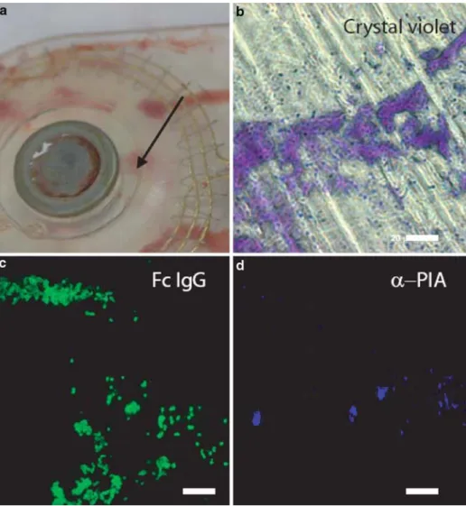
The biofilm was detected on the glutaraldehyde-fixed part of the implant containing the magnet pocket (Figure 1a). Cochlear implant pieces were successfully stained with CV in different zones visible to the naked eye. These colored zones were less abundant than areas reacting with the methylene blue stain. White-light microscopic obser-vations of CV-stained zones showed an association with cocci (Ø = 1 lm) (Figure 1b). Presence of adherent S. aureus on the implant was confirmed by the detection of protein A using a FITC-coupled
immuno-globulin (Figure 1c). The presence of the biofilm was postulated in these CV-stained zones and confirmed using specific antibodies raised against S. aureus PIA (Figure 1d). Surface PIA-positive zones were less abun-dant than CV-stained zones.
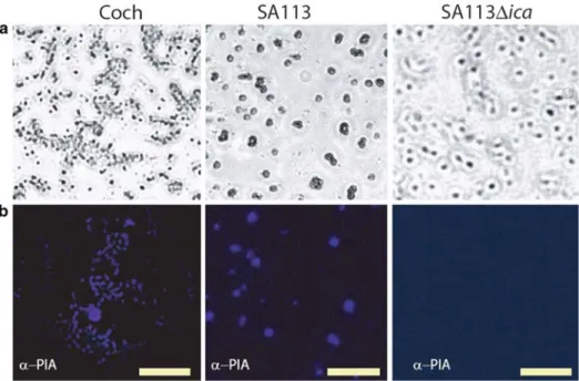
In vitro PIA-based Biofilm Formation
Coch strain produced PIA in vitro on glass coverslips at amounts quite similar to those of the control laboratory strain SA113 (Figure 2). Surface colonization to be ap-peared homogeneous for strain SA113 in vitro, whereas strain Coch showed punctual aggregates. The ica mutant was negative for PIA-specific fluorescence (Figures 1, 2).
Binding of Fluorescent Fc Fragments on the Electrode Wire
Immuno-detection performed on the electrode wire was positive for S. aureus on the first five proximal sections (corresponding to a 4.4-cm length from the implant side) as revealed by Fc-FITC binding (not shown). The three most distal fragments studied were negative.
Figure 1. Microscopy imaging of the removed cochlear implant. a) Magnet pocket of the explanted Clarion 90 K cochlear implant. The successfully stained zones depicted on panels b, c, d are localized near the recess surrounding the magnet (A ¼ 8 mm, see arrow). b) Crystal violet-stained zone associated with cocci-like structures. c) Immuno-fluorescence microscopy showing the presence of S. aureus protein A. d) Immuno-fluorescence
microscopy showing the presence of biofilm-associated
polysaccharide intercellular adhesion (PIA). Scale bars: 20 lm.
Discussion
Worldwide, only two cases of implant extrusion due to persistent infections by S. aureus have been clearly asso-ciated to biofilms. In these cases, identification was per-formed using scanning electron microscopy [1, 20]. We report here a third case of S. aureus biofilm infection of a Clarion HiRes 90 K cochlear implant. The microscopic appearance of the biofilm-related PIA matrix produced by the isolated S. aureus strain, denoted here as Coch, dif-fered between the in vitro and ex vivo experiments. CV staining is an easy indirect procedure that can be used to quantify biofilm in vitro [7, 21], but it has never been used for ex vivo biofilm detection. Our case is therefore the first time this procedure has been applied to detect bacterial biofilm on ex vivo materials as an initial evaluation of the surface of the implant. Cocci-like structures (A approx. 1 lm) were clearly visible in the microscopic observations. Taken together, PIA immuno-detection and CV staining confirmed the presence of a S. aureus biofilm on the sur-face of the implant inside the pocket of the removable magnet. Analysis of the magnet itself was impossible for technical reasons. We speculate that S. aureus contami-nated first a hematoma inside the muscle pouch made to hold the processor and subsequently the processor itself. Even though a treatment of amoxicillin/clavulanate fol-lowed by ciprofloxacin and rifampin was given as soon as a biofilm infection was suspected, the processor had to be ultimately explanted. This case illustrates how efficiently the bacterial community resisted the host immune re-sponse and the antibiotics that are effective when the same bacteria are in their planktonic form [3] and
con-the early stage of con-the disorder. There have been published cases of redness and tenderness around the processor followed by flap necrosis or dehiscence and, ultimately, rejection of the processor. These cases were suspected to be caused by an allergy to one of the implant components [16] because no germs could be detected. However, even in such cases, biofilm infections should not be excluded. Sub-clinical biofilm infections can persist for many years before they manifest [11]; therefore, a biofilm infection cannot be ruled out based on a negative culture of the material [14].
Antibiotic prophylaxis is recommended for surgeries involving implants, but the postoperative use of antibiot-ics is not [2]. This latter practice has, since this case, been abandoned by our center. Although we consider the case reported here to be unique among all the procedures carried out at our center, representing 0.5% of our im-plants, the post-operative use of antibiotics could have contributed to increasing the resistance of the infecting bacteria and the formation of biofilm.
The first processors implanted in the 1980s were sealed in a smooth ceramic case [13]. The contribution of bacterial biofilm-related infection to the frequency of implant removal is clearly under-reported in the literature as biofilm presence has not been not systematically as-sessed. In 2000, new processors constructed out of flexible silastic and containing a removable magnet pocket were developed by CochlearÒand by Advanced BionicsÒ. Two recent studies in which this type of implant was used have reported the presence of biofilm scattered over the entire the surface of the devices [1, 20]. The authors of another
Figure 2. In vitro production of PIA. Microscopic pictures of in vitro biofilms formed on glass coverslips of Coch (left column), SA113 (middle column), and SA113Dica (right column). Row A: Adherent bacteria visible using white light
microscopy. Row B: Pictures of PIA immuno-detection on the corresponding surfaces. Scale bars: 20 lm.
also reported higher counts of bacteria on implants har-boring an empty magnet pocket as compared to models without a magnet pocket.
As reported previously by other authors [1, 17, 20], we cannot confirm the presence of biofilm around the elec-trode wire. The tests performed after the removal of the processor revealed that there was S. aureus attached to the electrode wire up 4.4 cm distally from the body of the processor, but these are not specific for the detection of biofilm. In our case, during the removal surgery, the length of the wire that extended from the mastoid to the cochl-eostomy appeared to be normal. We arbitrarily decided to cut the wire at the level of the cochleostomy and leave the electrodes array inside the cochlea to avoid obliteration by fibrous tissue that could prevent a new implant.
Modern processors equipped with a removable mag-net represent a major improvement for patients suffering from chronic disease that requires regular follow-up with MRIs. The ability to remove the magnet removes the risk of magnet mobilization or demagnetization in patients requiring radiological examination. Unfortunately the pocket designed to encase the magnet seems to favor biofilm formation, and the possibility that this pocket is involved in biofilm formation should not be ignored. Technical developments are under way to avoid the for-mation of bacterial biofilms, such as surface treatment with antimicrobial molecules [6]. Clearly, such developments need careful evaluation in the clinical context [6, 12].
In conclusion, biofilm can cause resistant infections of cochlear implants that manifest many months after the surgery. The pocket of the removable magnet could be one niche facilitating biofilm, formation although coloni-zation of the electrode wire is not excluded.
Acknowledgments
This work was supported by grants from the Swiss National Science Foundation no. 112370/1 (JS) and no. 3100A0-116075/1 (PF), and the European Commission 6th framework program (MagRSA project no. 37957). We are grateful to Fried-rich Go¨tz for the gift of S. aureus SA113 and Dica and to Johannes Knobloch for providing us with the anti-PIA antibodies.
Reference
1. Antonelli PJ, Lee JC, Burne RA: Bacterial biofilms may contribute to persistent cochlear implant infection. Otol Neurotol 2004; 25: 953–957.
2. Bratzler DW, Houck PM: Antimicrobial prophylaxis for surgery: an advisory statement from the National Surgical Infection Prevention Project. Am J Surg 2005; 189: 395–404. 3. Costerton JW, Stewart PS, Greenberg EP: Bacterial biofilms:
a common cause of persistent infections. Science 1999; 284: 1318–1322.
4. Cunningham CD III, Slattery WH III, Luxford WM: Postoperative infection in cochlear implant patients. Otolaryngol Head Neck Surg 2004; 131: 109–114.
5. Darouiche RO: Treatment of infections associated with surgical implants. N Engl J Med 2004; 350: 1422–1429.
6. Darouiche RO: Antimicrobial coating of devices for prevention of infection: principles and protection. Int J Artif Organs 2007; 30: 820–827.
7. Djordjevic D, Wiedmann M, McLandsborough LA: Microtiter plate assay for assessment of Listeria monocytogenes biofilm formation. Appl Environ Microbiol 2002; 68: 2950–2958. 8. Francois P, Bento M, Renzi G, Harbarth S, Pittet D, Schrenzel J:
Evaluation of three molecular assays for rapid identification of methicillin-resistant Staphylococcus aureus. J Clin Microbiol 2007; 45: 2011–2013.
9. Galli J, Calo L, Ardito F, Imperiali M, Bassotti E, Fadda G, Paludetti G: Biofilm formation by Haemophilus influenzae iso-lated from adeno-tonsil tissue samples, and its role in recurrent adenotonsillitis. Acta Otorhinolaryngol Ital 2007; 27: 134–138. 10. Gotz F: Staphylococcus and biofilms. Mol Microbiol 2002; 43:
1367–1378.
11. Harada T, Ishida K, Endo M, Takahashi M, Sakai M: Recurrent extrusion of cochlear implant at an interval of 5 years. Otol Neurotol 2003; 24: 83–85.
12. Johnson TA, Loeffler KA, Burne RA, Jolly CN, Antonelli PJ: Biofilm formation in cochlear implants with cochlear drug delivery channels in an in vitro model. Otolaryngol Head Neck Surg 2007; 136: 577–582.
13. Kessler DK: The CLARION multi-strategy cochlear implant. Ann Otol Rhinol Laryngol Suppl 1999; 177: 8–16.
14. Klykken PC, Curtis JM: Re: ‘‘Silicone allergy: a new cause for cochlear implant extrusion and its management’’. Otol Neurotol 2007; 28: 1159–1161.
15. Knobloch JK, Horstkotte MA, Rohde H, Mack D: Evaluation of different detection methods of biofilm formation in Staphylo-coccus aureus. Med Microbiol Immunol 2002; 191: 101–106. 16. Kunda LD, Stidham KR, Inserra MM, Roland PS, Franklin D, Roberson JB Jr: Silicone allergy: a new cause for cochlear implant extrusion and its management. Otol Neurotol 2006; 27: 1078–1082.
17. Loeffler KA, Johnson TA, Burne RA, Antonelli PJ: Biofilm forma-tion in an in vitro model of cochlear implants with removable magnets. Otolaryngol Head Neck Surg 2007; 136: 583–588. 18. Morris DP, Hagr A: Biofilm: why the sudden interest? J
Otolar-yngol 2005; 34: S56–S59.
19. O’Donoghue GM, Nikolopoulos TP: Minimal access surgery for pediatric cochlear implantation. Otol Neurotol 2002; 23: 891–894.
20. Pawlowski KS, Wawro D, Roland PS: Bacterial biofilm formation on a human cochlear implant. Otol Neurotol 2005; 26: 972–975. 21. Tu Quoc PH, Genevaux P, Pajunen M, Savilahti H, Georgopoulos
C, Schrenzel J, Kelley WL: Isolation and characterization of biofilm formation-defective mutants of Staphylococcus aureus. Infect Immun 2007; 75: 1079–1088.
22. Vlastarakos PV, Nikolopoulos TP, Maragoudakis P, Tzagaroulakis A, Ferekidis E: Biofilms in ear, nose, and throat infections: how important are they? Laryngoscope 2007; 117: 668–673. 23. Yu KC, Hegarty JL, Gantz BJ, Lalwani AK: Conservative
man-agement of infections in cochlear implant recipients. Otolar-yngol Head Neck Surg 2001; 125: 66–70.