DOI 10.1007/s00402-008-0694-7
O R T H O P A E D I C S U R G E R Y
The anterior center edge angle in Lequesne’s false pro
Wle view:
interrater correlation, dependence on pelvic tilt and correlation
to anterior acetabular coverage in the sagital plane.
A cadaver study
P. O. Zingg · C. M. L. Werner · A. Sukthankar · M. Zanetti · B. Seifert · C. Dora
Received: 12 March 2008 / Published online: 16 July 2008 © Springer-Verlag 2008
Abstract
Introduction Lequesne’s vertical-center-anterior margin (VCA) angle measured on the false proWle view of the pel-vis aims at quantifying the anterior acetabular coverage of the femoral head. The anterior delimitation of the acetabu-lar roof is often deWned on the false proWle view but there are no data on its interrater reliability. Additionally, it is not known how pelvic tilt may inXuence this angle. Finally, the plane in which this angle is measured lies at an angle of 65° to the sagittal plane and we wondered if this angle would be transposable to the anterior acetabular coverage measured in the sagittal plane.
Methods Eight hips from four cadaver pelvises were investigated by means of a total of 72 false proWle views, each taken in deWned pelvic inclinations at 5° increments ranging from ¡20° to +20°, and the VCA angle measured by three independent raters. A computed tomography (CT) of each hip was performed in a neutral pelvic tilt position and a sagittal 2D reconstruction calculated in order to mea-sure anterior coverage in the sagittal plane. The interrater reliability of the VCA angles was assessed using the intra-class correlation coeYcient (ICC). The dependence of the VCA angle on pelvic tilt was assessed by regression
analysis. The Correlation between the VCA angle and ante-rior coverage in the sagittal plane of the CT was analyzed using a simple linear regression model.
Results The interrater reliability for measurements of the VCA angle was almost perfect (ICC:0.97). Regression analysis showed that each degree of pelvic tilt was accom-panied by a change of the VCA angle by a value of 0.63° (P < 0.001). A low correlation between the VCA angle measured in the false proWle view and the anterior coverage in the sagittal plane was statistically not signiWcant (r = 0.667, P = 0.06).
Conclusions Lequesne’s VCA angle has an excellent interrater reliability and represents a reliable measure of acetabular dysplasia for comparisons with published data. Lequesne’s VCA angle is inXuenced by pelvic tilt in a lin-ear manner. Performing the false proWle view in a stand-ing position may reduce the clinical relevance of this dependency on pelvic tilt. The correlation of Lequesne’s VCA angle to anterior acetabular coverage in the sagittal plane is low and therefore unsuitable to be transposed into the sagittal plane.
Keywords Hip · LEQUESNE’s false proWle view · Pelvic tilt · Anterior acetabular coverage
Introduction
Acetabular dysplasia can be quantiWed by measuring the amount of femoral head coverage by the acetabulum [6, 9]. Lateral coverage is measured using Wiberg’s center edge (CE) angle on an AP pelvic X-ray [6]. Because of the superposition of both hips on a true proWle view of the pelvis, Lequesne [6] proposed to measure anterior acetabular coverage on a false proWle view using the P. O. Zingg · C. M. L. Werner · A. Sukthankar · M. Zanetti ·
C. Dora (&)
Department of Orthopaedics, University of Zurich, Balgrist, Forchstr. 340, 8008 Zurich, Switzerland e-mail: [email protected]
P. O. Zingg
e-mail: [email protected]
B. Seifert
Biostatistics Unit, Institute of Social and Preventive Medicine, University of Zurich, Hirschengraben 84,
vertical-center-anterior margin (VCA) angle (Fig.1). This VCA angle is composed of a vertical line (parallel to the border of the Wlm) through the center of the femoral head and a second line through the center of the hip and the fore-most aspect of the condensed line of the acetabulum [6] (Fig.2). A VCA angle greater than 25° is widely accepted to be normal, whereas less than 20° abnormal [6], corre-sponding to acetabular dysplasia. Other investigators have disagreed on these normal values [2, 7]. However, they did not use exactly the same imaging protocol. In Lequesne’s original description, the sclerotic zone of the acetabulum only (i.e., without marginal osteophytes) is taken into con-sideration for VCA angle measurements. This was not the case in Milcan’s [7] and Crockarell’s [2] study and may be an explanation for this discrepancy. Nevertheless, the nor-mal values proposed by Milcan and Crockarrell diVered signiWcantly from each other (17.7° vs. 24.75°) despite using an identical technique. We wondered if these discrep-ancies were due to a lack of inter-observer reliability due to diYculties in deWning the anterior delimitation of the ace-tabulum or to diVering patient positions during X-ray acquisition, especially varying pelvic inclinations. The aim of the present study was, therefore, to analyze the interrater reliability of measuring the VCA angle on identical false proWle views, and to correlate VCA angle values with diVering pelvic inclinations.
The VCA angle may additionally be used for the preop-erative planning of reorienting procedures of the acetabu-lum [1, 4], especially as far as the anterior coverage is concerned. However, the false proWle view is taken at an angle of 65° to the sagittal plane, whereas surgeons prefer
the VCA angle of Lequesne with the acetabular coverage determined in the sagittal plane of a CT scan.
Materials and methods Cadaver specimens
The responsible investigational review board did not require a formal approval of this study. The cadaver pel-vises were obtained and utilized according to the institu-tional guidelines and with informed consent of the donors prior to death or appropriate family members.
Eight formalin Wxed hips in four cadaver specimens including the pelvis and the proximal parts of the thighs were available from the anatomic department for this inves-tigation.
Before inclusion in to the study, absence of osteoarthritis of the hip was conWrmed by an AP pelvic X-ray.
Each specimen was Wxed to a frame in a standardized pelvic tilt position; in analogy to Legaye et al. [5] 0° pelvic tilt position in this study was assumed when the line con-necting the promotorium and the center of the hip joint was parallel to the frame on a true proWle view. Each specimen was positioned in this standardized position using Xuoros-copy.
Image acquisition
Fig. 1 Schematic drawing of patient positioning according the
Lequesne [6] projection: the hip of interest is facing the detector while the pelvis is rotated by 65° in the axial plane. The axis of the ipsilateral
foot has to be parallel to the radiographic plate Fig. 2 The VCA angle measured on the false proWle view of Lequesne
[6] is suggested to correspond to anterior coverage of the femoral head. The VCA angle is measured between the vertical and the line extend-ing from the centre C of the head to the most anterior point of the ace-tabulum A. Normal values are greater than 25° [6]
False proWle views were acquired for each hip separately. According to Lequesne’s description, the X-ray beam was perpendicular to the longitudinal axis of the specimen and rotated by 65° around its longitudinal axis and centered exactly between the two rotational centers of both hips.
In order to imitate varying pelvic tilts the X-ray beam was rotated stepwise by 5° increments around the trans-verse axis through the rotational centers of the hip joints while leaving the Wxed specimen in an unchanged position, and the false proWle view repeated. This was performed for +5°, +10°, +15° +20° (inclination) and ¡5°, ¡10°, ¡15°, ¡20° (reclination). The same procedure was performed for all 4 cadaver specimens and resulted in 36 false proWle views of a right and 36 false proWle views of a left hip with deWned pelvic inclinations.
In a second step the Wxation frame together with the specimen was positioned on the horizontal table of a CT scanner (Siemens Somatom plus 4, Siemens Medical Solu-tion, Erlangen) in the supine position and 2-mm thick axial images were acquired. In order to imitate various pelvic tilts secondary 2-D sagittal reconstructions were calculated through the center of the femoral head.
Measurements
All images were transferred to a dedicated workstation (Advantage Windows Workstation, Version 4.1; General Electrics Medical Systems Europe, Buc, France) equipped with an electronic caliper allowing precise measurements on the digital images.
The center of the femoral head was identiWed on both the sagittal plane CT reformations through the center of the femoral head and false proWle views, by superimposing a circle of the same radius as the femoral head contour onto the femoral head.
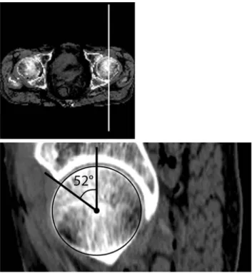
On the false proWle views, the VCA angles were mea-sured drawing a line parallel to the border of the Wlm though the center of the femoral head, i.e., the vertical line, and a line connecting the center of the femoral head with the most anterior delimitation of the acetabular dome scle-rotic line (Fig.3). These measurements were carried out by three independent observers on diVerent days. All observers were blinded for each others measurements and the inter-rater reliability calculated.
On the 2-D sagittal plane CT reformations the angle between the border of the Wlm (i.e., the vertical line) and the line connecting the center of the femoral head and the most anterior bony end of the acetabular roof was measured (Fig.4). These measurements were performed by means of consensus of the three investigators.
Fig. 3 X-ray measurement of VCA angle. The geometric center of the
femoral head was determined by superimposing a circle of the same ra-dius as the articular surface. Then a parallel line to the Wlm margin was drawn though the center of the femoral head, i.e. representing the ver-tical. Finally, the angle between the vertical and the anterior border of the sclerotic line was measured
Fig. 4 CT measurement of anterior acetabular coverage in the sagittal
plane. Measurements of the anterior acetabular coverage on CT scans, was performed by using a sagittal plane CT reformations through the center of the femoral head
Statistical analysis
Statistical analysis was performed by a statistical consultant from the Department of Biostatistics (SB).
Interrater reliability of the measured VCA angle was assessed in a four-way random ANOVA using the factors rater, side, cadaver, and pelvic tilt. Restricted maximum likelihood estimation was used to calculate variance com-ponents of the diVerent sources of variation. Interrater reli-ability was deWned as the part of variation not inXuenced by the rater. Thus, the interrater reliability is a direct gen-eralization of the intra-class correlation coeYcient (ICC) to this complex design. ICC values were interpreted as follows: ICC < 0.20 = slight agreement; 0.21–0.40 = fair agreement; 0.41–0.60 = moderate agreement; 0.61–0.80 = substantial agreement; and >0.80 = almost perfect agreement [8].
The dependence of VCA angle measurements on pelvic tilt was assessed in a three-way mixed ANCOVA (analysis of covariance) using the random factors rater, cadaver and Wxed factor side. Pelvic tilt was included as a covariate. Thus, the ANCOVA is a regression analysis addressing the correlation of measurements within the same pelvis and those performed by the same rater.
The correlation between the VCA angle and anterior coverage in the sagittal CT plane was analyzed using a sim-ple linear regression model.
All statistical analyses were performed using SPSS 13.0 for MacIntosh OS X. The signiWcance level was set at P < 0.05.
Results
All of the four specimens available (8 hips) could be included in the analysis since none of the 4 cadavers (2 males and 2 females) had radiological evidence of osteo-arthritis. Table1 summarizes the VCA angles of all hips in neutral pelvic tilt position. In 2 hips the VCA angle indi-cated acetabular dysplasia according to Lequesne’s criteria. Variance components of the factors rater, side, cadaver and pelvic tilt as well as their interactions are summarized in Table2. An almost perfect interrater reliability was found for measurements of the VCA angle (ICC = 0.97).
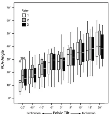
Analysis of covariance showed that each degree of pelvic tilt was accompanied by a linear change of the VCA angle by a value of 0.63° (P < 0.001) (Fig.5).
The correlation between the VCA angle of Lequesne and the corresponding angle measured in the sagittal plane of the CT was low and did not reach the signiWcance level of 5% with the numbers analyzed in the present study (r = 0.667, P = 0.06).
Discussion
The present study reveals a high interrater reliability for measurements of the VCA angle of Lequesne. DiYculties in deWning the anterior delimitation of the acetabulum in the false proWle view seems not to be an issue. Thus, Lequesne’s angle gives a reproducible measure of acetabu-lar coverage in the anterolateral region of the joint. Together with normal values published in the literature it is a reliable measure of acetabular dysplasia and its improve-ment after reorientation procedures.
As expected, pelvic tilt does have an inXuence on the values of the measured VCA angles. Fortunately this inXu-ence is linear. Every 1° of pelvic tilt increases the measured VCA angle by 0.63°. Two out of eight evaluated hips had a
Table 1 VCA angles of all hips in neutral pelvic tilt position
Cadaver 1 2 3 4
Hip 1 2 3 4 5 6 7 8
VCA angle 13a 32 29 41 16a 36 30 29
Table 2 Variances of VCA angle measurements
Covariance analysis illustrating the diVerent variances of the factors rater, side, cadaver and pelvic tilt as well as their interactions. Since statistical tests applied are covariance analyses the variance data have no units
a No units
Source of variation Variancea Interrater reliability of VCA angles
Cadaver 13.86 = intra-class correlation coeYcient (ICC) Side 38.29 = 0.97 Rater 0.52 Tilt 72.75 Cadavera side 54.20 Cadavera rater 0 Cadavera tilt 6.43 Sidea rater 0 Sidea tilt 0 Ratera tilt 1.11 Cadavera sidea rater 1.04 Cadavera sidea tilt 7.47 Cadavera ratera tilt 0.96 Sidea ratera tilt 0.13
conclude that measurements of normal and dysplastic hips are equally aVected by changes in pelvic inclination. This dependency on pelvic tilt should be kept in mind when using this angle to measure the degree of acetabular dyspla-sia. One measure to reduce this confounding factor is to perform the false proWle view in the standing and not in the lying position, expecting that the standing position would correspond to a more functional position of the pelvis.
However, the VCA angle of Lequesne had a low and nonsigniWcant correlation with the anterior acetabular cov-erage in the sagittal plane. This is in line with the anatomi-cal and radiologianatomi-cal investigation of Fabeck et al. [3] demonstrating that the VCA angle does not correspond to a real anatomical structure. However, the fact that only four whole body specimens (eight hips) were available in the present study may limit this Wnding and a greater number of specimens might have rendered this correlation signiWcant. Nevertheless, even if signiWcant with a higher number of specimens, the low correlation would be unlikely to have increased with higher numbers and we estimate the VCA angle of Lequesne unsuitable to be transposed into the sag-ittal plane and therefore unsuitable for guiding the surgeon
during reorientation procedures. Sagittal reconstruction of CT scans would be more appropriate for guiding reorienta-tion procedures with respect to anterior acetabular cover-age. However, CT scans are rarely used for the primary diagnosis of acetabular dysplasia. One reason might be the higher dose of radiation and the lack of normal values for anterior coverage in the sagittal plane. In addition CT scans would face the same positional problems of pelvic tilt.
In conclusion, Lequesne’s VCA angle measured on a false proWle view has an excellent intra- and inter-rater reli-ability and represents a reliable measure of acetabular dys-plasia when a comparison to published data needs to be made. However, Lequesne’s VCA angle is inXuenced by pelvic tilt. This inXuence is linear: 3° of pelvic tilt results in increasing the VCA angle by approximately 2°. Performing the false proWle view in a standing position may reduce the clinical relevance of this dependency on pelvic tilt. Unfor-tunately the correlation of Lequesne’s VCA angle to ante-rior acetabular coverage in the sagittal plane is low and therefore not very helpful in guiding the surgeon during reorientation procedures.
References
1. Crockarell J Jr, Trousdale RT, Cabanela ME, Berry DJ (1999) Early experience and results with the periacetabular osteotomy. The Mayo Clinic experience. Clin Orthop Relat Res (363):45–53 2. Crockarell JR Jr, Trousdale RT, Guyton JL (2000) The anterior
cen-tre-edge angle. A cadaver study. J Bone Joint Surg Br 82(4):532– 534. doi:10.1302/0301-620X.82B4.10063
3. Fabeck L, Farrokh D, Behets C, Delince P (2002) Anatomical and radiological correlation of Lequesne’s “false proWle”. Surg Radiol Anat 24(3–4):212–216. doi:10.1007/s00276-002-0038-1
4. Ganz R, Klaue K, Vinh TS, Mast JW (1988) A new periacetabular osteotomy for the treatment of hip dysplasias. Technique and pre-liminary results. Clin Orthop Relat Res (232):26–36
5. Legaye J, Duval-Beaupere G, Hecquet J, Marty C (1998) Pelvic incidence: a fundamental pelvic parameter for three-dimensional regulation of spinal sagittal curves. Eur Spine J 7(2):99–103. doi:10.1007/s005860050038
6. Lequesne M, de Seze S (1961) False proWle of the pelvis. A new radiographic incidence for the study of the hip. Its use in dysplasias and diVerent coxopathies. Rev Rhum Mal Osteoartic 28:643–652 7. Milcan A, Yildiz A, Oztuna V, Eskandari MM, Sahin G, Kuyurtar F
(2004) The anterior center edge angle: a study of 102 volunteers. Joint Bone Spine 71(3):221–223. doi:10.1016/S1297-319X(03)00121-0
8. Montgomery AA, Graham A, Evans PH, Fahey T (2002) Inter-rater agreement in the scoring of abstracts submitted to a primary care re-search conference. BMC Health Serv Res 2(1):8. doi:10.1186/ 1472-6963-2-8
9. Wiberg G (1939) Studies on dysplastic acetabula and congenital subluxation of the hip joint with special reference to the complica-tion of osteoarthritis. Acta Chir Scand Suppl (83):1–130
Fig. 5 Box-plots demonstrating the dependence of VCA angle
mea-sured on false proWle view on pelvic tilt and the excellent interrater reliability (ICC = 0.97) of VCA angle measurements. Analysis of covariance showed that each degree of pelvic tilt was accompanied by a linear change of the VCA angle by a value of 0.63° (P < 0.001)