ORIGINAL ARTICLE
In vitro evaluation of surface roughness, adhesion
of periodontal ligament fibroblasts, and Streptococcus
gordonii following root instrumentation with Gracey curettes
and subsequent polishing with diamond-coated curettes
Sigrun Eick&Philip Bender&Simon Flury&
Adrian Lussi&Anton Sculean
Received: 1 December 2011 / Accepted: 14 March 2012 / Published online: 17 April 2012 # Springer-Verlag 2012
Abstract
Objectives The objective of the study was to evaluate the efficacy of an additional usage of a diamond-coated curette on surface roughness, adhesion of periodontal ligament (PDL) fibroblasts, and of Streptococcus gordonii in vitro. Materials and methods Test specimens were prepared from extracted teeth and exposed to instrumentation with conven-tional Gracey curettes with or without addiconven-tional use of diamond-coated curettes. Surface roughness (Ra and Rz) was measured before and following treatment. In addition, the adhesion of PDL fibroblasts for 72 h and adhesion of S. gordonii ATCC 10558 for 2 h have been determined. Results Instrumentation with conventional Gracey curettes reduced surface roughness (median Ra before: 0.36 μm/ after: 0.25μm; p<0.001; median Rz before: 2.34 μm/after: 1.61 μm; p<0.001). The subsequent instrumentation with the diamond-coated curettes resulted in a median Ra of 0.31μm/Rz of 2.06 μm (no significance in comparison to controls). The number of attached PDL fibroblasts did not change following scaling with Gracey curettes. The addi-tional instrumentation with the diamond-coated curettes resulted in a two-fold increase in the number of attached PDL fibroblasts but not in the numbers of adhered bacteria.
Conclusions Treatment of root surfaces with conventional Gracey curettes followed by subsequent polishing with diamond-coated curettes may result in a root surface which provides favorable conditions for the attachment of PDL fibroblasts without enhancing microbial adhesion.
Clinical relevance The improved attachment of PDL fibro-blasts and the limited microbial adhesion on root surfaces treated with scaling with conventional Gracey curettes fol-lowed by subsequent polishing with diamond-coated cur-ettes may favor periodontal wound healing.
Keywords Curettes . Surface roughness . Adhesion of periodontal ligament fibroblasts . Adhesion of bacteria
Introduction
The primary goal in cause-related periodontitis treatment is to remove hard and soft bacterial deposits which should result in a smooth and biocompatible root surface to facili-tate fibroblast attachment and minimize bacterial adhesion.
Scaling and root planing are effective methods to remove supra- and subgingival bacterial deposits [1]. Findings from a systematic review have shown that subgingival mechani-cal debridement resulted in a mean attachment gain varying from 0.3 to 1.02 mm in pockets with an initial depth of up to 4 mm and in pockets with an initial depth of≥7 mm, a mean attachment gain of up to 1.58 mm was reported [2]. Despite the introduction of machine driven sonic, ultrasonic, and laser scalers, hand instruments (i.e., curettes) are still widely used in dental practice. Comparisons between hand instru-ments and sonic and ultrasonic scalers did not show a clear advantage for the machine driven instruments [3], and trau-matisation immediately after instrumentation was similar
S. Eick (*)
:
P. Bender:
A. SculeanDepartment of Periodontology, Laboratory of Oral Microbiology, School of Dentistry, University of Bern,
Freiburgstrasse 7,
CH-3010 Bern, Switzerland e-mail: sigrun.eick@zmk.unibe.ch S. Flury
:
A. LussiDepartment of Preventive, Restorative and Pediatric Dentistry, School of Dentistry, University of Bern,
[4]. Moreover, the use of hand instruments (i.e., convention-al Gracey curettes) yielded even greater improvements in some parameters (e.g., bleeding on probing) compared to instrumentation with an ultrasonic system [5]. Other studies have shown that the use of conventional Gracey curettes may result in higher substance loss but significantly better calculus removal [6–8] and smoother surfaces compared to sonic and ultrasonic instrumentation [6, 9, 10]. Thus, the clinical interest to improve the quality of root surface instru-mentation when using hand instruments remains. Very re-cently, the use of newly developed diamond-coated curettes used following scaling and root planing with conventional curettes has been suggested as a modality to additionally improve the quality of root surface debridement. These newly developed curettes are diamond coated at the working ends with 15 μm grit size of natural diamond granulate. Diamond-coated curettes have been introduced to be used in addition to conventional curettes for finalizing root planing. At the time being, however, there are no data evaluating the additional effect of the newly developed diamond-coated curettes used subsequently to debridement with convention-al hand instruments (i.e., Gracey curettes).
Therefore, the aim of the present study was to evaluate the in vitro efficacy of an additional usage of a newly developed diamond-coated curette on surface roughness, adhesion of periodontal ligament (PDL) fibroblasts and of Streptococcus gordonii as an early colonizer.
Materials and methods Specimens and instrumentation
Teeth extracted for periodontal reasons were selected in the study. Before the extraction, the patients were informed about the use of their teeth for research purposes and con-sent was obtained. After extraction, the teeth were placed in 0.5 % chloramine solution for disinfection overnight and thereafter stored in 0.9 % NaCl solution. The crowns of the teeth were removed and dentin slices of the buccal side of the roots were cut with diamond disks (~6×12 mm (thick-ness ~3 mm)). The surface properties of the buccal side of the dentin specimens were standardized by grinding with silicon carbide papers of #1,000 grit size, corresponding to an abrasive particle size of 18μm (Struers A/S, Ballerup, Denmark). The dentin specimens were mounted to the buc-cal site of an artificial incisor, exposing the standardized, ground side of the dentin specimen. To simulate in vivo conditions as far as possible, these teeth were placed into the artificial mouth of a mannequin head (Fig.1). For each of the three experiments (surface roughness, adhesion PDL fibroblasts, and adhesion of S. gordonii), 30 specimens were evaluated (n010 per curette treatment and n010 as a
control). Additionally, six (n02 per curette treatment and n02 as a control) were used for visualization of the results by means of scanning electron microscopy (SEM).
In the first of the three groups, the standardized speci-mens (ground with silicon carbide papers as described above) served as a control. In the second group, specimens were treated with Gracey curettes only (11GC12 Gracey curette, 13GC14 Gracey curette; both Deppeler SA, Rolle, Switzerland). In the third group, additional instrumentation was performed after treatment with Gracey curettes by using diamond-coated curettes (Intensiv PerioDiaCurette, PDC-11/12, Intensiv PerioDiaCurette, PDC-13/14; both Intensiv SA, Montagnola, Switzerland).
Specimens were scaled in an apical to coronal direction, during which they received 15 strokes by each site of the used curettes. Each side of the curette was used for five specimens and then replaced by a new curette. All treatments were performed by the same calibrated operator (P.B.).
Determination of surface roughness
Before and after treatment, the average surface roughness (Ra; μm) and the arithmetic mean height of the surface profile (Rz; μm) were profilometrically determined with a surface roughness meter (Perthometer S2; Mahr GmbH, Göttingen, Germany). Across the whole specimen, nine measurements were determined over a transverse length of Lt01.750 mm with a cutoff value of 0.25 mm. The speci-men was turned 45° after three measurespeci-ments (i.e., three measurements across the whole specimen at 0°, at 45°, and at 90°). From the nine Ra and Rz values, one mean Ra and one mean Rz value were calculated per specimen.
Adhesion of periodontal ligament fibroblasts
Human PDL fibroblasts were placed in T-25 cell culture flasks containing DMEM (Life Technologies/Invitrogen,
Fig. 1 Placement of the specimen in the artificial mouth and instru-mentation by using a Gracey curette
Paisley, UK) with 10 % fetal calf serum (FCS; Life Technologies/Invitrogen) to grow to confluency. At the start of the experiments, the fibroblasts were always in the third passage.
After a short autoclaving to avoid external contamina-tions, specimens were placed into 24-well plates and cov-ered with 25 % FCS overnight. Thereafter, PDL fibroblasts in DMEM with 10 % FCS were added at a density of 10,000 cells/well and incubated at 37 °C with 5 % CO2. At h, the
fibroblasts were fixed and stained for adhesion experiments with DAPI staining (Roche Diagnostics GmbH, Mannheim Germany). The attached fibroblasts were counted by using a fluorescent microscope (Olympus BX51, Tokyo, Japan). Three fields of 1 mm2per specimen were counted and one mean value was calculated per specimen, which was used for analysis.
Adhesion of S. gordonii
For bacterial assays, the autoclaved specimens were also covered with 25 % FCS overnight. The S. gordonii ATCC 10558 strain was kept frozen prior to the experiments. Then it was cultured on Tryptic soy agar plates (BioMerieux, Marcy l’Etoile, France) containing 8 % sheep blood 24 h before starting the experiments with 5 % CO2 at 37 °C.
After that, a suspension of OD660 nm01.0 was prepared in
cell culture media without serum and transferred into Petri dishes containing the specimens. After an incubation time of 2 h, the specimens were removed, shortly dipped into 0.9 % sodium chloride solution to remove non-adherent bacteria and then placed into other tubes containing 0.9 % sodium chloride solution. The tubes have been exposed to ultra-sonication of 160 W for 1 min. After a subsequent vortexing for 1 min, the numbers of bacteria were determined as colony forming units (cfu) after plating of 100 μl of the suspension on tryptic soy agar plates and cultivation.
Visualization of the results by means of scanning electron microscopy
Specimens were fixed in 2 % glutaraldehyde in cacodylate buffer for 30 min, washed twice with cacodylate buffer and dehydrated using a graded ethanol series (concentrations: 30, 40, 50 up to 100 %; 10 min per concentration). Following that, specimens were mounted on aluminum stubs, sputter-coated with gold/palladium (100 s, 50 mA) using a sputtering device (Balzers SCD 050, Balzers, Liechtenstein), evaluated under a Stereoscan S360 scanning electron microscope at 20 kV (Cambridge Instruments, Cambridge, UK), and digital SEM photographs were taken (Digital Image Processing System, version 2.3.1.0, point electronic GmbH, Halle, Germany).
Data analysis
The data were expressed as medians incl. range. The signif-icance of differences between all three groups was assessed using Kruskal–Wallis test adjustment. Mann–Whitney U test was used for comparison of two groups. A p value of <0.05 was considered to be statistically significant. PASW 18.0 (IBM SPSS Statistics, Chicago, IL, USA) was used for all statistical analyses.
Results
Surface roughness
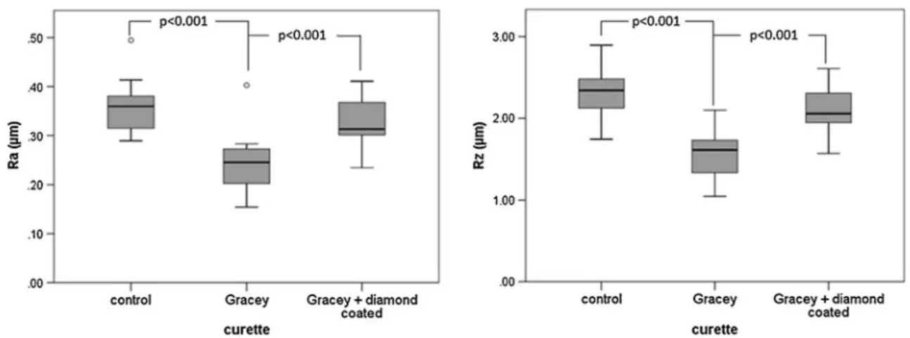
Instrumentation with conventional Gracey curettes remark-ably improved surface roughness. The median Ra values were reduced (p < 0.001) from 0.36 μm (range 0.28– 0.49 μm) of the standardized, ground surface (control) to 0.25μm (range 0.15–0.40 μm) after using Gracey curettes. The respective median Rz values were 2.34 μm (range 1.74–2.90 μm) before and 1.61 μm (range 1.05–2.10 μm) after treatment (p < 0.001). The subsequent usage of a diamond-coated curette resulted in a median Ra of 0.31 μm (range 0.23–0.41 μm) and a median Rz of 2.06 μm (range 1.57–2.61 μm), both being significantly higher (p<0.001) than after treatment with Gracey curettes only. Although the values were below those before treatment, these differences did not reveal significance. The differences of Ra and Rz between the three groups were significant (Kruskal–Wallis test, each p<0.001) (Fig. 2).
SEM photographs supported the results obtained by pro-filometry. The machined surface was rough with clear straight grooves. After application of Gracey curettes, the surface was smooth, although a smear layer was visible. The additional usage of diamond-coated curettes resulted in a more homogenous wave-like pattern with nearly no smear layer (Fig.3).
Adhesion of periodontal ligament fibroblasts
The numbers of attached fibroblasts differed significantly (p<0.001) between all three groups. In median, 27.5 PDL fibroblasts/mm2(range 20–35 fibroblasts/mm2) attached to the machined surface. These counts did not significantly change after exposure to Gracey curettes (median 28.5 fibroblasts/mm2, range 21.3–34.7 fibroblasts/mm2). After an additional instrumentation of a diamond-coated curette, about twice as much fibroblasts (median 55 fibroblasts/ mm2, range 43–90 fibroblasts/mm2) were counted on the surface in comparison to the machined and to Gracey cur-ettes treated surfaces (each p<0.001) (Fig.4).
SEM photographs showed that the fibroblasts were pre-dominantly oriented in one single direction on the speci-mens treated with Gracey and diamond-coated curettes than in the other groups (Fig.5).
Adhesion of S. gordonii
The numbers of the adhered bacteria were the highest on the specimens after treatment with Gracey curettes only (medi-an: 4.1×10⁴cfu/mm2, range 3.5–5.3×10⁴cfu/mm2). More bacteria adhered to the controls (median: 4.9×10⁴cfu/mm2, range 2.7–5.3×10⁴
cfu/mm2) and the specimens after
application of Gracey and diamond-coated curettes (me-dian: 4.4 × 10⁴ cfu/mm2, range 3.3–4.8×10⁴ cfu/mm2). The difference between the two types of treatment revealed significance (p00.021) (Fig. 6). In these experi-ments, differences are weakened due to the fact that only one of the six surfaces of the specimens was treated and the used method determined the cfu counts from the whole specimens.
SEM photographs showed the bacteria were well-spread on the surface after exposure to Gracey curettes only. The presence of bacteria seemed to be limited in the area of grooves at the other specimens (Fig.7).
Fig. 2 Average surface roughness (Ra) and the arithmetic mean height of the surface profile (Rz) of the dentin specimen before (control) as well after treatment with Gracey curettes and Gracey curettes followed by a diamond-coated curette
Fig. 3 Scanning electron microscope photographs of surfaces of the dentin specimen before (1; control) as well after treatment with Gracey curettes (2) and Gracey curettes followed by a diamond-coated curette (3) in 100-fold (A) and 300-fold (B) magnifications
Discussion
This in vitro study compared the effect of instrumentation with conventional Gracey curettes with and without the additional use of newly developed diamond-coated curettes. Standardized dentin specimens were prepared, incorporated into artificial teeth, and placed into the artificial mouth of a mannequin head. This allowed for an instrumentation which more closely mimicked the in vivo situation compared with studies where test teeth were incorporated into a plastic block [9] or where roots alone were used [6]. The Ra and Rz values obtained in the present study are somewhat lower than those reported before [9], but on the other hand, they are in line with the results obtained by others [11]. It has to be kept in mind that our study did not focus on removal of deposits but aimed to determine the effect of the additional
use of a diamond-coated curette on root surface properties. A recent study comparing the cutting capacity after extend-ed use of different curettes has failextend-ed to show any difference between the tested instruments [12]. In the present study, specimens with a standardized, relatively smooth surface without deposits were used. Further, the solid fixation of the specimens by screwing and the ideal working position might have contributed to the results. Unexpectedly, the additional use of diamond-coated curettes did not result in a smoother surface. Ra and Rz values were still below those of the standardized, ground controls but significantly higher than after using a Gracey curette only. SEM photographs showed a pattern of regular shallow grooves. These obser-vations appear to suggest that the type of the used curette may substantially influence surface roughness.
In removing deposits from implant abutments, plastic and titanium curettes yielded a smoother surfaces compared to those observed following the use of steel curettes [13]. Moreover, it appears that the observations made 30 years ago which have indicated that ultrasonic and rotating dia-mond instruments produced more uneven surfaces than hand curettes [14] are still valid today. It has been shown that after ultrasonic instrumentation, the surfaces appeared irregular and had grooves [15], and they were rougher compared to the surfaces obtained following the use of a curette [7]. Only one study reported about higher Ra values after using a hand curette in comparison to an ultrasonic system or Er:YAG laser [16]. As already described by others [14, 15], a smear layer after using Gracey curettes was always visible in the SEM photographs. In the present study, the smear layer was clearly reduced following the additional by use of the diamond-coated curette. A comparable effect was recently observed after ultrasonic instrumentation [15].
Fig. 4 Numbers of periodontal ligament (PDL) fibroblasts on surfaces of the dentin specimen before (control) as well after treatment with Gracey curettes and Gracey curettes followed by a diamond-coated curette after 72 h of incubation
Fig. 5 Periodontal ligament (PDL) fibroblasts on surfaces of the dentin specimen before (1; control) as well after treatment with Gracey curettes (2) and Gracey curettes followed by a diamond-coated curette (3) and after 72 h of incubation (DAPI staining, scale 1μm)
Following scaling and root planing in the context of non-surgical or non-surgical periodontal therapy, the instrumented root surfaces should facilitate cell attachment. Previous studies have indicated that fibroblast attachment was low on untreated, periodontally diseased root surfaces [17,18].
Several treatment modalities have been proposed to im-prove the biocompatibility of the instrumented root surfaces and to enhance fibroblast attachment. The application of diode laser did not increase the numbers of attached cells after scaling and root planing [19]. CO2laser in combination
with ultrasonic scaling was compared with hand scaling and root planing, and both promoted cell attachment being more pronounced for the additional application of laser [20]. In contrast, more PDL fibroblasts attached to surfaces pre-treated with Er:YAG laser or ultrasonic scalers than with SRP [21]. The last findings are in contrast to other reports where no difference between hand and ultrasonic instrumen-tation [17,22] was found or less attachment if the ultrasonic treatment without polishing was used [22]. The attachment rate did not also differ between using rotary instruments and hand scaling [11]. Our study has clearly shown an influence of surface properties on cell attachment. All specimens were
first covered with serum to simulate the in vivo situation in periodontal pockets; serum proteins are an essential compo-nent of gingival crevicular fluid [23]. Nearly no rounded fibroblasts were observed like in other studies [20, 21], suggesting that neither the specimens nor the treatment damaged the cells. The number of attached PDL fibroblasts was doubled after the additional instrumentation with the diamond-coated curette. Moreover, the fibroblasts were ori-ented suggesting that the moderate rough surface with straight shallow grooves is beneficial for cell attachment. This confirms the observation of Babay [18] that fibroblasts attach more to rough than to smooth surfaces. Surface morphology of apatite may alter cell shape and early gene response expression of PDL progenitor cells; a smooth surface reduced the expression of integrin cell surface medi-ators and fibronectin [24].
Initial bacterial adhesion preferably starts from irregular-ities [25]. A study comparing hand instrumentation with Er: YAG laser and ultrasonication on bovine roots found the roughest surfaces and more adhesion of Streptococcus san-guinis after hand instrumentation [26]. But in contrast to most studies, the rougher surface after the additional instru-mentation with diamond-coated curettes did not increase the numbers of initially adhered streptococci; the counts were reduced in comparison to the instrumentation with Gracey curettes only. S. gordonii was selected; that species is used as an early colonizer of plaque in several biofilm models [27,28]. A study on titanium surfaces found generally more adhered S. sanguinis on rough (Ra between 0.68 and 0.73 μm) than on smooth surfaces (Ra between 0.20 and 0.38μm); however, the analysis between the smooth surfa-ces did not reveal a difference [29]. In our study, the speci-mens were covered with serum first to simulate the in vivo conditions which may weaken surface irregularities. Saliva reduced the surface roughness of hydroxyapatite; further, the initial bacterial adhesion was lower at polished enamel having an extremely smooth surface in comparison to hy-droxyapatite [30]. In a clinical study using a split-mouth design placing titanium abutments with different roughness
Fig. 7 Scanning electron microscope photographs of surfaces of the dentin specimen before (1; control) as well after treatment with Gracey curettes (2) and Gracey curettes followed by a diamond-coated curette (3) and 2 h adhesion of S. gordonii ATCC 10558
Fig. 6 Numbers of viable bacterial cells (S. gordonii ATCC 10558) on surfaces of the dentin specimen before (control) as well after treatment with Gracey curettes and Gracey curettes followed by a diamond-coated curette after 2 h of incubation
(Ra00.05–0.21 μm), surface roughness in the range of Ra 0.05–0.13 μm did not show any influence on microbial composition within plaque [31]. A higher surface roughness of a titanium implant influences biofilm formation in the supragingival but not in the subgingival area [32]. This may raise the question whether the newly obtained surface fol-lowing subgingival (non-surgical or surgical) debridement has a more important role in promoting the attachment of PDL fibroblasts than in preventing bacterial adhesion. It may thus be speculated that the attachment of PDL cells might have an effect on the reduction of periodontopatho-gens. However, further studies are needed in order to clarify this issue.
Taken together, the present findings have shown that the additional use of diamond-coated curettes may lead to a surface which promotes the attachment of PDL fibroblasts without changing the properties for bacterial adhesion. The potential clinical relevance of these in vitro-results should, however, be validated in randomized controlled clinical studies.
Conclusions
Treatment of root surfaces with conventional Gracey cur-ettes followed by subsequent polishing with diamond-coated curettes may result in a root surface which favors the attachment of PDL fibroblasts but not the microbial adhesion.
Acknowledgments The authors would like to thank Bernita Bush (Department of Periodontology, Dental School, University of Bern) for supervising the instrumentation of the root surfaces as well as Regula Hirschi, Sabrina Ruggiero, and Marianne Weibel (Department of Peri-odontology, Laboratory of Oral Microbiology, Dental School, Univer-sity of Bern) for technical assistance. This study was financially supported by Intensiv SA, Montagnola, Switzerland and partially by Deppeler SA, Rolle, Switzerland.
Conflict of interests The authors declare that they have no conflicts of interest.
References
1. Apatzidou DA, Kinane DF (2010) Nonsurgical mechanical treat-ment strategies for periodontal disease. Dent Clin North Am 54:1– 12
2. Van der Weijden GA, Timmerman MF (2002) A systematic review on the clinical efficacy of subgingival debridement in the treatment of chronic periodontitis. J Clin Periodontol 29(Suppl 3):55–71, discussion 90-1
3. Tunkel J, Heinecke A, Flemmig TF (2002) A systematic review of efficacy of machine-driven and manual subgingival debridement in the treatment of chronic periodontitis. J Clin Periodontol 29(Suppl 3):72–81, discussion 90-1
4. Alves RV, Machion L, Casati MZ, Nociti FH Jr, Sallum EA, Sallum AW (2005) Clinical attachment loss produced by curettes and ultrasonic scalers. J Clin Periodontol 32:691–694
5. Christgau M, Manner T, Beuer S, Hiller KA, Schmalz G (2007) Periodontal healing after non-surgical therapy with a new ultra-sonic device: a randomized controlled clinical trial. J Clin Periodontol 34:137–147
6. Schmidlin PR, Beuchat M, Busslinger A, Lehmann B, Lutz F (2001) Tooth substance loss resulting from mechanical, sonic and ultrasonic root instrumentation assessed by liquid scintillation. J Clin Periodontol 28:1058–1066
7. Kocher T, Langenbeck M, Ruhling A, Plagmann HC (2000) Subgingival polishing with a teflon-coated sonic scaler insert in comparison to conventional instruments as assessed on extracted teeth. (I) Residual deposits. J Clin Periodontol 27:243–249 8. Braun A, Krause F, Frentzen M, Jepsen S (2005) Efficiency of
subgingival calculus removal with the Vector-system compared to ultrasonic scaling and hand instrumentation in vitro. J Periodontal Res 40:48–52
9. Ribeiro FV, Casarin RC, Nociti Junior FH, Sallum EA, Sallum AW, Casati MZ (2006) Comparative in vitro study of root rough-ness after instrumentation with ultrasonic and diamond tip sonic scaler. J Appl Oral Sci 14:124–129
10. Kocher T, Rosin M, Langenbeck N, Bernhardt O (2001) Subgingival polishing with a teflon-coated sonic scaler insert in comparison to conventional instruments as assessed on extracted teeth (II). Subgingival roughness. J Clin Periodontol 28:723–729 11. Kishida M, Sato S, Ito K (2004) Comparison of the effects of
various periodontal rotary instruments on surface characteristics of root surface. J Oral Sci 46:1–8
12. Sisera M, Hofer DJ, Sener B, Attin T, Schmidlin PR (2009) In vitro evaluation of three curettes with edge retention technology after extended use. Schweiz Monatsschr Zahnmed 119:1200–1208 13. Mengel R, Meer C, Flores-de-Jacoby L (2004) The treatment of
uncoated and titanium nitride-coated abutments with different instruments. Int J Oral Maxillofac Implants 19:232–238
14. Meyer K, Lie T (1977) Root surface roughness in response to periodontal instrumentation studied by combined use of micro-roughness measurements and scanning electron microscopy. J Clin Periodontol 4:77–91
15. Aspriello SD, Piemontese M, Levrini L, Sauro S (2011) Ultramorphology of the root surface subsequent to hand-ultrasonic simultaneous instrumentation during non-surgical peri-odontal treatments: an in vitro study. J Appl Oral Sci 19:74–81 16. de Mendonca AC, Maximo MB, Rodrigues JA, Arrais CA, de
Freitas PM, Duarte PM (2008) Er:YAG Laser, ultrasonic system, and curette produce different profiles on dentine root surfaces: an in vitro study. Photomed Laser Surg 26:91–97
17. Khosravi M, Bahrami ZS, Atabaki MS, Shokrgozar MA, Shokri F (2004) Comparative effectiveness of hand and ultrasonic instru-mentations in root surface planing in vitro. J Clin Periodontol 31:160–165
18. Babay N (2001) Attachment of human gingival fibroblasts to periodontally involved root surface following scaling and/or etch-ing procedures: a scannetch-ing electron microscopy study. Braz Dent J 12:17–21
19. Kreisler M, Meyer C, Stender E, Daublander M, Willershausen-Zonnchen B, d’Hoedt B (2001) Effect of diode laser irradiation on the attachment rate of periodontal ligament cells: an in vitro study. J Periodontol 72:1312–1317
20. Crespi R, Barone A, Covani U, Ciaglia RN, Romanos GE (2002) Effects of CO2 laser treatment on fibroblast attachment to root surfaces. A scanning electron microscopy analysis. J Periodontol 73:1308–1312
21. Schwarz F, Aoki A, Sculean A, Georg T, Scherbaum W, Becker J (2003) In vivo effects of an Er:YAG laser, an ultrasonic system and
scaling and root planing on the biocompatibility of periodontally diseased root surfaces in cultures of human PDL fibroblasts. Lasers Surg Med 33:140–147
22. Kishida M, Sato S, Ito K (2004) Effects of a new ultrasonic scaler on fibroblast attachment to root surfaces: a scanning electron microscopy analysis. J Periodontal Res 39:111–119
23. Tew JG, Marshall DR, Burmeister JA, Ranney RR (1985) Relationship between gingival crevicular fluid and serum antibody titers in young adults with generalized and localized periodontitis. Infect Immun 49:487–493
24. Dangaria SJ, Ito Y, Yin L, Valdre G, Luan X, Diekwisch TG (2011) Apatite microtopographies instruct signaling tapestries for progenitor-driven new attachment of teeth. Tissue Eng Part A 17:279–290
25. Quirynen M, Bollen CM (1995) The influence of surface rough-ness and surface-free energy on supra- and subgingival plaque formation in man. A review of the literature. J Clin Periodontol 22:1–14
26. Ota-Tsuzuki C, Martins FL, Giorgetti AP, de Freitas PM, Duarte PM (2009) In vitro adhesion of Streptococcus sangui-nis to dentine root surface after treatment with Er:YAG laser, ultrasonic system, or manual curette. Photomed Laser Surg 27:735–741
27. Periasamy S, Kolenbrander PE (2009) Mutualistic biofilm com-munities develop with Porphyromonas gingivalis and initial, early, and late colonizers of enamel. J Bacteriol 191:6804–6811 28. Valappil SP, Coombes M, Wright L, Owens GJ, Lynch RJ, Hope
CK, Higham SM (2012) Role of gallium and silver from phosphate-based glasses on in vitro dual species oral biofilm models of Porphyromonas gingivalis and Streptococcus gordonii. Acta Biomater. In press
29. Duarte PM, Reis AF, de Freitas PM, Ota-Tsuzuki C (2009) Bacterial adhesion on smooth and rough titanium surfaces after treatment with different instruments. J Periodontol 80:1824–1832 30. McConnell MD, Liu Y, Nowak AP, Pilch S, Masters JG, Composto RJ (2010) Bacterial plaque retention on oral hard materials: effect of surface roughness, surface composition, and physisorbed poly-carboxylate. J Biomed Mater Res A 92:1518–1527
31. Quirynen M, Bollen CM, Papaioannou W, Van Eldere J, van Steenberghe D (1996) The influence of titanium abutment surface roughness on plaque accumulation and gingivitis: short-term observations. Int J Oral Maxillofac Implants 11:169–178 32. Elter C, Heuer W, Demling A, Hannig M, Heidenblut T, Bach FW,
Stiesch-Scholz M (2008) Supra- and subgingival biofilm formation on implant abutments with different surface characteristics. Int J Oral Maxillofac Implants 23:327–334