• Aucun résultat trouvé

en fr

N/A
N/A
Protected

Academic year: 2021

Partager "en fr "

Copied!
159
0
0

Texte intégral

(1)

HAL Id: tel-01680009

https://tel.archives-ouvertes.fr/tel-01680009

Submitted on 10 Jan 2018

HAL is a multi-disciplinary open access archive for the deposit and dissemination of sci-entific research documents, whether they are pub-lished or not. The documents may come from teaching and research institutions in France or abroad, or from public or private research centers.

L’archive ouverte pluridisciplinaire HAL, est destinée au dépôt et à la diffusion de documents scientifiques de niveau recherche, publiés ou non, émanant des établissements d’enseignement et de recherche français ou étrangers, des laboratoires publics ou privés.

Role of the SUMO pathway in Acute Myeloid Leukemias

response to treatments

Hayeon Baik

To cite this version:

Hayeon Baik. Role of the SUMO pathway in Acute Myeloid Leukemias response to treatments. Human health and pathology. Université Montpellier, 2017. English. �NNT : 2017MONTT016�. �tel-01680009�

(2)

Délivré par

l’Université de Montpellier

Préparée au sein de l’école doctorale

CBS2

Sciences Chimiques et Biologiques pour la Santé

Et de l’unité de recherche

IGMM CNRS UMR5535

Institut de Génétique Moléculaire de Montpellier

Spécialité

Biologie-Santé

Présentée par

Hayeon BAIK

Soutenue le 29 Juin 2017 devant le jury composé de

Dr. Valérie Lallemand-Breitenbach

Directrice de Recherche, INSERM, Paris

Rapporteur Président du jury

Pr. Stefan Müller

Professeur, Université de Francfort

Rapporteur

Dr. Jean-Emmanuel Sarry

Chargé de Recherche, INSERM, Toulouse

Dr. Guillaume Bossis

Chargé de Recherche, CNRS, Montpellier

Dr. Marc Piechaczyk

Examinateur

Examinateur

Directeur de Thèse

Role of the SUMO pathway

in Acute Myeloid Leukemias response

to treatments

(3)
(4)

Acknowledgements

I dedicate this thesis to my Heavenly Father, for all the blessings he has bestowed upon me.

Foremost, I would like to acknowledge the members of the jury for evaluating my work and for coming to Montpellier: Valérie Lallemand-Breitenbach and Stefan Müller as ‘rapporteurs’, and Jean-Emmanuel Sarry as ‘examinateur’.

I would like to express my sincerest gratitude to Dr. Marc Piechaczyk, my thesis director, for having accepted me in his laboratory, for his strong support, and his precious discussions.

Sincere thanks go to Dr. Guillaume Bossis, for his sage advice, mentoring, and for his daily support. He made my Ph.D. experience productive and stimulated me to continue in research. It has been truly a privilege for me to have Marc and Guillaume as my supervisors in a project, which I really enjoyed.

I am also grateful to Dr. Jean-Emmanuel Sarry and Dr. Martin Villalba for their valuable advices and careful guidance in my annual thesis committee meeting. I also thank Dr. Jean-Emmanuel Sarry for his deep engagement, his contribution to in vivo experiments and for having accepted me in his laboratory to follow these experiments.

Great thanks to the “Oncogenesis and Immunotherapy” team. I was very happy to work with each of you. Your dedication, insightful criticism and help were so precious for me. Particular thanks go to Julie and Mathias for their contribution to my project. I wish to also express my appreciation to all members of the IGMM for being part of a wonderful working environment. In my daily work I have been really blessed with friendly and kind members of the IGMM.

I am also very indebted to Tamara and Claire for their sincere sharing, laughing, crying in times of difficulties and happiness. I would also like to thank Sung-Muk Choi and Young-Suk Kim for their encouragements and prayers.

Many people contributed to this thesis both for its scientific aspects as well as in daily life and I express my gratitude to them.

Words cannot express the feeling, love and thankfulness I have for my family who supported and encouraged me during all my studies in France and in Madagascar. Finally, thank you Stephan, for your special dedication and support during my last year of PhD. Your presence was countlessly important.

(5)
(6)

Table of Contents

! !"#$%&'()*(+($,-! "! ,!.'(/%0/"%$,($,-! #! '1-,/%0/01*23(-! $! '1-,/%0/,!.'(-! %&! 43(!+.'(! %%! '! '()*+,-.)'+(! %"! %! /01! %#! "#"! $%&'()!*+'(,%-%.+/./!($0!)+12+'.(! "3! "#"#"!*4567898:4;:;! "3! "#"#<!=>?;74@;!8A!B:AA4@4C7:67:8C;! "D! "#"#E!)4?F45:6! "G! "#<! (=1,+!'H+)%.0!)+12+'.(! <I! "#<#"!.CJ:B4CJ4!6CB!58@76>:7K! <I! "#<#<!(J?74!5K4>8:B!>4?F45:6! <"! "#<#E!(')!J>6;;:A:J67:8C! <"! "#<#E#6! L(M!J>6;;:A:J67:8C! <"!

"#<#E#N! =K78O4C47:J!J>6;;:A:J67:8C! <E!

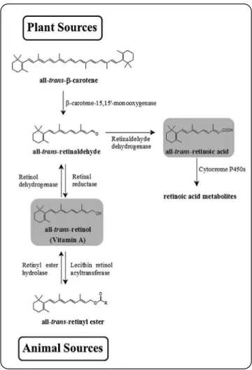
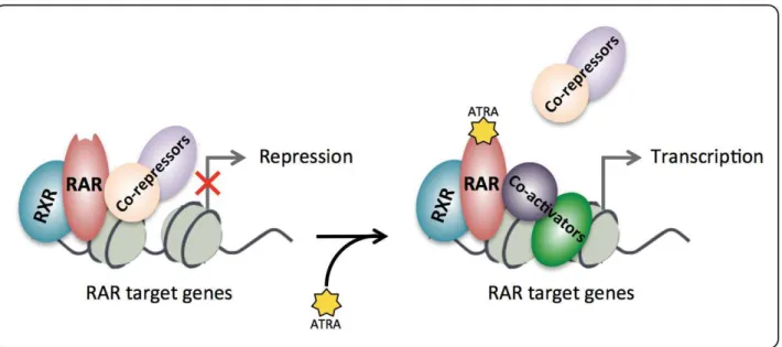
"#<#P!(')!7@46754C7! <3! "#<#P#6! /76CB6@B!JQ4587Q4@69K! <3! "#<#P#N! 0:AA4@4C7:67:8C!7Q4@69K!R(-)S!-')T&(&UV! <D! "#<#3!$8W4>!7Q4@694?7:J6>!699@86JQ4;! <X! "#<#Y!(')!J4>>!>:C4;! EI! 2! /)*/!'(,-.3,4*3/.)'5/)'+(!+6!,'663*3()'/)'+(!'(!/01! "%! <#"! 0.LL+&+$,.(,.%$!,*+&(-H! E"! <#"#"!&47:C8:B;!6CB!&47:C8:B!6J:B!@4J4978@;! E"! <#"#<!(,&(!:C!7Q4!7@46754C7!8A!(-)! EE! <#"#E!0:AA4@4C7:67:8C!7Q4@69KZ!6!9@85:;:CO!699@86JQ!:C!(')!7@46754C7! EE! <#<! 0.LL+&+$,.(,.%$!,*+&(-.+/!1/.$[!(,&(!.$!$%$T(-)!(')! EY!

(7)

<#<#"!+AA4J7!8A!@47:C8:B;!:C!C8CT(-)!(')! EY!

<#<#<!(,&(!:C!J85N:C67:8C!\:7Q!4]:;7:CO!JQ4587Q4@69K! ED!

<#<#E!+AA4J7!8A!(,&(!:C!;94J:A:J!(')!;?N7K94;! EX!

<#E! 0+&+[1)(,.%$/!%L!(,&(T'+0.(,+0!,&($/=&.-,.%$()!(=,.^.,H!.$!(')!=+))/!($0!-+&,.$+$=+!%L!.,/!

,(&[+,.$[! EG! <#E#"!.CQ:N:7:8C!8A!&(&!6J7:W:7K!NK!>4?F45:J!8CJ89@874:C;! EG! <#E#<!+9:O4C47:J!58B:A:J67:8C;!;KC4@O:_4!\:7Q!(,&(!:C!C8CT(-)!(')!NK!J8C7@8>>:CO!&(&! ;:OC6>:CO! PI! <#E#<#6! *:;78C4!(J47K>67:8C! P<! <#E#<#N! '47QK>67:8C! P<! "! 7-0+81/)'+(! 9#! E#"! 1M.`1.,.$! P3! E#<! /1'%Z!(!-%/,T,&($/)(,.%$()!'%0.L.+&!%L!,*+!1M.`1.,.$!L('.)H! PY! E#<#"!/1'%!9@874:C! PY! E#<#<!/?58K>67:8C!4C_K54! PD! E#<#E!04;?58K>6;4! PG! E#<#P!/1'%!J8C;4C;?;!587:A! 3I! E#<#3!/1'%!.C74@6J7:CO!'87:A!R/.'V! 3"!

E#E! ,(&[+,.$[!,*+!/1'%!-(,*a(H! 3<!

E#P! &+[1)(,.%$!%L!,&($/=&.-,.%$!MH!/1'%! 3E!

E#3! /1'%!.$!=($=+&! 3D! E#3#"!/1'%!:C!J6@J:C8O4C4;:;! 3D! E#3#<!/1'%!:C!(')! YI! E#3#<#6! /?58K>67:8C!:C!(-)! YI! E#3#<#N! /1'%!:C!C8CT(-)!(')! Y"! ''! *37-1)7! :#! 0/(-7.*';)!%! :<! '''!,'7.-77'+(7! $<! '5!+)=3*!;*+>3.)7! %&#!

(8)

%! )=3!*+7?7-0+!/@'7!.+()*'A-)37!)+!)=3!*37;+(73!+6!/.-)3!0831+',!13-B30'/!.3117!)+! .=30+)=3*/;3-)'.!,*-C7!D0/(-7.*';)!2E! %&<! 0/(-7.*';)!2! %&$! 2! *+13!+6!(/,;=!+@',/737!'(!/.-)3!0831+',!13-B30'/7!.=30+*37'7)/(.3! %2%! 5! *363*3(.37! %2"! -2++!35! %#F! !

(9)
(10)

List of Figures

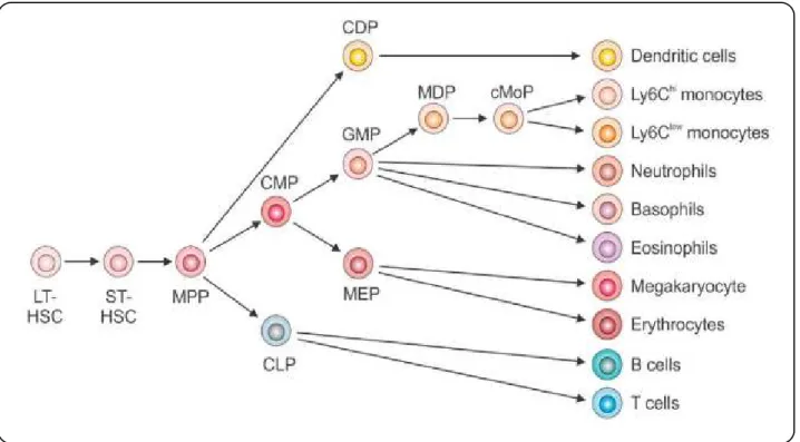
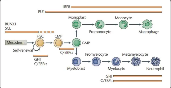
! L.[1&+!"Z!*+'(,%-%.+/./! "3! L.[1&+!<!Z!(!/,+-a./+!&+`1.&+'+$,!L%&!,&($/=&.-,.%$!L(=,%&/!01&.$[!'H+)%.0!! 0.LL+&+$,.(,.%$! "Y! L.[1&+!EZ!$%&'()!'%$%=H,.=!0+^+)%-'+$,!.$!M%$+!'(&&%a! "D! L.[1&+!PZ!$%&'()![&($1)%=H,.=!0+^+)%-'+$,!.$!M%$+!'(&&%a! "X! L.[1&+!3Z!$%&'()!+&H,*&%.0!0+^+)%-'+$,!.$!M%$+!'(&&%a! "X! L.[1&+!YZ!)+12+'.(!0+^+)%-'+$,! "G! L.[1&+!DZ!=%&&+)(,.%$!M+,a++$!0.LL+&+$,!(')!L(M!/1M,H-+/!($0!=0P3b//=!0%,!-)%,!! -(,,+&$/! <<! L.[1&+!XZ!=H,%[+$+,.=!=)(//.L.=(,.%$!%L!(=1,+!'H+)%.0!)+12+'.(! <P! L.[1&+!GZ!&+,.$%.0!'+,(M%)./'! E"! L.[1&+!"IZ!'+=*($./'!%L!,&($/=&.-,.%$!&+[1)(,.%$!MH!&(&/! E<! L.[1&+!""Z!.$01=,.%$!%L!(')!=+))!0.LL+&+$,.(,.%$!MH!$(,1&()!=%'-%1$0/!($0!! -*(&'(=+1,.=()/! E3! L.[1&+!"<Z!$1=)+%/%'+!/,&1=,1&+!($0!-&.$=.-()!'%0.L.=(,.%$!/.,+/!%$!*ES!*P!($0!0$(#! P"! L.[1&+!"EZ!,*+!1M.`1.,.$!-(,*a(H#!L! P3! L.[1&+!"PZ!().[$+'+$,!%L!('.$%!(=.0!/+`1+$=+/!%L!/1'%T"S!/1'%T<S!/1'%TES!/1'%TP!($0!! /1'%T3! PY! L.[1&+!"3Z!,*+!/1'%!-(,*a(H! PG! L.[1&+!"Y!Z!L%&'(,.%$!%L!/1'%!=*(.$! PG! L.[1&+!"DZ!/1'%!.$,+&(=,.$[!'%,.L/!R/.'/V! 3<! L.[1&+!"XZ!/1'%!&+[1)(,+/!,&($/=&.-,.%$!,*&%1[*!'1),.-)+!'+=*($./'/! 33! L.[1&+!"GZ!0+&+[1)(,.%$/!%L!,*+!/1'%!-(,*a(H!.$!^(&.%1/!,H-+/!%L!=($=+&! 3D! L.[1&+!<IZ!/1'%!=%$c1[(,.%$!&+[1)(,+/!,*+!L%&'(,.%$!($0!0./&1-,.%$!%L!-')T$M/! Y"! L.[1&+!<"Z!&%/b/1'%!-(,*a(H!./!.$^%)^+0!.$!,*+!=*+'%&+/./,($=+!(')!=+))/! Y<! L.[1&+!<<Z!/1'%T.$,+&(=,.$[!'%,.L!R/.'V!.$!=%&+/,"!./!&+`1.&+0!L%&!,&($/=&.-,.%$()!! &+-&+//.%$!%L!&(&!,(&[+,![+$+/! "I"! L.[1&+!<EZ!(,&(!,&+(,'+$,!0+=&+(/+/![)%M()!/1'%H)(,.%$!.$!(')!=+))/! "IE! L.[1&+!<PZ!'+(/1&+'+$,!%L!&%/! "<"! L.[1&+!<3Z!)+^+)!%L!&%/!.$!(')!0.([$%/./!-(,.+$,/! "<<!

(11)

List of Tables

,(M)+!"Z!L&+$=*T('+&.=($TM&.,./*!RL(MV!=)(//.L.=(,.%$!%L!(')!/1M,H-+/! <<! ,(M)+!<Z!^(&.($,!,&($/)%=(,.%$/!.$!(-)! <D! ,(M)+!EZ!&+=1&&+$,)H!'1,(,+0!%&!,&($/)%=(,+0![+$+/!a.,*!+-.[+$+,.=!L1$=,.%$!.$!(')! <G! ,(M)+!PZ!=H,%2.$+/!=($!.$01=+!0.LL+&+$,.(,.%$!%L!*1'($!'H+)%.0!)+12+'.(!=+))/! EP! ,(M)+!3Z!,*+!/1'%H)(,.%$!'(=*.$+&H!.$!/#!=+&+^./.(+!($0!'(''()/! PD! ,(M)+!YZ!=)(//.L.=(,.%$!%L!/+$-!./%-+-,.0(/+/! 3I! ,(M)+!DZ!/1'%H)(,.%$!.$*.M.,%&/! 3E!

(12)

Preamble

Acute myeloid leukemia (AML) is one of the four types of primary leukemia’s, which is characterized by large increase in the number of myeloid precursor cells. These precursors have lost their ability to differentiate and accumulate in the bone marrow. AML is a very heterogeneous disease and most subgroups are treated by chemotherapy composed of a combination of two genotoxics: one anthracycline such as daunorubicin and the nucleoside analogue Ara-C. Unfortunately, a large number of patients relapse. In spite of many efforts in the development of novel chemotherapy, no significant improvement was observed in the survival rates during the past 40 years. Nevertheless, one minor group defined as acute promyelocytic leukemia (APL), is the only one subtype, which is now cured by a differentiation therapy with very high survival rate. It has been shown recently that many cancers have impaired regulation of small-ubiquitin like modifier (SUMO) pathway and this post-translational modification might serve as a new target in the treatment of cancer.

When I arrived in the lab, I first participated to an ongoing study that concerned the role of the SUMO pathway in AML response to chemotherapeutic drugs and its involvement in chemoresistance. We have shown that chemotherapeutic drugs induce a massive desumoylation of cellular proteins through the ROS-dependent inactivation of the SUMO activating and conjugating enzymes. This desumoylation starts rapidly after drug addition and participates in the induction of apoptosis. In particular, we could show that this desumoylation is involved in the translational activation of DDIT3, a gene known to participate in genotoxics-induced apoptosis in AML. In chemoresistant AML, genotoxics do not induce ROS production and desumoylation. However reactivation of the ROS/SUMO axis, by pro-oxydants or pharmacological inhibition of the SUMO pathway restores expression of pro-apoptotoc genes and apoptosis in these cells (patients cells and mouse models). Thus, this work suggested that targeting the ROS/SUMO axis might be a way to overcome chemoresistance in AML and thus improve the treatment of this disease. My participation was to analyze the effects of the treatment with chemotherapeutic drugs on the sumoylation levels in both chemosensitive and chemoresistant AML cell lines. I also compared the sensitivity to Anacardic acid, an inhibitor of sumoylation, of both normal and AML cells. This work was published in 2014 and is detailed in Manuscript 2 (IVOther Projects).

Then, we were interested in the role of the SUMO pathway in AML differentiation therapy. This work constituted the main part of my PhD project. As mentioned previously, prognosis of APL patients is now very good thanks to the use of all-trans retinoid acid (ATRA). However its clinical

(13)

ATRA-responsive genes. Sumoylation plays a critical role in transcriptional regulation. In this context, using pharmacological inhibitor of sumoylation, 2D08 and Anacardic acid, I showed that inhibition of sumoylation enhances ATRA-induced differentiation in many AML cell lines (HL-60, U937, MOLM 14, THP1) and primary patient samples. To characterize the differentiation, I used flow cytometer to detect differentiating marker of myeloid and monocytic cell lines (CD11b, CD15 and CD14) and the activity of NADPH-oxidase was measured. Julie Kowalczyk, an intern of our lab, also confirmed the pro-differentiating action of sumoylation inhibitors by analysing the morphological changes of cells using May-Grumwald-Giemsa staining. To know if desumoylation could have a benefic effect on ATRA-induced differentiation of chemoresistant cells, I generated U937 resistant cell line to Ara-C by increasing gradually the concentration of Ara-C in the culture media. This also demonstrated that inhibition of sumoylation promotes sensitivity to ATRA even in chemoresistant AML cells. The main results obtained with inhibitors were also confirmed by genetically modulating the SUMO pathway through overexpression of desumoylases, which markedly increased their differentiation by ATRA or SUMO/Ubc9, which limited differentiation. The combination treatment composed of ATRA and inhibitors of sumoylation induce an arrest in AML cells proliferation in vitro and in vivo in NOD-Scid-IL2rgnull mice. Differentiation is a result of transcriptional reprograming process. I could show that inhibition of sumoylation facilitates the ATRA-induced expression of master genes of the myeloid differentiation using qRT-PCR on AML cell lines. Furthermore, I could show that inhibition of sumoylation increase the presence of H3K4Me3, a mark of active transcription, on the promoter of these genes using CHIP-qPCR. Altogether, this work suggests that targeting the SUMO pathway could constitute a promising approach to sensitize AML to differentiation therapies. This project is described in ‘Results’ part as Manuscript 1, which will be submitted soon.

(14)
(15)
(16)

1 AML

1.1 Normal hematopoiesis and leukemia

1.1.1 Hematopoiesis

Hematopoiesis is a lifelong highly regulated multistages process. Self-renewing hematopoietic stem cells (HSC) commit to specific lineage-committed progenitors. These progenitors have lost the ability to self-renew and commit to both common lymphoid progenitors generating T-and B-lymphocytes or natural killer (NK) cells, and common myeloid progenitors, which give rise to granulocyte-monocyte and megakaryocyte-erythrocyte progenitors (Figure 1) (Rieger and Schroeder 2012; Orkin and Zon 2008).

Figure 1: Hematopoiesis. Starting from the stem cell, hematopoiesis process gives two branches, myeloid and

lymphoid, which through downstream stages of differentiation are producing normal blood constituents, red blood cells, neutrophils and lymphocytes. LT-HSC: long-term hematopoietic stem cell; ST-HSC: short-term hematopoietic stem cells; MPP: multipotent progenitors; CMP: common myeloid progenitors; CDP: common dendritic cell progenitors; GMP: granulocyte-macrophage progenitors; MEP: megakaryocyte-erythrocyte progenitors and CLP: common lymphocyte progenitors (Schultze and Beyer 2016).

The initial steps of hematopoiesis start in the bone marrow and are tightly regulated by a network of cell extrinsic and intrinsic pathways, which control the HSC/progenitors cell self-renewal capacity and maturation into functional cells. The external signals come from the bone marrow microenvironment and are mediated by soluble cytokines and growth factors mainly provided by stromal cells, cell-cell interactions, and cell-extracellular matrix interactions

(17)

involves a massive transcriptional reprogramming through the expression of specific transcription factors (Figure 2), epigenetic changes, post-translational modifications of nucleosomal histone proteins, and the expression of small regulatory ncRNA, which all contribute to the irreversibility of cell maturation (Álvarez-Errico et al., 2015; Rosenbauer and Tenen 2007; Fazi and Nervi 2008; Orkin and Zon 2008; Zardo et al., 2008). Terminal differentiation processes that lead to the generation of mature cells take place in the blood or peripheral tissues. It depends on the exposure of precursor cells to cytokines, antigens and other factors (Geissmann et al., 2010).

Figure 2: A stepwise requirement for transcription factors during myeloid differentiation. The differentiation of

stem cells into the two main myeloid lineages, the monocytic and the neutrophilic lineages, is regulated by a hierarchical network of transcription factors. Runt-related transcription factor 1 (RUNX1) and stem-cell leukaemia factor (SCL) are required for the generation of haematopoietic stem cells (HSCs) whereas growth-factor independent 1 (GFI1) and CCAAT/enhancer binding protein-α (C/EBPα) function in self-renewal of existing HSCs. C/EBPα has another indispensable role in conferring the transition of common myeloid progenitors (CMPs) into granulocyte/monocyte progenitors (GMPs), GFI1, and similarly C/EBPε, are crucial for late-stage neutrophil production. Macrophage production depends on PU.1 and interferon-regulatory factor 8 (IRF8). In this process, PU.1 seems to be essential for all intermediate steps starting from HSCs. (Rosenbauer and Tenen 2007).

Another theory of hematopoiesis has recently emerged stating that lineage commitment during myelopoiesis is not linked to late-stage progenitors with multi-lineage potential. Using single cell transcriptomic of the myeloid progenitor cell compartment Paul et al. (Paul et al., 2015) suggest a much earlier commitment toward distinct lineages, even prior to the common myeloid progenitor state. This hypothesis was also confirmed by Schumacher and colleagues using single-cell fate mapping in vivo (Perié et al., 2015) and more recently by Velten et al., who questioned the stepwise progression of hematopoiesis (Velten et al., 2017).

(18)

1.1.2 Clusters of differentiations

Many efforts have been done to identify cell-type-restricted surface marker, called clusters of differentiation (CDs) proteins. This characterization enables the discrimination and enrichment of basically all different hematopoietic cell types by flow cytometry or magnetic cell sorting. As mentioned, differentiation and maturation can be monitored by changes in cytomorphology and immunophenotype. Here, I will develop in more detail the characterization by cell surface markers of monocytic, granulocytic and erythroid lineage differentiation stages (Terstappen et al., 1990).

" Normal Monocytic differentiation (van Lochem et al., 2004)

Macrophages are differentiated from myelo/monoblast, which becomes pro-monocyte, then monocyte expressing progressively CD11b, CD15 and at late stage, macrophage expressing CD14 (Figure 3).

Figure 3: Normal monocytic development in bone marrow. Differentiated macrophage comes from

myelo/monoblasts, which becomes pro-monocyte then monocyte. Mature macrophage differs from its precursors by the expression of CD14. Pro-monocyte and monocyte express CD11b and high CD15, which distinguishes them from myelo/monoblast.

" Normal granulocytic differentiation (van Lochem et al., 2004)

Granulocyte (neutrophil) derives from myelo/monoblast, which becomes promyelocyte, then myelocyte and metamyelocyte before being granulocyte. Differentiated granulocytes express CD11b and CD15 compared to the progenitor myelo/monoblasts, which don’t express these markers at all (Figure 4).

Myelo/monoblast Pro-monocyte Monocyte Macrophage CD34 CD117 HLA-DR CD13 CD33 HLA-DR CD13high CD33high CD11b CD15high HLA-DR CD13high CD33high CD11b CD15high CD14 HLA-DR CD13 CD33 CD11b CD14

(19)

Figure 4: Normal granulocytic development in bone marrow. Myelo/monoblast becomes neutrophil (granulocyte)

through a stepwise process. First it begins to express CD15 and becomes promyelocyte. Then appearance of CD11b classifies it as myelocyte. Then it becomes metamyelocytes expressing CD16 and differentiates finally into granulocyte.

" Normal erythroid development in bone marrow (van Lochem et al., 2004)

Erythrocyte arises from erythroblast, which is differentiated from pro-erythroblasts (Figure 5).

Figure 5: Normal erythroid development in bone marrow. Erythrocyte is derived from pro-erythroblast, which

becomes erythroblast before being erythrocyte. Pro-erythroblast express CD117, CD45 and CD71 and erythroblast loose CD117 and express CD235a. Finally erythrocyte expresses only CD235a.

As I mentioned previously, according to the classical hierarchical model of cell differentiation, the differentiation is governed by a major transcriptional reprogramming. Mutations in the genes encoding for many of the critical transcription factors, epigenetic regulators, and miRNA have been found mutated in hematological malignancies or involved in chromosomal rearrangement generating oncogenic fusion proteins responsible for these diseases (Radulovićet al., 2013; Schotte et al., 2012; Zardo et al., 2008). The acquisition of such mutations by leukemic stem cells (LSC) or progenitors impairs the maturation resulting in the generation of a neoplastic clone, expansion of immature progenitor in the bone marrow with abnormal growth properties, uncontrolled cell division, differentiation arrest, and cell death escape. This leads to the accumulation of early blood cell precursors known as blast cells and gives rise to leukemia (Figure 6).

Myelo/monoblast Promyelocyte Myelocyte Metamyelocytes Band Cell CD34 CD117 HLA-DR CD13high CD33high CD117 CD13high CD33high CD15 CD13high CD33high CD15high CD11b CD13 CD33high CD15 CD11b CD16 Neutrophil CD13high CD33 CD15 CD11bhigh CD16high

Pro-erythroblast Erythroblast Erythrocyte CD117

CD45dim

CD71 CD71

(20)

Figure 6: Leukemia development. Normal myeloid progenitors undergo rapid proliferation before they differentiate

into functional immune cells. The balance between proliferation and differentiation is well controlled. Oncogenic mutation or chromosome rearrangements of LSC or progenitors cause a block in their differentiation and a prolonged proliferation phase. This gives rise to the leukemia. HSC: hematopoietic stem cell; CMP: common myeloid progenitor; GMP: granulocyte/monocyte progenitor (Adapted from Rosenbauer and Tenen 2007).

1.1.3 Leukemia

Leukemias account for 3.8% of new cancers diagnosed annually and are divided into categorically different types of leukemia depending on the type of white blood cell affected (lymphoid vs. myeloid) and the characteristics of the disease (acute vs. chronic):

Acute leukemias are generally aggressive cancers where cancerous transformation occurs at the

early stages of the cell differentiation. Untreated, this disease can be rapidly fatal.

Chronic leukemias are characterized by a slower progression than acute leukemias, and most

patients can live with them for many years. However they are generally difficult to cure and the therapy is often conservative and aims at controlling symptoms.

Myeloid leukemiasaffect the myeloid lineage cells - white blood cells (other than lymphocytes),

red blood cells or megakaryocytes. They are also known as myelocytic, myelogenous or non-lymphocytic leukemias.

Leukemia

Mature myeloid cells Blocked differentiation Proliferation Differentiation Mutation Rearrangement CMP GMP Leukemic blasts HSC

(21)

Lymphocytic leukemias affect immature form of lymphocytes. They are also known as lymphoid

or lymphoblastic leukemias when they are developed in bone marrow, and lymphomas when they are found in lymph nodes or other organs.

Thus, we can distinguish 4 different types of leukemias: • Acute Lymphoblastic Leukemia (ALL)

• Acute Myeloid Leukemia (AML) • Chronic Lymphoid Leukemia (CLL) • Chronic Myeloid Leukemia (CML)

Acute Lymphoblastic Leukemia (ALL) is the most common form of leukemia diagnosed in

children. It affects B or T precursor cell and cells with B-cell type associated antigen. The incidence of ALL peaks between the ages of 3-7, falls by 10 years of age, and rises again after the age of 40.

Acute Myeloid Leukemia (AML)represents 10-15% of leukemias diagnosed in childhood and is

the most common type of acute leukemia diagnosed in adults.

Chronic lymphoid leukemia (CLL)is characterized by the accumulation of fully developed B or

T lymphocytes in the blood. These diseases are closely related to lymphomas, in which lymphocytes accumulate in lymph nodes and vessels. CLL mainly affects elderly individuals, with a peak incidence between 60 and 80 years of age. It is the most common form of leukemia in Western countries. CLL follows a variable course, with survival ranging from months to decades. Other types of chronic lymphoid leukemias include Prolymphocytic leukemia, hairy cell leukemia, Plasma cell leukemia, large granular lymphocytic leukemia and T-cell prolymphocytic leukemia.

Chronic Myeloid Leukemia (CML) accounts for approximately 15% of leukemias, and occurs

most frequently between the ages of 40 and 60 years. Laboratory tests reveal increased numbers of cells belonging to the myeloid cell line (monocytes, neutrophils, basophils, eosinophils) at various stages of development circulating in the blood stream.

1.2 Acute myeloid leukemia

1.2.1 Incidence and mortality

Acute myeloid leukemia is the most common acute leukemia in adults (Yamamoto and Goodman 2008) and is a primarily a disease of older adults (≥60 years), with a median age at diagnosis of 67 years (National Cancer Institute. SEER stat Fact Sheets: AML,

(22)

in United States in 2017, AML accounts 34% of all leukemia cases in adults 20 years of age and older (https://old.cancer.org/acs/groups/content/@research/documents/document/acspc-047079.pdf, 2016). The yearly incidence of AML in US is 4,5 cases per 100,000 individuals per year (Patel et al., 2012) and in European adults is 5 to 8 cases per 100,000 individuals with a steep increase in the population aged over 70 years where the incidence reaches 15-25/100,000 per annum and the yearly mortality figures in AML is 4 to 6 per 100,000 (Fey, Buske, and ESMO Guidelines Working Group 2013). The 5-year relative survival rate is 19% for AML (Visser et al., 2012). Of note, 70% of AML patient aged over than 65 years die in the first year after the diagnosis (Meyers et al. 2013).

1.2.2 Acute myeloid leukemia

AML are distinguished by the presence of more than 20% of leukemic blasts in the bone marrow, and AML patients exhibit signs and symptom of the disease. Usually the first step begins with a decrease in the number of normal blood cells, which results in varying degrees of anemia, thrombocytopenia, and neutropenia. Then, the rapid proliferation of immature cells along with their impairment to undergo apoptosis results in their accumulation in the bone marrow, the blood, and, frequently, the spleen and liver. The hyper-proliferation and block in differentiation involves the activation of abnormal genes through chromosomal translocations and/or mutations (Arber et al. 2016). AML diagnosis relies on morphological study of the blasts (FAB criteria (1.2.3.a)), signs of dysplasia as well as cytochemistry, immunophenotyping, cytogenetic study of the bone marrow (caryotype and FISH), and finally, molecular biology study (chromosome rearrangement).

1.2.3 AML classification 1.2.3.a FAB classification

AML is a heterogeneous group of malignancies with varying clinical, morphologic, immunologic, and molecular characteristics. One historical classification called ‘French-American-British’ (FAB) classification was established in 1976 (Bennett et al. 1976). It distinguishes 8 different subgroups of AML (M0 to M7) based on the morphological and cytochemistry features of leukemic cells (Table 1). Subtypes M0 through M5 are blocked at different stages of the myeloid lineage, M6 AML of the erythroid, while M7 AML arises from progenitors of cells that make platelets.

Beside, AML has a pattern of antigen acquisition seen in normal hematopoietic differentiation. Multiparameter flow-cytometry is a useful adjunct to morphology and cytochemistry and is an invaluable tool in the diagnosis of AML (Woźniak and Kopeć-Szlęzak 2008). Flow-cytometry of

(23)

detection of residual disease: on a CD45/SSC bi-dimensional plot blast, blast cells are located in the so-called “Bermude Area” (Figure 7).

Table 1: French-American-British (FAB) classification of AML subtypes.

Figure 7: Correlation between different AML FAB subtypes and CD45/SSC dot plot patterns. (Brahimi et al. 2014)

The FAB classification system is useful and is still commonly used to group AML into subtypes. However it doesn’t take into account several prognosis factors. Therefore, the World Health Organization (WHO) has developed a newer classification that includes some of these factors to try to better classify and stratify AML patients.

M1 M2 M3 M4 M5 M6 M7 Blasts Lymphocytes Granulocytes Monocytes Erythroblasts SSC CD45

(24)

1.2.3.b Cytogenetic classification

Recently, considerable progress has been performed to decipher AML molecular genetic and epigenetic to find novel diagnosis and prognosis markers. In 2001, WHO classification has been proposed. It distinguishes different subgroups of AML depending on the cytogenetic and genetic abnormalities. In 2016, this classification has been revised again and now includes the morphology, immunophenotyping as well as clinical presentation in order to divide 6 major AML subtypes (Arber et al. 2016).

1. AML with certain genetic abnormalities

• AML with t(8;21)(q22;q22) ; RUNX1-RUNX1T1

• AML with inv(16)(p13.1q22) or t(16;16)(p13.1q22) ; CBFB-MYH11 • APL with PML-RARA

• AML with t(9;11)(p21.3;q23.3) ; MLLT3-KMT2A • AML with t(6;9)(p23;q34.1) ; DEK-NUP214

• AML with inv(3)(q21.3q26.2) or t(3;3)(q21.3;q26.2) ; GATA2,MECOM • AML (megakaryoblastic) with t(1;22)(p13.3;q13.3) ; RBM15-MKL1 • AML with BCR-ABL1(provisional entity)

• AML with mutated NPM1

• AML with biallelic mutations of CEBPA • AML with mutated RUNX1 (provisional entity) 2. AML with myelodysplasia-related changes 3. Therapy-related myeloid neoplasms

4. AML not otherwise specified (This includes cases of AML that do not fall into one of the above groups, and is similar to the FAB classification.)

• AML with minimal differentiation (M0) • AML without maturation (M1)

• AML with maturation (M2)

• Acute myelomonocytic leukemia (M4) • Acute monocytic leukemia (M5) • Acute erythroid leukemia (M6) • Acute megakaryoblastic leukemia (M7) • Acute basophilic leukemia

• Acute panmyelosis with fibrosis

5. Myeloid sarcoma (also known as granulocytic sarcoma or chloroma) 6. Myeloid proliferations related to Down syndrome

(25)

Sometimes ALL with myeloid markers may be included in the AML group and are called AML with lymphoid markers, or mixed lineage leukemias or undifferentiated or biphenotypic acute leukemias (with both lymphocytic and myeloid features).

Acute promyelcytic leukemia (APL) constitutes a specific group of AML classified as M3 or AML3. APL is a rare condition, through extremely malignant because of its very rapid spontaneous evolution and occurrence of sudden hemorrhages mainly caused by coagulation disorders. APL is associated with specific chromosomal translocation that always involve the retinoic acid (RA) receptor α (RARα) gene on chromosome 17 to create a variety of X-RARα fusion, the most common one being t(15,17) translocation encoding the PML/RARα fusion (de Thé et al. 1990, 1991) which is associated with >98% of APL cases where PML/RARα is most often the only driving genetic alteration (Welch et al. 2011).

The WHO classification stratifies AML patients according to their prognosis factor: low risk group; intermediate risk group and poor risk group (Figure 8). Importantly the treatment depends on this stratification (1.2.4), age and the comorbidities of the patients. On note, most of the APLs are defined as a particular subtype having a very good prognosis factor (1.2.4.b). The patients in the low-risk cytogenetic group constitute 10-15% of all AML patients, intermediate group 50-60% and poor risk group accounts for 15-20%.

Figure 8: Cytogenetic classification of acute myeloid leukemia. CBF: Core Binding Factor; AML3: Acute Myeloid

Leukemia 3; NPM: Nucleophosmin; Flt3-ITD: Internal tandem duplication (ITD) of the fms-like tyrosine kinase 3 (Flt3). Dnmt3a: DNA methyl-transferase 3a.

!"#$%&'"(%%$(%%') *+,&-".&/(%%') !"&0$&1') 234.567)289:3!:.6)";/)8+3485<6=') *+,/"(%&(%-'>!"/$/'"(%&$(%-') !"-$?'".%/$(/@') !"-$&&'"(%1$(%/') A0>A1) B3+3=34<C)289:3!:.6) !"#$%&'(%#)"*)+,' AD>E#>E&&>E&/>E%&) !"?$&&'".%%$(%/') -".'/012) Mutations: NPM, Flt3-ITD, Dnmt3a… High Risk 15-20% AML3 10% CBF AML 10-15% Intermediate 60%

(26)

1.2.4 AML treatment

At diagnosis, the number of AML blasts in patients can reach up to 95% of total blood cells, which implies an urgent treatment. Despite important progresses in the molecular characterization and prognosis refinement of this disease (Network, The Cancer Genome Atlas Research. 2013), AML treatments have not significantly improved during the past 40 years except for the M3 AML subtype (APL).

1.2.4.a Standard chemotherapy

Front line induction therapy (<60 years)

The standard therapy of AML is an induction chemotherapy with cytarabine (Ara-C) and an anthracycline such as daunorubicin (DNR) or idarubicin, sometimes in association with other drugs. The standard combination is the ‘7+3’, with a 7 days continuous infusion of cytarabine at the dosage of 100 or 200mg/m2/day on days 1 to 7 and daunorubicin at 60-90 mg/m2 or idarubicin at 12 mg/m2/day on days 1 to 3.

After induction chemotherapy, most of the patients enter in a complete remission (CR). In AML, remission is defined as a normal peripheral blood cell count (absolute neutrophil count >1,000/mm3 and platelet count >100,000/mm3) (Cheson et al. 1990) and normocellular bone marrow with less than 5% blasts and no signs or symptoms of the disease. Recently a new definition of complete remission has been suggested, including the cytogenetic remission, in which a previously abnormal karyotype reverts to normal, and the molecular remission, in which interphase fluorescent in situ hybridization (FISH) or multiparameter flow cytometry are used to detect minimal residual disease (Cheson et al. 2003).

Low-risk cytogenetic group has a CR rate of 90-95% with an overall survival in 5 years of 50-60% and a longer median duration of remission. Intermediate-risk patients have CR rate of 70-85% and a 5-year overall survival between 24-45%. CR rate of high-risk patients is only between 25-50% and a 5 year overall survival around 20%. Relapses are largely due to the persistence of leukemic stem cells (LSCs) or leukemic progenitors, which are refractory to chemotherapeutic drug-induced cell death (Vergez et al. 2011).

To limit the occurrence of relapses, AML patients receive a post-remission therapy also called consolidation therapy, which differs depending on the AML subgroup.

(27)

Post-remission therapy

Standard consolidation after CR:

Low-risk patients, who reached CR, generally receive several dose of Aracytin during 5 days at day 1, 3, and 5. Unlike favorable group patients, high-risk patients receive 1 to 2 dose of consolidation therapy before undergoing Hematopoietic Stem Cell Transplantation (HSCT). In alternative protocols, patients undergo HiDAC (high Dose Ara-C) consolidation courses using cytarabine twice daily at a 3g/m2 dose on days 1,3 and 5 (Mayer et al. 1994). However the optimal cytarabine dose, schedule of administration, and number of cycles are still to be defined (Richard F. Schlenk 2014).

Allogeneic HSCT:

For patients with favorable-risk AML, the relapse risk may be low enough and the salvage rate high enough to postpone HSCT (Koreth et al. 2009). Most young patients with intermediate- and unfavorable-risk AML are generally considered candidates for allogenic HSCT from sibling or fully-matched unrelated donors after the first CR.

Treatment for older AML patients (>60)

For older patients, outcome remains dismal with lower CR rates and very few long-term survivors compared with younger patients. General health status and the presence of organ dysfunctions or comorbidities affect intensive chemotherapy tolerance. Similar to younger patients, their response depends on the cytogenetics characteristics of the AML. When it is possible, they receive intensive chemotherapy but in many cases they are not fit enough to tolerate it. Low-dose cytarabine (LDAC) has also been introduced as a possible standard treatment for elderly. However a clinical trial in adverse cytogenetics has not shown a significant benefit for this treatment (Burnett et al. 2007). More recently, new drugs are also used, in particular hypomethylating agents including decitabine and azacitidine, which result in a longer median and higher 1-year survival than those observed in LDAC arms, even if this did not result in a higher proportion of long-term survivors (Dombret et al., 2015). Many other new molecules are being investigated to treat older patients (1.2.5).

Refractory and Relapse therapy

Primary refractory AML and early relapse remain among the most important challenge in the management of AML. Primary refractory AML is defined by an absence of CR with 5% or more remaining blast count after several induction therapy (Cheson et al., 2003). Currently, treatment of

(28)

relapsed AML patients is not well defined. In most AML subsets (other than APL), the principal objective is to prepare the patients to receive HSCT through either targeted therapies such as FLT3 inhibitors or with standard intensive chemotherapy. If patients don’t enter in CR, HSCT is performed after 5 days of conditioning regimens including chemotherapy, monoclonal antibody therapy, and radiation to the entire body to prevent the patient's body from rejecting the transplanted cells, and to kill any remaining cancer cells.

1.2.4.b Differentiation therapy (APL, PML-RARα)

Among all AML subtypes, only APL receives a specific therapy called differentiation therapy. APL accounts for around 10% adult AML cases and APL is associated with chromosomal translocation that disrupts RARα gene located on the short arm of chromosome 17(q21) and results in an arrest of the early stage of granulocytic differentiation (promyelocytes) (H. de Thé et al. 1991). The genetic hallmark of 98% of APL is characterized by t(15;17)(q22;q11-12) that results in the generation of the PML-RARα onco-fusion protein that initiates the disease by promoting a block in myeloid differentiation and proliferation of the promyelocytic blasts (de Thé and Chen 2010). In 2% of morphologically defined APL, patients carry other variants of 17q chromosome translocation (Redner 2002)(Table 2).

Table 2: Variant translocations in APL. (Marchwicka et al., 2014 and

http://atlasgeneticsoncology. org/index.html)

In the last two decades, the treatment of APL with all-trans-retinoic acid (ATRA) (2.1.1) in combination with arsenic trioxide (ATO) has transformed this aggressive form of leukemia into a highly eradicable disease, (Lallemand-Breitenbach and de Thé 2013) (2.1.2 and 3.5.2.a). The current treatment results in long-term survival rates up to 90%, at least for low-risk APL patients. The high-risk patients are defined as those exhibiting >10 x 109/L white blood cells (WBC) at diagnosis. Their immunophenotypic features have been associated expression of CD34, CD56, T

(29)

cell antigen CD2. At the molecular level, the short PML/RARα isoform and FLT3-internal tandem duplication (ITD) mutations have also been associated with increased relapse risk. The same was found it the case of additional chromosomal abnormality such as trisomy 8 and abn(7q), abnormally expressed gene such as LEF1, ERG and mutation of the epigenetic modifier gene including DNMT3A, MLL, IDH1, IDH2, and TET2 (Testa and Lo-Coco 2016). For high-risk disease, current clinical trials leave open the issue of the most appropriate regimen (Cicconi and Lo-Coco 2016).

Even though APL is considered as a curable disease, there are still some complications and limitations in their treatment. Differentiation syndrome (DS) is a relatively common and potentially life-threatening complication that can occur during the first days or weeks after the beginning of ATRA and ATO. Several clinical signs and symptoms exist, most common are dyspnea, interstitial pulmonary infiltrates, unexplained fever etc. Therefore, ATRA-ATO regimens now include a steroid prophylaxis (Sanz and Montesinos 2014). Other complications are Pseudotumor cerebri (PTC), which is a peculiar complication of ATRA therapy. The common well-documented side-effect of ATO is cardiac death and hepatic toxicity (Cicconi and Lo-Coco 2016).

1.2.5 Novel therapeutical approaches

Current AML treatment still rely largely on intensive chemotherapy and allogeneic hematopoietic stem cell transplantation (HSCT), at least for younger patients who can tolerate such intensive treatment (Döhner et al. 2010). Unfortunately the outcome of most AML patients and especially those older than 60 years or with secondary AML after cancer therapy remain poor (Döhner et al. 2010). Therefore it is essential to develop new, effective and less-toxic agents that, either alone or in combination to increase the response rate and survival.

Many therapeutic strategies aim at changing the doses of cytotoxic chemotherapy or at incorporating new agents in combination with standard chemotherapy. Here are listed some examples:

• Liposomal formulation of cytarabine and daunorubicin in a 5:1 molar ratio, called CPX-351. • Vasaroxin. Vasaroxin is a quinolone derivative that inhibits topoisomerase II without the

production of oxygen free radicals, which causes cardiac toxicity particularly in patients with preexisting hear failure.

• Guasdecitabine. Hypomethylating agents are used for patients with AML who are ineligible for conventional cytotoxic induction chemotherapy. Study on the guadecitabine showed that it could be a supplant hypomethylating agents.

(30)

• Addition of third agents such as Purine analogs, Sorafenib (multikinase inhibitor), Gemtuzumab ozogamicin.

As AML is a heterogeneous disease presenting several mutations and translocations (Table 3), various molecularly targeted agents targeting the mutated proteins were investigated. Among others:

• Flt3 inhibitors. Flt3 internal tandem duplication (Flt3-ITDs) accounts almost 30% of patients with de novo AML and patient carrying these mutations have a very poor prognosis.

• IDH1 and IDH2 inhibitors. IDH2 and IDH1 mutations are seen in approximately 10-15% and 5-10% of AML patients respectively.

• DOT1L, BCL-2, BET bromodomain inhibitors, and histone deacetylase inhibitors.

Table 3: Recurrently mutated or translocated genes with epigenetic function in AML. (Wouters

(31)

Apart from introducing new chemotherapy agents or targeting oncogenic drivers, various other therapeutic strategies are under investigation. Among others:

• Stem cell targeting (Horton and Huntly 2012)

• Immunotherapy to target AML LSC (Snauwaert, Vandekerckhove, and Kerre 2013)

• Targeting aberrant glutathione metabolism and oxidative phosphorylation to eradicate human AML cells (Pei et al. 2013; Lagadinou et al. 2013).

1.2.6 AML cell lines

Immortalized cell lines are used in research in place of primary cells to study biological processes. These cell lines offer several advantages. They are cost effective, easy to use, provide an unlimited supply of material, bypass ethical and provide also a pure population of cells, which gives reproducible results. Numerous human cell lines were established according to several publications as well as American Type Culture Collection (ATCC) Cell biology Collection. ATCC consists of over 3,600 cell lines from over 150 different species.

Unfortunately, cell lines do not always accurately replicate the primary cells. Indeed, cell lines are genetically manipulated which can alter their phenotype, functions and their responsiveness to stimulis.

Several human AML cell lines were also established having different cytogenetic characteristics and mutations (Table 4:). These cell lines provide model systems to study for instance the differentiation as well as the normal myeloid development. During my thesis, I decided to use 4 different AML cell lines: HL60, Molm14, U937 and THP1. They are blocked at different steps of maturation and each of them has different and frequent cytogenetics and mutations. Moreover, they are one of most used in the literature to study the AML differentiation.

Table 4: AML cell lines used in the project

!"# $%&'()*)&+,- .'/),0/12

34567 !" !#$%&'()*+*,&-*./ 012&34%$$05"%$"6784%(96%:.'.;<=.>3%

?@)@-*./

.84.9:; !A%(.3-%!5B !CC1DEF EC86GH85

<=>? !A -IJKLJJMI(JALN"6M (96

(32)

2 ATRA induced-reactivation of differentiation in AML

In this part, I will develop why differentiation therapy is promising to treat non-APL AML using ATRA. For that, first, I will describe the mechanism of differentiation via ATRA and its own receptors, retinoic acid receptors (RAR) and its use in APL will be addressed. Then I will present current promising work using ATRA to treat non-APL AML. Finally I will describe the deregulations of RAR transcriptional activity in AML cells and the pertinence of their targeting.

2.1 Differentiation therapy

2.1.1 Retinoids and Retinoid acid receptors

Retinoids are a class of chemical compounds derived from vitamin A called also all-trans retinol (Figure 9) or are chemically related to it. All-trans retinol and retinyl esters are the most abundant retinoids in the diet and can be converted to all-trans retinaldehyde. Then retinaldehyde dehydrogenases can catalyze retinaldehyde oxidation to all-trans-retinoic acid (ATRA).

Figure 9: Retinoid metabolism. Retinoids are either

from plant source (all-trans-β-carotene) or from animal source all-trans-retinyl ester. All-trans-retinoic acid (ATRA) is converted from all-trans-retinaldehyde, which is from ß-carotene as well as from all-trans-retinol (vitamin A) provided from all-trans-retinyl ester of animal source. Finally, all-trans-retinoic acid is oxidized by cytochrome P450s to retinoic acid metabolites (di Masi et al. 2015).

(33)

ATRA is the differentiation agent used in APL as mentioned in the section 1.2.4.b. Here I will describe the mechanisms of ATRA-induced differentiation. ATRA is recognized by its own receptor called retinoic acid receptor (RAR). RAR has three different forms, RARα, RARβ, RARγ, which are critical regulators during myeloid differentiation (Collins 2008). Many RAR target genes are involved in myelopoiesis (Balmer and Blomhoff 2002), including CCAAT/enhancer-binding proteins (C/EBPs), PU.1 and HOX proteins. Many other are implicated in regulation of the cell cycle, and intrinsic and extrinsic apoptotic pathways such as p21, c-myc, several cyclin proteins, and FAS and FASL. All cytogenetic aberrations found in APL concern RARα gene, which plays a central role in APL pathogenesis (Redner 2002). RARα is a principal mediator of ATRA activity and regulates various transcription factors PU.1 (Iwasaki et al. 2005) and (C/EBPs): C/EBPα (Friedman et al. 2003), C/EBPβ (Duprez et al. 2003) and C/EBPε (Morosetti et al. 1997). In the absence of ligands, RAR dimerizes with RXR and tethers to target promoters a complex with co-repressor proteins that contain histone deacetylase activity. This complex modulates target chromatin structure and actively represses gene expression (Nagy et al., 1997). Upon ATRA binding, RAR undergoes a major change in conformation (Nagy et al., 1999). Co-repressor complex is thus released, and a domain is exposed that allows RAR to interact with co-activator complex. This results in the recruitment of RNA polymerase to initiate gene transcription implicated in the differentiation (Figure 10).

Figure 10: Mechanism of transcription regulation by RARs. Interaction of RARs with corepressors and coactivators

(34)

2.1.2 ATRA in the treatment of APL

In APL, PML-RARα antagonizes the transactivational function of wild-type RARα on retinoic acid (RA)-inducible promoters by homodimerizing through PML coiled–coil domains and acts as a dominant-negative for RARα signaling. This blocks the conformational change, which impairs the release of co-repressor complex (SMRT and N-CoR), methyltransferase, and histone desacetylases (HDACs) (de Thé and Chen 2010) and lead to histone H3 modifications (Saeed et al. 2011) which repress RARα target genes involved in differentiation. Thus, RARα loses its potential to respond to physiological concentrations of ATRA and acts as a constitutive repressor resulting in the inhibition of the differentiation of APL (de Thé and Chen 2010). It is only upon ATRA treatment that APL cells differentiate into matures granulocytes-like cells and enter into programmed cell death (Petrie et al., 2009). A breakthrough in APL treatment was the combination of ATRA with arsenic trioxide (ATO) (Lallemand-Breitenbach and de Thé 2013). The most important effect of ATO is the degradation of PML-RARα (de Thé et al., 2012). ATO binds to Cys residues of Zn-fingers located within the RBCC motif in PML-RARα and in PML (Jeanne et al. 2010) and induces PML and PML-RARα sumoylation (Tatham et al. 2008) which is followed by its ubiquitination and degradation by the proteasome (de Thé and Chen 2010) (more details about sumoylation of PML in APL, 3.5.2.a).

2.1.3 Differentiation therapy: a promising approach in AML treatment

AML chemotherapies are mostly aiming at inducing the death of highly proliferating cancer cells. However, as mentioned previously, in spite of efforts in the development of such chemotherapy, no significant improvement has been made during past 40 years except for APL and the relapse rate is still considerably high. Most malignant cells are blocked in their differentiation, and the success of differentiation therapies in APL has led to consider the potential effect of such therapies for other AMLs. The induction of tumor cell differentiation has been demonstrated to be effective in the in vitro and in vivo treatments of several types of cancer cells (Leszczyniecka et al., 2001). Many molecules have also been proposed to induce differentiation via several mechanisms of action. Here, I will mention rapidly the most common differentiating agents (other than ATRA):

• Vitamin D3

Vitamin D3 (VD) is the ligand of nuclear receptors called vitamin D receptors (VDR). VD has with a high differentiating efficacy but its use is limited to life-threatening cardiotoxicity. Many studies are carried out to develop this agents as well as derivatives that would limit its hypercalcemic side effect (Hughes et al. 2010) (for more details, 2.2.1).

(35)

• PPARgamma ligands

Peroxisome proliferator-activated receptor (PPAR) gamma ligands bind to specific receptors of the nuclear hormone receptor family, and PPAR heterodimerizes with RXR. It shows efficiency in vivo on myeloid leukemic cells (Konopleva et al. 2004). However clinical trials have not shown any significant effect so far (Veliceasa et al. 2008).

• G-CSF

Granulocyte colony-stimulating factor (G-CSF) can differentiate leukemia cells in vivo (Souza et al. 1986). However it is more often used to enhance immune defenses in leukemia rather than as a differentiating agent. Several cytokines were also shown in various cell lines (Table 5) to enhance differentiation. However, they were usually not efficient in vivo.

Table 5: Cytokines can induce differentiation of human myeloid leukemia cells.

(Koeffler, 2010)

• Tyrosine kinase inhibitors

Gefitinib, an epidermal growth factor receptor (EGFR) inhibitor targets the tyrosine kinase. Gefitinib or its analog R406 promote the differentiation of HL60 and U937 (Stegmaier et al. 2005). The spleen tyrosine kinase (SYK) inhibitor R406 can also induce differentiation in acute lymphoblastic leukemia (ALL) B lymphocytes (Wossning et al. 2006).

(36)

• Epigenetic modulating agents

DMSO, a polar planar compound, was discovered as inducer of differentiation and other polar compounds were introduced including hexamethylamine bisacetamide (HMBA) (Reuben et al. 1976) and also suberoylanilide hydroxamic acid (SAHA), a second-generation polar compound and HDAC inhibitor, which can induce differentiation (Richon et al. 1996). Chromatin modifying enzymes such as HDAC and DNA methyl-transferase play an important role in the regulation of gene transcription and hence differentiation (2.3.2.a). Drugs targeting these enzymes can have some anti-leukemic and anti-myelodysplasia syndrome (anti-MDS) effects, and both hypomethylating agents, azacitidine and decitabine, have been approved by the Food and Drug Administration (FDA) for treatment of advanced MDS.

In addition to these well-studied differentiating agents, many natural compound and pharmaceuticals have been shown to activate leukemia cell differentiation (Morceau et al. 2015) (Figure 11).

Figure 11: Induction of AML cell differentiation by natural compounds and pharmaceuticals. Several compounds

have been reported to be able of inducing AML cell differentiation, which leads to cell growth arest and/or apoptosis. Association arrows and sticks indicate the inducing or the inhibiting effect on differentiation, respectively. HSC: hematopoietic stem cell, MSC : mesenchymal stem cell, AML : acute myeloid leukemia; CML : chronic myeloid leukemia; MM: multiple myeloma; PIC : plasma cells; CMP : common myeloid progenitor; MP (GM): myeloid precursor (granulocyte-monocyte); Meg : megakaryoblast; Eryt : erythroblast, and ProM : promyelocyte. (1)1,alpha,25-Dihydroxyvitamin D3; (2)all-trans retinoic acid (ATRA); (3)Valproic acid; (4)Securinine; (5)5-aza-2’-deoxycytidine; (6)Cyclopamine; (7)Tomatidine; (8)Verticinone; (9)Tryptanthrin; (10)Cotylenin A; (11)Berberine; (12)Wogonine;

(37)

In conclusion, although many efforts were carried out to identify new differentiating therapies for the treatment of non-APL AML none of them are used in clinical practice yet.

2.2 Differentiation therapies using ATRA in non-APL AML

2.2.1 Effect of retinoids in non-APL AML

Because of the success of ATRA on APL patients with CR >90%, many studies intended to determine its efficacy in non-APL AML. Indeed ATRA has been already well known to effectively mediate the differentiation of non APL-AML cell lines (Brown and Hughes 2012) including HL60, U937, THP-1, MOLM14 and HF6 (Collins 2002). It was also shown to differentiate many other type of solid tumor cell, including osteosarcoma, glioma (Campos et al. 2010) and breast cancer (Ginestier et al. 2009) cells. In spite of broad differentiating activity of ATRA in vitro, results have been disappointing when used in vivo. Combining ATRA with other molecules has emerged as a more effective strategy.

For example, ATRA has been combined with ligand of peroxisome proliferator activator gamma (PPARγ). PPARγ is a nuclear receptor and functions as a ligand-dependent transcription factor responsible for lipid metabolism (Konopleva et al. 2004). Interestingly, its ligands can force cells to differentiate toward macrophages (Tontonoz et al. 1998). In particular ligand of PPARγ, troglitazone, can inhibit clonal proliferation of myeloid monocytic leukemic cells U937 in combination with ATRA and/or RXR ligands (Asou et al. 1999).

Retinoid X receptor is another important target in AML. RXRs are receptors for vitamin A metabolites like 9-cis-RA and interact with other members of the steroid/thyroid hormone receptor superfamily, including RARs, VDRs, and PPARs (Rowe 1997) to play a role in transcriptional activation. RXR agonist, bexarotene, was studied as an inhibitor of growth and inducer of differentiation toward neutrophils in HL60 and patient’s cells. Furthermore, Phase I clinical trials with bexarotene in non-APL patients demonstrated that co-stimulation of both RAR and RXR receptors could be involved in differentiation of non-APL AML (Tsai et al. 2008).

Vitamin D3 (VD), 1a, 25-dihydroxyvitamin D3 (carcitriol), and vitamin D derivatives (VDDs) are important differentiating agent (Marchwicka et al. 2014). Vitamin D receptor (VDR) heterodimerizes with RXR and turns on a variety of genes. VD can differentiate HL60 cells into macrophage-like cells (Hughes et al. 2010). However VD causes hypercalcemia in patients at clinical doses, which can lead to fatal heart failure (Krishnan et al. 2010). Therefore VD analogs were developed but the toxicity of VDDs still remains high. The idea of using VD or VDDs in

(38)

combination with RA has been investigated to limit the doses to use and hence their toxicity. The study was performed in HL60 and NB4 using VDD, 20-epi-22oxa-24a,26a,27a-tri-homo-1,25(OH)2D3 (KH1060) and 9-cis-RA. This combination promoted the differentiation and inhibited the growth of the cell lines, reduced anti-apoptotic bcl-2 and increased pro-apoptotic bax expression (Elstner et al. 1996). Other VDDs were also tested in combination with RA in vitro on HL60 cells revealing a pro-differentiating effect (Doré et al. 1994).

Securinine was also shown to enhance the differentiating activities of ATRA, as well as that of cytidine analog 5-aza-2’-deoxycytidine (decitabine or Dacogen) and VD on HL60 cells suggesting the benefic effect of natural alkaloid in a combination therapy (Gupta et al. 2011). Securinine triggers growth arrest in cell lines, patient samples and AML tumors in nude mice, confirming its clinical potential.

In same way, plant-derived steroidal jerveratrum alkaloid cyclopamine from the corn lily Veratrum californicum Durand improves HL60 cells differentiation in combination with ATRA by up-regulating T cell marker CD44. This effect was also observed in primary cells from patients with induction of the myeloid markers CD11b, CD14 and CD15 (Takahashi et al. 2011). The isosteroidal alkaloid verticinon from the bulbs of Fritillaria usuriensis Maxim was also shown to differentiate HL60 into granulocytic lineage and to increase the differentiating activity of ATRA (Pae et al. 2002).

All these observation, as well as many others not listed here, suggest that differentiation observed in APL through the RAR pathway might also occur even in non-APL AML by reactivating ATRA pathway. However the clinical efficiency of ATRA has been mostly limited to cell lines and/or was observed primarily under in vitro condition.

2.2.2 ATRA in combination with existing chemotherapy

ATRA as a part of the induction chemotherapy was tested in several clinical trials. However the results are controversial and disparate. Patients with relapsed or refractory AML were treated in a Phase II trial with idarubicin 10mg/d x 3d and cytarabine 1000 mg/m2 12h for 6 days with or without ATRA 45 mg/m2/d from day 1 until remission (Belhabri et al. 2002). No significant effect of ATRA was observed. Other 405 patients with high-risk AML were treated either with 2 courses of ADE (cytarabine 100 mg/m2 q12h d 1-10; daunorubicin 50 mg/m2 d1,3,5; etoposide 100 mg/m2 qd d1-5) vs 2 courses of FLA (fludarabine 30 mg/m2 d1-5; cytarabine 1 or 2 mg/m2qd d1-5), +/- ATRA 45 mg/m2 for a maximum of 90 days, +/- G-CSF. Again, no advantage for ATRA or G-CSF was observed (Milligan et al. 2006). Furthermore, low dose cytarabine (20 mg sq bid x 10d every

(39)

4-6 weeks) or hydrea +/- ATRA 45 mg/m2 qd for 60 days had no significant benefit in survival or remission rate (Burnett et al. 2007). 1075 patients were induced with daunorubicin 50 mg/m2 d1,3,5; cytarabine 100 or 200 mg/m2 d1-10 q12h; and thioguanine 100 mg/m2 d1-10. Then followed second induction cycle of 8 days duration +/- ATRA at a dose of 45 mg/m2 day 1-6 (Burnett et al. 2010). Again no benefic effect from ATRA addition was observed. Finally, randomized high risk patients received fludarabine 30 mg/m2 qd x 4 plus cytarabine 2 mg/m2/d d 1-4, and idarubicin 12 mg/m2 days 2-4 +/- G-CSF +/- ATRA 45 mg/m2/d day-2 though d7 with 53-55 patients each arm revealed here again no favorable effect (Estey et al. 1999).

However, in contradiction with the above results, some benefic effects of ATRA were observed in combination with existing chemotherapy. In a Phase III trial, 242 elderly AML patients were randomized to receive either conventional chemotherapy for induction and consolidation or the same regimen with ATRA. The ATRA receiving arm had a statistically significant improvement in the remission rate (38.0% vs. 27.5%) and overall survival (estimated median survival 11.3 versus 7 months). Interestingly among these patients, ATRA was more benefic for NPM1 mutated patients without FLT-ITD mutation (Schlenk et al. 2004). Moreover in younger AML patients with NPM1 mutation, response rate, event-survival, and overall-survival increased in the ATRA-treated cohort. Different trials were performed with similar association, but they could not reproduce the effect of ATRA treatment on NPM1 mutated patients (Burnett et al. 2010). Finally a trial with 63 patients including low and high-risk cytogenetics showed that the treatment with timed-sequential therapy comprising cytarabine, idarubicin and etoposide and ATRA 45 mg/m2 on day 1-6 had 60% of CR (Bolaños-Meade et al., 2003) which is very high compared to prior studies (Ma et al., 2017).

It is thus still difficult to conclude on the efficiency of ATRA as a part of induction chemotherapy because of conflicting results and clinical trials that differs in age, entry criteria, chemotherapy regimens, dose and duration of administration of ATRA. In spite of this contradictory results, certain patients may benefit from retinoid treatment.

2.2.3 Effect of ATRA in specific AML subtypes

The use of next generation sequencing identified new mutations in AML. As mentioned above, ATRA treatment might benefit to patients carrying specific mutations.

Isocitrate dehydrogenase (IDH) 1/2 mutations are found in 15% of AML patients (Mardis et al. 2009). IDH is a metabolic enzyme that converts isocitrate to α-ketoglutarate. The mutations found in AML result in the aberrant production of the oncometabolite (R)-2-hydroxyglutarate (2-HG) and leads to DNA hypermethylation (Figueroa et al. 2010; Turcan et al. 2012). This hypermethylation is

(40)

in particular observed in genes of the RA pathway (Chou et al., 2012). Interestingly, cells expressing IDH1 oncogenic mutation express a transcriptional program of ATRA-responsiveness (Boutzen et al., 2016). Accordingly, their treatment with ATRA enhances terminal granulocytic differentiation and death of AML cell lines, primary patient samples, and a xenograft mouse model-carrying mutant IDH1 (R132H) (Boutzen et al., 2016).

Combination of ATRA with ATO has strongly improved APL treatment (Lo-Coco et al. 2013). The question was addressed whether the ATRA/ATO strategy might also be used for non-APL leukemia and this idea was tested on AML with NPM1 mutation. NPM1 is a gene encoding a nucleolar shuttling protein and is frequently mutated in AML (30%). Even though this mutation has a favorable prognosis, relapses often occur. The oncogenic mutations cause the delocalization of NPM1 mutant from the nucleolus with a disorganization of PML nuclear bodies. ATRA/ATO treatment on NPM1 mutant cells has been showed to induce selective proteosomal degradation of the mutant NPM1 protein accompanied by a nucleolar redistribution of wile-type NPM1, apoptosis and/or differentiation. Importantly, this treatment induces also oxidative stress and p53 activation (Hajj et al. 2015; Martelli et al. 2015).

Flt3 mutation is another common mutation in AML with Flt3/ITD (20-25%) (Network, The Cancer Genome Atlas Research. 2013). Flt3 is a receptor tyrosine kinase and it is expressed during normal hematopoietic development. Its mutation leads to a constitutive activation and leads to a block in differentiation and increases proliferation. Several studies confirm the synergy in combining ATRA and Flt3 inhibitor (TKIs) in AML treatment. For instance, Ma et al., determined that TKIs with ATRA efficiently eliminate Flt3/ITD LSCs in vitro and also in patient samples. This was extended in mouse model, which revealed a prolonged survival of leukemic mice and engraftment of mutated patients cells in mice was reduced upon this combination therapy. They stated that the synergic effect is through the regulation of the antiapoptotic BCL2 (Ma et al. 2016). Some clinical trials are ongoing using sorafenib or midostaurin in combination with ATRA in Flt3/ITD mutated subgroup (Ramsingh et al., 2014; Guenounou et al., 2014).

2.3 Deregulations of ATRA-mediated transcriptional activity in AML cells and

pertinence of its targeting

2.3.1 Inhibition of RAR activity by leukemic oncoproteins

First argument why it is pertinent to focus on ATRA-mediated differentiation in AML is that many of the genes mutated or aberrantly expressed in AML impact RAR either directly or

Figure

Figure  1:  Hematopoiesis.  Starting  from  the  stem  cell,  hematopoiesis  process  gives  two  branches,  myeloid  and  lymphoid, which through downstream stages of differentiation are producing normal blood constituents, red blood cells,  neutrophils
Figure 2: A stepwise requirement for transcription factors during myeloid differentiation
Figure  3:  Normal monocytic  development  in  bone  marrow.  Differentiated  macrophage  comes  from  myelo/monoblasts, which becomes pro-monocyte then monocyte
Figure  4:  Normal  granulocytic  development  in  bone  marrow. Myelo/monoblast becomes neutrophil (granulocyte)  through  a  stepwise  process
+7

Références

Documents relatifs

The receptors for stem cell growth factor (CD117) and stro-1 expressed by mesenchymal stem cells are expressed by osteosarcoma cancer stem cells and were

Of these cancer cells, a subpopulation exhibits stemness markers and educates mesenchymal stem cells to release exosomes, which in turn increase the stem cell phenotype and

Although it remains unclear why the single IPA pathway mutants show increased signaling output, this phenotype reinforces the link between auxin signaling activity

Même s’ils sont des vecteurs techniques indispensables pour relier les métropoles entre elles et les connecter au reste du monde, les aéroports doivent donc de plus en plus

L’archive ouverte pluridisciplinaire HAL, est destinée au dépôt et à la diffusion de documents scientifiques de niveau recherche, publiés ou non, émanant des

l’utilisation d’un remède autre que le médicament, le mélange de miel et citron était le remède le plus utilisé, ce remède était efficace dans 75% des cas, le

We injected 30-35S imHSCs or the progeny of an equivalent numbers of progenitors obtained after culture with TPO and stromal cells into Rag2 –/– or into conventional

Therefore, space managers at CNES, scientists conceiving instruments and more generally the community of Earth scientists must come to terms with the novelty of handling