HAL Id: tel-02961064
https://hal.archives-ouvertes.fr/tel-02961064
Submitted on 8 Oct 2020HAL is a multi-disciplinary open access archive for the deposit and dissemination of sci-entific research documents, whether they are pub-lished or not. The documents may come from teaching and research institutions in France or abroad, or from public or private research centers.
L’archive ouverte pluridisciplinaire HAL, est destinée au dépôt et à la diffusion de documents scientifiques de niveau recherche, publiés ou non, émanant des établissements d’enseignement et de recherche français ou étrangers, des laboratoires publics ou privés.
Surface patterning strategies to dissect T-cell adhesion
and actin organization.
Aya Nassereddine
To cite this version:
Aya Nassereddine. Surface patterning strategies to dissect T-cell adhesion and actin organization.. Biophysics. Aix Marseille Université, 2019. English. �tel-02961064�
AIX-MARSEILLE UNIVERSITÉ
ED352 Physique et Sciences de la Matière
Centre Interdisciplinaire de Nanoscience de Marseille
(CINaM)/ AMU-CNRS, UMR 7325
Laboratoire Adhésion et Inflammation (LAI)/ INSERM UMR
1067, AMU-CNRS UMR 7333
Thèse présentée pour obtenir le grade universitaire de Docteur
Discipline: Biophysique
Aya NASSEREDDINE
Surface patterning strategies to dissect T-Cell
adhesion and actin organisation
Stratégies de surfaces pour décortiquer l’aire d’adhésion et l’organisation d’actine des cellules T
Sous la direction de Directeurs de thèse: Kheya SENGUPTA et Laurent LIMOZIN
Soutenance le 16/12/2019 devant le jury composé de:
Laurent VONNA Rapporteur Pablo PIEROBON Rapporteur Gerhardt SCHÜTZ Examinateur Nadine CANDONI Examinateur Kheya SENGUPTA Directeur de thèse Laurent LIMOZIN Directeur de thèse
iii
Abstract
Surface patterning strategies to dissect T-cell adhesion and actin organisation by Aya NASSEREDDINE
For an efficient immune response, an optimal interaction between T-cells and anti-gen presenting cells (APC) is required; it takes the form of a cell-cell contact in-volving different scales ranging from the molecular (1-10 nm) to the cellular (1-10 micrometer). The ligation of the special T cell receptors (TCR) to its ligands on an APC, leads to larger scale molecular reorganisation leading first to formation of TCR micro-clusters, and later to cell-scale restructuring of both the membrane and the cy-toskeleton. Patterning an artificial substrate with ligand-clusters that in turn induce TCR-clustering is an important tool to understand the link between the organisation of TCR and its ligand, the organisation of the actin cytoskeleton and the impact of both on overall cell behavior including adhesion and signaling. We developed a new nanotechnology based substrate (ligand-dot size down to 250 nm) and also used an alternative strategy based on colloidal self-assembly (700 or 400 nm) to show that TCR is clearly clustered on 700 nm dots but not on smaller 400 nm dots. Actin is homogeneously distributed in the form of a network in most cells but in a few of them, it appears as dots that co-localize with the ligand clusters. However, even for cells where the actin network appears homogeneous, a novel image analysis based on deep learning strategy, developed specifically for cells on the nanotech based substrates, show that the actin organisation on the dots and outside the dots may be different at least for some of the cells. While the nature of the difference is not clear from this analysis, finer observation using stochastic optical reconstruction mi-croscopy indicates that the dots may in fact be sites where actin bundles cross each other forming nodes that are not visible at lower resolution. This work confirms a close link between T cell receptor organisation and actin structure.
v
Acknowledgements
I remember the first day Kheya interviewed me, we discussed the thesis subject, for the biological part, I was excited, for the physical one, I was like what am I doing here! She believed in me (I think) and since I was accepted, now I am writing this page to thank all the people who contributed to making my thesis work possible and an unforgettable experience to me.
For that, I would first like to thank my thesis advisor Kheya Sengupta. The door to her office was always open whenever I ran into a trouble or had a question about my research. I express my sincere gratitude to her for the continuous support of my Ph.D study and research, for here patience, motivation, and immense knowledge. I will never forget her encouragement and advises, especially one sentence that caught my attention: ”Biology and Chemistry are important but physics is ubiquitous”.
I would like also to express my deep and sincere gratitude to my co-supervisor Lau-rent Limozin who instilled in me his knowledge, rigor, attention to detail, and taste of research. I will not forget his advice to be ”systematic”. It was a great privilege and honor to work and study under his guidance.
I thank my fellow lab-mates, in particular, I am grateful to Arif for enlightening me the first glance of research. My great thanks also go for Etienne and Alexis; we had interesting and long but enjoyable discussions. I will never forget how much we laughed during coffee breaks and the hiking adventures together. Special thanks to you Alexis for everything you did, you handled my bad times, during the writing of this thesis ,well and tried to make me smile and encourage me always with your special attitude.
I am also thankful to Annie, Manue, Anne, Didier, Carole, Cécile, Alex, Sébastien, Volkane, Anais, Arnaud, Emma, Celine, and Astrid, whom I shared with them great and interesting moments. I wish good luck to my mates who didn’t finish their the-sis yet: Mariem, Aubin, Cécile, Farah, Sree, and especially Ahmad who developed the deep learning part and whom I bothered with my many questions and Yann for trying new experiments on soft substrates. Also, thanks to my colleagues in LAI – Cristina, Jim, Jeoffrey, Nico, Xuan, Pierre Henrie, Philippe, and Olivier Theodoly. Thanks to Laurence Borge and Martine pelicot for the help you gave me with the cell culture. Thanks to Fred, Igor for your precious help in PLANETE, and also Damien, and Alain for helping in imaging. Never forgotten will be the discussions with my chinese friends in the lobby, the ping pong games, the UNO games: Ling, Wendy, zhenbin, Jiaxuan, and Dinesh. Thanks to my friends Batoul, Farah, Kazma, Riva, Ali, Ahmad, Maatouk, Rim, Zeinab and Issa who believed in me and encouraged me to keep going.
Finally, I must express my very profound gratitude to my family for providing me with unfailing support and continuous encouragement throughout my years of study and through the process of researching and writing this thesis. Najah, Moe, Amani, Maha, my helpful brother in Law Hus, and especially my little sister Elissar who with her moving to France added a lot of good memories to my life. This ac-complishment would not have been possible without them.
I would like to thank god for granting me the power to pass through this important phase of my life, hopefully, it will lead me to a career full of success.
vii
Contents
Abstract iii
Acknowledgements v
1 Introduction 1
1.1 The Immune System . . . 1
1.1.1 Overview . . . 1
1.1.2 T lymphocytes . . . 1
1.1.3 Antigen presenting cells (APC) . . . 5
1.1.4 Proteins on the surface of T cell and APC and their interaction 6 1.2 Immunological synapse . . . 6
1.2.1 Structure of the immunological synapse . . . 6
1.2.2 Signaling at immunological synapse . . . 7
1.3 Modulation of T cell signaling by the actin cytoskeleton . . . 8
1.3.1 Actin filament organisation . . . 8
1.3.2 The role of actin in T cell receptor triggering . . . 9
1.3.3 The role of actin in formation of the immunological synapse . . 10
1.4 Cells on artificial surfaces . . . 11
1.4.1 Patterned substrate . . . 11
1.4.2 Soft substrates . . . 12
1.5 T cells on engineered APCs . . . 13
1.5.1 Hybrid system for T cell studies . . . 13
1.5.2 Micro and nano patterned surfaces . . . 13
1.5.3 Patterned soft surfaces . . . 17
1.6 Technology for fabrication of biomimetic patterned surfaces . . . 18
1.6.1 Photolithography . . . 19
1.6.2 Contact printing . . . 19
1.6.3 Nanoimprint lithography . . . 20
1.6.4 Block Copolymer Micelles Nanolithography . . . 21
1.6.5 Nanosphere lithography . . . 22
1.7 Super resolution technology for visualisation of adhesion immune cells 23 1.8 Thesis outline . . . 26
2 Materials and Methods 27 2.1 Substrate preparation . . . 27
2.1.1 Substrate overview . . . 27
2.1.2 Cleaning procedures . . . 27
2.1.3 Preparation of primary colloidal bead masks . . . 29
2.1.4 Preparation of secondary aluminum mask . . . 29
2.1.5 Preparation of protein template. . . 30
2.1.6 Passivation with Polyethylene glycol . . . 31
2.1.7 Functionalization with antibody . . . 32
viii 2.2.1 Lymphocytes types . . . 32 2.2.2 Cell Culture . . . 34 2.2.3 Flow cytometry . . . 34 2.2.4 Cell preparation. . . 34 2.3 Microscopic observation . . . 36 2.3.1 Epifluorescence . . . 37
2.3.2 Reflection Interference Contrast Microscopy(RICM) . . . 38
2.3.3 Total Internal Reflection Microscopy (TIRF) . . . 39
2.3.4 TIRF 360 . . . 39
2.3.5 Stochastic Optical Reconsrtuction Microscopy (STORM) . . . . 40
2.4 Image and data analysis . . . 42
2.4.1 Substrate . . . 42
Manual analysis. . . 42
Automatized analysis . . . 43
Protein density quantification . . . 43
2.4.2 Cells . . . 44
Static description . . . 44
Statistical analysis . . . 45
3 Cellular scale characterisation of T cells on colloidal bead pattern 47 3.1 Characterisation of substrates . . . 47
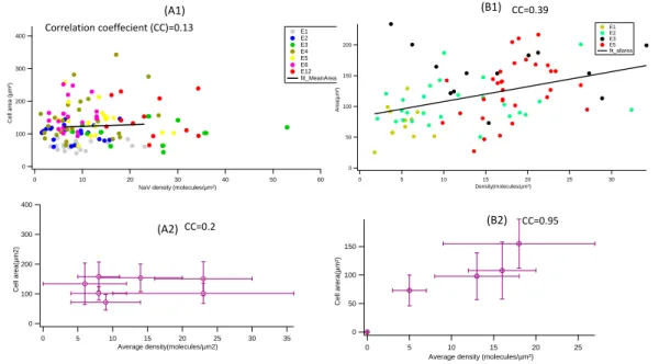
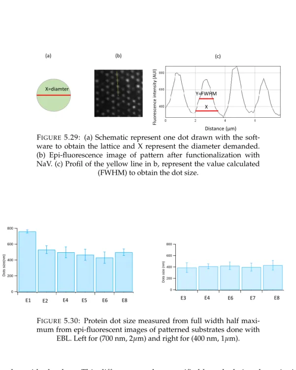
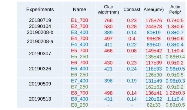
3.1.1 Protein dot size and spacing. . . 48
3.1.2 Fluorescence intensity and estimation of protein density . . . . 50
3.2 Static description of cells . . . 52
3.2.1 Spread area . . . 52
3.2.2 TCR centralisation . . . 55
3.2.3 Actin distribution . . . 56
3.3 Dynamic description of cells . . . 59
3.3.1 Dynamics RICM . . . 59
3.3.2 Time series experiments . . . 61
3.4 Conclusion . . . 65
4 Cluster scale characterisation of T cells on colloidal bead pattern 71 4.1 Membrane roughness. . . 71
4.2 TCR colocalisation . . . 71
4.3 Actin organisation. . . 73
4.4 Actin imaged by STORM. . . 75
4.5 Conclusion . . . 78
5 Pattern fabrication using electron beam lithography 79 5.1 Principle and previous work. . . 79
5.2 Basic Protocol . . . 80
5.2.1 Slide cleaning . . . 80
5.2.2 Resin spin coating . . . 80
5.2.3 E-beam lithography . . . 80 5.2.4 Development . . . 83 5.2.5 Evaporation . . . 83 5.2.6 Lift-Off . . . 84 5.2.7 Functionalization . . . 84 5.3 Limitations . . . 85
ix
5.4.1 Adhesion promotor layer . . . 87
5.4.2 Negative resin . . . 89
5.4.3 Reducing layer thickness: diluted resin . . . 91
5.4.4 Post lift-Off treatment . . . 92
5.5 Characterisation of patterned substrates . . . 95
5.5.1 Protein dot size . . . 95
5.5.2 Protein density . . . 95
5.6 Conclusion . . . 97
6 Cellular and cluster scale characterisation of T cells on e-beam lithography substrates 99 6.1 Cell scale analysis . . . 99
6.1.1 Adhesion area . . . 99
6.1.2 Actin organisation . . . 100
6.2 Dot scale analysis . . . 102
6.2.1 Simulated Lattice . . . 103
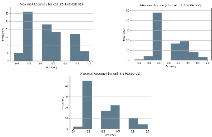
6.2.2 Deep learning in microscopy . . . 105
6.2.3 Classification of actin images . . . 106
6.3 Conclusion . . . 110
7 Cellular scale characterisation of T cells on soft substrates 113 7.1 Preparation and functionalization of soft substrates . . . 113
7.1.1 Polydimethylsiloxane elastomer (PDMS) . . . 113
7.1.2 Polyacrylamide gel . . . 115
7.2 T cells on Polydimethylsiloxane . . . 116
7.2.1 Cells on anti-CD3 and anti-LFA1 . . . 117
7.2.2 Comparing with anti-LFA1 and ICAM1 . . . 117
7.3 Cells behavior on polyacrylamide gel . . . 118
7.4 Conclusion . . . 119
8 Conclusion and perspective 123
xi
List of Abbreviations
T-cell TLymphocytes cell TCR T Cell Receptor
APC Antigen Presenting Cells IS Immunological Synapse
pMHC Peptide Molecular Histocpmatibility Complex SMAC Supramolecular Activation Compex
PLL Poly-L-Lysine
PEG Poly Ethylene Glycol
bBSA biotynalted Bovine Serum Albumin
NAV Neutravidin
APTES 3-Amino Propyl Tri Ethoxy Silane PBS Phosphate Buffered Saline
PFA ParaFormAldehyde
IPA Iso Propyl Alcohol
PA Poly Acrylamide
PDMS PolyDiMethylSiloxane
Al Aluminium
RICM Reflection Interference Contrast Microscopy TIRF Total Internal Reflection Fluorescence
STORM Stochastic Optical Reconstruction Microscopy SEM Scanning Electron Microscopy
SR Super Resolution
EBL Electron Beam Lithography FWHM Full Width at Half Maximum
xiii
To my father soul,
To my mother for her endless love and support,
To my sisters and brother
1
Chapter 1
Introduction
1.1
The Immune System
1.1.1 Overview
An important property of any immune system is the ability to discriminate between self and non-self. In vertebrates, this system is composed of two parts: the innate immunity (or non specific immune response) which represents first line of defense against pathogens [1,2]; and the adaptive immune response, also refereed to as ac-quired immunity (or specific immunity), which is the second line of defense, and is only found in vertebrates [3]. An important role of this interconnected network is to protect our body from external pathogens, like viruses and bacteria, but it also plays an important role against cancer. The innate immune response consists of physical, chemical and cellular defenses against pathogen. The main purpose of the innate system is to immediately prevent the spread and movement of foreign pathogens throughout the body [4]. It has the ability to recognize the majority of infections at an early stage, but the disadvantage of this strategy is that the response is not neces-sarily adapted, is short lived and does not confer pathogen memory to the host [4]. The adaptive immunity is specific to the pathogen presented [2]. It is implemented by white blood cells called lymphocytes. There are two broad classes of such re-sponse: antibody mediated and cell mediated [2], and they are carried out by differ-ent types of lymphocytes, called B cells and T cells respectively. When the organism is infected by a pathogen more than once, a quicker and enhanced response is gen-erated the second time onward due to the immunological memory created by the adaptive immunity [5]. It is an enormous challenge, leading to a very active field of fundamental and applied research, to understand how immune cells interpret and react to the specific signals received at their membrane to take a decision [6].
Currently, medical immunology is a very important branch of the medical and bio-logical sciences. Nowadays, it’s really important to understand the fundamentals of immunology for many reasons such as clinical or commercial application. In addi-tion to this, it really helps in discovering new diagnostics and treatments to minimize the diseases [7].
1.1.2 T lymphocytes
T lymphocytes can be considered as "soldiers" of the body [9]. They coordinate the activity of other immune cells and directly carry out specific immune functions [3]. Given these roles, T cells are implicated in many diseases, including cancer Fig.1.1. T cell manipulation has therefore been proposed as a therapeutic treatment for these diseases, for example by selecting T cells with receptors specific antigens presented to the T cell.
2 Chapter 1. Introduction
FIGURE 1.1: The cancer-immunity cycle. APC: antigen presenting
cell; MHC: major histocompatibility complex; TCR: T cell receptors. From [8].
T cells are able to detect their ligand with very great precision and accuracy. Both false negative, resulting in lack of correct immune response, and false positives, re-sulting in auto-immune diseases, are harmful. There have been a lot of theoretical work to understand how T cells can identify agonists and amplify the signal [10], [11], [12], [13] [14], [15], [16], [17] and [18]
Effector, memory, and regulatory cells are the three main T cell subtypes. Each sub-type implements a specific function during an immune response. They all express a distinct receptor, the T cell receptor (TCR) on their surface. The vast majority of T cells have TCRs with α and β chains, and are distinguished from each other by their surface markers: T helper cells, distinguished by the presence of CD4 (also known as CD4+ T cells) (as seen in Fig1.2) and cytotoxic T lymphocyte, distinguished by the presence of CD8 (also known as CD8+ T cells) [3]. Memory T cells are distinguished by the expression of a different type of the membrane glycoprotein CD45.
Role of T cells
Once activated by an APC in the lymph nodes, a CD4+ T lymphocyte will secrete interferon gamma (IFNγ) which will then activate macrophages. These will then secrete IL-12 which will activate other CD4+ T cells. The idea is that of a dialogue between different cell types (Fig.1.1). CD4+ T cells also provide support for CD8+ T cells which destroy virus-infected cells and tumor cells. They are also the cells implicated in transplant rejection. Memory T cells are a subset of antigen-specific T cells that persist long time after an infection, they provide the immune system with memory against past infections. Regulatory T cells are crucial for the maintenance of immunological tolerance that help control and limit immune responses by sup-pressing auto-reactive T cells. [19].
T cell receptor (TCR)
Receptors at the membrane of immune cells are the principle specific molecular ac-tors of immunity [6]. From this point of view, the presence of a TCR on the surface
1.1. The Immune System 3 of T cells confer them the property of recognizing antigenic peptide associated with the protein of the major histocompatibility complex (pMHC) (Fig.1.2). The TCR is membrane bound protein which is a disulfide linked heterodimer. It normally con-sists of α and β chains which form a complex with the chain molecules known as CD3 [3] (Fig.1.2). T cells expressing such TCR are called α β T cells. Some T cell types express another form of the receptor, consisting of γ and δ chains, referred to as γ δ T cells. Each chain is characterized by the following different regions (Fig.1.3) present at the two chains associated with one to the other by a disulfide bond: (i) a region V (for "variable") which will allow the recognition of the antigen and which will be at the origin of the polymorphism of the TCR. This region itself has hypervariable regions CDR (for "Complementary Determining Region") which are the areas of contact with the antigen. (ii) a region C ("for constant") which is proxi-mal to the cell membrane, (iii) a transmembrane region, and (iv) a very short intra-cytoplasmic region.
The activation of TCR upon binding to its ligand initiates a signaling cascade which ultimately controls the functioning of the T cell. The natural ligands of TCR are short peptides presented by MHC class I or II molecules (MHC I for CD8 T cells and MHC II for CD4 T cells) (Fig.1.2). However, the TCR by itself cannot activate downstream pathways to initiate T cell activation. So TCR requires CD3 and zeta chains to carry out the signal transduction in its place. In this way the MHC-TCR-CD3 interaction is necessary for the correct working of T cells. In addition, initiation of TCR signaling requires co-receptors (CD4 (Fig.1.2) for helper T cells and CD8 for cytotoxic T cells). These co-receptors act like cellular adhesion molecules and bind to MHC molecules, thus helping to sustain the interaction of T cells and antigen presenting cells [20]. Glycocalyx of T cells
As on most animal cells, on T cells membrane there are polysacharides collectively called glycocalyx. The glycocalyx is a complex layer which decorates cell mem-branes and plays an important role in cell adhesion. It is composed of a large variety of glycoproteins, glycolipids, and sugar chains dangling from the membrane and it builds a complex polymeric structure exhibiting highly variable density and various degrees of branching and/or entanglement [21].
Several studies support the view that the glycocalyx has an antiadhesive role. Re-moval of the glycocalyx by enzymes or genetic engineering facilitates cell adhesion. A prominent example of the role of repulsive molecules in the control of specific cell response concerns the activation of T cell during the interaction between surfaces of cells [22],[23].
In T cells, two important constituents of the glycocalyx are the membrane-anchored extracellular domain of CD45 [24] and the sialophorin or CD43 which is a transmem-brane cell surface protein [25]. CD45 (lymphocyte common antigen) is a receptor-linked protein tyrosine phosphatase that is expressed on all lymphocytes, and which plays a crucial role in the function of these cells (Fig.1.4). In T cells, the TCR is de-phosphorylated by CD45 if the two types of molecules are positioned close to each other. The expression of CD45 is essential for activating T cells via the TCR.[26], [27]. Cytoskeleton of T cells
The cytoskeleton is a key cellular structure that is essential for cellular structure, controled movement, mechano-sensing [31]. Also, cytoskeletal remodeling plays an essential role in coordinating molecular rearrangements in T cells interacting with
4 Chapter 1. Introduction
FIGURE1.2: Diagram of TCR engagement with the peptide antigen
MHC complex using the CD4 T cell as an example. From [28].
FIGURE1.3: A T cell receptor spans the membrane and projects
vari-able binding regions into the extracellular space to bind processed antigens via MHC molecules on APC. From [29].
APC [20] . All eukaryotic cells have at least two cytoskeletal elements, microtubules and actin fibers [32]. Animal cells have a third element called intermediate filaments (Fig.1.5). These three components of the cytoskeleton are long chains, or polymers of assembled protein subunits. Actin is a major cytoskeletal protein of most cells. In addition to providing structural support, microtubules and actin fibers enable the movement of substances within cells as well as changes in cell shape leading to cell spreading and migration.
Actin filaments play a critical role throughout different steps of T cell activation. A deeper discussion of the very important role of actin in T cells is presented in sec-tion1.3. Recent advances in the field have demonstrated that the actomyosin and microtubule networks are intertwined in many ways and provide a dynamic milieu for signaling cascades downstream of TCR in the context of polarized stimulation [33]; [34] and [35].
1.1. The Immune System 5
FIGURE1.4: Schematic representation of T cell receptor signal trans-duction. CD4-associated Lck is reciprocally regulated by CD45 and in turn phosphorylates CD3 chain immunoreceptor tyrosine-based ac-tivating motifs (ITAMs) and ZAP-70. ZAP-70 phosphorylates addi-tional downstream effectors, including the adaptors Slp-76 and Lat.
Yellow bands represent CD3 chain ITAM domains [30].
FIGURE1.5: Actin microfilaments form the cortex around the inner
surface of a cell; like rubber bands, they resist tension. Microtubules are found in the interior of the cell where they maintain cell shape by resisting compressive forces. Intermediate filaments are found
throughout the cell and hold organelles in place. From [36].
1.1.3 Antigen presenting cells (APC)
Antigen presenting cells (APC) are cell types that display antigens, ie. short amino acid sequences, complexed with major histocompatibility complexes (MHC) on their surfaces, dendritic cells and macrophages being prominent examples. After infec-tion, the APC process and present the captured antigens on MHC molecules, and migrate to draining lymph nodes. In the lymph nodes, T cells recognize the anti-genic peptides presented by the MHC with the help of TCR. However, T cell acti-vation also requires the interaction of co-stimulatory molecules on APC and their ligands on T cells.
Recent studies have shown that according to the APC nature and the stimuli they received, their rigidity as well as viscoelasticity may vary [37]; [38]. Also it has been shown that on APC, pMHC are presented in the form of clusters with a diameter between 200-300 nm. These clusters are observed independently of the interaction with T cells [39]. These discoveries highlight the importance of taking into account mechanical properties and ligand organization in the form of clusters to understand the T cell/APC interaction.
6 Chapter 1. Introduction 1.1.4 Proteins on the surface of T cell and APC and their interaction Understanding how receptors, together with co-receptors and adapter/signalling molecules, self-organise to induce specific recognition and controlled immune re-sponse remains a big challenge [40]. First, there are MHC molecules on APC mem-brane (Fig.1.2) carrying the antigenic peptide which will be recognized by T cells. For example, for the dendritic cells (types of APC), there is either MHC class I which will activate CD8+ T cells or MHC class II which will activate CD4+ T cells. This interaction is considered as the first signal of activation of T cells. Moreover this acti-vation regulates the expression of co-stimulatory molecules like B7-2 on APC which will interact with CD28 on T cells. B7-2 (CD80 or CD86) provides an important co-stimulatory signal binding CD28 on T cells [41]. This interaction is considered as second signal allowing a complete activation of the T cells.
Another adhesion molecule on APC, called ICAM-1 is seen in Fig.1.6. It permits the strengthening of the adhesion between T cells and APC during the antigen recogni-tion phase [42]. ICAM-1 (or CD54) is a highly glycosylated molecule that belongs to the immunoglobulin superfamily. It is composed of 5 extracellular domains which can interact with other adhesion molecules, one transmembrane domain and an in-tracytoplasmic tail which can interact with the cytoskeleton. ICAM-1 binds to the integrin (α5β1) Lymphocyte Function-associated Antigen 1 (LFA-1 or CD11a,CD18),
which belongs to the integrin family of cell adhesion molecules, and is present on the surface of T cells (Fig.1.6).
Antigen Presenting Cell T cell TCR pMHC LFA-1 CD28 CD45 Signal 1 Signal 2 Signal 3 Cytokines B7-1 or 2 ICAM1 or 2
FIGURE1.6: Schematic represents the major interactions between T
cells and APC and the three signals leading to activation and expan-sion of T cells. Designed for this thesis based on the literature.
1.2
Immunological synapse
1.2.1 Structure of the immunological synapse
The immunological synapse (IS) is formed as a result of the tight contact of a T cell with an APC. Traditionally the IS was considered to be the late time ’bull’s eye’ struc-ture of segregation proteins but it is known now that the triggering occurs earlier, prior to the formation of this concentric pattern [43]. The first model of the IS, sug-gesting molecular segregation, was proposed by Springer [44]. This organization in concentric segregated domains is supposed to be produced following the length
1.2. Immunological synapse 7 of the molecular complexes formed between the T lymphocyte and the APC. This concentric organization was observed by Monks et al. who studied the interaction between a T-lymphocyte and a B-lymphocyte in presence of antigens. It has since been validated by different researchers including those who replaced the B cell by a glass supported lipid bilayer [45]; [46]; [47]. Nowadays, the complete organization of the ’bull’s eye’ IS (monocentric) has been more or less identified. As we can see in Fig.1.7, the central zone (designed as cSMAC) is enriched in signaling molecules like the TCR on T cell surface which will interact with pMHC on APC. The cSMAC con-tains TCR-pMHC bond, surrounded by an adhesive ring of integrins LFA-1-ICAM bonds, and an external ring of CD45 [48]. Then, in the peripheral zone (designed as pSMAC), there are molecules like the co-receptors or LFA-1 which will stabilize the interaction by binding to the pMHC or ICAM-1. The last ring (designed as dSMAC) is enriched in CD43 and CD45 molecules. The exclusion of the large phosphatase CD45 from the TCR has been proposed as a mechanism of T cell activation, termed as the kinetic-segregation model [49]; [50].
FIGURE1.7: The formation of the immunological synapse:
cluster-ing patterns of TCR, CD3, LAT and LCK dictate spatiotemporal sig-nalling events. Event (a) shows LAT (red) clusters in vesicles being re-cruited to the membrane to join pre-existing LAT nanoclusters upon activation of the TCR (green). (b) TCR signalling occurs mainly in the dSMAC of mature synapses. (c) Micro- and nanoclusters of high, medium and low affinity LFA-1 occur in the pSMAC, binding to cog-nate ligand ICAM-1 present on the APC surface. (d) clusters of TCR, CD3 and LAT which have already undergone signalling are found in
the cSMAC. From [51].
1.2.2 Signaling at immunological synapse
Following proper engagement of TCR with the peptide-MHC complex, conforma-tional changes in the associated CD3 chains are induced. It creates a different or-ganisation in the IS. [52]; [53]. TCR form microclusters (MC) following their inte-gration with the agonist pMHC [47]. The TCR ζ chain is phosphorylated upon TCR engagement via their c-terminal immunoreceptor tyrosine-based activation motifs (ITAMs) by the leukocyte-specific tyrosine kinase (LCK). Phosphorylated CD3 then recruit and activate the Syk family kinase zeta-activated protein 70 kDa (ZAP70) [54]. Then, ZAP70 phosphorylates a membrane protein called LAT (linker for acti-vation of T cells) [55]. LAT in turn recruits a second molecular scaffold, SH2-domain containing leukocyte protein of 76 kDa (SLP-76). SLP-76 is then phosphorylated by ZAP70 and the resulting LAT-SLP-76 complex acts as a scaffold for the recruitment
8 Chapter 1. Introduction of signaling effector molecules [56]. (Fig.1.8).
Subsequently, ZAP70, actin adaptor proteins such as cytoplasmic protein NcK1, SLP-76 and actin polymerisation regulatory molecules such as Wiskott aldrich syn-drom protein (WASp) and the Wiskott aldrich synsyn-drome protein family member 2 (WAVE2) complex, are recruited to the TCR-CD3 complexes to regulate localised actin polymerisation (Fig.1.8) [47]. The signaling of TCR is still attracting the inter-est of researchers. New discoveries will no doubt explain to us more about how this signaling happen with current proteins involved in this signaling (or maybe new protein/interactions). A summary of the major aspects of TCR signaling is shown in Fig.1.8; [47].
FIGURE 1.8: Schematic illustration of some of the key proteins
in-volved in linking actin to TCR signaling. Engagement of TCR with agonist pMHC molecules leads to phosphorylation of the cytoplas-mic domains of CD3 by Lck. Subsequently, ZAP70, actin adaptor proteins such as cytoplasmic protein Nck1, the scaffold protein SLP-76, and actin polymerization-regulatory molecules such as WASp and the WAVE2, are recruited to the TCR–CD3 complexes to regulate lo-calized actin polymerization. Integrins such as leukocyte LFA-1 also regulate the actin cytoskeleton through linker proteins such as talin.
Adapted from [47].
1.3
Modulation of T cell signaling by the actin cytoskeleton
1.3.1 Actin filament organisationAs we discussed before, a major cytoskeletal protein of most cells is actin. Many molecules of globular [G] actin will join together to form a polymer called filamen-tous [F] actin as seen in Fig.1.9. Actin filaments have a distinct polarity and their
1.3. Modulation of T cell signaling by the actin cytoskeleton 9 ends (called the plus and minus ends) are distinguishable from each other. The actin G-actin binds ATP, which is hydrolyzed to ADP following filament assembly. The three dimensional structures of both individual actin molecules and actin filaments were determined in 1990 by Kenneth Holmes et al. [57].
Actin filaments are highly involved in the gross movement of the cell. They are dynamic, it means that they constantly polymerize at the barbed (plus) end and depolymerize at the pointed (minus) end, producing a so-called treadmilling haviour. They become longer in a process known as actin polymerization and be-come shorter in a process known as actin depolymerization. These two phenomena help to move the cell, and also to divide.
Within the cell, the actin filaments are regulated by actin-binding proteins [58]. One protein responsible for actin filament dissambley is called cofilin. It binds to actin filaments and enhances the rate of dissociation of actin monomers from the minus end. In addition, cofilin can severe actin filaments, generating more ends and fur-ther enhancing filament disassembly. However, profilin play a role in the exchange of bound ADP for ATP, which leads to the formation of ATP-actin monomers that can be repolymerized into filaments, including new filaments nucleated by the Arp2/3 proteins [59], (Fig.1.10).
G-actin Actin polymer
Actin filaments (F-actin)
Minus end Plus end
ADP ADP
ATP
ADP
Exchange of ATP for ADP ATP ATP
FIGURE 1.9: Schematic represents the steps of assembly and
struc-ture of actin filaments: Actin monomers (G-actin) polymerize to form actin filaments (F-actin). Designed for this thesis based on the
litera-ture.
1.3.2 The role of actin in T cell receptor triggering
Actin promotes early steps of TCR signaling in at least two levels: via effects on the TCR itself, and via the assembly of TCR micro clusters, which help amplify TCR signals. The actin cytoskeleton plays a major role in the signaling cascade. Actin ap-plies forces and re-organise molecular movements and lead to signaling, including Ca2+flux [60].
Yu et al. demonstrate that actin is essential to ensure the specificity and sensitivity of T cell triggering, by enabling the rapid association–dissociation of TCR–pMHC binding [47]. The pulling force from actin proposed is expected to increase the dis-sociation of TCR–pMHC, and this has been demonstrated experimentally in recent
10 Chapter 1. Introduction
Mother
filament ARP2/3complex Daughter filament
Plasma membrane Receptor
70°
2 3
FIGURE1.10: Cartoon representation of Activated Arp2/3 complex
binds to the side of a mother actin filament to form new daughter filament. The two branches continue to grow by addition of G-actin
to each Arp. Designed for this thesis based on the literature.
studies [61]; [62].
The second model adopts the idea that the mechanical forces applied on TCR, must be necessary to launch T cell signaling. It means that the forces provided by actin are transmitted to the TCR-CD3 complex [63]; [64].
A recent study shows that TCR engagement leads to a fast and short-lived polymeri-sation of actin in the nucleus in order to to produce a dynamic filament network in the nucleus of CD4+ T cells. Nuclear actin polymerization in response to TCR en-gagement gives rise to an elevation of Ca2+levels and involves the nuclear Arp2/3 complex [65]. The transmission of forces to the nucleus by actin is a subject of lot of current actin research.
1.3.3 The role of actin in formation of the immunological synapse
The importance of actin in T cell function is not limited to dictating the cell mor-phology and protein transportation. During the last decade our understanding of actin in T cell activation expanded broadly. Indeed, actin dynamics function plays an essential role in coordinating T cell signaling events at the IS.
Additionally, the actin cytoskeleton (precisely F-actin) is dynamic and plays an im-portant role during the formation of the IS. Indeed, protein clusters are driven by the actin centripetal flow in order to form the SMAC. In a mature synapse, actin is depleted from the center where TCR microclusters accumulate (Fig. 1.11) [47]. At the IS, actin dynamics is characterised by polymerization in the lamellipodium, cen-tripetal flow, and filament disassembly in the central region.
Dynamic studies demonstrated that the cSMAC structure is the result of the fusion of TCR microclusters which migrated toward the center in a dependent manner [66]; [67].
It was also shown that there is a relation between the pushing force of actin retro-grade flow in the dSMAC and the pulling force of actomyosin in the pSMAC. These phenomena drive the centripetal transport of TCR micro clusters at the IS [68].
1.4. Cells on artificial surfaces 11
FIGURE1.11: A mechanistic overview of actin in T cell activation. (A) After T cell triggering, the actin cytoskeleton promotes cell polariza-tion, maintains cell–cell contact and facilitates TCR signaling across the plasma membrane. TCR signaling is amplified and sustained in distinct microclusters. Actin acts as a scaffold for the clustering of proteins, drives their centripetal translocation and spatially organizes the microclusters to different domains to form the IS. In a mature IS, actin is depleted from the central area where TCR microclusters accu-mulate. (B) Transport of TCR microclusters and actin retrograde flow are temporally coordinated during T cell activation. In a T cell trig-gered by a stimulatory supported lipid bilayer, the TCR and actin are imaged simultaneously by using total internal reflection fluorescence
microscopy (TIRFM) [47].
1.4
Cells on artificial surfaces
1.4.1 Patterned substrateIn the last decades, new approaches combining physical and biological sciences emerged, in order to deepen the knowledge concerning cellular behavior. Such ap-proaches have also let to advances in biomedicine, which has stimulated the de-velopment of strategies for production of diverse biocompatible and cell mimetic surfaces, of which surfaces patterned with bio-molecules is an important example. Micro/nano fabrication techniques, originally developed to manufacture integrated circuits, were adapted early-on for patterning biomolecules on surfaces [69] and [70]. They are now increasingly used in other fields including biology, biotechnology and biomedical science and engineering [71]; [72] and [73]. In the field of cell biology, combined with surface chemistry and material science knowledge, micro/nano fab-rication techniques have provided new tools to further explore and guide cellular processes such as adhesion, spreading, motility, and proliferation [72] and [74]. A wide number of techniques have been developed for producing patterned surfaces at the micro and nano scales. These techniques will be described in section1.6. Using micropatterns of adhesive patches of different size, the group of Ingber and White-sides showed that cells could be switched from growth to apoptosis by controlling
12 Chapter 1. Introduction their adhesion [75]. Moreover, they showed also that cells can vary their shape while maintaining the total cell-ECM area constant by decreasing the size and spacing be-tween the focal adhesion points (Fig.1.12)[75]. Following this study, it became clear that localization of focal adhesion points can be controlled by patterning patches of adhesive proteins [76]. Micro-patterning is now a standard technique in different domains of biology and biophysics [77] and [78].
FIGURE 1.12: Effect of ligands geometry on cell adhesion and
growth (A) Different sizes of square adhesive islands coated with fribronectin. (B) Final shapes of enthodelial cells adhering to the ad-hesive islands. Cells adapt their shape and adhesion area according to the adhesive island. (C) Substrates used to explore the shape varia-tion of the cell independently of the cell-ECM contact area. (D) Phase contrast images of cell spreading on the patterns shown in (C). (D) Im-muno fluorescence staining of fibronectin or vinculin for cell
spread-ing on (C). [75]
1.4.2 Soft substrates
Recent decades have seen considerable progress in our understanding of the mechanosen-sitivity of cells. Studies from the 1980s and 90’s [79] and [80] definitively established that cells apply forces to substrates they interact with. More recent development in traction force microscopy allows quantification of these forces [81]. Cells apply forces and in turn react to the resistance offered by the environment. Using a hard surface such as glass as substrate for cell adhesion does not fully represent the soft physiological state of cells. Therefore, in order to better mimic this state, and to un-derstand the role of mechanics in controlling cell behavior, soft substrates became an important tool [82]; [83].
The relative stiffness of the extra cellular matrix (ECM) is an important mechan-ical parameter for cell functions and behavior. Mechanmechan-ical forces are shown to play a major role in a wide range of biological and pathogenic processes [84] and [85], and mechanobiology is becoming an emerging field in biomedical research. A wide number of experiments were reported on the capacity of the cells to sense
1.5. T cells on engineered APCs 13 the mechanical properties of their environment. This ability is called mechanosens-ing. Mechanosensing is carried out by applying forces through adhesion proteins on their surface, and traducing the force into biochemical signals in response. It is now well recognized that cells respond actively to the stiffness of their underlying sub-strate (Fig. 1.13). Experiments to explore the stiffness dependence of cell behaviour are usually performed with non adhesive polyacrilamide (PAA) or polydimethyl-siloxane (PDMS) coated with ECM proteins or other integrin ligands such as the tripeptide Arg-Gly-Asp (RGD).
FIGURE 1.13: Schematic representation of different setups for
trac-tion force microscopy. (a) Thin films buckle under cell tractrac-tion, there-fore this setup is difficult to evaluate quantitatively. (b) The stan-dard setup are thick films with embedded marker beads. (c) Pillar arrays are local strain gauges and do not require any deconvolution; however, they also present topographical and biochemical patterns to
cells. Adapted from [86]
1.5
T cells on engineered APCs
1.5.1 Hybrid system for T cell studiesTo study T cell adhesion and signalling at the immune synapse, it is common to form an interface between a living cell and a cell-mimetic engineered surface and to image it with advanced optical microscopy techniques. In the context of T cells, the APC has traditionally been mimicked by a supported lipid bilayer (SLB) that carries diffusing ligands, typically for the TCR complex and for integrins [87], [88], [89]. Many of the results reported above for T cell/APC interactions were in fact discovered using this kind of hybrid system. The role of ligand mobility in a SLB which was clearly demonstrate recently [90]. Using a fluid supported lipid bilayer which mobile or immobilised ligands (anti-CD3 as TCR ligand), it was shown that cell spreading area as well as actin organization are influenced by the ligand mobility (Fig.1.14) [90]; [91].
1.5.2 Micro and nano patterned surfaces
Interaction of cells with nano and micro-patterns have attracted a lot of attention [92],[93], [94], A new challenge in cellular context comes from the fact that cells are
14 Chapter 1. Introduction
FIGURE1.14: Ligand mobility determines the T cell spread area and
actin distribution Row 1: Scheme of the functionalization of the sub-strates with monobiotin fluorescent Anti-CD3 coupled via Neutra-vidin. Fix: immobilized Anti-CD3 on Supported Lipid Bilayer (SLB). Mob: mobile Anti-CD3 on SLB. Row 2: RICM images of Jurkat T cells after 15 min engagement on substrates. Row 3: TIRF images after la-belling actin with Rhodamine Phalloidin. Scale bars: 5 µm. Adapted
from [91]
additionally sensitive to the compliance of their immediate environment. Further, because in cell-cell interactions the relevant proteins are embedded in the cell mem-brane, synthetic supported lipid bilayer (SLB) membranes are often used as surro-gate cells. Patterning of soft substrates at the nanoscale, or an SLB even at the mi-croscale, is a current engineering challenge.
A lot of progress understanding the importance of spatial cues was made with the the strategy of Groves lab, who designed an hybrid live-cell-supported membrane configuration, in which a solid supported lipid bilayer (SLB) takes the place of the APC [92]; [95]; [96]; [93] . They aimed to understand how the organization of recep-tors and signaling molecules at the cell-cell interface could have an impact on the activity of lymphocytes. For example, they have demonstrated, by confining TCR ligands within a micrometer perimeter using a barrier in the bilayer (Fig. 1.15), that receptors reorganization via actin toward the center is important for T cell signaling [97].
Bio-mimetic surface bearing small protein clusters of controlled size can be made of various kinds of synthetic substrates: either protein-covered patterns on glass [90]; [98]; [99], or nano motifs in the context of protein clusters on a supported bilayer [47]; [97]; [98]; [87].
1.5. T cells on engineered APCs 15
FIGURE 1.15: Schematic of a hybrid live-cell-supported membrane
junction. pMHC, ICAM1, and possibly other molecules such as CD80 can be incorporated into a supported membrane where they are free to diffuse laterally and engage their cognate receptors on the live T cell. Structures, at nanometer-scale, may be fabricated onto the un-derlying substrate to corral and guide the motion of these supported membrane molecules. Then, through specific receptor-ligand interac-tions, molecules within the living T cell become subject to the same
physical constraint. From [97]
An early work by Doh and Irvine [98] used photolithography to immobilize anti-CD3 into different micro-patterns. The authors studied T cell activation on sub-strates composed of antibodies directed against the CD3 molecule of the TCR com-plex (anti-CD3) surrounded by the adhesion molecule ICAM-1. They were able to create different patterns which mimic the molecular organization found at the IS by presentation of the TCR ligands in the form of large cell scale clusters (of several mi-crons) or the characteristic motif of the synapse, with the TCR ligands in the center surrounded by adhesion molecules (Fig. 1.16). They found that T cells can be fully activated when the TCR ligands are arranged in the form of a circle that can sustain the cSMAC at the center of the interacting surface, but on other configurations the T cell struggles to spread and be activated.
Later, group of Kam [100] have shown that T cells produce IL-12 when anti-CD3 dots are surrounded by CD28 whereas when both are co-localized they did not. These re-sults highlight the importance of the organization of the ligands on the formation of the IS and the activation of the T cell.
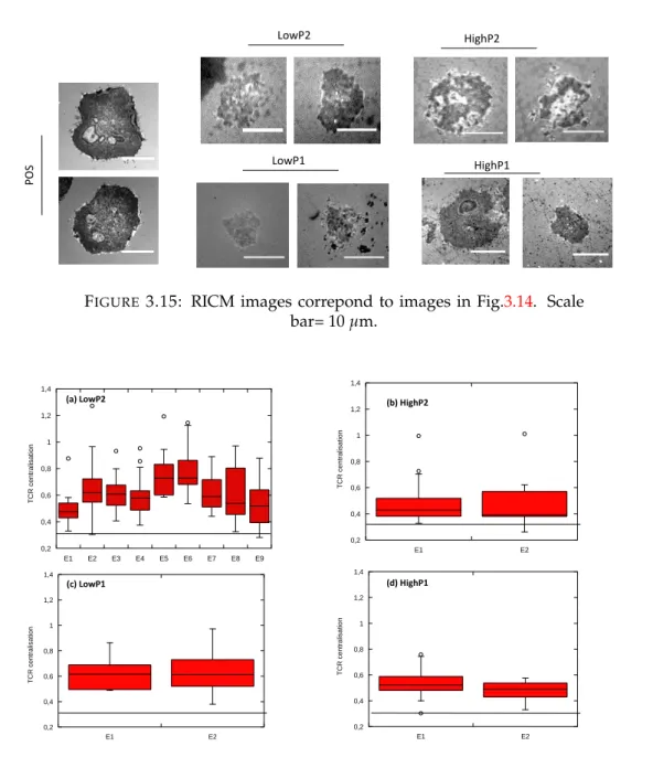
In my host laboratory, another technique to build sub-micropatterned surface to mimic the APC was developped to look at the molecular organization at the T cell-APC interface [101]. This technique is based on microsphere lithography. With this technique, the spacing between ligand is well controlled by the diameter of the beads, and the size of the motif can be varied to some extent [102]. Moreover, the ab-sence of metal on this substrate facilitates imaging with sensitive surface microscopy technique like TIRF, RICM or super-resolution techniques. These kinds of studies on T cell show that different molecules are influenced by dot geometry, such as TCR and kinases ZAP-70 as well as cell membrane topography (Fig.1.17) [88].
16 Chapter 1. Introduction
FIGURE1.16: Synapse array patterns template T cell surface receptor
and intracellular signaling molecule accumulation at the cell-surface contact site. Top: Schematic of IS array surface pattern. In each panel there is a schematic representation of the anti-CD3/ICAM-1 substrate pattern and representative immunofluorescence images at the cell-substrate contact plane. A and B Immunostaining of PKC-θ (green) and talin (red) (A) or TCR and LFA-1 on focal anti-CD3 patterns (B). C and D Immunostaining of PKC-θ(green) and LFA-1(red) on multi-focal patterns (each anti-CD3 spot 2µm in diameter) (C) or annular
anti-CD3 patterns (D)(Scale bars: 5µm.) [98]
Furthermore, in our lab, another kind of substrate was created: APC-mimetic syn-thetic patterned substrate in which the dots containing anti-CD3 are surrounded by a fluid supported lipid bilayer (SLB) [103]. They showed that for T cell adhesion mediated by TCR alone, in the patterned, but not in the homogeneous controls, the TCR, ZAP-70 and actin are present in the form of cluster that colocalize with the lig-and dots [87].
Moving to nano patterning, Spatz et al. developed a substrate composed of a nanoscale protein pattern on gold nano-dots whose, molecular density and distribution were controlled [104]. They studied the roles of the substrate’s geometrical properties and the presentation of nanoscale ligands on adhesion and T cell activation. This nano-pattern is obtained by a nanolithography technique block copolymer micellar nanolithography (BCML) which allows gold nanoparticles to be disposed on glass in an organized manner on which the proteins of interest are directly immobilized [105] and [100] (discussed again in subsection1.6.4. Similar experiments were done by Deel et al. [99]. Although this substrate confirms the existence of a relationship between molecular density, ligand organization and cell adhesion and activation (Fig.1.18), it does not allow, to our knowledge, the observation of the molecular or-ganization at the T cell-APC interface due to its incompatibility with the TIRF (total internal reflection fluorescence) or super-resolution microscopy, especially at high densities of the gold dots.
1.5. T cells on engineered APCs 17
FIGURE1.17: Distribution of TCR on submicro-patterned substrates.
Row 1: RICM images of Jurkat cells allowed to interact with dots. B2M: dot pitch of 2µm with dot size= 640±30 nm. B4: dot pitch of 4µm with dot size= 1600±100 nm. Row 2: TIRF images of TCR clus-ters. On B2M, the TCR clusters are clearly visible and are usually well localized on the site of the underlying antibody dots as evidenced by FFT in the inset. On B4, the TCR clusters are usually localized on the dots but a closer inspection shows that each dot often recruits several clusters. Insets show FFT which reflects the ordering (or not) of the TCR clusters. Scale is set by the pitch of the dots(left: 2 µm, right: 4
µm. On Row 3: Underlaying TCR ligand dots. [88]
1.5.3 Patterned soft surfaces
Since the physiological state of cells is closer to soft substrates more than hard, re-searchers have started creating soft surfaces with pattern that better mimic the real environment of the cell. One recent technique is based on patterning soft hydro-gels by employing block copolymer micelles nanolithography for the production of surface patterned with gold nanoparticles on a solid template, then transferring the pattern to a polymeric hydrogel [106]; [107] and [27]. These nanoparticles are used for immobilization of active biomolecules. But this is limited by its application only to hydrogels, also by incompatibility between microscopy and gold chemistry. In our group, Pi and colleagues, reported a NL based technique, creating nanodots
18 Chapter 1. Introduction
FIGURE1.18: Nano-patterned antigen arrays mimicking APC surface
during T cell activation. Substrates are biofunctionalized with pMHC molecules following a spatial arrangement, local density, and orien-tation that are presumed to have a major impact on T cell activation.
[99]
whose size could be tuned independently of the spacing, and used this strategy to pattern glass as well as soft elastomer (PDMS). They proceeded as follows: a primary silica bead mask was self-assembled on an hydrophilic glass substrate, resulting in two dimensional monolayer of colloidal beads, then a secondary aluminum mask was deposited by sputtering, resulting after removing of the beads, a well defined array of pits giving access to the glass substrate for further deposition1. They ob-served that by tuning the thickness of the deposited aluminum layer, the later size of the pits could be tuned. The resulting substrate was further bio-functionalized with proteins and used to study T cell adhesion or to transfer the protein pattern to the surface of soft PDMS elastomer by contact printing [108] . This technique removes the constraint of using gold particles, and provides a powerful tool for cell biology studies.
1.6
Technology for fabrication of biomimetic patterned
sur-faces
In order to fabricate biomimetic patterned surfaces different techniques were first developed (micro/nano) to make the integrated circuits but then it has also evolved for applications in other fields like biology, biotechnology, biomedical and engineer-ing [71]. In cell biology, this technique is used to further study cellular processes like cell adhesion, spreading, motility, proliferation and, in general, cell interaction with their environment [74]. A patterned surface can be achieved by two types of approaches either top-down or bottom-up [109]. The top-down approach consist of creating micro/nano scale structures at a macroscopic scale (e.g Photolithography) and the bottom-up approach consist of assembling small structures to form a bigger one (e.g molecular self-assembly). This section describes some of the well known techniques used to fabricate micro/nano patterned surface.
1.6. Technology for fabrication of biomimetic patterned surfaces 19 1.6.1 Photolithography
Two approaches are possible in order to make pattern surfaces with the lithography technique: either the substrate is coated with a positive photoresist or a negative one. Then, the surface is exposed to UV light through a mask with the desired pattern. In the case of a positive photoresist, the area exposed to the UV light will depolymerize and be chemically removed during the development step. However, in the case of a negative photoresist, the area exposed to the UV light will polymerize and remain during the development process (Fig. 1.19). The remaining pattern can be used as a mask (for deposition or etching techniques) or as mold to shape polymers such as PDMS. It has several advantages such as reproducibility and the high resolution of the pattern. But it needs clean room facilities which may not be easily accessible, as well as the incompatibility with chemical functionalities and delicate ligands which will be denatured or deactivated.
Traditionally, only larger features could be done by photolithography. However, now much smaller features and multi scale patterns may be possible using deep UV laser patterning [110].
FIGURE1.19: Photolithography using positive or negative
photore-sist. Adapted from [111].
1.6.2 Contact printing
In 1993 Whitesides et al. developed micro-contact printing [112], which has proved to be very popular for biological studies. Its concept is based on transferring molecules from an elastomeric stamp which is micro/nano structured to another surface. To do so, an elastomeric stamp (usually obtained using a mold fabricated by lithogra-phy) is coated with molecules of interest, typically a simple protein, which are then transferred to another surface by putting the two surfaces in contact [70]; [113] (Fig.
1.20). The non-coated regions can also be functionalized with another molecule [98]. The substrate need to have highly attractive interaction with the transferred mate-rial compared to the stamp to achieve the transfer of the molecules. It has several advantages as the simplicity of the processing/using and the capacity of patterning large area on various materials. But, it is limited by the possibility to denature the
20 Chapter 1. Introduction protein if they stay dry too long before printing or if they are particularly fragile.
FIGURE1.20: Schematic representation of the micro-contact printing
technique (a) Liquid polymer is cast on the structured master sur-face. (b) Cure of the stamp (c) The elastomeric stamp after curing, (d) the stamp is inked with the solution containing the bio molecules to be printed. (e) Transfer of molecules by printing onto the substrate. (f) Removal of the stamp. (g) depicts the surface being treated with the passivating molecular solution which back-fills the bare glass.
Adapted from [114].
1.6.3 Nanoimprint lithography
Nanoimprint Lithography (NIL) technique is a mechanical deformation of a spread layer of imprint resist (it is a mixture of a polymer that can be changed (cured) by electron beam or UV light) [115]. To do so, a hard mold is pressed on a thin layer of resist. Afterwards, a patterned topography is created on the resist by removing this mold. Finally, a reactive ion etching (RIE) process is used to remove the residual resist present on the compressed area (Fig. 1.21). Three usable types of NIL: either thermoplastic, photo or electrochemical methods. The thermoplastic-NIL consists of using a thermoplastic polymer. By pressing the mold and heating up above the glass transition temperature of the polymer, in order soften the polymer film. The photo-NIL, meanwhile, use a photo curable liquid resist and a transparent mold. By pressing the mold and the substrate together, the resist is exposed to UV light and becomes solid. An RIE application can subsequently remove the residual re-sist present on the compressed area. The third one, the electrochemical-NIL utilizes
1.6. Technology for fabrication of biomimetic patterned surfaces 21 a conductive mold and applying a high voltage when the mold and the substrate are pressed together. In that case, an electrochemical etching is applied in order to pattern the surface. Here, the final imprint resist is suitable for a substrate with 3D topography [116] or for a contact printing stamp [117]. However, the obtained pat-tern quality is uncertain due to a lack of control during the transfer step. Very small topographical features may also be deformable or fragile.
FIGURE1.21: Schematic representation of the Nanoimprint
Lithogra-phy technique. Adapted from [116].
1.6.4 Block Copolymer Micelles Nanolithography
Block Copolymer Micelles Nanolithography is a simple technique to fabricate nano-scale structures over large areas [118]. The basis of this approach is the self-assembly of polymer micelles into uniform monomicellar films on solid supports such as Si-wafer or glass cover slips. The block copolymer is composed of two polymer chains - one hydrophilic and one hydrophobic - attached by a covalent bond. These molecules form micelles in aqueous solution. Typically, a metal salt is included on the solution which is precipitated such that in the micelle a metal nano-particle is em-bedded. By spin coating or dipping into the micelles solution, the micelles will form a uniform hexagonally close-packed pattern on the surface. Afterwards, a plasma treatment is used in order to remove the polymer shell leaving the metal nanoparti-cle pattern (Fig.1.22). In general, gold metallic nanoparticle arrays are obtained. The distance between each particle can be changed by changing the molecular weight of the block copolymer [119]. The limitation of the separation distance between in-dividual dots or the pattern geometry is overcome by combining self-assembly of diblock copolymer micelles with pre-structures formed by photo or e-beam lithog-raphy. Also, the particle size can be independently changed by combining this tech-nique with electroless deposition [120]. This technique is used in cell biology for example to study the impact of clustering and density of ligand on cell adhesion and activation (Fig. 1.23) [99]; [105]. Various advantages of this technique are no-table like the versatility of the technique, the easiness in using, the capacity of nano-patterning large area on different materials. Nevertheless, some disadvantages are also present, like the use of metallic particles which are incompatible with high sen-sitive surface microscopy technique (especially at higher density where plasmons may form) which are essential in cell biology.
22 Chapter 1. Introduction
FIGURE1.22: Schematic representation of the Block Copolymer
Mi-celles Nano-lithography technique. A scheme of stimulatory nanoar-ray fabrication process including substrate nanopatterning by BCML,
passivation, and biofunctionalization. Adapted from[98].
FIGURE1.23: Scanning electron microscopy image of Au pattern
ob-tained with BCML technique. Scanning electron microscopy of (a) an extensive continuous Au nanopattern and (b) a micronanopattern; (c) fluorescent microscopy image of pMHC labeled with a fluorescent Atto-655 dye and specifically immobilized on a micronanopattern; (d) fluorescent microscopy image of the border between a functionalized Au-patterned and a nonpatterned area; fluorescently labeled pMHC only binds to the area with embedded Au particles (bottom bright
side). From [99]
1.6.5 Nanosphere lithography
Nanosphere lithography (NSL) was reported for the first time by Fisher and Zing-sheim in 1981. In this technique, a colloidal sphere suspension is used to form an or-dered monolayer on glass, which plays the role of a lithographic mask for platinum nanostructure preparation. One of the requirements of this technique is the forma-tion of a regular and homogeneous patterned distribuforma-tion of nanoparticles. After a
1.7. Super resolution technology for visualisation of adhesion immune cells 23 colloidal sphere (like polystyrene or silica particles) deposition on the substrate (e.g glass) and a drying step, molecules of polymers or proteins are deposited through the holes of the bead-mask. Finally when the spheres are removed, nanodots array on the surface of the substrate is formed (Fig.1.24).
Being metal free, this nano-scale technique is compatible with TIRF and RICM. How-ever a serious disadvantage is that the nanospheres diameter determines the size and the inter-dot spacing, which means that the dot size cannot be independently changed in the same sample. Unfortunately, that point is an important requirement for cell studies. Different strategies have been tried to overcome this issue [121]; [111] but without any conclusive outcome. In our group, a new NSL approach was developed creating nano-dots whose size can be changed, to a certain extent, inde-pendently of the spacing [102].
FIGURE1.24: Schematic representation of the Nanosphere
Lithogra-phy technique (a) Cleaning of the substrate and deposition of the col-loidal suspension. (b) Self assembly of the colcol-loidal suspension on the substrate. (c) Deposition of the material of interest. (d) Removal
of the spheres. Adapted from [122]
1.7
Super resolution technology for visualisation of adhesion
immune cells
In the last two decades, it has become possible to image beyond the diffraction limit, for which E. Betzig, S. Hell and W. Moerner were given the Nobel Prize in Chem-istry 2014. This led to the evolution of commercially available super-resolution mi-croscopy techniques. This has democratized these technologies and biologists can
24 Chapter 1. Introduction do their research in the nanoworld-fields using the superesolution (SR) microscopy. For cell biology applications, super-resolution microscopy has already made consid-erable progress.
The different types of SR microscopy include photoactivated localization microscopy (PALM), stimulated emission depletion (STED) microscopy, stochastic optical recon-struction microscopy (STORM) and Light sheet fluorescence microscopy (LSFM*)2. Recapitulative table as seen in (Fig.1.25and1.26) for the principle of each one and some characteristics. These techniques go beyond the diffraction limit of resolution and can be used to analyse localisation of proteins within structures with precise de-tail to the 10 nm scale.
TIRF Microscopy STED PALM and STORM
Electron microscopy
FIGURE1.25: Spatial resolution of biological imaging techniques. In-spired from [123].
Overview of discoveries in T cells with super resolution techniques
In the applied super resolution (SR) and biophysics, IS has proved ground for SR technique development. Cell biology has profited from the system as progress to understand the role of nanoscale molecular organization of the TCR [126], [127], LFA-1 [128], [129], LAT [130], [131] and the nanoscale meshworks formed like actin. Hu et al. [132] showed for the first time that T cell receptors form nanoclusters in activated cells, which forms a group together to amplify a signal, allowing T cells for activation. Recently, it has been shown that T cell adhesion which is based around LFA1 integrin is small around 100 nm in dimater [129]. Adhesions which are an-chored to the ligand outside the cell and to the actin inside remain engaged from the leading edge to the lamella, but have a total life of less than 1 min. One prospect is that actin and the membrane work together to split into small units the nano-adhesions, a concept promoted by Kusumi [133], [134] and suggested for T cells by Lillemeier [132].
Lattice light sheet microscopy (LLSM) is characterised by its high spatiotemporal resolution using low laser power. IS was studied in a 3D matrix between a T cell and a dendritic cell [135] as well as microvilli [136].
2Lighsheet and lattice lighsheet offer superb temporal resolution but remain diffraction limited, and
therefore do not qualify as a super-resolution technique but here I mention it because it still brings us new information
1.7. Super resolution technology for visualisation of adhesion immune cells 25
STED PALM STORM LSFM*
Concept Simulated emission
depletion
Photo-activatable fluorescent proteins
Photoswitchable dyes, blinking fluorophores
laser beam which is focused only in one
direction Illumination Lase scanning confocal Wide-field (epi/TIRF) Wide-field (epi/TIRF) Single plane
illumination
Typical resolution 40-80 nm 10-20 nm 10-20 nm 250-500 nm
Advantages Highest frame
rates(with limited fields of view) -Maximal lateral resolution -Single molecule information -Maximal lateral resolution -Single molecule information -selective illumination
Disadvantages -Setup complexity -Bleaching and
phototoxicity
-Large data processing -Specialised fluorophores required -difficulty in targeting specific locations
-Large data processing -Specialised fluorophores required
-image registration and stitching
FIGURE 1.26: Super resolution Microscopy: A comparison of
com-mercially available options with some characteristics. Source various including [124], [125]. Lighsheet and lattice lighsheet offer superb temporal resolution but remain diffraction limited, and therefore do not qualify as a super-resolution technique but here I mention it still
brings us new information
Baumgart et al.[128] showed that PALM and dSTORM are inclined to give artefac-tual protein clustering because of the inhomogeneous stochastic fluorophore blink-ing and therefore detection of clusters due to overcountblink-ing. While it was thought that the kinase Lck forms a cluster with diameters of 50 nm [137], Baumgart demon-srated that Lck is distributed homogeneously in both resting and activated T cells. When Schütz and colleagues applied this approach to the TCR, they did not observe overt receptor clustering in non-activated CD4+ T cells in dSTORM and PALM ex-periments [138]. Using STORM, Zhuang et al. showed individual actin filaments in cells and divulged three-dimensional ultrastucture of the actin cytoskeleton. They observed two vertically separated layers of actin networks with distinct structural organizations in sheet-like cell protrusions [139].
Another lab used PALM microscopy revealed the organisation of several molecules in active jurkat T cells with a focus on LAT complexes. They found that LAT was sig-nificantly pre-clustered in the inactivated state, whereas TCR activation caused an increase in the extent and size of clustering. Similar results were seen for the TCR, images using TCR-PAmCherry [126].
Group of fritzsche, using STED microscopy, measured the density distribution of the cortical actin cytoskeleton and the distance between the actin cortex and the mem-brane in live Jurkat T cells. They found an asymmetric cortical actin density distri-bution. Their results suggest that in some regions the cortical actin is closer than 10 nm to the membrane and a maximum of 20 nm in others [140].
In addition, the group of Fritzsche was interested in resting and activated T cells. They showed -using 3D STED to visualize the actin cortex, a presence of distinct filamentous structure at the contact center. They revealed a new ramified actin net-work in the center of activated T cells that is involved in IS formation and occur with actin reorganizations and global rearrangements at the IS interface [141].