HAL Id: hal-03011418
https://hal.archives-ouvertes.fr/hal-03011418
Submitted on 20 Nov 2020
HAL is a multi-disciplinary open access
archive for the deposit and dissemination of
sci-entific research documents, whether they are
pub-lished or not. The documents may come from
teaching and research institutions in France or
abroad, or from public or private research centers.
L’archive ouverte pluridisciplinaire HAL, est
destinée au dépôt et à la diffusion de documents
scientifiques de niveau recherche, publiés ou non,
émanant des établissements d’enseignement et de
recherche français ou étrangers, des laboratoires
publics ou privés.
Jérôme Bernard, Abdulaziz Al-Mogeeth, Abdul-Rahman Allouche, Lin Chen,
Guillaume Montagne, Serge Martin
To cite this version:
Jérôme Bernard, Abdulaziz Al-Mogeeth, Abdul-Rahman Allouche, Lin Chen, Guillaume Montagne,
et al.. Photo-dissociation of naphthalene dimer cations stored in a compact electrostatic ion
stor-age ring. Journal of Chemical Physics, American Institute of Physics, 2019, 150 (5), pp.054303.
�10.1063/1.5055939�. �hal-03011418�
ion storage ring
Cite as: J. Chem. Phys. 150, 054303 (2019); https://doi.org/10.1063/1.5055939
Submitted: 12 September 2018 . Accepted: 14 January 2019 . Published Online: 07 February 2019 J. Bernard , A. Al-Mogeeth, A.-R. Allouche , L. Chen, G. Montagne, and S. Martin
ARTICLES YOU MAY BE INTERESTED IN
Vacuum ultraviolet excited state dynamics of small amides
The Journal of Chemical Physics
150, 054301 (2019);
https://doi.org/10.1063/1.5079721
The effect of Coulomb repulsion on the space-time resolution limits for ultrafast electron
diffraction
The Journal of Chemical Physics
150, 054201 (2019);
https://doi.org/10.1063/1.5060673
On the choice of coordinates in anharmonic theoretical vibrational spectroscopy: Harmonic
vs. anharmonic coupling in vibrational configuration interaction
Photo-dissociation of naphthalene dimer
cations stored in a compact electrostatic
ion storage ring
Cite as: J. Chem. Phys. 150, 054303 (2019);doi: 10.1063/1.5055939
Submitted: 12 September 2018 • Accepted: 14 January 2019 • Published Online: 7 February 2019
J. Bernard,a) A. Al-Mogeeth, A.-R. Allouche, L. Chen, G. Montagne, and S. Martin
AFFILIATIONS
Institut Lumière Matière, UMR5306 Université Lyon 1-CNRS, Université de Lyon, Villeurbanne Cedex 69622, France
a)Author to whom correspondence should be addressed:jerome.bernard@univ-lyon1.fr
ABSTRACT
Naphthalene dimer cations [C10H8]2+have been produced by using an electron cyclotron resonance plasma ion source and stored
in a compact electrostatic ion storage ring. We show that the radiative cooling of these cations is much slower than the isolated monomer naphthalene cations. We also report on photo-dissociation studies in the gas phase of naphthalene dimer cations at high internal energy. The dissociation energy is estimated to 0.5 eV in close agreement with previous measurements but a factor of 2 smaller than recent (density functional theory (DFT) and ab initio) theoretical studies. As uncertainties on theory as well as on the experiment cannot be as large as this difference, we conclude that this discrepancy may be due to temperature effects with possible isomerization. As an interpretation of the photo-dissociation spectrum of naphthalene dimer cations, we propose a tentative simple analytical model based on effective Morse potentials. These effective potentials are expected to “average” temperature effects that would apparently result in a smaller energy difference between the fundamental and dissociation states due to the twisting vibration modes of the naphthalene dimer cations.
Published under license by AIP Publishing.https://doi.org/10.1063/1.5055939
I. INTRODUCTION
Numerous observational (as for instance recent observa-tions with the Spitzer space telescope1–5), laboratory6–15and theoretical16,17studies on Polycyclic Aromatic Hydrocarbons (PAHs) have been motivated by the now commonly accepted presence of these large complex molecular systems in the interstellar medium.18,19However, after a couple of decades of intensive research in this field, it has still not been pos-sible to attribute any spectral feature, neither in absorption nor in emission, to a specific molecule of this family. Nonethe-less, it has been estimated that the typical sizes of the PAHs are expected in the range of 50–150 carbon atoms.18The key question of the growth of PAHs to such sizes is also a mat-ter of debate. The first investigation by Frenklach and Feigel-son suggested that most of the PAHs may be produced in the circumstellar shells of carbon-rich red giant stars.20More recent studies suggest that small clusters of PAHs may form
initial nuclei in the soot formation of hydrocarbon com-bustions and in the formation of carbonaceous particles in astrophysical environments.21 On the other hand, in Photo-Dissociation Regions (PDRs) of nebulae, free flying PAHs are expected to result from the dissociation of very small carbona-ceous grains (VSGs) under UV radiation.22,23Possible candi-dates for these VSGs could be PAH clusters. This led Rapaci-oli et al.24 to study theoretically the formation and destruc-tion of neutral PAH clusters in the PDRs. Their results show that these clusters should be evaporated faster than those reformed under a certain limit size. However, these authors also mention that cationic PAH clusters may be more stable than neutral ones.24
Small clusters of PAHs have been studied experimentally in neutral,25anionic,26and cationic forms and theoretically24 in the past two decades. The very first photo-dissociation spectra of naphthalene dimer cations were obtained in solu-tion by Badger and Brocklehurst27 and in the gas phase by
Inokuchi et al.28The photo-absorption spectrum of naphtha-lene dimer cations exhibits two features: the most intense in the near infrared region is attributed to the charge reso-nance (CR) transition, and the other one in the visible range is attributed to the local excitation (LE) transition. The dis-sociation energy leading to the separation into a neutral monomer and a charged monomer had then been roughly esti-mated as half of the CR transition energy as shown in the energy diagram ofFig. 1. Other methods have been used to measure the dissociation energy of the naphthalene dimer cations: thermodynamics coupled to mass spectrometry29and two-color two-photon measurements of the ionization and appearance energies.30Concerning the structure of the naph-thalene dimer cations in the ground state, several theoretical papers31 have shown that the sandwich parallel structure is favored. Ion mobility measurements have also confirmed this result.32However, there is still some dispersion in the exper-imental measurements (up to ±0.3 eV)27,29,30 as well as in the theoretical values of the dissociation energy of the naph-thalene dimer cation (seeTable I). The systematic difference between experimental and theoretical values of about a fac-tor of two could hardly be attributed to uncertainties nor to too strong approximations in the calculations. It is notewor-thy that the configurations of naphthalene dimer cations are very sensitive to the internal energy, leading to possible iso-merization even at rather low internal energies. For instance, for pyrene dimer cations, Dontot et al. have shown that the potential energy curves (PECs) are expected to change signifi-cantly with the twisting angle between the two monomers (see Fig. 4 in Ref.31).
FIG. 1. Approximate diagram of electronic energy level of naphthalene and
naphthalene dimer cations.
TABLE I. Theoretical (in sandwich configuration)31 and experimental dissociation energy for the2B
1gground state of the naphthalene dimer.
Method (Ref.) De(eV)
Theory DFTB-CI31 0.9 CAS(1/1)PT2/B131 1.17 Valence-band model 0.86 potential58 TPSS0/def2-qzvpp/D3BJ 1.18 (this work) Experiment Absorption spectroscopy 0.59 (solution)27 Appearance energy 0.68 ± 0.04 (gas phase)30 Thermochemistry 0.77 ± 0.03 (gas phase)29
Photo-dissociation (gas phase) 0.5 ± 0.1 (this work)
Electrostatic storage rings and traps have now proved to be powerful devices to study the dynamics of molecular ions in the gas phase over a wide time range from microseconds to seconds at room temperatures or down to ultra-cold con-ditions.8,33–40In this work, we used the Mini-Ring,41a home-designed electrostatic ion storage ring to store up to 100 ms ion beams in order to characterize the dynamics of dissocia-tion and of radiative relaxadissocia-tion of stored naphthalene dimer cations and to record as well their dissociation spectrum. We have evidenced experimentally that prompt dissociation is only observed by giving an upper limit of the dissociation lifetime after laser absorption. We have also confirmed experi-mentally that dissociation can only occur via separation in two monomers using a coincidence detection. Finally, we propose a model for the recorded photo-dissociation spectrum using “effective” Morse potential energy curves that average the effects of the set of real isomer-dependent potential energy hypersurfaces.
II. EXPERIMENT
A. Production and formation of naphthalene dimer cations in the ECR ion source
electron cyclotron resonance (ECR) ion sources are widely employed in low and high energy accelerator facil-ities.42–44 They are well-known to produce high intensity beams of singly charged to highly charged ion beams (Ar18+,
Xe30+, . . .). In short, a gas to be ionized is injected at low
pres-sure (about 10−6 mbar) in a chamber surrounded by magnets
providing strong magnetic fields for plasma confinement. A high frequency (HF) field is injected such that a resonance surface between the electron gyromagnetic movement and the HF is formed. At this resonance surface, electrons can be accelerated up to keV energies depending on several fac-tors: amplitude of the HF, electron mean-free path, etc. We used a Nanogan I ECR ion source (purchased from the Pan-technik company), using optimized settings for the formation
of a rather cold plasma in order to gently ionize molecules, i.e., in order to avoid dissociation in the source. The naph-thalene, C10H8, powder was purchased from Fluka Chemicals
(purity 99%) and used as delivered. This powder was placed into a reservoir with a hole of 1 mm in diameter and con-nected to the ECR source via a valve whose opening could be controlled in order to optimize the naphthalene gas pres-sure in the source (we let the naphthalene powder sublimate under vacuum without any heating). The real pressure inside the source could not be directly measured. However, it is expected that the pressure measured by the vacuum gauge located in the next chamber of the beam line was rather pro-portional to the pressure in the source in the considered 1–30 ×10−6mbar range (free molecular flow regime). Knowing the
pumping speed and the conductance determined by the sizes of the tube and holes between the source and the next cham-ber (plasma electrode with a hole of 2 mm in diameter), we could estimate the pressure in the source to be typically in the range of 10−1–10−2mbar. The ECR ion source was biased to
12 kV and connected to a beam line (via an insulating flange) at the ground potential in order to produce 12 keV singly charged ion beams.
A 90◦magnet was used to select the naphthalene dimer
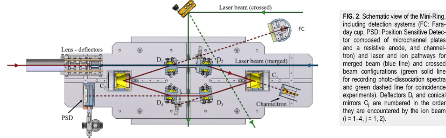
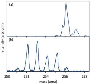
cations. Mass spectra were recorded by scanning the selec-tion magnet and by letting a continuous ion beam complete a single revolution in the Mini-Ring and recording the beam current collected by a Faraday cup (FC inFig. 2). The opti-mization of the ECR source operation was made by recording mass spectra. Two typical mass spectra are shown inFig. 3. With the combination of the selection magnet (velocity anal-ysis) and the Mini-Ring used as an electrostatic energy ana-lyzer (detection after one revolution), the mass resolution was improved (compared to the magnetic selection only) up to about m/∆m = 3000. The mass spectrum ofFig. 3(a) shows only intact naphthalene dimer cations in the displayed mass range (and a peak resulting from dimers with one isotope13C).
In contrast, the mass spectrum ofFig. 3(b)shows mostly frag-mented dimer cations, corresponding to the loss of one or several H atoms. These spectra have been recorded for two different naphthalene gas pressures in the ECR source. As
the pressure could not be measured directly in the source, it would be rather difficult to model quantitatively the depen-dence of the plasma temperature on the naphthalene pres-sure. However, it may be qualitatively explained as follows. At high pressure, the mean free path of the electrons is rather short such that they cannot be much accelerated (by the electron cyclotron resonance effect) between two collisions. Consequently, most of the electrons of the plasma could not acquire sufficient kinetic energies to ionize and dissociate naphthalene neutral molecules. Hence, a high source pres-sure results in a low plasma temperature and no dissociated naphthalene dimer cations were observed. Oppositely, a lower pressure induced a higher electronic temperature and con-sequently the observation of dimers with missing hydrogen atoms. These observations also support the following forma-tion mechanism of the dimer caforma-tions: naphthalene molecules were ionized and possibly dissociated by emission of one or several H atoms; then, those cation monomers may encounter another neutral and intact monomer in the plasma to form the dimer. There is a long distance attractive force between the cationic and the neutral monomers due to polarization lead-ing to the formation of the dimer. We may exclude the case of intact ionized monomers sticking with fragmented neu-tral monomers because the latter are expected to be present in the plasma in much smaller quantities compared to the intact ones that are constantly injected in the plasma at room temperature.
B. The Mini-Ring and neutral detection systems The Mini-Ring has been extensively described in Refs. 8 and41. Briefly, it is composed of four parallel-plate elec-trostatic deflectors, labelled D1–D4 in the order they are
encountered by the ion beam, and 2 conical shaped elec-trodes, labelled C1and C2, used as electrostatic focusing
mir-rors (Fig. 2). Ion bunches of 1–5 µs are prepared in the beam line between the selection magnet and the Mini-Ring by chop-ping the continuous beam with a homemade chopper com-posed of two parallel plates. In usual operation, dissociation events of the circulating molecular ions occurring in between
FIG. 2. Schematic view of the Mini-Ring,
including detection systems (FC: Fara-day cup, PSD: Position Sensitive Detec-tor composed of microchannel plates and a resistive anode, and channel-tron) and laser and ion pathways for merged beam (blue line) and crossed beam configurations (green solid line for recording photo-dissociation spectra and green dashed line for coincidence experiments). Deflectors Diand conical
mirrors Cj are numbered in the order
they are encountered by the ion beam (i = 1–4, j = 1, 2).
FIG. 3. Mass spectra (in the range 250–258 amu) recorded for a measured
pres-sure of 2 × 10⁻⁵mbar (a) and 5 × 10⁻⁶mbar (b). Depending on the pressure, the
temperature change of the plasma in the ECR ion source for these two pressures lead to the appearance of fragmented dimer cations.
the deflectors D3 and D4 can lead to neutral fragments that
were detected by microchannel plates equipped with a resis-tive anode. This system provided time and position sensi-tive detection (PSD). The PSD was used for two purposes. First, optimized storage parameters were obtained with tests beams (e.g., Ar+) by reducing the spot size and the deviation
of the center of the spot from turn to turn (i.e., reducing the amplitude of the betatron oscillations). Second, kinetic energy release (KER) could be estimated from the comparison of the spot size of the stored molecular ion beam with that of the test beam.45
Recently, the Mini-Ring has been equipped with a chan-neltron at the extension of the straight line between D2and
C1for detecting the neutral fragments that could go through
the hole of C1. Although not fully used in this work, the
ben-efits brought by this detector are multiple: improved resolu-tion of the decay time measurements, access to very short decay times (shorter than the half of a period) and coincidence experiments.
The vacuum chamber surrounding the Mini-Ring was equipped with several glass and quartz windows allowing for laser irradiation of the ion bunch parallel to the D1-D2straight
line in order to study laser-induced delayed dissociation and perpendicular to the ion bunch trajectory between D3 and
D4 in order to study prompt laser-induced dissociation. We
used the wavelength-tunable (260–2000 nm) optical paramet-ric oscillator (OPO) output of a 3 ns pulsed laser (EKSPLA model NT242) to record the photo-dissociation spectrum of the stored naphthalene dimer cations in the visible and near infrared range with photon energies from 0.5 to 5 eV.
III. EXPERIMENTAL RESULTS
A preliminary study with the naphthalene dimer cations was to confirm that the naphthalene dimer cations dissociate
into two fragments of the same mass. To this purpose, we per-formed a coincidence experiment between neutrals detected by the channeltron and ions detected by the PSD. In this case, we used the Mini-Ring for half a revolution setting C1and D3
voltages to half of their nominal values and D4to 0 V to drive
the cationic monomer fragment to the PSD. The laser beam (2nd harmonics of the Nd:YAG at 532 nm) was deflected to cross the ion beam between D2and C1to enhance the number
of dissociation events. The electronic signal from the channel-tron was used as a start of a time-to-digital converter and the PSD as a stop. With these settings, we confirmed, as expected, that the naphthalene dimer cations dissociated into two frag-ments of equal masses. By scanning the voltages of C1 and
D3, from 0 V to their nominal value (2 kV and 16 kV,
respec-tively) we could not observe any other dissociation scheme for naphthalene dimer cations.
It is very instructive to study the “natural” decay due to the dissociation of molecular ions of high internal energy in a storage ring.46,47 The natural decay due to the dissocia-tion of naphthalene dimer cadissocia-tions of high internal energy was recorded up to 100 ms storage time (Fig. 4). It could be fitted by two contributions: (i) a t−1 law, typical of a broad
inter-nal energy distribution and48 (ii) an exponential decay law, typical of dissociation due to collisions between the circu-lating ions and the residual gas (the residual pressure in the Mini-Ring vacuum chamber was of about 1 × 10⁻⁹mbar). This
result differs from our previous studies on the anthracene monomer cation49 showing a t−0.8initial decay law and, after
1 ms of storage, a significantly steeper slope compared to this initial power decay. This steeper decay was attributed to the quenching of the dissociation process by a fast radiative cooling process due to fluorescence from thermally excited electronic states (also referred as Poincaré fluorescence50,51 or recurrent fluorescence34,35,52). Consequently, the present
FIG. 4. Red dots: neutral counts on the PSD detector due to the natural decay
of hot naphthalene dimer cations in the Mini-Ring and to collisions with the rest gas. The green dotted line is an exponential fit to neutral counts at long storage time accounting for neutrals resulting from collisions with the rest gas. The blue dashed curve is a t−1power law fit to the difference between the neutral and the
exponential fit (considered as an estimation of the background at short storage times).
t−1decay law without any quenching shows that naphthalene
dimer cations do not cool efficiently via a fast radiative cooling process such as the Poincaré fluorescence. However, vibra-tional infrared cooling may occur but on a much longer time scale compared to the Poincaré Fluorescence.
The absence of such Poincaré fluorescence is most prob-ably due to the much smaller dissociation energy in the case of dimer cations (about 0.5 eV) compared to monomers (about 4.5 eV). Consequently, there is no energy matching between high vibrationally excited state of the ground electronic state and any low-lying bound electronic state that could fluoresce to the ground electronic state (seeFig. 1). Therefore, the pop-ulation of any bound electronic excited states via the inverse internal conversion process, which is a necessary step for the fast radiative cooling via the Poincaré fluorescence, is highly improbable. Moreover, the first excited electronic state is dis-sociative.31In case it can be thermally populated, dissociation only may occur. This is most probably the origin of the natural decay observed at short storage time.
Figure 5displays a typical time spectrum of the neutral counts on the PSD for naphthalene dimer. The bunched struc-ture (period of 7.92 µs and width of about 1.5 µs) of the stored ions can be seen in the inset ofFig. 5. The narrow peak at 1833 µs corresponds to the huge enhancement of the neutral counts due to laser absorption (2nd harmonic—532 nm) in the crossed beam configuration in between D3 and D4. The laser
power was set to about 80 µJ/pulse in order to avoid multi-ple photon absorption (measurement made downstream the whole laser pathway after the cross-beam region). This laser
FIG. 5. Typical time of flight spectrum. The raw neutral counts were recorded as
a function of the storage time. The peak at about 1833µs is due to the neu-trals produced after laser absorption at 532 nm (second harmonic output of the Nd:YAG laser). This dissociation signal was used to perform the photo-dissociation spectrum by scanning the laser wavelength. The inset shows a zoom-in view around the photo-dissociation signal for better visualization of the bunched structure of the stored ions and of the signal to-noise ratio.
pulse energy is of the same order of magnitude as the pulse energies in most of the visible range of the OPO laser that is used in the following. However, in the infrared range, the max-imum OPO laser intensity is about one order of magnitude smaller than in the visible range. The storage time of about 2 ms prior laser irradiation has been chosen to reduce the con-tributions of the natural decay and of the collisions with resid-ual gas (both considered as noise in the photo-dissociation spectrum) to the laser induced signal. Longer storage times would have the inconvenience to reduce the repetition rate of the experiment for a rather small gain on the signal to noise ratio. It is noteworthy that no delayed dissociation is observed onFig. 5. It shows that the lifetime of the naphthalene dimer cations after absorption is much smaller than one of their rev-olution periods in the ring. Prompt dissociation only has been observed whatever the wavelength used in the visible and near infrared ranges. This absence of delayed dissociation suggests a non-statistical dissociation process for which the absorption in the CR band (near infrared range) and in the LE line (visible
FIG. 6. (a) Experimental photo-dissociation spectrum (this work) obtained in hot
conditions (blue circles). Simulated photo-dissociation spectra for the CR (red plain curve) and LE (orange dashed curve) including the contributions from v = 0 to v = 4 vibrational states calculated with effective Morse PEC parameters ofTable III. (b) Experimental photo-dissociation spectrum (cold conditions) digitized from Ref.28 (black squares). Simulated photo-absorption spectra for the CR (red plain curve) and LE (plain green line) using the v = 0 vibrational state only. The LE simulated curve with v = 0 (green plain line) of figure (b) has been also displayed in figure (a) for easy comparison. In (a) and (b), the dotted curves are the simulated CR lines calculated from the PECs of Ref.31for Boltzmann temperatures of 400 K and 10 K, respectively. All the spectra have been normalized to 1.
range) band would probably populate directly the dissociative excited states.
In order to record the photo-dissociation spectrum of the stored naphthalene dimer cations [Fig. 6(a)], the laser wave-length was scanned in the 400–690 nm and 800–2000 nm ranges (the laser intensity was too small in between these two ranges). An appropriate time gate was used to record the laser-induced neutral counts in order to reduce background counts due to collisions with the rest gas and the remaining natural decay. As expected,28 the photo-dissociation spec-trum shows two broad peaks corresponding to the CR and LE bands centered at about 1 eV and 2.2 eV. Hence, we estimate the dissociation energy to half of the CR transition energy, i.e., about 0.5 eV in rather good agreement with other mea-surements (Table I). The CR band we have recorded is much broader than in the photo-dissociation spectrum of naphtha-lene dimer cations recorded by Inokuchi et al.28[displayed in
Fig. 6(b)]. This is most probably due to a much broader internal energy distribution of the stored naphthalene dimer cations in our experiment. Indeed, naphthalene dimer cations are pro-duced in rather hot conditions in the plasma of the ECR ion source compared with a cold jet source as in the work of Inokuchi et al.28 Since there is no efficient radiative cooling for these species (see the above discussion about the natu-ral decay), a 2 ms storage time is not long enough in order to have a significant change of the internal energy distribu-tion. Another spectrum recorded after 20 ms storage time (not shown here), with less statistics though, did not show any significant change in the CR and LE peak widths. How-ever, when comparing the spectra ofFigs. 5(a)and5(b), it is interesting to remark that the LE band is not much broad-ened in the hot conditions, as least not as much as the CR band.
IV. MODELLING AND DISCUSSION
We have tentatively used the following strategy in order to test the PECs of Dontot et al.31 and to model the experi-mental photo-dissociation both for our study and the earlier one by Inokuchi et al.:28
(i) Starting parameters were taken from PECs resulting from a fit with Morse potentials to ab initio calculations of Dontot et al.31 The PECs along the inter-molecular separation of the 8 lowest electronic states of the naph-thalene dimer cations in the sandwich configuration given in Ref.31could be nicely fitted by Morse poten-tials, assuming that the separation of the two monomers could be modelled in a similar way as for a diatomic molecule.Figure 7displays the resulting fit for the2B
1g
and 2A
u states (CR transition). Very small differences
between the Morse fit and the ab initio calculation of Ref.31of the2B
1gand2AuPECs can be barely seen only
at rather large inter-monomer distance (greater than 5.5 nm).
(ii) Morse PECs present the advantage of having analytical solutions to calculate the vibrational energy levels and eigenstates.53 Hence, the Franck-Condon factors for
FIG. 7. PECs for the2B
1gand2Austates. Light blue diamonds (2B1g) and red
circles (2A
u): digitized from Ref.31. Black dashed lines: fits with Morse PECs on
digitized data from Ref.31. Blue (2B
1g) and black (2Au): modified Morse PECs
used to model the CR line inFig. 6.
the absorption transition to the2A
udissociative state
could be estimated following Ref.54, p. 392 by the over-lap of the wave functions obtained with parametrized PECs as shown inFig. 8. As an example, the absorption line profile from v = 0 of the2B
1gground state to the 2A
ustate is given by the red curve on the left side of
Fig. 8.
(iii) We calculated the contributions of the five first vibra-tional levels (v = 0 to v = 4) to the CR and the LE bands using Boltzmann distributions of the internal energy. The contribution of higher vibration levels has been neglected. These states may contribute mostly to the sides of the bands and might improve slightly the agreement between the model and experimental band widths.
Besides, we have calculated the energies of all the vibration modes of the naphthalene dimer cations using TPSS055 (Tao, Perdew, Staroverov, and Scuseria (TPSS)56 meta hybrid with 25% of exact exchange) density functional theory (DFT) func-tional with the def2-qzvpp basis set, including the disper-sion via the D3 verdisper-sion of Grimme’s approach with Becke-Johnson damping.57 The energies of the six lowest modes, corresponding to intermolecular motions between the two monomers, were found to be between 9.7 and 13.2 meV (78.1 and 106.1 cm−1). They are in the same order of
mag-nitude as the one calculated from the Morse potential used in the following, hυ0 ≈ 7.8 meV, with the parameters of
FIG. 8. Morse potential energy curves used in the calculations to simulate the
photo-absorption spectra ofFig. 6. For illustration purpose, the shape of the vibra-tion wave funcvibra-tions for v = 0 and v = 5 of the electronic ground state2B
1g, the
shape of the wave function in the continuum of the2A
gdissociative state are
dis-played. On the left side of the vertical scale are shown the simulated absorption function for the CR and LE states (relative amplitudes of the CR and LE curves are arbitrary).
Table I for the 2B
1g ground state. Intramolecular vibration
energies were found at significantly higher energies (e.g., the lowest one was found at 23.2 meV). The rather small vibrational energy of the intermolecular motions shows that small internal energies may be sufficient to induce motions that are not accounted for in the one-coordinate PECs of Ref.31.
Using directly the PECs resulting from the fit to those of Ref. 31, the v = 0 vertical absorption transition from the ground state,2B
1g, to the first excited dissociative state,2Au
state, would correspond to a CR transition of about 1.23 eV slightly larger than the experimental values 1.05 eV in gas phase ofTable II. Moreover, with these PECs, we could not model satisfactorily the CR band of both photo-dissociation spectra as shown by the dotted curves ofFigs. 6(a)and6(b). An optimization procedure of the parameters of the Morse potentials has been used in order to minimize the difference between the modeled and experimental CR bands. The result-ing parameters of the Morse potentials are given inTable III
TABLE II. Energies of CR and LE transitions.
CR (eV) LE (eV)
Gas phase28 1.05 2.17
Liquid phase59 1.21 Liquid phase60 1.24
This work 1.0±0.2 2.15
TABLE III. Parameters of the Morse potentials used in the model: dissociation energy (De), equilibrium distance (re), and minimum potential energy at re(Ve).
2B
1g 2Au 2B1u 2Ag 2B2u
De(eV) 0.5 0.05 0.1 0.5 0.5
re(Å) 3.21 4.37 4.33 3.22 3.196
Ve(eV) −0.55 −0.1 0.7 0.3 1.4
and the resulting PECs, displayed onFig. 7, are rather different compared to those of Dontot et al.31
Including only the v = 0 vibrational state of the 2B 1g
ground state, the line profiles obtained with the adjusted Morse PECs fit nicely to experimental absorption curves obtained by Inokuchi et al.28 in cold conditions both for the CR and LE bands. This supports that it was possible to find a set of parameters for the2B
1gand2Austates leading to PECs
with optimized shapes around the equilibrium distance of the ground state. It is notable that the width of the model peak profile is very sensitive to the slope of the 2A
u dissociative
state.
For higher vibration levels, the probability of presence is maximum at distances close to the edge of the potential well, as illustrated in Fig. 8 (see Ref.54). Consequently, for the higher temperature conditions of our photo-dissociation spectrum [Figs. 6(a)], v > 0 levels mostly contribute to the sides of the line profiles. In order to reach a satisfactory model-experiment agreement, the high temperature spec-trum gave more constraints in order to determine the shapes of the effective PECs of the 2B
1g and 2Au states. We could
reach a rather good agreement between the modeled and the experimental CR bands for our spectrum using a Boltzmann temperature of 400 K. Using these effective potentials with a Boltzmann of 400 K, the center of the modelled CR band is slightly shifted towards lower energy at 400 K compared to the low temperature spectrum. This shift estimated to about −0.02 eV is slightly smaller than the experimental shift but in the right direction though.
However, this shift due to the population of v > 0 vibration level cannot explain the difference between the experimental CR lines and those obtained from the PECs from Dontot et al.31 The reason of this disagreement is most likely that these PECs are valid for motions in one coordinate only. As expected from our calculations of the vibration energies, rather small internal energies may lead to motions along other coordinates, such as, for instance, the aforementioned twisting motion calculated by Dontot et al. in the case of pyrene dimer cations. As shown in Fig. 4 of Ref.31for pyrene dimer cations, the gap between the 2B
1g and 2Au states depends significantly on this twist
angle such that the required energy for photo-dissociation is expected to be reduced with increasing twist angle up to 20◦.
Moreover, during the dissociation process, the relative motion between the two monomers may be rather complex involv-ing other motions that are not included in the one-coordinate PECs. The estimation of the effect of several intermolecu-lar (and possibly intramolecuintermolecu-lar) vibration motions would be needed in a full calculation. The Morse PECs resulting from
the above adjustment procedure are expected to be “effective” potentials that average all temperature and possible isomer-ization effects such as the twisting angle between the two monomers.
The same method has been repeated for the LE absorp-tion band. For the cold condiabsorp-tions ofFig. 6(b), the position and width of the model peak was found to be in good agreement with the experimental LE peak profile. However, for our spec-trum [Fig. 6(a)], using the same temperature of 400 K as for the CR transition, the broadening of the model LE peak due to the inclusion of v > 0 states is about twice as broad as the experimental profile. InFig. 6(a), we clearly see that the exper-imental LE band is not as broad as the CR band, but slightly broader than the LE band of the cold conditions as if, for the LE transition, the dimer had less vibrational energy. The diatomic model using effective Morse potentials for a single coordinate is possibly too crude to describe correctly the shape of the LE absorption peak profile. However, we may invoke two possible explanations for the small broadening of the LE peak at high temperature:
(i) It can be expected that the photon would be absorbed by one of the monomers. Therefore, the vibrational broad-ening would involve the intramolecular C–H and C–C vibrations of this monomer only. As these intramolec-ular vibrations have been calculated at much higher energy compared to the intermolecular vibrations, it can be expected that at the temperature of 400 K deduced from the CR broadening, the v > 1 lev-els of the intramolecular vibrations are very weakly populated.
(ii) The photo-absorption may not populate directly the dissociative state 2B
1u, but the 2B2u bound state. This
state may undergo statistical dissociation via inter-nal conversion leading to a thinner profile compared to the model the LE curve of Fig. 6(a). This tentative explanation is supported by the rather high oscillator strength of transitions at 2.1 and 2.2 eV calculated by Dontot et al.31
More investigations would be needed to come to a more definitive conclusion about the evolution of the width of the LE band as a function of the temperature.
V. CONCLUSION
In summary, we have stored in the Mini-Ring beams of naphthalene dimer cations produced in an ECR ion source. Special tuning conditions, i.e., low HF power and high pres-sure, of this ion source have been discussed. Oppositely to monomer naphthalene cations, no fast reduction of the internal energy of the naphthalene dimer cations could be evidenced, demonstrating that the Poincaré (or recurrent) fluorescence is not an efficient process for the naphthalene dimer cations. The reason is that the low dissociation energy of the dimers do not allow any energy match between high vibrational states of the ground electronic state and the lower vibrational level of the first electronic excited state.
A photo-absorption spectrum of the naphthalene dimer cations has been recorded after 2 ms storage time in the Mini-Ring. CR and LE lines have been clearly identified as in previous studies in colder conditions. Effective Morse PECs for the ground and the first dissociative states have been used to compute a model photo-dissociation spectrum. It is expected that the proposed PECs average in some way the complexity induced by all the possible intermolecular motions. A more accurate modelling would require a con-siderable theoretical effort to sample the whole potential energy hypersurface in order to account for all possible vibra-tion and rotavibra-tion movibra-tions of all possible isomers. Neverthe-less, additionally to the expected broadening, a shift towards lower energy of the CR absorption band was found with increasing temperature and was implicitly accounted for in the present modelling by choosing an appropriate param-eter for the Morse PECs. However, this shift is not suffi-cient to explain the discrepancy between the experimental band centers and those obtained from the ab initio PECs. We expect that, even at rather low temperatures, motions in all inter-molecular coordinates should be included as the cor-responding vibration energies are rather small. More exper-imental and theoretical efforts would be needed in order to investigate thoroughly the temperature effects in PAH dimer cations.
ACKNOWLEDGMENTS
This work was supported by Grant No.ANR-10-BLAN-0426of
the ANR (Agence Nationale pour la Recherche). The authors would like to thank Christine Joblin and Mathias Rapaci-oli for valuable and intensive discussions concerning PAH dimers.
REFERENCES
1E. Peeters et al., “The rich 6 to 9 µm spectrum of interstellar PAHs,”Astron.
Astrophys.390(3), 1089–1113 (2002).
2E. Peeters, C. W. Bauschlicher, L. J. Allamandola, A. G. G. M. Tielens, A. Ricca, and M. G. Wolfire, “The PAH emission characteristics of the reflection nebula NGC 2023,”Astrophys. J.836(2), 198 (2017).
3D. J. Stock et al., “Polycyclic aromatic hydrocarbon emission in spitzer/IRS maps. I. Catalog and simple diagnostics,”Astrophys. J.819(1), 65
(2016).
4D. J. Stock and E. Peeters, “Polycyclic aromatic hydrocarbon emission in spitzer/IRS maps. II. A direct link between band profiles and the radiation field strength,”Astrophys. J.837(2), 129 (2017).
5E. Peeters, “The infrared emission bands,”Proc. Int. Astron. Union9(S297), 187–196 (2013).
6A. Leger, L. D’Hendecourt, P. Boissel, and F. X. Desert, “Photo-thermo-dissociation. I. A general mechanism for destroying molecules,” Astron. Astrophys. 213, 351–359 (1989).
7J. Montillaud, C. Joblin, and D. Toublanc, “Evolution of polycyclic aromatic hydrocarbons in photodissociation regions: Hydrogenation and charge states,”Astron. Astrophys.552, A15 (2013).
8S. Martin et al., “Fast radiative cooling of anthracene observed in a compact electrostatic storage ring,”Phys. Rev. Lett.110(6), 063003 (2013).
9T. Giesen et al., “Molecular Spectroscopy,” in Laboratory Astrochemistry, edited by S. Schlemmer, H. Mutschke, T. Giesen, and C. Jäger (Wiley-VCH Verlag GmbH & Co. KGaA, 2014), pp. 13–108.
10J. Szczepanski, M. Vala, D. Talbi, O. Parisel, and Y. Ellinger, “Electronic and vibrational-spectra of matrix-isolated anthracene radical cations— Experimental and theoretical aspects,” J. Chem. Phys. 98(6), 4494–4511
(1993).
11D. M. Hudgins, S. A. Sandford, and L. J. Allamandola, “Infrared spec-troscopy of polycyclic aromatic hydrocarbon cations. 1. Matrix-isolated naphthalene and perdeuterated naphthalene,”J. Phys. Chem.98(16), 4243–
4253 (1994).
12D. M. Hudgins and L. J. Allamandola, “Infrared spectroscopy of matrix-isolated polycyclic aromatic hydrocarbon cations. 2. The members of the thermodynamically most favorable series through coronene,”J. Phys. Chem. 99(10), 3033–3046 (1995).
13J. Oomens, A. G. G. M. Tielens, B. G. Sartakov, G. von Helden, and G. Meijer, “Laboratory infrared spectroscopy of cationic polycyclic aromatic hydrocarbon molecules,”Astrophys. J.591(2), 968 (2003).
14C. Joblin, P. Boissel, A. Leger, L. D’Hendecourt, and D. Defourneau, “Infrared spectroscopy of gas-phase PAH molecules. II. Role of the tem-perature,” Astron. Astrophys. 299, 835 (1995).
15A. J. Huneycutt, R. N. Casaes, B. J. McCall, C.-Y. Chung, Y.-P. Lee, and R. J. Saykally, “Infrared cavity ringdown spectroscopy of jet-cooled polycyclic aromatic hydrocarbons,”ChemPhysChem5(3), 321–326 (2004).
16G. Malloci, C. Joblin, and G. Mulas, “On-line database of the spectral prop-erties of polycyclic aromatic hydrocarbons,” e-printarXiv:astro-Ph0701254
(2007).
17G. Malloci, G. Mulas, G. Cappellini, and C. Joblin, “Time-dependent density functional study of the electronic spectra of oligoacenes in the charge states −1, 0, +1, and +2,”Chem. Phys.340(1-3), 43–58 (2007).
18A. G. G. M. Tielens, “The molecular universe,”Rev. Mod. Phys.85(3), 1021– 1081 (2013).
19A. Leger and J. L. Puget, “Identification of the ‘unidentified’ IR emission features of interstellar dust?,” Astron. Astrophys. 137, L5–L8 (1984). 20M. Frenklach and E. D. Feigelson, “Formation of polycyclic aromatic hydrocarbons in circumstellar envelopes,”Astrophys. J.341, 372–384 (1989).
21H. Sabbah, L. Biennier, S. J. Klippenstein, I. R. Sims, and B. R. Rowe, “Exploring the role of PAHs in the formation of soot: Pyrene dimerization,”
J. Phys. Chem. Lett.1(19), 2962–2967 (2010).
22M. Rapacioli, C. Joblin, and P. Boissel, “Spectroscopy of polycyclic aro-matic hydrocarbons and very small grains in photodissociation regions,”
Astron. Astrophys.429(1), 193–204 (2005).
23P. Pilleri, J. Montillaud, O. Berné, and C. Joblin, “Evaporating very small grains as tracers of the UV radiation field in photo-dissociation regions,”
Astron. Astrophys.542, A69 (2012).
24M. Rapacioli, F. Calvo, C. Joblin, P. Parneix, D. Toublanc, and F. Spiegelman, “Formation and destruction of polycyclic aromatic hydrocarbon clusters in the interstellar medium,”Astron. Astrophys.460(2), 519–531 (2006).
25P. Benharash, M. J. Gleason, and P. M. Felker, “Rotational coherence spec-troscopy and structure of naphthalene trimer,”J. Phys. Chem. A103(11),
1442–1446 (1999).
26J. K. Song, N. K. Lee, J. H. Kim, S. Y. Han, and S. K. Kim, “Anion clusters of anthracene, Ann–(n=1–16),”J. Chem. Phys.119(6), 3071–3077 (2003).
27B. Badger and B. Brocklehurst, “Formation of dimer cations of aromatic hydrocarbons,”Nature219(5151), 263 (1968).
28Y. Inokuchi, K. Ohashi, M. Matsumoto, and N. Nishi, “Photodissociation spectrum of naphthalene dimer cation,”J. Phys. Chem.99(11), 3416–3418
(1995).
29M. Meot-Ner, “Dimer cations of polycyclic aromatics. Experimental bond-ing energies and resonance stabilization,”J. Phys. Chem.84(21), 2724–2728
(1980).
30T. Fujiwara and E. C. Lim, “Binding energies of the neutral and ionic clus-ters of naphthalene in their ground electronic states,”J. Phys. Chem. A 107(22), 4381–4386 (2003).
31L. Dontot, N. Suaud, M. Rapacioli, and F. Spiegelman, “An extended DFTB-CI model for charge-transfer excited states in cationic molecular clusters: Model studies versus ab initio calculations in small PAH clusters,”Phys. Chem. Chem. Phys.18(5), 3545–3557 (2016).
32T. Beitz, R. Laudien, H.-G. Löhmannsröben, and B. Kallies, “Ion mobility spectrometric investigation of aromatic cations in the gas phase,”J. Phys. Chem. A110(10), 3514–3520 (2006).
33H. T. Schmidt et al., “First storage of ion beams in the double elec-trostatic ion-ring experiment: DESIREE,” Rev. Sci. Instrum.84(5), 055115
(2013).
34V. Chandrasekaran et al., “Determination of absolute recurrent fluores-cence rate coefficients for C6–,” J. Phys. Chem. Lett. 5(23), 4078–4082 (2014).
35G. Ito et al., “Cooling dynamics of photoexcited C6–and C6H–,”Phys. Rev.
Lett.112(18), 183001 (2014).
36S. P. Møller, “ELISA, and electrostatic storage ring for atomic physics,”
Nucl. Instrum. Methods Phys. Res., Sect. A394(3), 281–286 (1997).
37D. Zajfman et al., “Electrostatic bottle for long-time storage of fast ion beams,”Phys. Rev. A55(3), R1577–R1580 (1997).
38R. von Hahn et al., “The cryogenic storage ring CSR,”Rev. Sci. Instrum.
87(6), 063115 (2016).
39C. R. Calvert et al., “LIAD-fs scheme for studies of ultrafast laser inter-actions with gas phase biomolecules,” Phys. Chem. Chem. Phys. 14(18),
6289–6297 (2012).
40T. Doussineau, R. Antoine, M. Santacreu, and P. Dugourd, “Pushing the limit of infrared multiphoton dissociation to megadalton-size DNA ions,”
J. Phys. Chem. Lett.3(16), 2141–2145 (2012).
41J. Bernard et al., “A ‘tabletop’ electrostatic ion storage ring: Mini-ring,”Rev.
Sci. Instrum.79(7), 075109 (2008).
42R. Geller, Electron Cyclotron Resonance Ion Sources and ECR Plasmas (CRC Press, 1996).
43R. Geller, “ECRIS: The electron-cyclotron resonance ion sources,”Annu.
Rev. Nucl. Part. Sci.40, 15–43 (1990).
44L. Maunoury et al., “LIMBE: A new facility for low energy beams,”Rev. Sci.
Instrum.73(2), 561–563 (2002).
45M. Ji, G. Montagne, R. Bredy, J. Bernard, L. Chen, and S. Martin, “Ion beam characterization in the mini-ring and kinetic energy release measure-ment of unimolecular decay of anthracene cations,”Phys. Scr.T156, 014092
(2013).
46L. H. Andersen, “Thermionic electron emission from SF6−,”Phys. Rev. A
78(3), 032512 (2008).
47J. U. Andersen et al., “Power-law decay of collisionally excited amino acids and quenching by radiative cooling,”Eur. Phys. J. D25(2), 139–148
(2003).
48K. Hansen, J. U. Andersen, P. Hvelplund, S. P. Møller, U. V. Pedersen, and V. V. Petrunin, “Observation of a 1/t decay law for hot clusters and molecules in a storage ring,”Phys. Rev. Lett.87(12), 123401 (2001).
49J. Bernard et al., “Cooling of PAH cations studied with an electrostatic storage ring,”Nucl. Instrum. Methods Phys. Res., Sect. B408, 21–26 (2017).
50A. Léger, P. Boissel, and L. d’Hendecourt, “Predicted fluorescence mech-anism in highly isolated molecules: The Poincaré fluorescence,”Phys. Rev. Lett.60(10), 921–924 (1988).
51P. Boissel, P. de Parseval, P. Marty, and G. Lefevre, “Fragmentation of iso-lated ions by multiple photon absorption: A quantitative study,”J. Chem. Phys.106(12), 4973–4984 (1997).
52Y. Ebara et al., “Detection of recurrent fluorescence photons,”Phys. Rev.
Lett.117(13), 133004 (2016).
53P. M. Morse, “Diatomic molecules according to the wave mechanics. II. Vibrational levels,”Phys. Rev.34(1), 57–64 (1929).
54G. Herzberg, Molecular Spectra (D. Van Nostrand Company, Inc., 1950), Vol. I.
55S. Grimme, J. Antony, S. Ehrlich, and H. Krieg, “A consistent and accu-rate ab initio parametrization of density functional dispersion correc-tion (DFT-D) for the 94 elements H-Pu,”J. Chem. Phys. 132(15), 154104
(2010).
56J. Tao, J. P. Perdew, V. N. Staroverov, and G. E. Scuseria, “Climbing the density functional ladder: Nonempirical meta-generalized gradient approx-imation designed for molecules and solids,”Phys. Rev. Lett.91(14), 146401
57S. Grimme, S. Ehrlich, and L. Goerigk, “Effect of the damping function in dispersion corrected density functional theory,”J. Comput. Chem.32(7),
1456–1465 (2011).
58B. Bouvier, V. Brenner, P. Millié, and J.-M. Soudan, “A model potential approach to charge resonance phenomena in aromatic cluster ions,”J. Phys. Chem. A106(43), 10326–10341 (2002).
59T. N. Das, “Monomer and dimer radical cations of benzene, toluene, and naphthalene,”J. Phys. Chem. A113(23), 6489–6493 (2009).
60X. Cai, S. Tojo, M. Fujitsuka, and T. Majima, “Photodissociation of naph-thalene dimer radical cation during the two-color two-laser flash photol-ysis and pulse radiolphotol-ysis–laser flash photolphotol-ysis,”J. Phys. Chem. A110(30),