Abstract Multiple mass spectra (MS1 to MS6) of 55 phenylalkylamine derivatives were recorded with ion-trap mass spectrometry employing electrospray (ESI) and atmospheric pressure chemical ionization (APCI). Frag-mentation patterns were studied in detail and a generally applicable scheme was established for elucidation of the structures of phenylalkylamine derivatives. HPLC com-bined with ion-trap multiple mass spectrometry was used to identify the structure of reaction by-products in ecstasy samples from the “black” market. Low nanogram amounts were sufficient for on-line HPLC–MSnstructure
elucida-tion of unknowns.
Keywords Designer drugs · Dimethoxyamphetamine · LC–MSn· MDMA · Phenethylamines ·
Trimethoxyamphetamine
Introduction
During the last five years LC–MS has become a widely used tool for the analysis of thermally labile and/or polar molecules. The introduction of LC–MS ion-trap instru-ments in 1996 enabled a structure elucidation with low nanogram amounts. These instruments enable on-line gen-eration of a consecutive series of product-ion spectra by sequential isolation/fragmentation of selected ions, result-ing in a series of MS2, MS3to MSnspectra, which contain
highly structure-significant information. However, so far no general rules have been established for elucidation of a structure from the observed fragments, as for electron-im-pact ionization. Therefore, our group has started to develop structure-elucidation schemes for different compound classes such as carbonyl-dinitrophenylhydrazones [1, 2], trichothecenes [3], aconite alkaloids [4], or oligomers of bisphenol A diglycidyl ether [5].
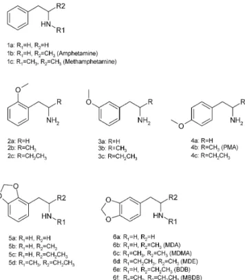
Many important neurotransmitters such as dopamine, adrenaline, or noradrenalin and synthetic drugs such as dex-amine, amphecloral, fenflurdex-amine, orthoxine, or methoxy-phenamine contain the phenethylamine ethanamine, Fig. 1, 1a) or amphetamine (1-phenyl-2-propanamine, Fig. 1, 1b) substructure. Mescaline (3,4,5-trimethoxy-phenethylamine, Fig. 2, 13a) is also the active compound in different species of cactus regarded as most sacred by the native American culture. The effects on hu-mans have been investigated for more than 150 phenyl-alkylamines [6]. The structures of the 55 phenylalkyl-amine derivatives studied in this work are shown in Figs. 1 and 2.
Stephan Kölliker · Michael Oehme
Structure elucidation of nanogram quantities
of unknown designer drugs based on phenylalkylamine derivates
by ion trap multiple mass spectrometry
DOI 10.1007/s00216-003-2411-2
Received: 8 September 2003 / Revised: 5 November 2003 / Accepted: 7 November 2003 / Published online: 21 January 2004 O R I G I N A L PA P E R
S. Kölliker · M. Oehme (✉)
Organic Analytical Chemistry, University of Basel, Neuhausstr. 31, 4057 Basel, Switzerland
e-mail: Michael.Oehme@unibas.ch © Springer-Verlag 2004
Fig. 1 Structures of the unsubstituted, monomethoxy- and
Phenylalkylamine derivatives attract considerable pub-lic attention, since the US food and drug administration (FDA) has classified N-methyl-3,4-methylendioxyamphet-amine (MDMA, Fig. 1, 6c) as a most restricted Class 1 compound in 1985–86. MDMA is the main representative of a class of pharmacologically active compounds called empathogens. Dosages of 50 to 100 mg MDMA induce a state of increased empathy, and feelings of centeredness and inner peace [7]. Empathogens continue to be suggested as potential therapeutic agents in facilitating psychother-apy [8]. Considerable benefits and few undesirable side effects have been reported from restricted medical use in Switzerland during the period 1989–1993 [9], and strong anti-Parkinson effects were described recently [10]. How-ever, long-term abuse of high dosages of illicit ecstasy seems to be related to increased depression and reduced short-term memory [11].
Although MDMA is regarded as relatively safe (Refs. [12, 13] and Bernhard Meili, Bundesamt f. Gesundheits-wesen, Bern, Switzerland; personal communication), re-lated designer drugs PMA (4-methoxyamphetamine, Fig. 1, 4b), PMMA (N-methyl-4-methoxyamphetamine), and 4-MTA (4-methylthioamphetamine) have caused several casualties during recent years [14, 15, 16]. In addition,
other compounds such as 2C-B (Fig. 2, 16a), 2C-I (Fig. 2, 17), 2C-T2 (Fig. 2, 18), 2C-T7 (Fig. 2, 19), TMA-2 (Fig. 2, 14b) and TMA-6 (Fig. 2, 15b) have been identified in pills sold in Switzerland (unpublished results). Moreover, new illicit psychoactive phenylalkylamine derivatives are ex-pected to appear on the market which might not have the effect of MDMA [17]. This can then lead to overdose af-ter attempts to reach the expected state and, consequently, increase the risk of acute damage or even death.
More than one hundred articles about the analysis of MDMA-related compounds have been published during the past five years [18, 19]. However, none describe rapid and reliable elucidation of the structure of completely un-known phenylalkylamine derivatives. Detailed investiga-tions of methoxyphenyl [14, 20] and methylendioxyphenyl compounds [21, 22, 23] revealed that retention time and EI or CI mass spectra of isomers often are identical. There-fore, elucidation of the structure of closely related ana-logues remains tricky and time-consuming. Recently pub-lished examples are N-methyl-1-phenethylamine [24], PMMA [25], 4-MTA [26], and 2C-B (4-bromo-2,5-di-methoxyphenylethylamine) [27].
The aim of this work was to study the possibility of us-ing multiple mass spectrometry (MSn) techniques to
iden-Fig. 2 Structures of the dimethoxy-,
trimethoxy-, and further substituted phenylalkylamines investigated
tify rapidly and unequivocally unknown and/or new de-signer drugs in pills. The MS1 to MS4 fragmentation of model compounds is described in detail and their impor-tance for structure elucidation discussed. Finally, the po-tential of the identification scheme was tested by elucidat-ing the structure of impurities in pills.
Experimental
Chemicals
Benzaldehyde, 2-methoxybenzaldehyde, 3-methoxybenzaldehyde, 4-methoxybenzaldehyde, 2,3-methylendioxybenzaldehyde, 3,4-meth-ylendioxybenzaldehyde, 2,3-dimethoxybenzaldehyde, 2,4-dimeth-oxybenzaldehyde, 2,5-dimeth2,4-dimeth-oxybenzaldehyde, 3,4-dimethoxy-benzaldehyde, 3,5-dimethoxy3,4-dimethoxy-benzaldehyde, 2,3,4-trimethoxybenz-aldehyde, 2,4,5-trimethoxybenz2,3,4-trimethoxybenz-aldehyde, 2,4,6-trimethoxybenzal-dehyde, and 3,4,5-trimethoxybenzaldehyde were purchased from Fluka (Buchs, Switzerland). All compounds were of “purum” qual-ity (97–98%).
Nitromethane (puriss., 98.5%), nitroethane (pract. 90–95%), 1-nitropropane (pract. 90–95%), acetic acid (puriss.), ammonium acetate (puriss., 99%) n-butylamine (purum, 97%), LiAlH4
(pu-rum, 97%), and tetrahydrofuran (THF, puriss. absolute over mo-lecular sieve) were purchased from Fluka (Buchs, Switzerland). Ultrapure water was from a Elga Maxima HLPC water supply sys-tem (Elga, High Wycombe, Bucks, UK). Methanol of “pesticide residue analysis” grade was purchased from sds (Peypin, France), and dichloromethane was from Romil (Cambridge, England). Synthesis of reference compounds
Most reference compounds were synthesized with the general method given in Ref. [6]. The method was modified for micro amounts. The aldehyde (10 mg) was added to a mixture of 8 mg ni-tromethane, 1 mg acetic acid, and 1 mg n-butylamine (catalyst) in a 5-mL vial. The reaction mixture was heated for 1–3 h at 75 °C until the color was deep-yellow. After cooling to room tempera-ture, 1 mL H2O was added. The formed yellow nitrostyrene was
extracted with 1.5 mL CH2Cl2. The organic layer was separated
and evaporated to dryness. Sometimes the residual yellow oil crys-tallized spontaneously.
The nitrostyrenes were reduced to the corresponding phenethyl-amines as follows. The nitrostyrene (approx. 1 mg) was dissolved in 0.5 mL dry THF in a 1-mL vial, and 5 mg LiAlH4was added.
The vial was closed air-tight and the reaction mixture was left for 1–2 h at 40 °C to form the phenethylamine. After cooling, 1–3 drops H2O containing 10% of ammonium acetate were added to destroy
hydride residues. The reaction mixture was transferred to a 5-mL Teflon syringe equipped with a 0.45-µm (25 mm i.d.) nylon filter (Scientific Resources, Eatontown NJ, USA) and a few more milli-liters of water were added. After filtration the reaction mixture was diluted approximately tenfold with a 50:50 mixture of CH3OH and
H2O containing 1% ammonium acetate. This solution was injected
directly into the mass spectrometer. The purity of the synthetic product was usually 80–90%. The most abundant impurity was the corresponding N-hydroxystyrene. Nitromethane was replaced by nitroethane for synthesis of amphetamines, and by 1-nitropropane for 1-phenyl-2-butane amines.
Mass spectrometry
All reference mass spectra were recorded with a ThermoFinnigan LCQ-G2 ion-trap mass spectrometer equipped with electrospray (ESI) or atmospheric pressure chemical ionization (APCI) ion sources. Solutions containing 10–100 ngµL–1of phenylalkylamine
derivatives in 50:50 CH3OH–H2O were injected with the syringe
pump at a flow rate of 5µL min–1. MS settings were: spray voltage,
4.5 kV; heated capillary temperature, 150 °C; capillary voltage 47 V, tube-lens offset –5 V, octapole 1 offset –3 V, lens voltage –16 V, octapole 2 offset –5 V, multipole RF amplitude 400 V, automatic gain control on, full MS target 5×10⁷. The scan range was usually
50–500 u; isolation width, 1.4 u (MS2, isolation efficiency 99.9%
for the 12C peak) or 2 u (MS3to MS6). Collision-induced
dissocia-tion (CID) was carried out at 22% (MS2) or 30% trap energy (MS3
to MS6).
Analysis of pills
Compounds in pills were determined by coupling a Rheos 2000 HPLC pump (flux instruments, Basel, Switzerland) and a PAL au-tosampler (CTC Analytics, Zwingen, Switzerland) in front of the mass spectrometer. The pill was dissolved in 100 mL water, acidi-fied with one drop conc. HCl, by ultrasonication for 5 min. A few milliliters of the resulting solution were filtered though a 0.45-µm (25 mm i.d.) nylon filter (Scientific Resources, Eatontown NJ, USA). Filtrate (50µL) was transferred to a 5-mL vial and 4.95 g water (acidified with a few drops HCl per liter) was added. The re-sulting drug concentration was in the range 1–15 ngµL–1. HPLC
separation was performed on a Nucleosil HD C18phase (125 mm
length, 3 mm i.d., 3µm particle size, 120 Å pore size; Macherey– Nagel, Oensingen, Switzerland). Elution was performed with a gradient prepared from 50 mmol L–1ammonium acetate (solvent A)
and methanol (solvent B, pesticide grade, sds, Peypin, France) at a flow rate of 500µL min–1. The gradient was: 0 min (70% A, 30% B),
2 min (70% A, 30% B), 25 min (20% A, 80% B), 26 min (70% A, 30% B), 30 min (70% A, 30% B).
Results and discussion
Selection and synthesis of reference compounds
From the literature [6, 28] it was concluded that future phenylalkylamines introduced on the black market will be substituted mainly at three positions, at the amino group (R1 in Fig. 1), at the carbon atomα to the amino group (R2 in Fig. 1) and at the aromatic ring. Substituents at the amino group are usually –H, –CH3, –CH2CH3, and –(CH3)2, and at theαcarbon atom –H, –CH3, or –CH2CH3. Numer-ous substituents are introduced into the aromatic ring. Most common are –H, –OCH3, and –O–CH2–O– (methylene-dioxy) groups in different combinations, but also –CH3, –CH2CH3, –OCH2CH3, –SCH3, –SCH2CH3, –Cl, –Br, –I, and others are described in the literature [6, 17, 26, 28, 29].
The corresponding phenethylamines, amphetamines and 1-phenyl-2-butane amines were synthesized from 15 com-mercially available aldehydes as described in the experi-mental section. Nine deuterium-labeled compounds were also prepared. Labeling of both the carbon skeleton and the nitrogen was necessary to identify clearly the origin of fragments in the MSnspectra. In addition, eleven
phenyl-alkylamine derivatives were donated by a variety of insti-tutions. A complete list of all the compounds studied is given in Figs. 1 and 2.
Recording of reference spectra
More than 300 MSnspectra were recorded from 19
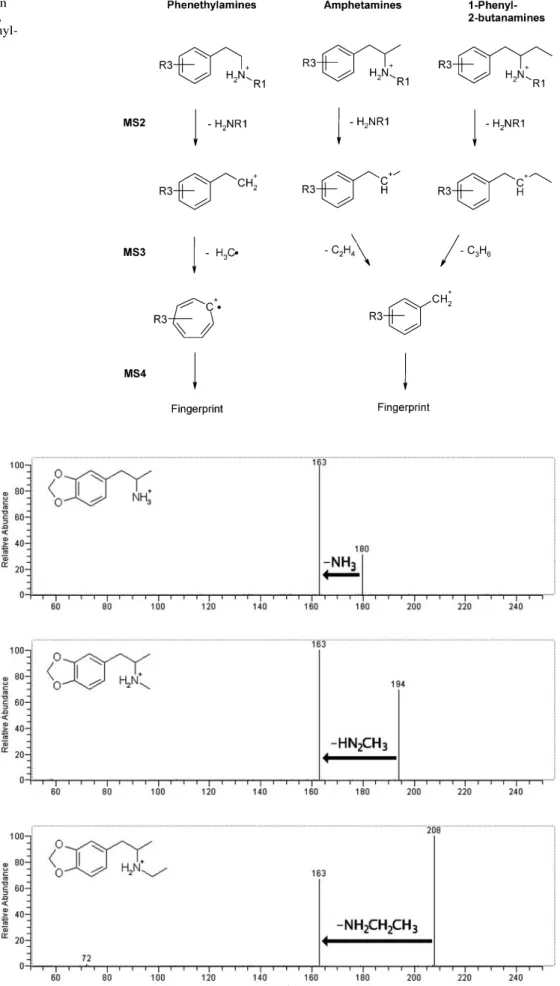
1-phenyl-2-bu-Fig. 3 Principal MSnfragmentation
behavior of phenethylamines (left), amphetamines (middle) and 1-phenyl-2-butanamines (right)
Fig. 4 MS2spectra of
3,4-methylenedioxyampheta-mine (MDA, top; loss of NH3),
N-methyl-3,4-methylene-dioxyamphetamine (MDMA, middle; loss of CH3NH2), and
N-ethyl-3,4-methylenedioxy-amphetamine (MDE, bottom; loss of CH3CH2NH2). Scan
tanamines. Amounts of 10 ng were sufficient for record-ing MS–MS or MSn spectra when injected with the
sy-ringe pump. The information from the recorded MS1 to MS4spectra can be summarized as follows (see also Fig. 3).
MS spectra
ESI and APCI spectra recorded in the positive-ion mode contained a pronounced [M+H]+peak for all compounds, enabling determination of the molecular mass. The maxi-mum temperature of the heated capillary should not ex-ceed 180–200 °C, however, otherwise, thermal degrada-tion occurs, resulting in reduced abundance of [M+H]+. Typical isotope signals also enabled identification of het-ero atoms such as S, Cl, or Br.
MS2spectra
In this step the amino function is lost together with its substituents, forming [M+H–NH2R]+, which is always the base peak. Other fragments have a relative intensity of <5%. Therefore, N-substitution of an unknown can be identified directly from the MS2 spectrum. Loss of 17 u (NH
3) is
typical for primary amines, loss of 31 u (H2N–CH3) for N-methylated compounds, and loss of 45 u for N-ethyl-ated or N,N-dimethylN-ethyl-ated compounds. MS2 spectra of 3,4-methylendioxyamphetamine (MDA), N-methyl-3,4-methylenedioxyamphetamine (MDMA), and N-ethyl-3,4-methylenedioxyamphetamine (MDE) are given in Fig. 4 as examples. As expected, the MS3spectra of the formed m/z 163 ion are identical for MDA, MDMA, and MDE.
MS3spectra
MS3spectra enable determination of the substitution at the
αcarbon atom, because of fragmentation of the saturated side-chain (see Fig. 5 for typical spectra). Consequently, 1-phenyl-2-butanamines lose 42 u (C3H6), amphetamines 28 u (C2H4), and phenethylamines 15 u (CH3). For 1-phenyl-2-butane amines the [M+H–NH2R–C3H6]+ion formed is always the base peak, and other ions have relative abun-dances of <30%. For amphetamines [M+H–NH2R–C2H4]+ is most abundant, but [M+H–NH2R–15]+ also often has high intensity.
[M+H–NH2R–15]+is usually the base peak for phen-ethylamines. It is often accompanied by a pronounced [M+H–NH2R–30]+ion. Peak intensity of [M+H–NH2R–28]+ Fig. 5 Typical MS3spectra of
phenethylamines (top, loss of 15 u); amphetamines (middle, loss of 28 u), and (2-amino-butyl)benzenes (bottom, loss of 42 u). Here the MS3spectra
of the 3,4,5-trimethoxy-compounds are shown. Scan sequence: [M+H]+→[M+H–NH
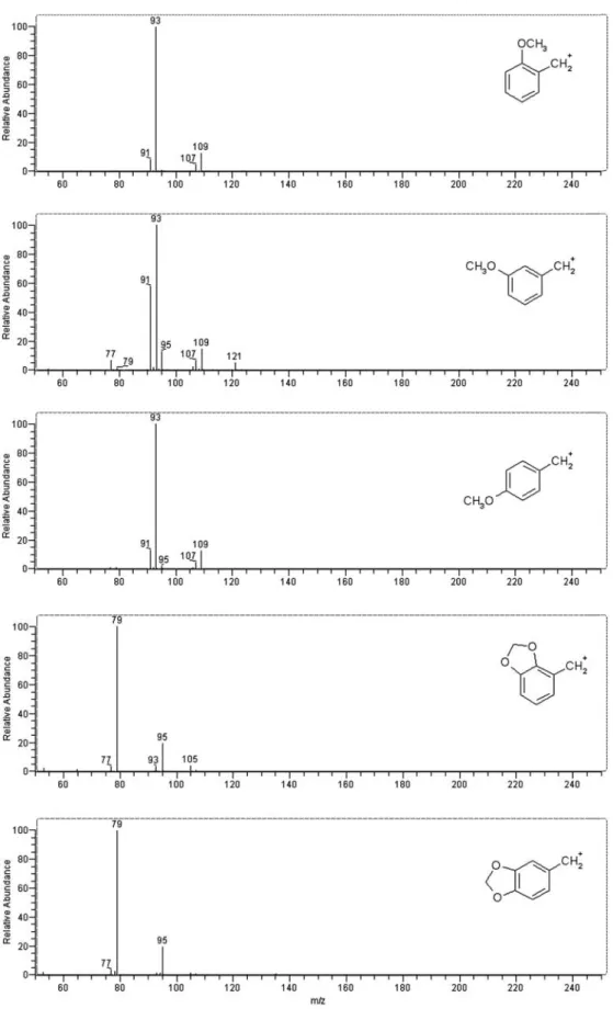
Fig. 6 MS4spectra of
monomethoxyamphetamines (top) and 1-(methylene-dioxyphenyl)-2-butanamines (bottom). Scan sequence: [M+H]+→[M+H–NH
3]+→
[M+H–NH3–C2H4]+→scan
(for amphetamines) and [M+H]+→[M+H–NH
3]+→
[M+H–NH3–C3H6]+→scan
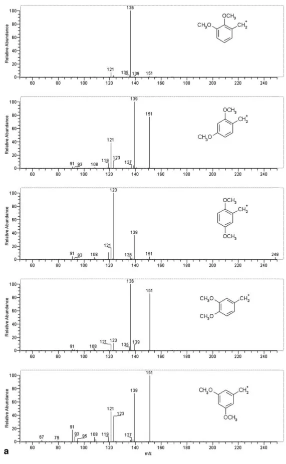
Fig. 7 MS4spectra of
di-methoxyamphetamines (a) and trimethoxyamphetamines (b). Scan sequence:
[M+H]+→[M+H–NH 3]+→
[M+H–NH3–C2H4]+→scan.
The strange fragments at m/z 139 (for dimethoxyam-phetamines) or m/z 169 (for trimethoxyamphetamines) probably result from addition of water to m/z 121 or m/z 151, respectively
and [M+H–NH2R–42]+ is <10%, which therefore enables differentiation of phenethylamine derivatives from 1-phenyl-2-butanamines and amphetamines. Deuterium-labeling showed that the [M+H–NH2R–15]+ion is formed both by cleavage of the ethyl C–C bond and loss of –CH3from a methoxy group.
MS4spectra
MS4fragmentation formed the C
7H6+radical ion (for phen-ethylamines) or the benzylium ion (amphetamines and 1-phenyl-2-butanamines; Fig. 3). The resulting fingerprint spectra contain detailed information about aromatic ring
substitution. This is especially useful for di- and trisubsti-tuted compounds and enables identification of different isomers:
– Methoxy-substitution at the 2- and 4-positions gives identical MS4spectra (Fig. 6) but the 3-position is dis-tinguishable.
– 2,3- or 3,4-methylendioxy-substitution results in identi-cal MS4spectra (Fig. 6) but differentiation by use of the MS3spectra is possible.
– The MS4 spectra of 2,3-, 2,4-, 2,5-, 3,4-, and 3,5-di-methoxy isomers are all different (Fig. 7).
– The same is valid for 2,3,4-, 2,4,5-, 2,4,6-, and 3,4,5-tri-methoxy-compounds (Fig. 7).
– Other studied substitution patterns such as 4-bromo-, 4-iodo-4-bromo-, 2,5-dimethoxy-4-methyl-, and 4-propylthio-2,5-dimethoxy- also gener-ated different MS4spectra.
Some phenethylamines are indistinguishable in the MS4, MS5, and MS6 spectra. Examples are the pairs 2,3- and 3,4-dimethoxyphenethylamine, 2,4- and 3,5-dimethoxy-phenethylamine, and 2,3,4- and 3,4,5-trimethoxyphen-ethylamine.
Moreover, some MS4spectra revealed the formation of ion molecule adducts with H2O. It seems that their forma-tion is indicative of two methoxy groups in the meta posi-tion. The mechanism of formation was not studied further, however [2, 30].
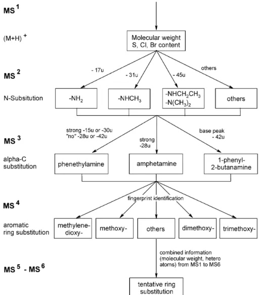
Structure elucidation scheme
A generally applicable structure elucidation scheme was developed on the basis of the recorded MS (see Fig. 8). It enables step-by-step elucidation of the structure of
un-known phenylalkylamines. The molecular mass and the presence of hetero atoms such as Cl, Br, and S can be easily determined by MS. The fragments formed during MS2give information about N-substitution, whereas theα -C-substi-tution can be derived from MS3spectra. Information about aromatic ring substitution is part of the “fingerprint patterns” in the MS4 to MS6 spectra and requires comparison with reference spectra of the corresponding positions. If no ref-erence spectrum matches the unknown the structure can of-ten be derived from the combined MS1to MS6information.
Reproducibility
MS–MS spectra and those of higher order (MS3–MS6) were highly reproducible. Of course, the relative intensities of the precursor and product ions was influenced by chang-ing the experimental conditions (collision energy, colli-sion gas pressure, software). However, the relative inten-sity of product ions vary within a few percent only even over several years and between different LCQ instruments and different software.
Fig. 8 Scheme for elucidation of the
structure of unknown phenylalkyl-amines by means of multiple mass spectrometry (MSn)
A change of ion-trap pressure (helium, cooling gas) is much less critical than for triple quadrupole instruments (argon collision gas), because the ion-trap collision en-ergy is normally applied to the mass of the selected pre-cursor ion only.
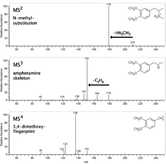
Application to unknowns
Elucidation of the structure of two impurities was per-formed by means of the selective fragmentation processes described above to demonstrate the applicability of the approach. The mass spectra obtained from one example are given in Fig. 9. A by-product present in an illicit MDMA sample furnished an [M+H]+ion at m/z 210. Loss of 31 u (m/z 179), typical of an N-methylated compound, was observed in the MS2spectrum. The base peak in the MS3 spectrum was m/z 151 (loss of 28 u) and identified the compound as an amphetamine derivative. The MS4 fingerprint was identical to the MS4fingerprint of 3,4-di-methoxyamphetamine, revealing the overall structure was N-methyl-3,4-dimethoxyamphetamine.
In a sample of 4-bromo-2,5-dimethoxyphenethylamine (2C-B, 16a in Fig. 2) the following impurities were identi-fied in a similar manner:
N-hydroxy-4-bromo-2,5-di-methoxyphenethylamine (HPLC signal intensity approx. 0.05%), 2,5-dimethoxyphenethylamine (intensity approx. 0.5%), two different dibromo-dimethoxyphenethylamines (intensity 0.2% each) and a second bromodimethoxy-phenethylamine (intensity 0.3%). A compound with [M+H]+ at m/z 246 was also found; this could be 4-bromo-2-hy-droxy-5-methoxyphenethylamine or 4-bromo-5-hydroxy-2-methoxyphenethylamine.
Conclusion
The highly structure-selective fragmentations described enable nearly unequivocal elucidation of the structures of unknown designer drugs by multiple mass spectrometry. Amounts of 10 ng are usually sufficient for recording of interpretable spectra down to MS6. The method is, there-fore, at least two orders of magnitude more sensitive than NMR, and only very simple sample pretreatment is neces-sary. Analysis and data interpretation leading to the struc-ture of a compound is possible within 15 min after some training. This study also indicates that structure elucida-tion and quantificaelucida-tion of primary drug metabolites (e.g. glucuronide and sulfate conjugates) and very polar neuro-transmitters might be possible with low nanogram amounts. Fig. 9 MS2to MS4spectra
of an impurity, identified as N-methyl-3,4-dimethoxyam-phetamine, in an illicit MDMA sample. Because the spectra of this compound were recorded with prototype LCQ software several years ago, the intensi-ties of the precursor ion peaks are different from those in the reference spectra
Acknowledgements This work was conducted under license no.
SHI-Betm.-AB 8/5–240 of the Swiss Federal Institute of Health, for which we are grateful. Financial support was also obtained from the Swiss National Science foundation under project no. 2000.064817.01. The donation of reference samples is gratefully acknowledged (Dr Franz Dussy, Institut für Rechtsmedizin beider Basel, Basel, Switzerland; Dr Peter Rösner, Landeskriminalamt Kiel, Germany; Smart Stuff GmbH, Zürich, Switzerland; Reseachem GmbH, Burgdorf, Switzerland). We would, moreover, like to thank numerous persons for valuable discussions.
References
1. Kölliker S, Oehme M, Dye C (1998) Anal Chem 70:1979– 1985
2. Kölliker S, Oehme M, Merz L (2001) Rapid Commun Mass Spectrom 15:2117–2126
3. Berger U, Oehme M, Kuhn F (1999) J Agric Food Chem 47: 4240–4245
4. Chen Y, Kölliker S, Oehme M, Katz A (1999) J Nat Prod 62: 701–704
5. Berger U, Oehme M, J AOAC Int (2000) 83:1367–1376 6. Shulgin AT, Shulgin A (1991) PIHKAL. A chemical love story.
Transform Press, Berkeley, CA
7. Adamson S (1985) Through the gateway of the heart. Four Trees, San Francisco, CA
8. Clark CR, DeRuiter J, Valaer A (1995) J Chromatogr Sci 33: 328–337
9. Widmer S (2000) Ins Herz der Dinge lauschen. Nachtschatten, Solothurn
10. Schmitt WJ, Mayerhofer A, Meyer A, Kovar KA (2002) Neu-rosci Lett 330:251–254
11. Parrott AC (2001) Hum Psychopharmacol Clin Exp 16:557– 577
12. Helmlin H, Bracher K, Bourquin D, Vonlanthen D, Brenneisen R, Styk J (1996) J Anal Toxicol 20:432–439
13. Vollenweider F, Gamma A, Liechti M, Huber T (1998) Neuro-psychopharmacology 19:241–251
14. Dal Cason TA (2001) Forensic Sci Int 119:168–194 15. Elliott S (2000) J Anal Toxicol 24:85–92
16. Decaestecker H, De Letter E, Clauwaert K, Bouche MP, Lambert W, Van Bocxlaer J, Piette M, van den Eeckhout E, van Peteghem C, De Leenheer A (2001) J Anal Toxicol 25: 705–710
17. Trachsel D (2002) Helv Chim Acta 85:3019–3026
18. Gentili S, Torresi A, Marsili R, Chiarotti M, Macchia T (2002) J Chromatogr B 780:183–192
19. Bogusz MJ, Krüger KD, Maier RD (2000) J Anal Toxicol 24: 77–84
20. Noggle FT, Clark CR, McMillian CL, DeRuiter J (1989) J Chro-matogr Sci 27:607–611
21. Borth S, Hänsel W, Rösner P, Junge T (2000) Forensic Sci Int 114:139–153
22. Borth S, Hänsel W, Rösner P, Junge T (2000) J Mass Spectrom 35:705–710
23. Aalberg L, DeRuiter J, Noggle FT, Sippola E, Clark CR (2000) J Chromatogr Sci 38:329–337
24. O’Connell D, Heffron JJA (2000) Analyst 125:119–121 25. Blachut D, Wojtasiewicz K, Czarnocki Z (2002) Forensic Sci
Int 127:45
26. De Boer D, Egberts T, Maes RAA (1999) Pharm World Sci 21:47–48
27. Giroud C, Augsburger M, Rivier L, Mangin P, Sadeghipour F, Varesio E, Veuthey JL, Kamalaprija P (1998) J Anal Toxicol 22:345–354
28. Trachsel D, Richard N (2000) Psychedelische Chemie. Nacht-schatten, Solothurn
29. Parker MA, Marona-Lewicka D, Kurrasch D, Shulgin AT, Nichols DE (1998) J Med Chem 41:1001–1005