ARTICLE
Prevalence of isolates with reduced glycopeptide susceptibility
in orthopedic device-related infections due to methicillin-resistant
Staphylococcus aureus
P. Vaudaux&T. Ferry&I. Uçkay&P. François&
J. Schrenzel&S. Harbarth&A. Renzoni
Received: 28 June 2012 / Accepted: 12 July 2012 / Published online: 26 July 2012 # Springer-Verlag 2012
Abstract We evaluated, by an improved susceptibility testing method, the prevalence and significance of low-level glycopep-tide resistance in methicillin-resistant Staphylococcus aureus (MRSA) isolates, which belonged to a previously described, retrospective cohort of patients treated for orthopedic device-related infections (ODRI) at the Geneva University Hospital between 2000 and 2008. Fifty-seven individual or multiple isolates were retrieved from 41 ODRI patients for glycopeptide susceptibility and clonality studies, including 20 patients with prosthetic joint (PJ) and 21 with osteosynthesis (OS) MRSA infections. Low-level glycopeptide resistance was detected by elevated teicoplanin or/and vancomycin minimum inhibitory concentrations (MICs≥4 mg/L), as determined by a previously validated combination of macrodilution and agar dilution
assays of improved sensitivity. MRSA isolates with elevated teicoplanin MICs were detected in 20/41 (49 %) ODRI patients at the onset or during the course of glycopeptide therapy, namely, in 10 of 20 patients with PJ and 10 of 21 patients with OS infections. Only one isolate developed a concomitant in-crease in vancomycin MIC during therapy. 13/20 (65 %) glycopeptide-intermediate S. aureus (GISA)-infected patients, including 7/10 (70 %) with PJ and 6/10 (60 %) with OS, experienced treatment failure. In contrast, therapy failed in only 5/21 (24 %) ODRI patients with non-GISA isolates (p00.012), including 2/10 (20 %) with PJ and 3/11 (27 %) with OS infections. The emergence of low-level teicoplanin resistance could not be explained by teicoplanin administration, since only four patients received teicoplanin. The evaluation of
This study was presented, in part, as a poster (P1335) at the 22nd Congress of the European Society of Clinical Microbiology and In-fectious Diseases, London, UK, 31 March to 3 April 2012. P. Vaudaux (*)
:
I. Uçkay:
J. Schrenzel:
S. Harbarth:
A. Renzoni Service of Infectious Diseases, Geneva University Hospital and Medical School,4 Rue Gabrielle Perret-Gentil, 1211 Geneva 14, Switzerland e-mail: [email protected] P. Vaudaux
e-mail: [email protected] T. Ferry
Infectious Disease Ward, Hospices Civils de Lyon, Lyon, France
T. Ferry
Université Claude Bernard Lyon 1, Lyon, France
T. Ferry INSERMU851, Lyon, France
I. Uçkay
Service of Orthopaedic Surgery, Geneva University Hospital and Medical School,
Geneva, Switzerland P. François
:
J. SchrenzelGenomic Research Laboratory, Geneva University Hospital and Medical School,
Geneva, Switzerland J. Schrenzel
Central Laboratory of Bacteriology, Geneva University Hospital and Medical School,
Geneva, Switzerland S. Harbarth
Infection Control Program, Geneva University Hospital and Medical School,
low-level teicoplanin resistance may improve the detection of GISA isolates. Further studies are warranted to evaluate the impact of low-level teicoplanin resistance on the outcome of glycopeptide therapy.
Introduction
Factors reported to increase the risk of glycopeptide treatment failure against invasive methicillin-resistant Staphylococcus aureus (MRSA) infections are: (1) the difficulty to reach adequate tissue levels at the true sites of MRSA infections, (2) the moderate bactericidal activity of glycopeptides, and (3) the emergence of glycopeptide resistance [1–8].
Phenotypic detection of vancomycin-intermediate S. aure-us (VISA) isolates, which are defined by vancomycin mini-mum inhibitory concentration (MIC) breakpoints of≥4 mg/L and <16 mg/L, and the absence of any vancomycin or teico-planin resistance determinants (vanA, vanB, or vanC) found in vancomycin-resistant Enterococcus faecalis or high-level vancomycin-resistant (vancomycin MIC: 16 mg/L) S. aureus isolates (VRSA) [5–12] is frequently problematic [1,2,4–8]. Since VISA isolates are generally cross-resistant to teicopla-nin [4,13], they are also designated glycopeptide-intermediate S. aureus (GISA). In contrast to vancomycin, teicoplanin susceptibility breakpoints in S. aureus vary from 2 mg/L by the European Committee on Antimicrobial Susceptibility Testing (EUCAST) [14] to 8 mg/L by the Clinical and Labo-ratory Standards Institute (CLSI) [15].
Detection of the GISA phenotype is particularly difficult for isolates displaying heterogeneous resistance to either or both glycopeptide(s) (hGISA), in which only a subset of the micro-bial population can express glycopeptide resistance [7, 9,
14–18], and which are likely precursors of GISA isolates under the selective pressure of glycopeptides [6,19]. The unsuccess-ful detection of hGISA isolates by standard microbiological methods has triggered the development of alternative assays, such as the modified population analysis profile (PAP) area under the curve (AUC), the Etest macromethod, and the gly-copeptide resistance determination (GRD) test [6,7,20–22].
The predictive value of vancomycin and teicoplanin sus-ceptibility breakpoints on the outcome of glycopeptide ther-apy is still debated, despite their recent adjustment by the CLSI and EUCAST [8,16]. While higher rates of vanco-mycin treatment failures were frequently reported in bacter-emic patients infected with MRSA isolates for which vancomycin MICs were 2 mg/L, compared to those with lower vancomycin MICs (<2 mg/L) [1,2,8,16,23–26], as confirmed by a recent meta-analysis [27], these data might be explained, at least in part, by the low sensitivity of some MIC testing methods, such as the broth microdilution and agar dilution assays, which lead to a significant underdetec-tion of some GISA or hGISA isolates [28–30].
While numerous studies evaluated the impact of glyco-peptide MICs and low-level resistance on the outcome of glycopeptide therapy in MRSA bacteremic patients [5–8,
24–26, 31], a single report linked the presence of GISA/ hGISA isolates with a negative outcome of vancomycin therapy for MRSA-infected, orthopedic patients [32].
We recently reported a high prevalence of GISA isolates in patients with persistent or recurrent MRSA bacteremia [33], in which low-level glycopeptide resistance was detected by elevated teicoplanin or/and vancomycin MICs (≥4 mg/L), as determined by a previously validated combi-nation of macrodilution and agar dilution assays, allowing more sensitive detection of slow-growing, glycopeptide-“re-sistant” subpopulations [30]. Using this improved suscepti-bility testing method, we now present data on the prevalence and potential significance of low-level glycopeptide resis-tance in MRSA isolates from patients treated for orthopedic device-related infections (ODRI) at the Geneva University Hospital [34].
Materials and methods
Clinical and microbiological data collection
Fifty-seven MRSA isolates from 41 patients with MRSA ODRI, who were treated at the Geneva University Hospital between 2000 and 2008, were routinely stored in skimmed milk/glycerol at−80 °C [34]. These ODRI patients belonged to a retrospective cohort study, and their major clinical char-acteristics and risk factors for treatment failure have been previously described in detail [34]. MRSA isolates were es-sentially intra-operative specimens and aspirated synovial flu-id [34]. In five patients, blood isolates that were clonally related to intra-operative isolates were also analyzed.
Twenty of the 41 ODRI patients had prosthetic joint (PJ) and 21 osteosynthesis (OS) MRSA infection [34]. MRSA infections were considered persistent if the patient’s clinical status required further surgery 5 days after the initiation of antimicrobial therapy, with isolation of the same MRSA isolate by intra-operative specimen [34]. Recurrence was defined as resurgence of the infection with a clonally related MRSA isolate after the end of antimicrobial therapy [34]. Determination of glycopeptide MICs
Vancomycin and teicoplanin MICs were determined by a slightly modified, previously described tube macrodilution assay, using cation-adjusted Mueller–Hinton broth and stan-dardized inocula of 106colony-forming units (CFU)/mL [30]. MIC endpoints were read after 48 h incubation at 37 °C, to improve the detection of slow-growing, glycopeptide-“resis-tant” subpopulations [30]. This procedure was combined with
a modified agar MIC testing method, which was used for confirmatory testing of glycopeptide MICs recorded by mac-rodilution, as previously described [30,33]. In this modified agar MIC testing method, residual viable counts of each antibiotic-containing agar plate inoculated with ca. 106CFU were scored after 48 h incubation at 37 °C in a semi-quantitative manner as confluent, semi-confluent, or ≤103
CFU, as previously described [30]. Glycopeptide MIC was defined as the lowest antibiotic concentration leading to a ≥99.9 % reduction in viable counts (≤103CFU) on brain–heart infusion agar from the uniformly applied inoculum of 106CFU, as previously described [30].
To be scored as GISA, all MRSA isolates had to display elevated teicoplanin or/and vancomycin MICs (≥4 mg/L) by both modified macrodilution and agar testing assays [30,33]. Molecular typing
The clonality of consecutive MRSA isolates from patients with persistent or recurrent ODRI was assessed by a variable-number tandem repeat (VNTR) genotyping method [35], as previously described [34]. Strain pairs with >85 % similarity in the dendrogram were considered to be clonally related (Bioanalyzer Experiments Clustering Software) [35]. Statistical analyses
Microbiological, demographic, and clinical characteristics of GISA- and non-GISA-infected ODRI patients were com-pared by the Fisher’s exact test for categorical variables or the Mann–Whitney test for continuous variables ( http://vas-sarstats.net/). Relationships were considered to be signifi-cant when the two-sided p-value was≤0.05.
Results
Prevalence of GISA in ODRI patients
MRSA isolates showing elevated teicoplanin MICs (≥4 mg/L) were detected in 20 (49 %) of the 41 ODRI patients, including 10 of 20 (50 %) PJ-infected and 10 of 21 (48 %) OS-infected patients. In contrast, none of the 41 ODRI pretherapy isolates displayed elevated vancomycin MICs (≥4 mg/L).
For two patients, teicoplanin MICs were 2 mg/L in pre-therapy isolates and increased to 4 mg/L in subsequent isolates. For the remaining 18 patients, teicoplanin MICs were already elevated in pretherapy isolates and remained constant in subsequent isolates, except for two patients in whom teicoplanin MICs increased from 4 to 8 mg/L in consecutive isolates. In a single patient with low-level teicoplanin-resistant isolates, vancomycin MICs increased from 2 to 4 mg/L from baseline to subsequent isolates.
Clonality of GISA isolates in ODRI patients
All MRSA isolates with elevated teicoplanin MICs belonged to the hospital-acquired South German MRSA clone ST228 (SCCmec type 1 and agr type 2), which became predominant in our institution after 1998 [36]. This MRSA clone was previously reported to have infected 35 of the 41 ODRI patients (85 %) by spa and VNTR typing methods [34]. All available sequential isolates from patients with persistent or recurrent ODRI were clonally related (data not shown). In contrast, none of the six residual isolates belonging to other clonotypes (ST1, ST5, ST8, ST80, ST239), including other SSCmec (III, IV, V) and agr (1, 3) types [34], exhibited low-level glycopeptide resistance. The prevalence of isolates displaying low-level glycopeptide resistance was significantly higher in the ST228 clonotype compared to other clonotypes (p00.021; Table1).
Prevalence of GISA isolates and treatment outcomes in different subgroups of ODRI patients
Thirteen of 20 GISA-infected ODRI patients (65 %) expe-rienced MRSA-linked treatment failure compared to 5/21 (24 %) patients infected with non-GISA isolates (p00.012) (Table 1). Treatment failure was significantly (p00.032) higher in GISA-infected patients with persistent ODRI epi-sodes compared to GISA isolates. There were also non-significant trends toward higher rates of recurrent ODRI episodes or multiple treatment failures in GISA-infected compared to non-GISA-infected patients.
Higher failure rates were observed in both subgroups of GISA-associated ODRI patients, namely, in 7/10 (70 %) PJ and 6/10 (60 %) OS patients, compared to 2/10 (20 %) PJ (p00.070) and 3/11 (27 %) OS patients with non-GISA infections (p00.198; Table1). Higher treatment failure rates were recorded in GISA- compared to non-GISA-infected ODRI patients with implant retention, as well as in patients with implant removal (Table1), but these differences did not reach statistical significance due to the small sample sizes.
In brief, there was a consistent trend toward higher treat-ment failure rates in all subgroups of GISA- compared to non-GISA-infected ODRI patients.
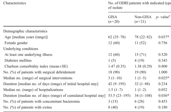
Comparison of GISA- and non-GISA-infected patients The major demographic and clinical characteristics of ODRI-treated patients are presented in Table2. Noteworthy, GISA-infected patients were significantly younger (p00.037) than non-GISA-infected patients. While underlying conditions and rates of surgical debridement were similar in GISA- and non-GISA-infected patients, there was a trend for longer duration of hospital stay, which reached significance for the cumulated duration of hospital stay (p00.036) in GISA- versus
non-GISA-infected patients. While the rates of bacteremic episodes concomitant to ODRI were not significantly different in GISA-and non-GISA-infected patients, there was a non-significant trend (p00.180) for a higher overall mortality in GISA-infected (40 %; n08) compared to non-GISA-GISA-infected (19 %; n04) patients. Most of those deaths were unrelated to MRSA-linked ODRI or bacteremia. Only two GISA-infected patients died from MRSA-linked ODRI or bacteremia, but death occurred only 77 and 185 days, respectively, after ODRI onset. Treatment modalities for GISA- and non-GISA-infected patients
The potential impact of therapeutic regimens on the treat-ment outcomes of GISA- compared to non-GISA-infected ODRI patients was evaluated. Besides one patient who was treated with trimethoprim–sulfamethoxazole, all other patients initially received intravenous regimens of vanco-mycin, either in monotherapy (n017) or combined with other agents (n023). Altogether, the administration of addi-tional antibiotic regimens to ODRI patients after initial intravenous therapy led to a total of eight different antimi-crobial regimens (Table 3). This great diversity and the small sample size in each group precluded any detailed comparison of the impact of each antimicrobial regimen on GISA- compared to non-GISA-infected ODRI patients.
There was a trend for higher failure rates recorded in GISA-infected patients (78 %) initially treated with vanco-mycin monotherapy (n09), with or without additional anti-microbial regimens, compared to the rate (38 %) in non-GISA-infected patients (n08), which did not reach signifi-cance (p00.153). In contrast, there were no significant dif-ferences in the daily doses and durations of antimicrobial regimens (data not shown) administered to GISA- versus non-GISA-infected patients.
Several regimens of combined therapy involving vancomy-cin associated with either rifampin, trimethoprim–sulfamethox-azole, or fusidic acid were administered to 23 ODRI patients (Table 3). The impact of each therapeutic regimen on the outcome of GISA- versus non-GISA-infected ODRI patients could not be analyzed in detail due to low sample sizes.
Interestingly, teicoplanin administration was shown to play no significant role in the presence of GISA isolates in ODRI patients. While only four ODRI patients received teicoplanin as subsequent antimicrobial therapy (Table 3), none of these patients were infected with GISA isolates.
Discussion
Despite a continuously increasing number of clinical and microbiological reports, as summarized in [6, 7, 27], the Table 1 Prevalence of
glycopeptide-intermediate Staphylococcus aureus (GISA) isolates and treatment outcomes in different subgroups of orthopedic device-related infection (ODRI) patients
a
Significance of differences in the characteristics of GISA-versus non-GISA-infected ODRI patients
*p<0.05
Characteristics No. of ODRI patients with indicated
type of isolate GISA (n020) Non-GISA (n021) p- valuea Microbiological characteristics
No. (%) of isolates belonging to the ST228 clonotype 20 (100) 15 (71) 0.021* No. (%) of ODRI patients with MRSA-linked treatment failures 13 (65) 5 (24) 0.012* No. (%) of patients with persistent ODRI episodes 8 (40) 2 (10) 0.032* No. (%) of patients with recurrent ODRI episodes 5 (25) 3 (14) 0.454 No. (%) of patients with multiple treatment failures 7 (35) 3 (14) 0.159
No. (%) of PJ patients 10 (50) 10 (50) 1.000
No. (%) of PJ patients with MRSA-linked treatment failures 7 (70) 2 (20) 0.070 No. (%) of PJ patients with removed implants 4 (57) 3 (43) 1.000
No. (%) of treatment failures 3 (75) 0 (<25) 0.142
No. (%) of PJ patients with implant retention 6 (46) 7 (54) 1.000
No. (%) of treatment failures 4 (75) 2 (29) 0.286
No. (%) of OS patients 10 (48) 11 (52) 1.000
No. (%) of OS patients with MRSA-linked treatment failures 6 (60) 3 (27) 0.198 No. (%) of OS patients without debridement 2 (10) 2 (10) 1.000 No. (%) of OS patients with removed implants 3 (43) 4 (57) 1.000
No. (%) of treatment failures 1 (25) 0 (<25) 0.429
No. (%) of OS patients with implant retention 5 (50) 5 (50) 1.000
impact of low-level glycopeptide resistance on the outcome of GISA infections is still debated. This unclear situation mostly results from technical issues in characterizing the low-level glycopeptide resistance phenotype in MRSA isolates, which is frequently heterogeneous and, thus, escapes detection by standard MIC assays. Furthermore, there is no molecular assay for detecting low-level
glycopeptide resistance, whose molecular basis is likely multifactorial and may even display some strain-specific variability [37]. While a recent meta-analysis supports the concept that slightly elevated vancomycin MICs, scored at the higher end of the susceptibility range by micro-dilution or Etest, were associated with worse outcomes of glycopeptide therapy in MRSA bloodstream infections, Table 2 Demographic and
clinical characteristics of patients with GISA and non-GISA ODRI episodes
aSignificance of differences in
the characteristics of GISA-versus non-GISA-infected ODRI patients
*p<0.05
Characteristics No. of ODRI patients with indicated type
of isolate GISA (n020) Non-GISA (n021) p- valuea Demographic characteristics
Age [median years (range)] 62 (35–78) 78 (22–82) 0.037*
Female gender 12 (60) 11 (52) 0.756
Underlying conditions
At least one underlying illness 12 (60) 15 (71) 0.520
Diabetes mellitus 1 (5) 4 (19) 0.343
Charlson comorbidity index (mean±SE) 1.47 (0.35) 1.38 (0.29) 0.800 No. (%) of patients with surgical debridement 18 (90) 19 (90) 1.000 Median no. (range) of surgical interventions 3 (1–10) 1 (1–5) 0.025* Duration [median no. of days (range) of initial hospital stay] 42 (9–195) 32 (1–88) 0.234 Median no. (range) of hospitalizations 1.5 (1–7) 1 (1–2) 0.052 Duration [median no. of days (range) of cumulated hospital stay] 53.5 (23–195) 34 (1–108) 0.036* No. (%) of patients with concomitant bacteremia 3 (15) 6 (28) 0.453
No. (%) of patients with exitus 8 (40) 4 (19) 0.180
Table 3 Treatment regimens of patients with GISA and non-GISA ODRI episodes
NA not applicable; LZD line-zolid; RIF rifampin; SXT tri-methoprim–sulfamethoxazole; VAN vancomycin; TEC teicoplanin
a
Significance of differences in the treatment regimens and out-comes of GISA- and non-GISA-infected ODRI patients
b
Two GISA and two non-GISA-infected patients received SXT and three non-GISA-infected patients received TEC
cTwo GISA and one
non-GISA-infected patients received SXT, two GISA and eight non-GISA-infected patients RIF + fusidic acid, one non-GISA patient RIF + LZD, and one non-GISA pa-tient received TEC
Initial antibiotic regimen No. of ODRI patients with indicated type of isolate
GISA
(n020) Non-GISA(n021) p- value
a
No. (%) of patients with VAN monotherapy 9 (45) 8 (38) 1.000
Failure rate (%) 7 (78) 3 (38) 0.153
No. (%) of patients with VAN alone 7 (35) 3 (14) 0.159
Failure rate (%) 5 (71) 1 (33) 0.500
No. (%) of patients with VAN + subsequent antibiotic therapyb 2 (10) 5 (24) 0.410
Failure rate (%) 2 (100) 2 (40) 0.429
No. (%) of patients with SXT monotherapy 1 (5) 0 (<5) NA
Failure rate (%) 1 (100) 0 (NA) NA
No. (%) of patients with VAN in combination 10 (50) 13 (62) 0.538
Failure rate (%) 5 (50) 2 (15) 0.169
No. (%) of patients with combined VAN + SXT therapy 1 (5) 1 (5) NA
Failure rate (%) 1 (100) 1 (100) NA
No. (%) of patients with combined VAN + RIF therapy 9 (45) 12 (57) 0.538
Failure rate (%) 4 (44) 1 (8) 0.119
No. (%) of patients with VAN + RIF + subsequent oral antibiotic therapyc
4 (20) 10 (48) 0.100
the contribution of hGISA could not be evaluated in this context [27].
To facilitate the detection of hGISA or GISA by standard MIC criteria, we developed a modified macrodilution MIC assay by using higher inocula than broth microdilution MIC and extending the incubation period to 48 h at 37 °C, which is mandatory for detecting the slowly growing resistant subpopulations [30]. The increased sensitivities of the mod-ified macrodilution assay combined with a modmod-ified agar MIC method markedly increased the detection rates of iso-lates with slightly elevated teicoplanin and vancomycin MICs (≥4 mg/L), by ca. 10-fold and 4-fold, respectively, compared to broth microdilution [30]. Most recently, the modified macrodilution MIC assay allowed detecting a higher prevalence of MRSA isolates with slightly elevated vancomycin or teicoplanin MICs in patients with persistent or recurrent episodes of MRSA bacteremia compared to those with single episodes [33]. Collectively, these data strongly suggest that a significant proportion of MRSA bloodstream isolates scored with slightly elevated vancomy-cin MICs (2 mg/L) by microdilution in previous studies [24,
27,38] could have been undetected VISA or hVISA. Since our previous contributions indicated that >95 % of isolates with elevated vancomycin MICs displayed cross-resistance to teicoplanin, elevated teicoplanin MIC was con-sidered to be a reliable marker of the GISA phenotype, which could facilitate the screening of isolates with low-level vancomycin resistance [13,30]. Indeed, in our recent study of patients with persistent or recurrent episodes of MRSA bacteremia, 63 % of isolates displaying low-level resistance to teicoplanin were concomitantly resistant to vancomycin [33].
In contrast to patients with MRSA bloodstream infec-tions, vancomycin cross-resistance was a rare event in teicoplanin-resistant isolates from ODRI patients. Indeed, only one patient among all of the teicoplanin-resistant iso-lates detected in 20 patients displayed concomitant resis-tance to vancomycin. Noteworthy, teicoplanin-resistant isolates were already present in 90 % of GISA-infected ODRI patients at the onset of antimicrobial therapy, while the emergence of low-level teicoplanin resistance during therapy occurred in only 10 % of the patients. Remarkably, the administration of teicoplanin therapy was not associated with the emergence of low-level teicoplanin-resistant iso-lates, since none of the four teicoplanin-treated patients were infected with resistant isolates.
A most intriguing finding of this study was that ODRI patients infected with MRSA isolates displaying low-level teicoplanin resistance but still showing in vitro susceptibility to vancomycin seemed to respond poorly to antimicrobial regimens involving vancomycin compared to non-GISA-infected patients These observations lead us to speculate that in vivo conditions prevailing in ODRI patients might
significantly alter the metabolic parameters of MRSA in such a way that teicoplanin resistance mechanisms may compromise MRSA susceptibility to vancomycin therapy. Previous studies performed in an animal model of chronic, implant-associated MRSA infection indicated a drastic loss of susceptibility to antibiotic killing in bacteria directly removed from infected foci [39–42]. Other important parameters that may contribute to bacterial survival in MRSA chronic infections are intracellular persistence [43], which may be further promoted by teicoplanin resistance determinants [44], and bacterial adherence, leading to biofilm formation and colonization of artificial surfaces [41,45,46].
Recent molecular studies indicated that the emergence of vancomycin and teicoplanin endogenous resistance is likely a stepwise process involving several mutations in key reg-ulatory genes [6, 13,37]. While the molecular differences between isolates displaying combined low-level resistance to both teicoplanin and vancomycin versus those resistant to teicoplanin alone have not been elucidated, molecular stud-ies performed in our laboratory suggest that resistance to teicoplanin alone might represent an early step of low-level glycopeptide resistance, which may eventually lead to van-comycin resistance via additional mutations [37]. In this context, the exclusive presence of GISA isolates in a single MRSA clonotype, namely, the South German clone ST228 or relatives, which became predominant in ODRI MRSA infections reported in our institution from 2000 to 2008 [34,
36], as opposed to its absence in other clonotypes, is re-markable. It is possible that the ST228 clonal family, which is characterized by SCCmec type 1 and agr type 2, may contain some phenotypically silent mutations predisposing to the emergence of glycopeptide resistance.
The complexity of the clinical data, in particular, the high diversity of surgical procedures and antimicrobial regimens administered to ODRI patients, did not allow a detailed risk factor analysis of GISA- compared to non-GISA-infected ODRI patients. Our previous report analyzing the overall impact of antimicrobial therapy on the outcome of all MRSA-infected ODRI patients revealed the significant ben-efit on patient outcome of a combined rifampin–fusidic acid regimen administered after initial rifampin–vancomycin therapy [34]. Controlled trials involving larger groups of MRSA ODRI patients are needed in order to evaluate the efficacy of the rifampin–fusidic acid combination over other regimens, against GISA- as well as non-GISA-infected patients.
This study has some limitations. This was a single-center, retrospective cohort study, which required extended follow-up periods for the clinical and microbiological evaluations of ODRI patients. Failure of glycopeptide therapy in MRSA ODRI patients is clearly multifactorial, as found in several studies, being influenced by several demographic and clin-ical risk factors in addition to the emergence of reduced
susceptibility to glycopeptides. The difficulty in recruiting adequate numbers of GISA- and non-GISA-infected ODRI patients for comparative studies leads to small sample sizes that prevent detailed analysis of risk factors. In some parts of our study, both OS and PJ patients with persistent and recurrent GISA ODRI episodes had to be analyzed collec-tively in view of the small numbers.
In conclusion, the development of simple, more sensitive MIC assays for detecting GISA isolates should prompt the development of a multicenter, prospective study for evalu-ating the impact of the GISA phenotype and clinical risk factors on the outcome of glycopeptide therapy in MRSA ODRI patients. In particular, multicenter controlled studies are needed so as to define the most adequate antibiotic regimen(s) for such infections.
Acknowledgments This study was supported, in part, by research grants 3200B0-108401 (D.P.L.) and 31003A-124717/1 (J.S.) from the Swiss National Science Foundation (SNSF), and by SNSF (grant 4049-40-106294/1) and the European Commission under the Life Health Priority of the 6th Framework Program (MOSAR network contract LSHP-CT-2007-037941) (S.H.). We thank E. Huggler and Myriam Girard for assaying the glycopeptide MICs and providing the VNTR data for MRSA isolates, respectively.
Conflict of interest There is no conflict of interest.
References
1. Deresinski S (2007) Counterpoint: vancomycin and Staphylococ-cus aureus—an antibiotic enters obsolescence. Clin Infect Dis 44:1543–1548
2. Mohr JF, Murray BE (2007) Point: vancomycin is not obsolete for the treatment of infection caused by methicillin-resistant Staphy-lococcus aureus. Clin Infect Dis 44:1536–1542
3. Moise PA, Sakoulas G, Forrest A, Schentag JJ (2007) Vancomycin in vitro bactericidal activity and its relationship to efficacy in clearance of methicillin-resistant Staphylococcus aureus bacter-emia. Antimicrob Agents Chemother 51:2582–2586
4. Liu C, Chambers HF (2003) Staphylococcus aureus with hetero-geneous resistance to vancomycin: epidemiology, clinical signifi-cance, and critical assessment of diagnostic methods. Antimicrob Agents Chemother 47:3040–3045
5. de Kraker ME, Wolkewitz M, Davey PG, Koller W, Berger J, Nagler J, Icket C, Kalenic S, Horvatic J, Seifert H, Kaasch AJ, Paniara O, Argyropoulou A, Bompola M, Smyth E, Skally M, Raglio A, Dumpis U, Kelmere AM, Borg M, Xuereb D, Ghita MC, Noble M, Kolman J, Grabljevec S, Turner D, Lansbury L, Grund-mann H; BURDEN Study Group (2011) Clinical impact of anti-microbial resistance in European hospitals: excess mortality and length of hospital stay related to methicillin-resistant Staphylococ-cus aureus bloodstream infections. Antimicrob Agents Chemother 55:1598–1605
6. Howden BP, Davies JK, Johnson PD, Stinear TP, Grayson ML (2010) Reduced vancomycin susceptibility in Staphylococcus au-reus, including vancomycin-intermediate and heterogeneous vancomycin-intermediate strains: resistance mechanisms, labora-tory detection, and clinical implications. Clin Microbiol Rev 23:99–139
7. van Hal SJ, Paterson DL (2011) Systematic review and meta-analysis of the significance of heterogeneous vancomycin-intermediate Staphylococcus aureus isolates. Antimicrob Agents Chemother 55:405–410
8. Gould IM, Cauda R, Esposito S, Gudiol F, Mazzei T, Garau J (2011) Management of serious meticillin-resistant Staphylococcus aureus infections: what are the limits? Int J Antimicrob Agents 37:202–209
9. Hiramatsu K, Aritaka N, Hanaki H, Kawasaki S, Hosoda Y, Hori S, Fukuchi Y, Kobayashi I (1997) Dissemination in Japanese hospitals of strains of Staphylococcus aureus heterogeneously resistant to vancomycin. Lancet 350:1670–1673
10. Tenover FC, Lancaster MV, Hill BC, Steward CD, Stocker SA, Hancock GA, O’Hara CM, McAllister SK, Clark NC, Hiramatsu K (1998) Characterization of staphylococci with reduced susceptibil-ities to vancomycin and other glycopeptides. J Clin Microbiol 36:1020–1027
11. Hiramatsu K (2001) Vancomycin-resistant Staphylococcus aureus: a new model of antibiotic resistance. Lancet Infect Dis 1:147–155 12. Fridkin SK, Hageman J, McDougal LK, Mohammed J, Jarvis WR, Perl TM, Tenover FC; Vancomycin-Intermediate Staphylococcus aureus Epidemiology Study Group (2003) Epidemiological and microbiological characterization of infections caused by Staphylo-coccus aureus with reduced susceptibility to vancomycin, United States, 1997–2001. Clin Infect Dis 36:429–439
13. Renzoni A, Kelley WL, Vaudaux P, Cheung AL, Lew DP (2010) Exploring innate glycopeptide resistance mechanisms in Staphylo-coccus aureus. Trends Microbiol 18:55–56
14. European Committee on Antimicrobial Susceptibility Testing (EUCAST) (2009) Glycopeptides—EUCAST clinical MIC break-points, 2009-09-29 (v 2.0). EUCAST, Basel, Switzerland.http:// www.srga.org/eucastwt/mictab/MICglycopeptides_v2.html
15. Clinical and Laboratory Standards Institute (CLSI) (2009) Meth-ods for dilution antimicrobial susceptibility tests for bacteria that grow aerobically; approved standard. M07-A8, 8th edn. CLSI, Wayne
16. Tenover FC, Moellering RC Jr (2007) The rationale for revising the Clinical and Laboratory Standards Institute vancomycin mini-mal inhibitory concentration interpretive criteria for Staphylococ-cus aureus. Clin Infect Dis 44:1208–1215
17. Clinical and Laboratory Standards Institute (CLSI) (2009) Perfor-mance standards for antimicrobial susceptibility testing, 19th in-formational supplement. M100-S19. CLSI, Wayne
18. van Hal SJ, Wehrhahn MC, Barbagiannakos T, Mercer J, Chen D, Paterson DL, Gosbell IB (2011) Performance of various testing methodologies for detection of heteroresistant vancomycin-intermediate Staphylococcus aureus in bloodstream isolates. J Clin Microbiol 49:1489–1494
19. Maor Y, Rahav G, Belausov N, Ben-David D, Smollan G, Keller N (2007) Prevalence and characteristics of heteroresistant vancomycin-intermediate Staphylococcus aureus bacteremia in a tertiary care center. J Clin Microbiol 45:1511–1514
20. Walsh TR, Bolmström A, Qwärnström A, Ho P, Wootton M, Howe RA, MacGowan AP, Diekema D (2001) Evaluation of current methods for detection of staphylococci with reduced susceptibility to glycopeptides. J Clin Microbiol 39:2439–2444
21. Wootton M, Howe RA, Hillman R, Walsh TR, Bennett PM, MacGowan AP (2001) A modified population analysis profile (PAP) method to detect hetero-resistance to vancomycin in Staph-ylococcus aureus in a UK hospital. J Antimicrob Chemother 47:399–403
22. Yusof A, Engelhardt A, Karlsson A, Bylund L, Vidh P, Mills K, Wootton M, Walsh TR (2008) Evaluation of a new Etest vanco-mycin–teicoplanin strip for detection of glycopeptide-intermediate Staphylococcus aureus (GISA), in particular, heterogeneous GISA. J Clin Microbiol 46:3042–3047
23. Hidayat LK, Hsu DI, Quist R, Shriner KA, Wong-Beringer A (2006) High-dose vancomycin therapy for methicillin-resistant Staphylococcus aureus infections: efficacy and toxicity. Arch In-tern Med 166:2138–2144
24. Sakoulas G, Moise-Broder PA, Schentag J, Forrest A, Moellering RC Jr, Eliopoulos GM (2004) Relationship of MIC and bactericidal activ-ity to efficacy of vancomycin for treatment of methicillin-resistant Staphylococcus aureus bacteremia. J Clin Microbiol 42:2398–2402 25. Lodise TP, Graves J, Evans A, Graffunder E, Helmecke M,
Lomaestro BM, Stellrecht K (2008) Relationship between vanco-mycin MIC and failure among patients with methicillin-resistant Staphylococcus aureus bacteremia treated with vancomycin. Anti-microb Agents Chemother 52:3315–3320
26. Soriano A, Marco F, Martínez JA, Pisos E, Almela M, Dimova VP, Alamo D, Ortega M, Lopez J, Mensa J (2008) Influence of vanco-mycin minimum inhibitory concentration on the treatment of methicillin-resistant Staphylococcus aureus bacteremia. Clin Infect Dis 46:193–200
27. van Hal SJ, Lodise TP, Paterson DL (2012) The clinical signifi-cance of vancomycin minimum inhibitory concentration in Staph-ylococcus aureus infections: a systematic review and meta-analysis. Clin Infect Dis 54:755–771. doi:10.1093/cid/cir935
28. Hsu DI, Hidayat LK, Quist R, Hindler J, Karlsson A, Yusof A, Wong-Beringer A (2008) Comparison of method-specific vancomycin min-imum inhibitory concentration values and their predictability for treatment outcome of meticillin-resistant Staphylococcus aureus (MRSA) infections. Int J Antimicrob Agents 32:378–385
29. Prakash V, Lewis JS 2nd, Jorgensen JH (2008) Vancomycin MICs for methicillin-resistant Staphylococcus aureus isolates differ based upon the susceptibility test method used. Antimicrob Agents Chemother 52:4528
30. Vaudaux P, Huggler E, Bernard L, Ferry T, Renzoni A, Lew DP (2010) Underestimation of vancomycin and teicoplanin MICs by broth microdilution leads to underdetection of glycopeptide-intermediate isolates of Staphylococcus aureus. Antimicrob Agents Chemother 54:3861–3870
31. Maclayton DO, Suda KJ, Coval KA, York CB, Garey KW (2006) Case–control study of the relationship between MRSA bacteremia with a vancomycin MIC of 2 microg/mL and risk factors, costs, and out-comes in inpatients undergoing hemodialysis. Clin Ther 28:1208–1216 32. Ariza J, Pujol M, Cabo J, Peña C, Fernández N, Liñares J, Ayats J, Gudiol F (1999) Vancomycin in surgical infections due to methicillin-resistant Staphylococcus aureus with heterogeneous resistance to vancomycin. Lancet 353:1587–1588
33. Uçkay I, Bernard L, Buzzi M, Harbarth S, François P, Huggler E, Ferry T, Schrenzel J, Renzoni A, Vaudaux P, Lew DP (2012) High prevalence of isolates with reduced glycopeptide susceptibility in persistent or recurrent bloodstream infections due to methicillin-resistant Staphylococcus aureus. Antimicrob Agents Chemother 56:1258–1264. doi:10.1128/AAC.05808-11
34. Ferry T, Uçkay I, Vaudaux P, François P, Schrenzel J, Harbarth S, Laurent F, Bernard L, Vandenesch F, Etienne J, Hoffmeyer P, Lew D (2010) Risk factors for treatment failure in orthopedic
device-related methicillin-resistant Staphylococcus aureus infection. Eur J Clin Microbiol Infect Dis 29:171–180
35. François P, Huyghe A, Charbonnier Y, Bento M, Herzig S, Topolski I, Fleury B, Lew D, Vaudaux P, Harbarth S, van Leeuwen W, van Belkum A, Blanc DS, Pittet D, Schrenzel J (2005) Use of an automated multiple-locus, variable-number tandem repeat-based method for rapid and high-throughput genotyping of Staph-ylococcus aureus isolates. J Clin Microbiol 43:3346–3355 36. François P, Harbarth S, Huyghe A, Renzi G, Bento M, Gervaix A,
Pittet D, Schrenzel J (2008) Methicillin-resistant Staphylococcus aureus, Geneva, Switzerland, 1993–2005. Emerg Infect Dis 14:304–307
37. Renzoni A, Andrey DO, Jousselin A, Barras C, Monod A, Vaudaux P, Lew D, Kelley WL (2011) Whole genome sequencing and complete genetic analysis reveals novel pathways to glyco-peptide resistance in Staphylococcus aureus. PLoS One 6:e21577 38. Moise-Broder PA, Forrest A, Birmingham MC, Schentag JJ (2004) Pharmacodynamics of vancomycin and other antimicrobials in patients with Staphylococcus aureus lower respiratory tract infec-tions. Clin Pharmacokinet 43:925–942
39. Chuard C, Lucet JC, Rohner P, Herrmann M, Auckenthaler R, Waldvogel FA, Lew DP (1991) Resistance of Staphylococcus aureus recovered from infected foreign body in vivo to killing by antimicrobials. J Infect Dis 163:1369–1373
40. Schaad HJ, Chuard C, Vaudaux P, Waldvogel FA, Lew DP (1994) Teicoplanin alone or combined with rifampin compared with van-comycin for prophylaxis and treatment of experimental foreign body infection by methicillin-resistant Staphylococcus aureus. Antimicrob Agents Chemother 38:1703–1710
41. Vaudaux P (1998) Phenotypic antibiotic tolerance of Staphylococ-cus aureus in implant-related infections: relationship with in vitro colonization of artificial surfaces. Drug Resist Updat 1:352–357 42. Vaudaux P, François P, Berger-Bächi B, Lew DP (2001) In vivo
emergence of subpopulations expressing teicoplanin or vancomycin resistance phenotypes in a glycopeptide-susceptible, methicillin-resistant strain of Staphylococcus aureus. J Antimicrob Chemother 47:163–170
43. Clement S, Vaudaux P, François P, Schrenzel J, Huggler E, Kampf S, Chaponnier C, Lew D, Lacroix JS (2005) Evidence of an intracellular reservoir in the nasal mucosa of patients with recurrent Staphylococ-cus aureus rhinosinusitis. J Infect Dis 192:1023–1028
44. Renzoni A, Huggler E, Kelley WL, Lew D, Vaudaux P (2009) Increased uptake and improved intracellular survival of a teicoplanin-resistant mutant of methicillin-resistant Staphylococcus aureus in non-professional phagocytes. Chemotherapy 55:183–188 45. Chuard C, Vaudaux P, Waldvogel FA, Lew DP (1993)
Suscepti-bility of Staphylococcus aureus growing on fibronectin-coated surfaces to bactericidal antibiotics. Antimicrob Agents Chemother 37:625–632
46. Chuard C, Vaudaux PE, Proctor RA, Lew DP (1997) Decreased susceptibility to antibiotic killing of a stable small colony variant of Staphylococcus aureus in fluid phase and on fibronectin-coated surfaces. J Antimicrob Chemother 39:603–608