ARTICLE
Risk factors for treatment failure in orthopedic
device-related methicillin-resistant
Staphylococcus aureus infection
T. Ferry&I. Uçkay&P. Vaudaux&P. François&J. Schrenzel&S. Harbarth&F. Laurent&L. Bernard&
F. Vandenesch&J. Etienne&P. Hoffmeyer&D. Lew
Received: 22 June 2009 / Accepted: 1 November 2009 / Published online: 28 November 2009 # Springer-Verlag 2009
Abstract The purpose of this study was to determine the clinical and microbiological risk factors for treatment failure of methicillin-resistant Staphylococcus aureus (MRSA) orthopedic device-related infection (ODRI). A retrospective cohort study of patients with MRSA ODRI who were treated at Geneva University Hospitals between 2000 and 2008 was undertaken. Stored MRSA isolates were retrieved for genetic characterization and determination of the vancomycin
minimum inhibitory concentration (MIC). Fifty-two patients were included, of whom 23 (44%) had joint arthroplasty and 29 (56%) had osteosynthesis. All 41 of the retrieved MRSA isolates were susceptible to vancomycin (MIC≤2 mg/L) and 35 (85%) shared genetic characteristics of the South German clone (ST228). During a median follow-up of 391 days (range, 4–2,922 days), 18 patients (35%) experienced treatment failure involving MRSA persistence or recurrence.
This study has been presented at the 49th ICAAC Meeting (12–15
September 2009, San Francisco, CA, USA) as a poster presentation (Poster K-963).
T. Ferry
:
I. Uçkay:
P. Vaudaux:
J. Schrenzel:
S. Harbarth:
D. Lew
Infectious Diseases Unit,
Geneva University Hospitals and Faculty of Medicine, Geneva, Switzerland
T. Ferry
Infectious and Tropical Diseases Unit, Croix-Rousse Hospital, Hospices Civils de Lyon, France
T. Ferry
:
F. Laurent:
F. Vandenesch:
J. EtienneINSERM U851, Lyon, France
T. Ferry
:
F. Laurent:
F. Vandenesch:
J. EtienneUniversité Claude Bernard, Lyon 1, France
I. Uçkay
:
P. HoffmeyerOrthopaedic Surgery Unit,
Geneva University Hospitals and Faculty of Medicine, Geneva, Switzerland
P. François
:
J. SchrenzelGenomic Research Laboratory,
Geneva University Hospitals and Faculty of Medicine, Geneva, Switzerland
J. Schrenzel
Central Laboratory of Bacteriology,
Geneva University Hospitals and Faculty of Medicine, Geneva, Switzerland
S. Harbarth
Infection Control Program,
Geneva University Hospitals and Faculty of Medicine, Geneva, Switzerland
L. Bernard
Infectious Diseases Unit, Bretonneau University Hospital, CHRU of Tours,
Tours, France
T. Ferry (*)
Service de Maladies Infectieuses et Tropicales, Hôpital de la Croix-Rousse,
93 Grande rue de la Croix-Rousse, 69004 Lyon, France
Microbiological factors such as infection with the predom-inant clone and a vancomycin MIC of 2 mg/L were not associated with treatment failure. Using a Cox proportional hazards model, implant retention (hazard ratio [HR], 4.9; 95% confidence interval [CI], 1.3–18.2; P=0.017) and single-agent antimicrobial therapy (HR, 4.4; 95% CI, 1.2–16.3; P=0.025) were independent predictors of treat-ment failure after debridetreat-ment. Therapy using a combina-tion of antimicrobials should be considered for patients with MRSA ODRI, especially when implant removal is not feasible.
Introduction
Orthopedic device-related infections (ODRI, including joint arthroplasty infections and infections of other orthopedic hardware, such as osteosynthesis) are infrequent, but are potentially severe and costly [1–3]. Staphylococcus aureus, which can persist within the implant site by producing a biofilm or variant microcolonies, is one of the most frequently associated bacteria with ODRI [4–8]. These infections are difficult to cure and relapse can occur many years after the initial episode [1–3,9–11].
The management of ODRI, whatever the type of ODRI (i.e., joint arthroplasty infections or infections of other orthopedic hardware), globally includes surgery (debride-ment with or without implant removal) and lengthy antimicrobial therapy [2]. Treatment failure is nine times more frequent in patients with prosthetic joint infections due to hospital-acquired methicillin-resistant S. aureus (MRSA) than in patients suffering from methicillin-susceptible S. aureus (MSSA) infection [12]. MRSA ODRI is considered to be difficult to treat, as: (1) the bacterium is usually resistant to many clinically important non-beta-lactam drugs, such as fluoroquinolones and clindamycin, that have excellent bone penetration and are usually recommended for the treatment of staphylococcal bone and joint infections; and (2) vancomycin, which is largely used to treat MRSA infections, has slow bactericidal activity, and treatment failure is not uncommon, even when strains are fully susceptible (minimum inhibitory concen-tration [MIC]≤2 mg/L) [13]. Moreover, it has been recently suggested that pandemic MRSA clones (usually character-ized by multilocus sequence typing [MLST]) responsible for such hospital-acquired ODRI might have advantageous virulence properties (such as an enhanced biofilm produc-tion, as described for the predominant clone in Brazil) that may facilitate infection and hinder eradication [14–16]. The influence on the outcome of particular clonal character-istics, as well as the pre-therapy vancomycin MIC and the different treatment options, is poorly documented in patients with MRSA ODRI.
The objectives of this study were: (1) to describe the clinical characteristics, surgical and medical therapy, and outcome of patients with MRSA ODRI managed at our institution; (2) to genetically characterize each MRSA isolate and to determine the vancomycin MIC at the onset of therapy in order to identify microbiological and clinical risk factors for treatment failure.
Materials and methods Patients and setting
Geneva University Hospitals is a 2,200-bed institution admitting about 40,000 patients annually. We conducted a retrospective cohort study of patients who had at least one episode of MRSA ODRI between 2000 and 2008. The databases of the bacteriology laboratory, the orthopedic sepsis cohort study, the arthroplasty cohort study, and the hospital’s administrative coding system were used for patient selection. The study was approved by the local ethics committee, waiving the need for informed consent. Data collection
Data were collected from medical reports and nursing charts. In order to limit loss to follow-up, patients or their family were contacted by telephone and interviewed about the outcome of their infection. If direct contact was not possible, outcome information was sought through health-care providers.
Inclusion criteria and definitions
Patients fulfilling all of the following criteria were included in the study: (1) local and/or systemic clinical signs of acute or chronic bone infection (pain and/or tenderness, fever, swelling, heat, erythema, purulent discharge, sinus tract); (2) presence of an implanted device at the site of infection; and (3) MRSA culture from a preoperative specimen (such as aspirated synovial fluid, needle aspirate of a sinus tract, or blood culture associated with clinical evidence that the implant was the primary site of infection) or from intra-operative specimens. Histological confirmation was not required for the diagnosis of bone infection. The infection was considered to be ‘acute’ when symptoms lasted ≤30 days and ‘late’ when occurring more than 30 days prior to admission [2]. Hematogenous infection was diagnosed when the implant site became infected following MRSA bacteremia associated with another initial site of infection. Persistent MRSA infection was recorded if the patient’s clinical status required further surgery five days after initial therapy, with isolation of the same MRSA strain
by intraoperative specimen culture. Recurrence was defined as resurgence of the infection with the same MRSA strain after the end of antimicrobial therapy. Treatment failure was recorded in case of persistent infection, recurrence, super infection (infection during treatment of the initial episode), or reinfection (infection after successful treatment of the initial episode) by another pathogen, limb loss, or death from ODRI. Treatment failure involving MRSA was defined as persistent infection, recurrence, limb loss due to MRSA ODRI, or death directly related to MRSA ODRI. The Charlson comorbidity index was calculated as de-scribed elsewhere [17]. Combination antimicrobial therapy was defined as a combination of two MRSA-active agents administered for at least one day during the initial ODRI episode. The defined daily dose (DDD) of each adminis-tered drug was calculated using current guidelines for the treatment of MRSA bone and joint infections [18]. Microbiological methods
MRSA was identified according to Clinical and Laboratory Standard Institute (CLSI) recommendations [19]. We retrieved MRSA isolates associated with implant-associated infections that had been stored in skimmed milk/glycerol at−80°C. A dendrogram was constructed for MRSA isolates responsible for the initial infection by using an automated variable number of tandem repeats (VNTR) method (Bioanalyzer Experiments Clustering Software) [20]. Isolates were further genotyped in terms of the accessory gene regulator (agr) allele and SCCmec typing, as appropriate (this analysis was restricted to a minimal number of strains when isolates were considered to be clonal according to the dendrogram) [21–23]. spa typing was performed with the Ridom Staph Type standard protocol (http://www.ridom.de) and the Ridom SpaServer, which assigns spa types (http://spa.ridom.de/index.shtml) and related sequence types (STs). Isolates sharing spa type t041 or relatives, agr type 2, and SCCmec type I were considered to belong to the so-called‘South German’ clone ST228 [14, 24, 25]. MRSA isolates isolated during persistent or recurrent ODRI were considered to be identical to the isolate responsible for the initial episode when they had a percentage of similitude of 90% or above. The vancomycin MIC was determined for all pre-therapy isolates by using the Mueller–Hinton broth macrodilution method, as recommended by the CLSI [19]. Isolates with vancomycin MICs≥4 mg/L and <16 mg/L were defined as glycopeptide-intermediate S. aureus (GISA).
Statistical analysis
In the descriptive analysis, the Chi-square test or Fisher’s exact test was used for categorical variables, as appropriate.
For the percentage calculation of each variable, the number of missing values were excluded from the denominator. The non-parametric Mann–Whitney test was used for continu-ous variables. Kaplan–Meier failure curves were compared between groups by using the log-rank test. Independent risk factors for treatment failure involving MRSA (i.e., persis-tence or recurrence of the MRSA infection) were deter-mined by using a stepwise Cox proportional hazards model. Variables with P-values <0.15 were included in the multi-variate model. Variables were checked for interaction, confounding, and collinearity. To avoid overfitting, a ratio of 10 failures per independent variable was adopted. The model was validated by testing the proportional hazards assumption [26]. Statistical analysis was performed with SPSS software version 15.0 (SPSS, Chicago, IL).
Results and discussion Patient characteristics
Fifty-two patients met the inclusion criteria. All but two of the patients had previously undergone implant surgery in Geneva. Twenty-three patients (44%) had had joint arthro-plasty (18 hip and five knee prostheses) and 29 patients (56%) had other implants (including 23 internal fixation devices, three centromedullar nails, and three external fixation pins) infected by MRSA. The median follow-up was 391 days (range, 4–2,922 days)
Comparison of patient groups
Patients with joint arthroplasty MRSA infection were older (P=0.009) and had greater comorbidity (P=0.015) than patients with osteosynthesis material-related MRSA infec-tion (Table 1). Patients with osteosynthesis had longer surgery (P=0.041) and more frequent emergency surgery (P=0.038). No significant differences in surgical treatment, antimicrobial therapy, or outcome were noted between these two populations, which were merged for the analysis of risk factors for treatment failure (Table2).
Microbiology
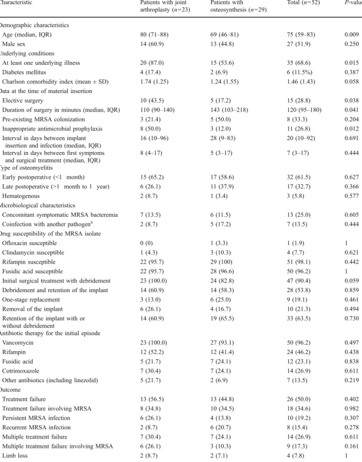
MRSA was retrieved from 41 (79%) of the 52 patients with implant-associated infections. The sources were mainly intraoperative specimens (30 isolates), blood (five isolates), aspirated synovial fluid (four isolates), an abscess (one isolate), and sterile aspiration of a sinus tract (one isolate). Thirty-five isolates (85%) were clonally related and shared microbiological characteristics of the South German MRSA clone (spa type t041 or relatives, SCCmec type I, agr type 2; Fig. 1). All of these isolates shared a similar
suscepti-Table 1 Characteristics and outcome of patients with methicillin-resistant Staphylococcus aureus (MRSA) orthopedic device-related infection (ODRI) and comparison of patients with joint arthroplasty and patients with osteosynthesis
Characteristic Patients with joint
arthroplasty (n=23)
Patients with osteosynthesis (n=29)
Total (n=52) P-value
Demographic characteristics
Age (median, IQR) 80 (71–88) 69 (46–81) 75 (59–83) 0.009
Male sex 14 (60.9) 13 (44.8) 27 (51.9) 0.250
Underlying conditions
At least one underlying illness 20 (87.0) 15 (53.6) 35 (68.6) 0.015
Diabetes mellitus 4 (17.4) 2 (6.9) 6 (11.5%) 0.387
Charlson comorbidity index (mean ± SD) 1.74 (1.25) 1.24 (1.55) 1.46 (1.43) 0.058
Data at the time of material insertion
Elective surgery 10 (43.5) 5 (17.2) 15 (28.8) 0.038
Duration of surgery in minutes (median, IQR) 110 (90–140) 143 (103–218) 120 (95–180) 0.041
Pre-existing MRSA colonization 3 (21.4) 5 (50.0) 8 (33.3) 0.204
Inappropriate antimicrobial prophylaxis 8 (50.0) 3 (12.0) 11 (26.8) 0.012
Interval in days between implant insertion and infection (median, IQR)
16 (10–96) 28 (9–83) 20 (10–92) 0.691
Interval in days between first symptoms and surgical treatment (median, IQR)
8 (4–17) 5 (3–17) 7 (3–17) 0.444
Type of osteomyelitis
Early postoperative (<1 month) 15 (65.2) 17 (58.6) 32 (61.5) 0.627
Late postoperative (>1 month to 1 year) 6 (26.1) 11 (37.9) 17 (32.7) 0.366
Hematogenous 2 (8.7) 1 (3.4) 3 (5.8) 0.577
Microbiological characteristics
Concomitant symptomatic MRSA bacteremia 7 (13.5) 6 (11.5) 13 (25.0) 0.605
Coinfection with another pathogena 2 (8.7) 5 (17.2) 7 (13.5) 0.444
Drug susceptibility of the MRSA isolate
Ofloxacin susceptible 0 (0) 1 (3.3) 1 (1.9) 1
Clindamycin susceptible 1 (4.3) 3 (10.3) 4 (7.7) 0.621
Rifampin susceptible 22 (95.7) 29 (100) 51 (98.1) 0.442
Fusidic acid susceptible 22 (95.7) 28 (96.6) 50 (96.2) 1
Initial surgical treatment with debridement 23 (100.0) 24 (82.8) 47 (90.4) 0.059
Debridement and retention of the implant 14 (60.9) 14 (58.3) 28 (53.8) 0.859
One-stage replacement 3 (13.0) 6 (25.0) 9 (19.1) 0.461
Removal of the implant 6 (26.1) 4 (16.7) 10 (21.3) 0.494
Retention of the implant with or without debridement
14 (60.9) 19 (65.5) 33 (63.5) 0.730
Antibiotic therapy for the initial episode
Vancomycin 23 (100.0) 27 (93.1) 50 (96.2) 0.497
Rifampin 12 (52.2) 12 (41.4) 24 (46.2) 0.438
Fusidic acid 5 (21.7) 7 (24.1) 12 (23.1) 0.838
Cotrimoxazole 7 (30.4) 7 (24.1) 14 (26.9) 0.611
Other antibiotics (including linezolid) 5 (21.7) 2 (6.9) 7 (13.5) 0.219
Outcome
Treatment failure 13 (56.5) 13 (44.8) 26 (50.0) 0.402
Treatment failure involving MRSA 8 (34.8) 10 (34.5) 18 (34.6) 0.982
Persistent MRSA infection 6 (26.1) 4 (13.8) 10 (19.2) 0.307
Recurrent MRSA infection 2 (8.7) 6 (20.7) 8 (15.4) 0.278
Multiple treatment failure 7 (30.4) 7 (24.1) 14 (26.9) 0.611
Multiple treatment failure involving MRSA 6 (26.1) 3 (10.3) 9 (17.3) 0.161
bility pattern, being resistant to gentamicin, erythromycin, lincomycin, and fluoroquinolones. No GISA strain was detected.
Surgical treatment and antimicrobial therapy
Surgical treatment consisted of debridement in 47 patients (90%), with implant retention in 28 patients, device explantation in ten patients, and one-stage exchange in nine patients. None of the patients had two-stage exchange, as the four patients scheduled for two-stage exchange experienced treatment failure or had other conditions that prevented reimplantation. Twenty-six patients (50%) re-ceived only single-agent antimicrobial therapy (vancomycin alone, cotrimoxazole alone, or vancomycin alone followed by cotrimoxazole or linezolid). The other 26 patients (50%) received combination antimicrobial therapy: 12 patients received rifampin plus fusidic acid (with vancomycin-rifampin as the initial therapy), eight patients received vancomycin plus rifampin, four patients received rifampin plus cotrimoxazole (with vancomycin-rifampin as the initial therapy), and two patients received vancomycin plus cotrimoxazole.
Univariate and multivariate survival analyses
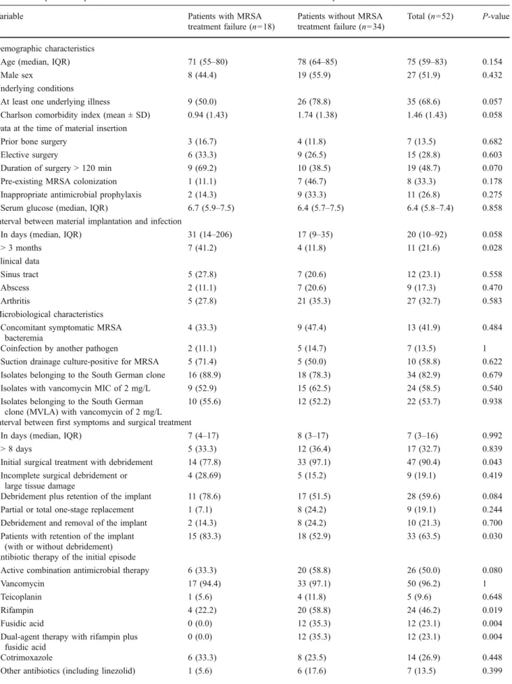
Patients with and without treatment failure are compared in Table 2. The Kaplan–Meier probability estimates of the
two-year failure rate were higher when the implant was left in place than when it was removed (log-rank test: P=0.036; Fig. 2, panel A). The estimates were lower in patients receiving rifampin plus fusidic acid than in patients receiving single-agent therapy and in patients receiving other combinations (log-rank test: P=0.036 and P=0.010, respectively; Fig. 2, panel B). In the subpopulation of patients who underwent debridement with implant reten-tion, the two-year probability of treatment failure was 83%
with single-agent therapy and 22% with combination therapy (log-rank test: P=0.020; Fig. 3). The incidence rate of failure involving MRSA in patients who had debridement with implant retention was 3.1 per 100 patient-months in patients treated with single-agent therapy and 1.4 per 100 patient-months in patients treated with an antimicrobial combination, giving an incidence rate ratio of 2.3 (confidence interval [CI], 0.55–13.51; P=0.11). There was a non-significant trend towards a higher probability of treatment failure in patients infected by the predominant South German clone ST228 in comparison with patients infected by sporadic MRSA strains. There was no differ-ence in the likelihood of failure according to the vancomy-cin MIC (2 mg/L versus <2 mg/L).
In multivariate Cox analyses, after exclusion of the five patients who did not receive a surgical debridement, implant retention and single-agent therapy were the only two independent variables associated with treatment failure involving MRSA at two years (hazard ratio [HR], 4.90; 95% CI, 1.32–18.17; P=0.017 and HR, 4.43; 95% CI, 1.20–16.33; P=0.025, respectively) (Table3).
Discussion
In this retrospective cohort study of patients with orthopedic device-related MRSA infection during the period 2000– 2008, most isolates belonged to the South German clone (ST228) and were fully susceptible to vancomycin (MIC≤ 2 mg/L). Only single-agent antibacterial therapy and implant retention were identified as independent risk factors for treatment failure involving MRSA persistence or recurrence at two years, after a median follow-up of 391 days.
Most nosocomial infections worldwide are due to a few hospital-acquired MRSA clones [15]. In our institution, the South German clone is endemic since 1999 [24, 25]. Amaral et al. recently found evidence that the predominant
Table 1 (continued)
Characteristic Patients with joint
arthroplasty (n=23)
Patients with osteosynthesis (n=29)
Total (n=52) P-value
Death 9 (39.1) 4 (13.8) 13 (25.0) 0.054
Death from implant-associated infection 2 (8.7) 2 (6.9) 4 (7.7) 1
Follow-up
Follow-up in days (median, IQR) 181 (77–596) 490 (232–1,011) 391 (110–879) 0.058
Follow-up of at least three months 14 (60.9) 25 (86.2) 39 (75.0) 0.054
Data are numbers (%) unless otherwise indicated. Missing values were excluded from the calculation ofpercentages and the statistical analysis. SD standard deviation; IQR interquartile range
aIncluding two coagulase-negative staphylococci, two Pseudomonas aeruginosa, one Enterobacter cloacae, one Streptococcus spp., and one
Table 2 Comparison of patients with MRSA ODRI with and without treatment failure directly attributed to MRSA
Variable Patients with MRSA
treatment failure (n=18)
Patients without MRSA treatment failure (n=34)
Total (n=52) P-value
Demographic characteristics
Age (median, IQR) 71 (55–80) 78 (64–85) 75 (59–83) 0.154
Male sex 8 (44.4) 19 (55.9) 27 (51.9) 0.432
Underlying conditions
At least one underlying illness 9 (50.0) 26 (78.8) 35 (68.6) 0.057
Charlson comorbidity index (mean ± SD) 0.94 (1.43) 1.74 (1.38) 1.46 (1.43) 0.058
Data at the time of material insertion
Prior bone surgery 3 (16.7) 4 (11.8) 7 (13.5) 0.682
Elective surgery 6 (33.3) 9 (26.5) 15 (28.8) 0.603
Duration of surgery > 120 min 9 (69.2) 10 (38.5) 19 (48.7) 0.070
Pre-existing MRSA colonization 1 (11.1) 7 (46.7) 8 (33.3) 0.178
Inappropriate antimicrobial prophylaxis 2 (14.3) 9 (33.3) 11 (26.8) 0.275
Serum glucose (median, IQR) 6.7 (5.9–7.5) 6.4 (5.7–7.5) 6.4 (5.8–7.4) 0.858
Interval between material implantation and infection
In days (median, IQR) 31 (14–206) 17 (9–35) 20 (10–92) 0.058
> 3 months 7 (41.2) 4 (11.8) 11 (21.6) 0.028 Clinical data Sinus tract 5 (27.8) 7 (20.6) 12 (23.1) 0.558 Abscess 2 (11.1) 7 (20.6) 9 (17.3) 0.470 Arthritis 5 (27.8) 21 (35.3) 27 (32.7) 0.583 Microbiological characteristics Concomitant symptomatic MRSA
bacteremia
4 (33.3) 9 (47.4) 13 (41.9) 0.484
Coinfection by another pathogen 2 (11.1) 5 (14.7) 7 (13.5) 1
Suction drainage culture-positive for MRSA 5 (71.4) 5 (50.0) 10 (58.8) 0.622
Isolates belonging to the South German clone 16 (88.9) 18 (78.3) 34 (82.9) 0.679
Isolates with vancomycin MIC of 2 mg/L 9 (52.9) 15 (62.5) 24 (58.5) 0.540
Isolates belonging to the South German clone (MVLA) with vancomycin of 2 mg/L
10 (55.6) 12 (52.2) 22 (53.7) 0.938
Interval between first symptoms and surgical treatment
In days (median, IQR) 7 (4–17) 8 (3–17) 7 (3–16) 0.992
> 8 days 5 (33.3) 12 (36.4) 17 (32.7) 0.839
Initial surgical treatment with debridement 14 (77.8) 33 (97.1) 47 (90.4) 0.043
Incomplete surgical debridement or large tissue damage
4 (28.69) 5 (15.2) 9 (19.1) 0.419
Debridement plus retention of the implant 11 (78.6) 17 (51.5) 28 (59.6) 0.084
Partial or total one-stage replacement 1 (7.1) 8 (24.2) 9 (19.1) 0.244
Debridement and removal of the implant 2 (14.3) 8 (24.2) 10 (21.3) 0.700
Patients with retention of the implant (with or without debridement)
15 (83.3) 18 (52.9) 33 (63.5) 0.030
Antibiotic therapy of the initial episode
Active combination antimicrobial therapy 6 (33.3) 20 (58.8) 26 (50.0) 0.080
Vancomycin 17 (94.4) 33 (97.1) 50 (96.2) 1
Teicoplanin 1 (5.6) 4 (11.8) 5 (9.6) 0.648
Rifampin 4 (22.2) 20 (58.8) 24 (46.2) 0.019
Fusidic acid 0 (0.0) 12 (35.3) 12 (23.1) 0.004
Dual-agent therapy with rifampin plus fusidic acid
0 (0.0) 12 (35.3) 12 (23.1) 0.004
Cotrimoxazole 6 (33.3) 8 (23.5) 14 (26.9) 0.448
MRSA clone in Brazil exhibited particular virulence properties [16]. In this study, by comparison with sporadic MRSA isolates, isolates belonging to the Brazilian clone were more adhesive and had a higher capacity to produce biofilm in vitro. Here, we detected a non-significant trend toward worse outcome among patients with isolates belonging to the South German clone. Common MRSA isolates are usually fully susceptible to vancomycin (MIC≤
2 mg/L), but small increases in the vancomycin MIC, remaining within the range of susceptibility (from 1 to 2 mg/L, for example), were recently shown to influence the outcome of bacteremia [13, 27]. Studies on vancomycin pharmacodynamics revealed that such an MIC increase in the range of susceptibility might have implications for localized orthopedic infections, as vancomycin penetration into bone is not optimal (bone-to-serum ratio 0.3) [28,29].
Table 2 (continued)
Variable Patients with MRSA
treatment failure (n=18)
Patients without MRSA treatment failure (n=34)
Total (n=52) P-value
Duration of antimicrobial therapy in days
(median, IQR)a
48 (35–87) 60 (41–88) 57 (41–88) 0.575
Duration of combination antimicrobial
therapy in days (median, IQR)a
34 (6–80) 29 (21–78) 29 (19–78) 0.629
Follow-up
Follow-up in days (median, IQR) 429 (136–1,481) 270 (95–837) 391 (110–878) 0.295
Data are numbers (%) unless otherwise indicated. Missing values were excluded from the calculation ofpercentages and statistical analysis. IQR interquartile range
a
After exclusion of the six patients with persistent MRSA infection for whom the duration of antimicrobial therapy priorto persistence were not taken into account
Fig. 1 Genetic characterization and vancomycin minimum inhibitory concentrations (MICs) of the 41 methicillin-resistant Staphylococcus aureus (MRSA) isolates responsible for orthopedic device-related
infection (ODRI) in Geneva between 2000 and 2008. Isolates in bold were considered to belong to the South German clone
However, we found no significant difference in outcome according to the vancomycin MIC.
Surgery is the cornerstone of the treatment of implant-associated orthopedic infections. Retention of the implant
is considered, nowadays, as a possible surgical option. This surgical procedure: (1) has to be performed if the pathogen is fully susceptible to antimicrobial agents; (2) has to be reserved for patients with a duration of symptoms <3 weeks and with a stable implant without soft-tissue damage nor sinus tract involvement; and (3) requires a rigorous debridement [2,3]. For staphylococcal ODRI, only a few studies are available and most of them included methicillin-susceptible isolates. For instance, in the study performed by Brandt et al. that included 33 patients with S. aureus prosthetic joint infections treated by debridement with prosthesis retention, the two-year probability of treatment failure was 69%, but only one isolate was methicillin-resistant [30]. More recently, Marculescu et al. found a two-year treatment failure rate of 40%, but, again, only one of the 32 S. aureus infections was due to MRSA [31]. Even though data on MRSA ODRI are lacking, complete implant removal is strongly recommended for MRSA ODRI [1–3]. Our study, which exclusively involved MRSA, definitively demonstrated
Fig. 3 Kaplan–Meier probability estimates of the 2-year failure rate in
the 28 patients with MRSA ODRI after surgical debridement with implant retention, treated with single-agent or combination antimicro-bial therapy
Fig. 2 Kaplan–Meier
probabili-ty estimates for the 2-year fail-ure rate in the 52 patients with MRSA ODRI. A Treated with implant retention (including five patients without surgical de-bridement) or implant removal. B Treatment with single-agent therapy or with rifampin-fusidic acid or with another combina-tion antimicrobial therapy, regardless of the surgical treat-ment (*P=0.010 and **P= 0.036, log-rank test)
that implant removal is required for the treatment of MRSA ODRI, as implant retention clearly emerged as an independent risk factor for treatment failure.
Antimicrobial therapy should always be combined with surgery for the treatment of ODRI [1–3]. Only one randomized double-blind placebo-controlled trial has dem-onstrated the superiority of combination therapy with rifampin (plus ciprofloxacin) over single-agent therapy (ciprofloxacin) for the treatment of staphylococcal ODRI [32]. It is noteworthy that, in this study, only two isolates (two coagulase-negative staphylococci) were resistant to methicillin. Since this landmark study, and since it has been demonstrated that rifampin was also effective on bacteria embedded in biofilm, rifampin-based combinations have been considered as standard therapy for MRSA ODRI [2,3,
33]. Few studies have compared different rifampin-based regimens in staphylococcal ODRI. Drancourt et al. demon-strated that a combination of rifampin and fusidic acid or ofloxacin was similarly effective and well tolerated during staphylococcal ODRI, but all of the isolates were methicillin-susceptible [34]. To our knowledge, different rifampin-based regimens have not been compared in MRSA ODRI. In our study, it is noteworthy that none of the patients who received combination therapy with rifampin plus fusidic acid experienced treatment failure. Controlled trials are needed to confirm the superiority of the rifampin-fusidic acid combination for the treatment of MRSA ODRI.
This study has some limitations. First of all, the combination of joint arthroplasty infections with other orthopedic hardware infections is criticable. Indeed, the type, the surface of the hardware, the long-term surgical implications, and the outcome might be different in these
two subgroups of patients. However, our cohort of patients is microbiologically homogeneous, as all of them were infected with MRSA, and guidelines for the initial treatment of joint arthroplasty or other orthopedic hardware infections are globally similar (i.e., surgery including debridement with or without implant retention with antimi-crobial therapy) [2]. Secondly, our study has the inherent limitations of all retrospective observational cohort studies. This was a single-center study, and the surgical and medical management of ODRI likely evolved during the eight-year study period. Thirdly, many patients were considered to be lost to follow-up at two years, as a quarter of them, enrolled after 2006, did not have a complete follow-up at the end of the study in 2008. Finally, patients with treatment failure occurring in another hospital may have been undetected. However, since the Geneva University Hospitals is, by far, the largest hospital in the area, we consider this latter possible selection bias as minimal.
In contrast, to our knowledge, this is the first study that examined microbiological and clinical risk factors of treatment failure specifically for MRSA ODRI. Indeed, the few previous reports of risk factors for treatment failure in staphylococcal ODRI included mainly MSSA isolates.
In conclusion, we observed a treatment failure rate of 35% in a cohort of patients with orthopedic device-related MRSA infection. Implant retention and single-agent anti-microbial therapy were the only independent risk factors for treatment failure. Microbiological factors, such as infection by the South German clone and a vancomycin MIC of 2 mg/L, were not associated with treatment failure. Therapy using a combination of antimicrobials should be considered for patients with MRSA ODRI, especially when implant removal is not feasible.
Table 3 Multivariable Cox regression model after exclusion of the five patients who did not receive a surgical debridement: independent factors associated with treatment failure involving MRSA in patients with MRSA ODRI
Variable HR 95% CI P-value Adjusted HR 95% CI P-value
Agea 0.98 0.958–1.009 0.726
Charlson comorbidity indexb 0.71 0.477–1.066 0.099
Sinus tracta 0.93 0.327–2.625 0.886
Occurrence of ODRI >3 months after material insertion 2.47 0.936–6.494 0.068 2.43 0.784–7.511 0.124
Duration of symptoms prior to ODRI surgery >8 daysa 1.67 0.591–4.718 0.333
Duration of surgery for the episode of ODRI >120 mina 2.19 0.673–7.146 0.192
Glycemia in the 24 h prior to ODRI surgerya 0.72 0.452–1.153 0.173
Retention of the implant after surgical debridement 3.07 0.853–11.067 0.086 4.90 1.322–18.166 0.017
Intermittent vancomycinainfusion 1.79 0.676–4.741 0.241
Single-agent antimicrobial therapy 2.39 0.896–6.386 0.082 4.43 1.203–16.329 0.025
HR hazard ratio; CI confidence interval
a
Variables not included in the multivariate Cox regression model (P>0.15)
b
Acknowledgments This work was supported by Fondation pour la Recherche Médicale, Paris, France. We are indebted to Elzbieta Huggler, Myriam Girard, Hélène Meugnier, Michele Bes, Colette Nicollier, Christine Courtier, Christine Cardon, Céline Spinelli, and Caroline Bouveron for the isolate characterization. We thank Nathalie Vallier for assistance with the statistical analysis, Abel Ferry for technical assistance, and David Young for editorial guidance.
References
1. Lew DP, Waldvogel FA (2004) Osteomyelitis. Lancet 364:369– 379
2. Widmer AF (2001) New developments in diagnosis and treatment of infection in orthopedic implants. Clin Infect Dis 33(Suppl 2):
S94–S106
3. Zimmerli W, Trampuz A, Ochsner PE (2004) Prosthetic-joint
infections. N Engl J Med 351:1645–1654
4. Gristina AG (1987) Biomaterial-centered infection: microbial
adhesion versus tissue integration. Science 237:1588–1595
5. Kilgus DJ, Howe DJ, Strang A (2002) Results of periprosthetic hip and knee infections caused by resistant bacteria. Clin Orthop
Relat Res 404:116–124
6. Proctor RA, von Eiff C, Kahl BC et al (2006) Small colony variants: a pathogenic form of bacteria that facilitates persistent and recurrent infections. Nat Rev Microbiol 4:295–305
7. Tsukayama DT, Wicklund B, Gustilo RB (1991) Suppressive antibiotic therapy in chronic prosthetic joint infections. Orthope-dics 14:841–844
8. Wilson MG, Kelley K, Thornhill TS (1990) Infection as a complication of total knee-replacement arthroplasty. Risk factors
and treatment in sixty-seven cases. J Bone Joint Surg Am 72:878–
883
9. Berbari EF, Hanssen AD, Duffy MC et al (1998) Risk factors for
prosthetic joint infection: case–control study. Clin Infect Dis
27:1247–1254
10. Betsch BY, Eggli S, Siebenrock KA et al (2008) Treatment of joint prosthesis infection in accordance with current
recommen-dations improves outcome. Clin Infect Dis 46:1221–1226
11. Lentino JR (2003) Prosthetic joint infections: bane of orthopedists, challenge for infectious disease specialists. Clin Infect Dis
36:1157–1161
12. Salgado CD, Dash S, Cantey JR et al (2007) Higher risk of failure of methicillin-resistant Staphylococcus aureus prosthetic joint infections. Clin Orthop Relat Res 461:48–53
13. Soriano A, Marco F, Martínez JA et al (2008) Influence of vancomycin minimum inhibitory concentration on the treatment of methicillin-resistant Staphylococcus aureus bacteremia. Clin
Infect Dis 46:193–200
14. Ferry T, Bes M, Dauwalder O et al (2006) Toxin gene content of the Lyon methicillin-resistant Staphylococcus aureus clone com-pared with that of other pandemic clones. J Clin Microbiol
44:2642–2644
15. Oliveira DC, Tomasz A, de Lencastre H (2002) Secrets of success of a human pathogen: molecular evolution of pandemic clones of meticillin-resistant Staphylococcus aureus. Lancet Infect Dis
2:180–189
16. Amaral MM, Coelho LR, Flores RP et al (2005) The predominant variant of the Brazilian epidemic clonal complex of methicillin-resistant Staphylococcus aureus has an enhanced ability to produce biofilm and to adhere to and invade airway epithelial cells. J Infect Dis 192:801–810
17. Charlson ME, Pompei P, Ales KL et al (1987) A new method of classifying prognostic comorbidity in longitudinal studies:
devel-opment and validation. J Chronic Dis 40:373–383
18. Zimmerli W, Ochsner PE (2003) Management of infection associated with prosthetic joints. Infection 31:99–108
19. Clinical and Laboratory Standards Institute (CLSI) (2007) Methods for dilution antimicrobial susceptibility tests for bacteria that grow aerobically; approved standard M7-S17, Wayne, PA 20. Francois P, Huyghe A, Charbonnier Y et al (2005) Use of an
automated multiple-locus, variable-number tandem repeat-based method for rapid and high-throughput genotyping of
Staphylo-coccus aureus isolates. J Clin Microbiol 43:3346–3355
21. Dauwalder O, Lina G, Durand G et al (2008) Epidemiology of invasive methicillin-resistant Staphylococcus aureus clones
col-lected in France in 2006 and 2007. J Clin Microbiol 46:3454–
3458
22. Jarraud S, Mougel C, Thioulouse J et al (2002) Relationships between Staphylococcus aureus genetic background, virulence factors, agr groups (alleles), and human disease. Infect Immun
70:631–641
23. Kondo Y, Ito T, Ma XX et al (2007) Combination of multiplex PCRs for staphylococcal cassette chromosome mec type assign-ment: rapid identification system for mec, ccr, and major differ-ences in junkyard regions. Antimicrob Agents Chemother 51:264– 274
24. François P, Harbarth S, Huyghe A et al (2008)
Methicillin-resistant Staphylococcus aureus, Geneva, Switzerland, 1993–
2005. Emerg Infect Dis 14:304–307
25. Sax H, Posfay-Barbe K, Harbarth S et al (2006) Control of a cluster of community-associated, methicillin-resistant
Staphylo-coccus aureus in neonatology. J Hosp Infect 63:93–100
26. Concato J, Feinstein AR, Holford TR (1993) The risk of determining risk with multivariable models. Ann Intern Med
118:201–210
27. Moise PA, Sakoulas G, Forrest A et al (2007) Vancomycin in vitro bactericidal activity and its relationship to efficacy in clearance of methicillin-resistant Staphylococcus aureus bacteremia.
Antimi-crob Agents Chemother 51:2582–2586
28. Knudsen JD, Fuursted K, Raber S et al (2000) Pharmacodynamics of glycopeptides in the mouse peritonitis model of Streptococcus pneumoniae or Staphylococcus aureus infection. Antimicrob Agents Chemother 44:1247–1254
29. Peetermans WE, Hoogeterp JJ, Hazekamp-van Dokkum AM et al (1990) Antistaphylococcal activities of teicoplanin and vancomy-cin in vitro and in an experimental infection. Antimicrob Agents
Chemother 34:1869–1874
30. Brandt CM, Sistrunk WW, Duffy MC et al (1997) Staphylococcus aureus prosthetic joint infection treated with debridement and
prosthesis retention. Clin Infect Dis 24:914–919
31. Marculescu CE, Berbari EF, Hanssen AD et al (2006) Outcome of prosthetic joint infections treated with debridement and retention
of components. Clin Infect Dis 42:471–478
32. Zimmerli W, Widmer AF, Blatter M et al (1998) Role of rifampin for treatment of orthopedic implant-related staphylococcal infec-tions: a randomized controlled trial. Foreign-Body Infection (FBI)
Study Group. JAMA 279:1537–1541
33. Widmer AF, Frei R, Rajacic Z et al (1990) Correlation between in vivo and in vitro efficacy of antimicrobial agents against foreign body infections. J Infect Dis 162:96–102
34. Drancourt M, Stein A, Argenson JN et al (1997) Oral treatment of Staphylococcus spp. infected orthopaedic implants with fusidic acid or ofloxacin in combination with rifampicin. J Antimicrob