CLINICAL ARTICLE - NEUROSURGICAL TECHNIQUES
Intraoperative fabrication of patient-specific moulded
implants for skull reconstruction: single-centre experience
of 28 cases
Lennart Henning Stieglitz&Nicolas Gerber&Thomas Schmid&Pasquale Mordasini&
Jens Fichtner&Christian Fung&Michael Murek&Stefan Weber&Andreas Raabe&
Jürgen Beck
Received: 1 October 2013 / Accepted: 4 December 2013 / Published online: 18 January 2014 # Springer-Verlag Wien 2014
Abstract
Background Intraoperatively fabricated polymethylmethacrylate (PMMA) implants based on computer-designed moulds were used to improve cosmetic results after hard tissue
replacement. To assess the implant’s cosmetic and
func-tional results we performed both subjective and objective assessments.
Methods This retrospective analysis was performed using a cohort of 28 patients who received PMMA implants between February 2009 and March 2012. The cosmetic and functional results were assessed using a patient questionnaire.
Further-more an objective volumetric subtraction score (0–100) was
applied and implant thickness, as well as gaps and tiers, were measured.
Results Patients mainly judged their cosmetic result as “good”. Two of the 28 patients found their cosmetic result unfavourable. The functional result and stability was mainly judged to be good. Measurements of implant thickness
showed a very high correlation with the thickness of the contralateral bone. Volumetric subtraction led to a median quality of 80 on a scale from 0 to 100. Median gaps around the margins of the implant were 1.5 mm parietally, 1.7 mm frontally and 3.5 mm fronto-orbitally, and median tiers were 1.2 mm, 0 mm and 0 mm respectively. The overall rate of surgical revisions was 10.7 % (three patients). Two patients suffered from wound healing disturbances (7.1 %). The over-all complication rate was comparable to other reports in the literature.
Conclusions Implantation of intraoperatively fabricated patient-specific moulded implants is a cost-effective and safe technique leading to good clinical results with a low compli-cation rate.
Keywords hemicraniectomy . PMMA . PSI . PSMI . Skull reconstruction
Introduction
Advances in neurosurgery and intensive care over the last 2 decades have achieved great improvement in the outcomes of patients with severe head trauma, stroke, brain tumours and infectious diseases affecting the skull. Surgical treatment often involves a large osteoclastic craniotomy or delayed reimplan-tation of the autologous bone flap. In many of these cases a reconstruction of the skull is necessary because of (1) destruc-tion of the bone as result of trauma, (2) infecdestruc-tion, (3) tumour infiltration, or (4) aseptic necrosis and resorption of the bone
flap [20].
There are several techniques and materials available for
preservation of the explanted bone-flap and its replacement [2,
5,8,13,14,17–19]. These techniques differ in functionality,
L. H. Stieglitz (*)
:
J. Fichtner:
C. Fung:
M. Murek:
A. Raabe:
J. BeckDepartment of Neurosurgery, Bern University Hospital, Freiburgstrasse 10, 3010 Bern, Switzerland
e-mail: Lennart.stieglitz@insel.ch L. H. Stieglitz
e-mail: lennart@stieglitze.de N. Gerber
:
S. WeberARTORG-ISTB, University of Bern, Bern, Switzerland T. Schmid
University of Bern, Bern, Switzerland P. Mordasini
Diagnostic and Interventional Neuroradiology, Bern University Hospital, Bern, Switzerland
cosmetic result, costs and spectrum of complications involved in the procedures. Among the artificial materials, polymethylmethacrylate (PMMA) is the least expensive and
most commonly used [2,18]. The material is usually applied
directly to the patient and moulded to the skull defect using free-hand cranioplasty, which often results in a suboptimal
cosmetic result (Fig.1).
Better cosmetic and very good functional results can be achieved using preoperatively designed patient specific
im-plants (PSIs) that have a low complication rate [5,19].
Dif-ferent materials have been used for this purpose, all with good results. The most commonly used are titanium and
polyether-ether-ketone (PEEK) [13]. To achieve optimal cosmetic and
functional results at a similar cost as the PMMA implants we developed a technique of intraoperative moulding of a PMMA
implant on a patient-specific mould [12]. To evaluate the
functional and cosmetic outcome of these patients, we per-formed a retrospective analysis of a consecutive series of 28 patients.
Methods and materials
Patients
Between February 2009 and March 2012 we performed 28 plastic reconstructions of the skull using the patient-specific moulded implant (PSMI) technique. Of these patients, 11 were female and 17 male. Indications for cranial reconstruction using artificial material were: resorption of the reimplanted
autologous bone flap in 12 patients (42.8 %), infection of the autologous bone so it could not be reimplanted in nine patients (32.1 %), infiltration by tumour in three patients (10.7 %), loss of the autologous bone following inter-hospital transfer in three patients (10.7 %), and relevant resorption followed by instability of the reimplanted autologous bone flap in one patient (3.6 %). One case was a bifrontal defect involving the brow, one a combined bifrontal and lateral decompression, 24 were hemicraniectomies involving one half of the forehead and the temporal region; only in two cases were visible parts of the face affected.
Creation of mould template
The mould template was created using high-resolution com-puted tomography (CT) scans of each patient. Two different techniques were used for making the mould: Subtraction of post-explantation CT from a pre-explantation CT, if available, or subtraction from the mirrored contralateral side. If available scans were of adequate quality (at least 2.5-mm slice thick-ness), no new scans were acquired to reduce costs and the
patients’ radiation doses. In most of the cases an emergency
CT scan of adequate quality (1.27 mm slice thickness), which was acquired before hemicraniectomy, was available and served as pre-explantation CT.
There were two variants for creation of the mould, referred to as Variant A and Variant B.
Variant A The CT scans were loaded into the BrainLab iPlan neuronavigation software (Version 3.0; BrainLab, Feldkirchen, Germany). After image fusion of the available scans a three-dimensional (3D) volume rendering was performed using Hounsfield thresholding (200>I[x, y, z]>3,071). Partially re-sorbed bone, drainages and other objects were manually erased from the object until a perfect 3D model of the skull and the bony defect was created.
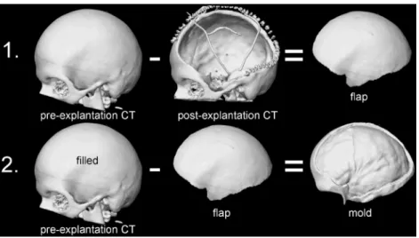
Using the same procedure a second 3D model of the intact skull was created using the pre-explantation CT. Then the second model was dig-itally subtracted from the first, resulting in a 3D model of the intended PSMI. The model of the intact skull was digitally filled out and the model of the PSMI was subtracted. This resulted in a digital version of the mould for creation of the
Palacos-PSMI (Fig.2). To reduce material needed
for 3D printing, non-required parts such as the contralateral side were removed, leading to a mould that showed about one1 cm of the surface of the surrounding bone.
Variant B If no pre-explantation CT was available, the 3D model of the skull with the bony defect was Fig. 1 Postoperative CT scan of a free-hand PMMA cranioplasty—a
ideal shape of the cranioplasty; b the achieved shape of the cranioplasty is too flat
mirrored and manually adjusted to meet the shape of the opposite side using iPlan. Then the resulting mirrored object was shrunk by 3–4 mm (the intended thickness of the resulting PSMI) and fused to the non-mirrored 3D model, resulting in a mould for PSMI creation. The mould was then isolated from the intact parts of the skull, which are irrelevant for the creation of the PSMI, using the split-tool of iPlan.
The resulting object was exported as an STL file (“Surface Tesselation Language” 3D file for-mat) and transferred to a 3D printer (Spectrum Z™ 510 printer; Z-Corporation, Rock Hill, SC, USA), a high-speed printing device allowing the production of pieces up to 300×200×300 mm with a resulting printing resolution of 0.1 mm. It uses standard inkjet printing technology to create parts layer-by-layer by depositing a liquid binder
onto thin layers of powder. After completion of the printing process and drying of the binder solution, excessive powder material around the template model was removed and the mould was infiltrated with polyurethane.
Intraoperative workflow
The mould was brought into the sterile area of the operating room (OR) in an airtight sterile plastic bag, in which plastic tubing was previously inserted and connected to a vacuum
suction device (Fig.3A). After activation of the suction, the
plastic bag neatly covered the surface of the mould (Fig3B).
The PSMI was then formed from polymethylmethacrylate bone cement (PMMA) on the sterile surface of the mould
(Fig.3C). After hardening of the PSMI a series of holes was
drilled into it to allow exchange of fluid or blood between the Fig. 2 Creation of mould
template. Step 1: The post-explantation CT is subtracted from the pre-explantation CT. The result is a 3D model of the required PSMI (flap). Step 2: The 3D model of the flap is subtracted from the pre-explantation CT, which is then digitally“filled out”. After elimination of non-required parts such as the contralateral side and zygoma the result is a 3D model of the mould
Fig. 3 Intraoperative creation of patient-specific moulded implant (PSMI). AThe mould was brought into the OR in an air-tight sterile plastic bag (a), which was connected with the vacuum suction using a flexible tube (b). B After removal of the air inside the plastic bag using the vacuum suction the plastic neatly covered the mould surface. C The PSMI was created from PMMA on the mould’s surface. D After hardening of the PMMA, holes were drilled into the PSMI. E The implant was placed into the defect and was fixed using low-profile titanium plates and screws
epidural and the subgaleal space and to facilitate fixation of the dura at the implant through tissue growth into the holes
(Fig.3D). The PSMI was then fixed into the bony defect using
titanium plates and screws (Fig.3E). To prevent subgaleal and
epidural haematoma, a subgaleal drainage was implanted in all cases.
Time investment in the procedure
The whole procedure, from planning of the 3D mould until implantation, could be performed within 24 h if necessary, which was done in one case. Creating the digital 3D mould using BrainLab iPlan took, depending on the quality of the CT images, size and location of the bone defect, between 1 and 2 h in the hands of an experienced person. Rapid prototyping of
the mould took approximately 2.4 h for a volume of 350 cm3.
The binder solution required drying overnight and infiltration with polyurethane. The process of creating a PSMI implant using the mould took approximately 30 min altogether.
Postoperative neuroimaging
Postoperative CT was performed in case of clinical necessity (e.g. headache onset) to rule out complications such as epidu-ral haematomas or hygromas requiring revision. In cases with perfect clinical and cosmetic results, we dispensed with post-operative imaging to reduce unnecessary application of radi-ation to the patients.
Patients’ subjective assessments of implant quality
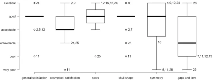
To judge the cosmetic results of the PSMI implantations, all patients were asked to answer a catalogue of questions concerning their subjective estimation of their status at least 3 months postoperatively. Seventeen patients (61 %) filled in the questionnaire. One patient could not be contacted postop-eratively as he moved to an unknown address. The questions were the following: general satisfaction with the result of the surgery, satisfaction with the cosmetic result, peculiarity of scars, satisfaction with skull shape, symmetry, and palpable
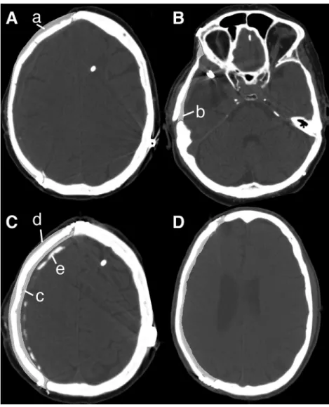
Fig. 4 Volumetric bone flap subtraction examples. AThe 3D reconstruction of the bone after implantation of a PSMI (in white) is subtracted from the 3D model of the ideal bone flap. The resulting volume (yellow) is representative for the“quality” of the PSMI shape. In this example the PSMI is a little too flat in the frontal region, which results in a considerable volume after subtraction (a). B In this example the PSMI is not in the perfect position, resulting in a small subtraction volume on the inside (b). C In the third example the PSMI is quite well shaped, but very thin compared with the contralateral side. This results in an interior (c) and exterior (d) volume after subtraction. The dura shows signs of calcification (e). D In the last example the shape and position of the PSMI are perfect. The outer surface of the implant is optimal. The thickness does not correspond with the contralateral side everywhere, but is absolutely sufficient
gaps or tiers around the implant (each indicated with an ordinal six-step scale from very poor to excellent). Finally, the patients were asked to judge their general condition pre-operatively and postpre-operatively (six-step scales from very poor to excellent). Statistical significance was tested using two-sample Wilcoxon test. P values less than 0.05 were con-sidered significant.
Objective assessments of implant quality—Volumetric bone flap subtraction
The available postoperative CT scans for 19 patients (68 %) were closely examined and parameters were measured to judge the objective quality and fit of the implant. A volumetric reconstruction of the implanted PSMI and surrounding skull (Hounsfield units 200–3,071) was subtracted from the 3D
model of the ideal flap (result of step 1 in Fig.2). In case of
a perfectly sized, shaped and implanted PSMI, this should lead to a complete extinction of the ideal flap model, with a
volume of 0 cm3. The other extreme, in which there was no
overlap of the two structures, would be the full volume of the flap model. The resulting volume was set in relation to the volume of the ideal flap model to achieve a result independent
from the size of the implanted PSMI (Fig.4). The result is
subtracted from 1 and multiplied by 100 to generate a“quality
grade” ranging from 0 (no overlapping between PSMI and
ideal flap model) to 100 (perfect size, shape and implantation). Gaps between bone and PSMI implant were measured in axial planes at three spots: Postero-superior (parietal), antero-superior (frontal) and antero-inferior (fronto-orbital), in millimetres. There were no measurements in the temporal region, as the implants were mostly not designed to cover the inferior portion of the temporal decompression. In addition to gaps, the thickness of tiers was measured in millimetres. The thickness of the PSMI was measured in millimetres via axial CT in the centre of the implant and on the corresponding contralateral side.
Objective assessments of implant quality—Calculation of the dice similarity coefficient
A standardised procedure to measure identity of two volumet-ric objects is calculation of the dice similarity coefficient
(DSC) [21] according to the following formula:
DSC¼ 2 A∩Bð Þ= A þ Bð Þ
DSC Dice similarity coefficient
A Volume (cm2) of the ideal flap model
B Volume (cm2) of the implanted PSMI
Values range between 0 (no overlapping of two objects) and 1 (perfect match).
Table 1 Complications after PSMI implantation
n Patient IDs Infection of unknown origin 0
Infection after wound healing disturbance 2 (7.1 %) 4, 25
Haematoma 2 (7.1 %) 5, 23
Hygroma 2 (7.1 %) 13, 16
Subcutaneous CSF leak (transient) 1 (3.6 %) 24 Subcutaneous CSF leak (persistent) 2 (7.4 %) 1
Hydrocephalus 1 (3.7 %) 13
Incidence of complications after implantation of a PSMI. The patient IDs correspond to the cases in Table2
Fig. 5 Cosmetic outcome after PSMI implantation. Patients were asked postoperatively to judge the cosmetic results on ordinal 6-step scales from very poor to excellent. The numbers represent the cases in Table2
Results
Surgical outcome and complications
All patients recovered well from surgery. One case of postop-erative epidural haematoma required surgical revision. One patient with delayed epidural haematoma was readmitted from rehabilitation because of new hemiparesis and deterioration of vigilance; she was successfully treated by trepanation and evacuation of a chronic haematoma at the centre of the PSMI and implantation of an epidural drain for 48 h. In one patient, a subcutaneous cerebrospinal fluid (CSF) collection was allevi-ated after transdermal puncture and evacuation. In another patient, the CSF collection slowly regressed without treat-ment. In one further patient, conservative treatment of a sub-cutaneous CSF collection was not successful and the patient recovered well after implantation of a VP shunt on the con-tralateral side. Two patients experienced wound-healing dis-turbances with opening of the scar over the titanium plates. Both showed no signs of local or systemic infection. In both patients the PSMI was removed and a new one was created using the same mould 4 weeks later. One of them recovered well, but the other suffered repeated wound healing distur-bances and required repeated revision of the implant and
closure of the skin using free tissue transplants (Table1).
Cosmetic results
In general, the patients judged the surgical results as good
(Fig.5). Both patients with wound healing disturbances found
their result ‘good’. Two of the patients with postoperative
subcutaneous CSF collections found their result even
‘excel-lent’. Most patients found their cosmetic result acceptable and
scars, skull shape and symmetry of the head‘good’. Figure6
shows 3D reconstructions of case 25, who judged skull shape,
symmetry and gaps and tiers to be‘very poor’. However, to
the observer the result does not look bad.
Other clinical results
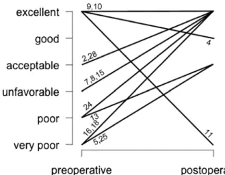
There was a significant improvement in the general condition
in our cohort of PSMI patients (p=0.024, Fig. 7). The one
patient who reported that his general condition deteriorated postoperatively, from excellent to very poor, is the same
patient who judged his cosmetic results as poor (Fig.6).
Objective assessment of PSMI quality in postoperative CT
scan—Volumetric bone flap subtraction
The median volume of the ideal flap was 98.3 cm3(SD 34.9).
After subtraction of the volumetric reconstruction of the im-planted PSMI and surrounding bone the median overlapping
volume was 22.3 cm3 (SD 18.5). The median calculated
objective quality grade was 80 (SD 18.0, Table2).
Gaps and tiers were smallest in the postero-superior (parietal) region, where an average gap of 2.2 mm (SD 2.1)
Fig. 6 Illustrative 3D views of case 25. The 3D reconstructions show the fit of the PSMI of case 25 (see Table2), who judged gaps and tiers“very poor”. A Lateral view; B frontal view; C posterior view—a titanium
low-profile microfixation plates, b small gap cranial of the petrous bone, c gap from dehiscent lamdoid suture resulting from the initial trauma; d recon-struction artefact (scan did not cover the most posterior parts of the skull) Fig. 7 Patients’ general conditions before and after PSMI implantation. Patients were asked preoperatively and postoperatively to judge their condition on ordinal six-step scales from very poor to excellent. The numbers represent the cases in Table2. Only one patient (11) encountered a relevant subjective deterioration
and a median of 1.5 mm were measured. Tiers were mainly inward (average 0.9 mm, SD 0.9, median 1.2 mm). In the antero-superior (frontal) location the average gap was 3.5 mm (SD 5.7, median 1.7 mm) and the tier was 0.2 mm outward (SD 1.1, median 0 mm). In the antero-inferior (fronto-orbital) location the average gap was 3.8 mm (SD 2.7, median 3.5 mm) and the average inward tier 0.1 mm (SD 1.0, median
0.0 mm, Table2).
The thickness of the PSMIs corresponded well with the thickness of the contralateral bone. The average PSMI thick-ness was 4.0 mm (SD 0.8, median 3.8 mm, range 3.2–6 mm). On the contralateral, side the average bone thickness was 4.2 mm (SD 0.9, median 4.0 mm, range 2.7–6.1 mm). The
average difference between PSMI and contralateral bone
thickness was −0.2 mm (SD 0.9, median 0.0 mm, range
−2.9 to 1.3 mm, Table2).
Discussion
Improvements in intensive care as well as neuroimaging and microsurgical techniques have led to a growing number of indications for hard tissue replacement (HTR) in cranial neu-rosurgery. There are numerous materials available for this purpose, all of which have their individual advantages. The most commonly used and cheapest is polymethylmethacrylate
Table 2 Objective judgement of PSMI quality
No. Volumetric assessment Thickness at centre
Missing bone volume (“ideal flap”) (cm3
)
Overlap (cm3)
Quality grade
(0–100) DSC (0–1) PSMI (mm) Contra-lateral bone(mm)
Relation (%) 1 80.6 6.7 92 0.72 3.5 3.5 100 2 53.3 18.9 65 0.66 3.2 3.2 100 3 88.7 41.9 53 0.6 3.5 3.7 95 4 No postop CT available 5 67.9 18.2 73 0.69 6.0 4.7 128 6 No postop CT available 7 93.4 12.1 87 0.69 3.3 2.7 122 8 137.5 24.5 82 0.84 3.2 6.1 52 9–11 No postop CT available 12 99.7 49.9 50 0.54 3.7 3.7 100 13 125.1 24.8 80 0.71 3.9 4.1 95 14 No postop CT available 15 128.3 75.6 41 0.38 4.4 4.3 102 16 98.8 7.9 92 0.8 3.6 4.4 82 17 98.3 5.72 94 0.76 5.8 4.0 145 18 No postop CT available 19 130.0 18.1 86 0.63 3.5 3.9 90 20 46.3 1.1 98 0.77 4.3 4.8 90 21 No postop CT available 22 91.9 28.1 69 0.66 3.9 3.9 100 23 124.0 21.9 82 0.7 4.0 5.2 77 24 177.7 35.1 80 0.76 4.5 4.6 98 25 135.7 46.1 66 0.68 4.8 5.1 94 26 No postop CT available 27 88.7 42.6 52 0.52 5.1 3.5 146 28 40.9 22.3 45 0.47 4.1 4.2 98 Mean 100.3 26.4 73 0.66 4.1 4.2 101 Median 98.3 22.3 80 0.69 3.9 4.1 98 SD 34.9 18.5 18 0.12 0.8 0.8 22
The result was evaluated by volumetric subtraction of the PSMI from a 3D object representing the missing bone. The overlapping volume was then put in relation with the volume of the missing bone. The resulting quality grade ranges from 0 to 100. The median result of our cases was 80. The median dice similarity coefficient (DSC) was 0.69. The thickness of the implanted PSMI was measured and put in relation with the contralateral bone. The median thickness was 98 %
Ta b le 3 M at eri als for har d tiss u e re p la cement: summary of literature Author Date Study desig n P athology Region n M at er ial O u tcome C ompl ica tion s M ate ria l cost s Ar tic o et al. 2003 1 R S Benign tumours and tumour -like les ions C rani o -f ac ia l 1 5 (8 f. 7 m) succ ess ful in 1 1 /15 cases Autologo us bo ne (split graft) co sme tic : 7 ex ce lle nt, 5 good 1 hypertensive pneumocephalus No cos ts Cabraj a et al . 2009 2 R S *1 3t ra u m a, *5C V I, * 4 m eni nge om a, * 1 dysplasia, * 1 bone erosion, * 1 h erpes encephalitis, * 1 b ra in absc ess * 1 1 fro nto-temporo-par iet al *1 b il at er al *4 b if ro n ta l 26 (11 f. 1 5 m ) T itanium * 1 7 excellent, * 6 good, *2f ai r, * 0 % poor results 88 % sat isf ied w ith co sme tic res u lt Me an 3,733 € Chim and Gos ain 2008 3 R S Cr ani o fa cia l irregularities Cranio-facial 2 5 * 5 hydrox ylapatite onlay * 3 calcium dahllite onlay (Norian C R S ; S ynthes, Pa o li, P A , U SA ) * 3 B ioactive glas s inlay (Nova Bone; P orex S u rg ic al, F air burn , GA , U S A ) * 7 D emineralis ed bone pa st e * 1 D emineralis ed bone st ru ts * 6 p re fa bric at ed po ly me rs (Medpor/porou s p o lyet hylene ) * 1 posto p infectio n re qui ring p ar ti al im pla n t removal (onlay patients ) * 1 posto p infectio n (p re fa bri cat ed polymers), conservative tre atmen t Da vid et al . 2005 4 R S S.a. Cranio-s ynosthosis Cranial d efects 8 (6 m , 2 f) Hydroxyapatite Al l adequate aesthetic an d m ec hani cal re pai r * 1 m ic ro fr agment ati o n during h ardening process * 1 w ound dehiscence with loose bone cement fra gme n ts De an et al . 2003 5 P S V arious lar g e skul l d ef ec ts 5 (5 m ) P SMI (PMMA formed af te r a com pute r-g enerated m ould) Duc ic, 2002 6 R S * 1 2 frontal, * 5 temporal, *2 p ar ie ta l, * 1 occipital 20 T itanium mesh and h ydroxyapatite bone ce me nt Goo d mec h anic al outcome 0 infections Dur h am et al . 2003 8 R S *4t ra u m a, * 2 fib rous dysplasia, * 2 infected bone flaps, * 1 tumour Fron toparietal 8 (4 m , 4 f) T itanium mesh and h ydroxyapatite bone cemen t E x cel lent co smeti c re sult s 2 infections requirin g removal Approx. US$ 7 ,00 0 Eppl ey et al . 2002 9 R S * 1 2 secondary recons truction s: includ ing bone flap in fe cti on, tr aumati c 14 (9 m, 5 f) h ar d ti ssue repla cemen t po ly me r P MI * 1 3 v ery good 0 infections
Ta b le 3 (continued ) Author Date Study desig n P athology Region n M at er ial O u tcome C ompl ica tion s M ate ria l cost s injury , con genital abs ence of the sphenoid w ing * 2 p rimary reconstructions: F ibrous d y splas ias * 1 requ ire d secon d ar y alloplastic temporal augmentation F at h ie ta l. 2008 10 TN * 1 bone infiltrating tumour *1d ec o m p re ss iv e cr ani ect omy w it h epi dura l empyema frontoparietal 2 (2 f) P MMA formed o n the explanted bone flap 2 excellent cos m etic re sult s 0 infections Friedman et al . 2000 11 R S * 3 4 sur gically induced defects * 6 posttraumatic defects Fron tal sinus and fronto-facial 40 (29 m , 1 1 f) H ydroxyl apa tite * 3 expl anta tion s for infe ct ions * 4 explantations for sur gical re-exp lora tion * 3 explantations for m ucous cyst s K ri eg ele ta l. 2007 15 R S * 1 9 d ecompress ive cr ani ect omi es; * 1 5 infection; * 2 h ead tr auma 6 1 *3 6f re eh an dP M M A ; * 2 5 T utoplast p roce sse d autografts PMMA : *2 6s at is fa ct o ry re sult s * 5 not satis factory results * 5 unknown T u toplas t: Al l sat isf act ory re sult s PM MA : *4d ie d *1 4s ev er el yd is ab le d *1 8s at is fa ct o ryr ec o v er y * 2 removal due to PMMA related complications T u toplast: *1d ie d *1 8s ev er el yd is ab le d *6s at is fa ct o ryr ec o v er y * 1 re m ova l due to inf ec tion * 1 removal for resorption No di ff er enc e in inf ec tion rat e (13. 9 % PMM A , 8.3 % T u toplast) 1 1 .8 % resorption rate in T u toplast g roup PMMA : 80 € Tu to p la st 44 –920 € (C era m ic or titanium PS I: 4,00 0– 8,000 €) Le e et al. 2009 16 R S D ecompr essi ve cr ani ect omi es Tr au m a CVI 131 *91: 26 f. 62 m * 23: 9 f. 1 4 m * 17: 5 f. 1 2 m ) *9 1f re sh fr o ze n autografts, *2 3f re eh an dP M M A , * 1 7 PSM I (computer -assis ted PMMA) * 5 infections of 91 in autogr af t ca ses * 3 infections of 23 in fr eeha n d P MM A ca ses *1i n fe ct io no f1 7i n PSMI ca ses No significant impro v ement by moulded d esig n of implan ts. PSMI: US$ 800 Nor m al PMMA : US $ 80 CVI cerebrovascular incident, RS retrospective study , PS prosp ective study , TN technical note, PMMA polymeth y lmethacrylate, PM SI patient specific moulded implant Th e table shows an overview over the literature co n cer ning the u se of di ff er en t m at er ial s fo r h ar d ti ssue repl ace me nt. T here w as only one pros pective study , w hich included five p atients and did not report abo u t complications related to the te chnique. T here is no study known to u s tha t systematically compares dif feren t m aterials concerning their b enef it s. RS re tr ospec tive stu dy , PS prospective stu dy , TN technical note, PM MA polymethylmethacrylate, PMSI pa tie nt spec if ic moulded implant
(PMMA) bone cement. It can be formed intraoperatively to fill the bone defect, and is biocompatible and very stable. A disadvantage is that the manually formed bone replacements often have a suboptimal shape, leading to an inferior cosmetic result. An alternative is the use of autologous bone (split
grafts) [1], but this is only available for small defects. Other
artificial bone replacements can be made of hydroxylapatite (HA) bone cement, which is usually formed on a matrix of titanium mesh (onlay technique). HA has the benefit that bone can grow into the margins of the bone replacement and im-prove the stability of the implant over time. Disadvantages are the high costs and the high infection rate in published studies
[3,4,6,8, 11,15,16]. Patient-specific implants (PSIs) are
usually designed based on CT scans of the patients and are provided by specialised companies. They can be made from titanium, polyether-ether-ketone (PEEK), other polymers, PMMA, HA or biocompatible glass ceramic. What they all have in common is that they usually fit into the defect per-fectly and lead to an optimal cosmetic result. Unfortunately, the costs for all of them are relatively high and range from US$ 3,500 to more than US$ 5,000 per piece, depending on
the dimensions of the implant and the material [2, 3, 9].
Titanium, PEEK and ceramics can be resterilised in case of surgical revision. Titanium has the disadvantage that it con-ducts cold temperature far better than bone, which can be
uncomfortable for patients (Table3) [2].
Alternatives to commercial PSIs
The only artificial alternatives to commercially available PSIs are hand-made bone replacements from PMMA or HA bone cement. PMMA is particularly inexpensive (less than US$ 500 per piece) and is therefore frequently used even for replacement of large bone defects. To achieve better cosmetic results using this material, several different attempts were made. One very simple approach was to form the bone
re-placement directly on the explanted bone flap [10]. This
technique has the potential disadvantage that a possibly in-fected bone flap must be brought into the OR and even into direct contact with the PMMA, leading to a risk of infection. Though increased infection was not shown in the small num-ber of published cases using this technique, it remains a possible serious threat for patients. Another idea is to combine the advantages of computer-assisted design with the benefits of intraoperatively formed PMMA implants. In addition to the PMMA material, the costs for this technique are relatively low (Z-Corporation Spectrum T510 3D printer is offered for about US$ 10,000 and material for printing is below US$ 100 per mould). Two publications in the literature describe the use of
computer-designed moulds [5,16]. We independently
devel-oped a comparable technique and this report describes the largest consecutive series operated upon using this method.
Furthermore, we provide patients’ assessments of the
cosmetic results and evaluation of postoperative CT scans for a majority of the patients.
Results of patient-specific moulded implants
Complications were comparable with those reported for other techniques, including both commercial PSIs and
non-commercial implants [7]. Infections related to wound healing
over the titanium plates and the screws were easily treated. No complications were directly attributed to the material used or to the technique. Patients were mainly satisfied with the cosmetic results. Few patients complained about asymmetry of their skull and palpable gaps and tiers around the implant. Atrophy of the temporal muscle and poor overall neurological condition or depression might have contributed to this result, but it remains possible that commercial PSIs lead to slightly better cosmetic results. A standard for assessment of the postoperative result does not currently exist, which makes it impossible to compare the outcomes of the different tech-niques that have been described in the literature. We per-formed volumetric subtractions of the postoperative CT scans from the ideal bone flap models, resulting in an objective quality scale ranging from 0 to 100. While the median quality measured by this method was 80, four patients had values of more than 90, indicating nearly absolute identity of the
im-planted PSMI with what would be an“ideal” implant. Eight
patients had values below 70, usually indicating a too thin or too flat PSMI. Of these, five patients completed the question-naire. Only one of them (case 25) judged his cosmetic result as unfavourable. The others found it good or, in one case (case 2), even excellent. Only two of the patients (cases 11 and 25) showed clear dissatisfaction with the PSMI implant. In one of them (case 25) the postoperative CT scan corresponded with a relatively low objective rating. In the other case (case 11) we unfortunately did not have a postoperative CT scan available for evaluation. Calculations of the dice similarity coefficient (DSC) corresponded well with the quality scale. Cases with values above 0.7 showed optimal shape and fit of the implant. Weaknesses of the present study are the low rate of returned patient questionnaires and the fact that not all patients received a postoperative CT scan. A prospective study comparing the results of commercially available PSIs with the PSMI
tech-nique reported here and described previously by Lee et al. [16]
and Fathi et al. [10] is needed to identify advantages and
disadvantages of the techniques.
Conclusions
Implantation of intraoperatively fabricated patient-specific moulded implants is a cost-effective and safe technique that leads to good clinical results with a low complication rate. Calculation of the DSC and the volumetric subtraction
technique presented here were appropriate for judging implant shape and fit.
Disclosure of funding No specific funding was received for this research.
Conflicts of interest None
References
1. Artico M, Ferrante L, Pastore FS, Ramundo EO, Cantarelli D, Scopelliti D, Iannetti G (2003) Bone autografting of the calvaria and craniofacial skeleton: historical background, surgical results in a series of 15 patients, and review of the literature. Surg Neurol 60: 71–79
2. Cabraja M, Klein M, Lehmann TN (2009) Long-term results follow-ing titanium cranioplasty of large skull defects. Neurosurg Focus 26: E10
3. Chim H, Gosain AK (2009) Biomaterials in craniofacial surgery: experimental studies and clinical application. J Craniomaxillofac Surg 20:29–33
4. David L, Argenta L, Fisher D (2005) Hydroxyapatite cement in pediatric craniofacial reconstruction. J Craniomaxillofac Surg 16: 129–133
5. Dean D, Min KJ, Bond A (2003) Computer aided design of large-format prefabricated cranial plates. J Craniomaxillofac Surg 14:819– 832
6. Ducic Y (2002) Titanium mesh and hydroxyapatite cement cranioplasty: a report of 20 cases. J Oral Maxillofac Surg 60:272–276 7. Dünisch P, Walter J, Sakr Y, Kalff R, Waschke A, Ewald C (2013) Risk factors of aseptic bone resorption: a study after autologous bone flap reinsertion due to decompressive craniotomy. J Neurosurg 118: 1141–1147
8. Durham SR, McComb JG, Levy ML (2003) Correction of large (>25 cm2) cranial defects with“reinforced” hydroxyapatite cement: technique and complications. Neurosurgery 52:842–845, discussion 845
9. Eppley BL, Kilgo M, Coleman JJ 3rd (2002) Cranial reconstruction with computer-generated hard-tissue replacement patient-matched
implants: indications, surgical technique, and long-term follow-up. Plast Reconstr Surg 109:864–871
10. Fathi AR, Marbacher S, Lukes A (2008) Cost-effective patient-specific intraoperative molded cranioplasty. J Craniomaxillofac Surg 19:777–781
11. Friedman CD, Costantino PD, Synderman CH, Chow LC, Takagi S (2000) Reconstruction of the frontal sinus and frontofacial skeleton with hydroxyapatite cement. Arch Facial Plast Surg 2:124–129 12. Gerber N, Stieglitz L, Peterhans M, Nolte LP, Raabe A, Weber S
(2010) Using rapid prototyping molds to create patient specific polymethylmethacrylate implants in cranioplasty. Conf Proc IEEE Eng Med Biol Soc 2010:3357–3360
13. Goiato MC, Anchieta RB, Pita MS, dos Santos DM (2009) Reconstruction of skull defects: currently available materials. J Craniomaxillofac Surg 20:1512–1518
14. Klammert U, Gbureck U, Vorndran E, Rodiger J, Meyer-Marcotty P, Kubler AC (2010) 3D powder printed calcium phosphate implants for reconstruction of cranial and maxillofacial defects. J Craniomaxillofac Surg 38:565–570
15. Kriegel RJ, Schaller C, Clusmann H (2007) Cranioplasty for large skull defects with PMMA (Polymethylmethacrylate) or Tutoplast processed autogenic bone grafts. Zentralbl Neurochir 68:182–189 16. Lee SC, Wu CT, Lee ST, Chen PJ (2009) Cranioplasty using
polymethyl methacrylate prostheses. J Clin Neurosci 16:56–63 17. Marbacher S, Andereggen L, Erhardt S, Fathi AR, Fandino J, Raabe
A, Beck J (2012) Intraoperative template-molded bone flap recon-struction for patient-specific cranioplasty. Neurosurg Rev 35:527– 535, discussion 535
18. Marchac D, Greensmith A (2008) Long-term experience with methylmethacrylate cranioplasty in craniofacial surgery. J Plast Reconstr Aesthet Surg 61:744–752, discussion 753
19. Saringer W, Nobauer-Huhmann I, Knosp E (2002) Cranioplasty with individual carbon fibre reinforced polymere (CFRP) medical grade implants based on CAD/CAM technique. Acta Neurochir (Wien) 144:1193–1203
20. Schuss P, Vatter H, Oszvald A, Marquardt G, Imohl L, Seifert V, Guresir E (2013) Bone flap resorption: risk factors for the d e v e l o p m e n t o f a l on g - t e r m c o m p l i c a t i o n f o l l o w i n g cranioplasty after decompressive craniectomy. J Neurotrauma 30:91–95
21. Zou KH, Warfield SK, Bharatha A, Tempany CM, Kaus MR, Haker SJ, Wells WM 3rd, Jolesz FA, Kikinis R (2004) Statistical validation of image segmentation quality based on a spatial overlap index. Acad Radiol 11:178–189