HAL Id: tel-00836921
https://tel.archives-ouvertes.fr/tel-00836921
Submitted on 21 Jun 2013
HAL is a multi-disciplinary open access archive for the deposit and dissemination of sci-entific research documents, whether they are pub-lished or not. The documents may come from teaching and research institutions in France or abroad, or from public or private research centers.
L’archive ouverte pluridisciplinaire HAL, est destinée au dépôt et à la diffusion de documents scientifiques de niveau recherche, publiés ou non, émanant des établissements d’enseignement et de recherche français ou étrangers, des laboratoires publics ou privés.
Fonctionnalité et dynamique des microvillosités
intestinales : rôles clés des protéines de liaison à l’actine
Florent Ubelmann
To cite this version:
Florent Ubelmann. Fonctionnalité et dynamique des microvillosités intestinales : rôles clés des pro-téines de liaison à l’actine. Biologie cellulaire. Université Pierre et Marie Curie - Paris VI, 2012. Français. �NNT : 2012PAO66649�. �tel-00836921�
THESE DE DOCTORAT DE L’UNIVERSITE PIERRE ET MARIE CURIE
Spécialité : Complexité du Vivant
Présentée par M. Florent Ubelmann Pour obtenir le grade de
DOCTEUR de l’UNIVERSITÉ PIERRE ET MARIE CURIE
Fonctionnalité et dynamique des microvillosités intestinales : Rôles clés des protéines de liaison à l’actine
Pivotal role of actin binding proteins for proper microvilli function and plasticity Thèse effectuée à l’Institut Curie, Paris
Equipe de Morphogenèse et Signalisation cellulaires UMR 144 CNRS/Institut Curie
soutenue le 18 septembre 2012
devant le jury composé de :
Pr Germain Trugnan Président du jury
Pr Inke Näthke Rapporteur
Dr Florence Niedergang Rapporteur
Dr Nadine Cerf-Bensussan Examinateur
Dr Edith Brot Laroche Examinateur
Dr Delphine Delacour Examinateur
Pr Daniel Louvard Examinateur
Mots clés : actine, microvillosités, lésions, plasticité, physiologie de l’intestin. Keywords : actin, microvilli, wound healing, plasticity, intestinal physiology
Résumé
Les microvillosités du pôle apical des cellules intestinales, sont de fines expansions membranaires structurées par un réseau de filaments d’actine organisé par plusieurs protéines de liaison à l’actine. L’objectif de ce travail de thèse consiste à mieux comprendre comment ces protéines de liaison à l’actine régulent deux fonctions critiques de ces organelles : leur fonctionnalité et leur plasticité.
Nous nous sommes tout d’abord intéressés aux trois protéines de liaison à l’actine qui organisent le réseau de microfilaments soutenant les microvillosités : villine, espine et plastine 1. Nous avons montré que ces protéines ne sont pas essentielles à la formation des microvillosités. En revanche, elles assurent l’agencement d’une fine architecture d’actine qui est requise pour la rétention à la membrane des microvillosités des protéines nécessaires pour la physiologie intestinale.
En plus de sa fonction structurale, la villine fragmente les filaments d’actine, suggérant un rôle central dans la dynamique du cytosquelette de la microvillosité. Celles-ci sont en effet sujettes à des réorganisations morphologiques majeures lors de stress intestinaux. Nous avons apporté les preuves définitives montrant que la villine joue un rôle critique au cours de la réparation tissulaire en participant au processus de migration cellulaire par l’initiation du désassemblage du pôle apical. Le pool d’actine microvillositaire est alors remobilisé pour assurer la formation de structures migratoires efficaces.
Ces travaux de thèse documentent ainsi deux aspects importants de la biologie de la cellule épithéliale intestinale: l’établissement de la fonctionnalité du pôle apical et son remodelage dynamique permettant une réponse rapide à un stress.
Abstract
Covering the apex of intestinal cells, microvilli are microscopic membrane protrusions structured by a network of actin filaments organized by actin binding proteins. The objective of my thesis consists on investigating how the microvillar actin binding proteins regulate two key features of these organelles: their functionality and plasticity.
From a structural point of view, the actin filaments supporting microvilli are tightly packed together by three actin bundling proteins: villin, espin and plastin-1. Using a combined knockout approach, we demonstrate that these three proteins are dispensable to enable and maintain microvilli protrusions. They however confer the appropriate actin organization required for the apical retention of proteins essential for normal intestinal physiology.
Besides its structural role, villin also nucleates, caps, and severs actin filaments in a calcium dependent manner, suggesting a central role in microvilli cytoskeleton dynamics. The brush border is indeed subject to major morphological remodeling upon various intestinal stresses (nutrition, pathogen infection, mechanical stress, injury....), which highlights the importance of such cell shape changes. We provide evidences that villin plays a critical role during tissue healing through the initiation of apex disassembly. Such cellular remodeling releases a pool of actin that is remobilized to build efficient migratory structures.
Overall, this thesis work documents two important aspects of the epithelial cell biology of the intestine: how the functionality of the apical cellular domain is established and how it can be dynamically remodelled allowing the enterocytes to rapidly respond to a stress.
Acknowledgments
I would like to start by deeply acknowledging Inke and Florence for their time and their valuable comments that helped to significantly improve this thesis manuscript. I also thank Germain Trugnan, Nadine, Delphine and Edith for having kindly accepted to be part of the jury.
This work would not have been possible without the support, help and advices of many persons. Above all, I would like to express my immense gratitude to Sylvie, for your trust, your enthusiasm, the constant support and the exceptional atmosphere that you were able to build in your group. Thank you very much as well for your expertise and for the sufficient degree of liberty that allowed me to learn how to independently think. I owe to you a lot. I am also profoundly grateful to Pr. Daniel Louvard for the opportunity to work in his lab, an environment of high scientific and technical knowledge. Thank you very much for your priceless advices and support.
I would like to particularly thank Céline, who I followed in the lab. Thank you very much for teaching me the basics of cell biology. Thank you for the continuous help and support throughout these years. I also express special thanks to Delphine Delacour for teaching me everything about immunohistochemistry and proteomics and for the numerous fruitful and friendly discussions. Thank you also Edith, for your time and expertise on mouse intestinal physiology. I am especially grateful to Mathias Chamaillard for the fruitful collaboration on colonoscopy and for giving me the opportunity to stay for a few days in Lille. I would like to additionally thank Gyorgy Hutvagner and Leggy Arnold for making me discover research, for teaching me how to work independently and for the very enriching stays in Dundee and in Memphis.
I warmly acknowledge all the past and current members of Sylvie’s group that I had the chance to meet: Fati, Silvia, Cédric, Mathilde, Maia, Jeanne, Giuseppe and Silvina. Thanks a lot for your advices and your friendship. I also thank Monique, Evelyne, Dominique and Renata for their very kind help, advices and comments. Thank you Danijela for “adopting” me in your labmeetings and for your precious help. I am very grateful to Toto, for your help, your enthusiasm and your friendship. I am also profoundly indebted to Ilse for her exceptional skills and for the beautiful EM imaging. I thank the mass spectrometry team, for
their very important help and advices, the animal facility team and especially Stéphanie and Virginie and the FACS team. And to Marie Ange and Julie, I really appreciated your advices and support. l also thank all the members of the 2nd floor for the very friendly and pleasant working atmosphere and especially Nadia, Alex, Sara, Shannon, Daniel, Dora, Véro, Carole, Maria, Claudia, Olga, Priscilla and Davide for our discussions and your friendship. Thank you also to the “bombardier” team, Guimo, Patou, Loredana, Florent and to the members of the ADIC.
I also thank my dear friends from university Manue, Pau, Mica, Mag, Hadley, and Lolo for all the adventures we had together and for their support. Thank you Ana for the time we spent together, for our long-lasting deep friendship and for making me discover Colombia. Teresa, I am very grateful for your time, our numerous discussions, your invaluable support during the writing and before and also for widening my mind - tudo vale a pena se a alma
não é pequena -. I wish you the best for your future. And I finally would like to thank my
family, my mom and dad, who I admire a lot, my grandparents and Anaïs, Gabrielle and Thomas. Thank you all for your support and for your understanding. I wish I was more present during these four years.
Foreword
This thesis manuscript describes the scientific works achieved during my four years of PhD that I conducted in the Institut Curie under the supervision of Sylvie Robine, in the Morphogenesis and Cell Signaling team led by Pr. Daniel Louvard. Since many years, our laboratory is interested in the biology of the intestine and more specifically has become specialized in a cellular domain found on the apex of intestinal cells: the brush border. The intestinal brush border is constituted by numerous microvilli, which are microscopic membrane protrusions supported by a network of actin filaments organized by several actin binding proteins. This structure maximizes the absorptive and digestive functions of the intestinal epithelium. The important contribution of Daniel Louvard and Sylvie Robine regarding the cell biology of the microvillus has set the basis for this study. Indeed, they were the first to report that each of the proteins organizing the microvillar actin filaments is not individually required to maintain microvilli. In addition, they could reveal an unexpected role for one of these proteins, villin, in cell reorganization elicited by various signals.
During my PhD, I explored how these actin binding proteins ensure two important characteristics of the epithelial cells: the establishment of the apical domain functionality and its dynamic remodeling, which allows the cells to rapidly respond to a stress. In particular, the analysis of the synergic contribution of these proteins revealed that their function is not to enable the microvillar protrusions, as often assumed, but to confer the appropriate actin organization for the apical retention of proteins essential to microvilli physiological functions. This study was already initated when I join the lab. In a second aspect, I pursued the investigations on the villin role in cell plasticity and could demonstrate unambiguously its central role during wound healing process in the intestine. More specifically, results obtained using a combination of cell culture and animal models show that villin actively participates in cell migration, a critical process during wound healing, through microvilli disassembly. Such cell reorganization releases an important pool of actin monomers that is remobilized to build efficient migratory structures.
In the introductory part, I will briefly depict the cytoskeletal elements which shape the cells. I will then focus on the establishment and maintenance of polarity in polarized epithelial cells such as the enterocytes. Polarity implies the generation of asymmetrical membrane domains of distinct morphological and functional characteristics. These aspects will hence be discussed before
giving a description of the model used in this study: the intestinal epithelium. I will particularly focus on the brush border that covers each intestinal cell.
The core of the manuscript is subdivided in two parts, each of them treating the different aspects that I developed during my PhD.
Finally in the appendix, I enclosed the reprints of the different scientific reports which resulted from this study. The main part of the work is represented by two articles, one of them already in press and the other one in the process of being published. I also had the opportunity to write a chapter focused on the structure and functionality of microvilli for the book “Cellular domains”. In addition, I could participate as a collaborator to a study demonstrating the importance of actin dynamics to ensure vesicle fission on highly tensed membrane such as found in the intestinal brush border.
Index
INTRODUCTION ... 12
A. The cytoskeleton: the building block of cell specializations ... 12
B. Elaboration of complex specialized domains ... 18
C. The epithelia: a polarized interface ... 24
D. The intestine: a specialized organ ... 29
E. How is the cytoskeleton structuring the brush border organized? ... 33
OBJECTIVES: ... 43
I/ AN UNEXPECTED ROLE FOR THE ACTIN ARCHITECTURE IN RETENTION OF MEMBRANE PROTEINS IN MICROVILLI ... 45
A. Actin bundling proteins and morphogenesis of actin-based protrusions ... 45
B. Results ... 49
C. Discussion ... 66
D. Limits and perspectives ... 75
II/ VILLIN AND CELL PLASTICITY ... 78
A. Villin, a multifunctional actin-binding protein... 78
B. Villin and cellular motion ... 81
C. Material and methods ... 89
D. Results ... 96
E. Discussion ... 110
F. How to envision the role of villin in epithelial cell migration? Limits and perspectives ... 118
III/ GENERAL CONCLUSIONS ... 120
REFERENCES ... 124
Figures index
Figure 1 : The three cytoskeletal filamentous elements of the cell ... 13
Figure 2 : Actin filament assembly ... 15
Figure 3 : A cohort of actin binding proteins regulates actin dynamics and organization ... 17
Figure 4 : The different types of actin networks building specialized cellular domains ... 20
Figure 5 : Model for nucleation and treadmilling at the lamellipodium ... 21
Figure 6 : Intracellular organization of an epithelial cell ... 25
Figure 7 : Vesicular trafficking in epithelia ... 26
Figure 8 : Organization of the intestinal epithelium ... 31
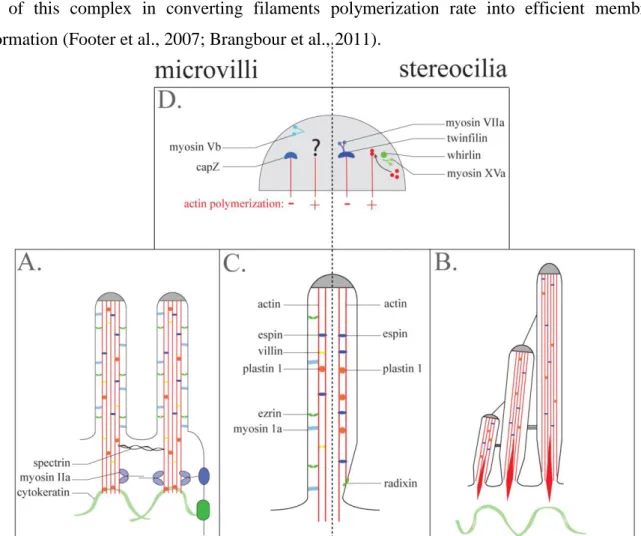
Figure 9 : Description of the different molecular components involved in the shape regulation of intestinal microvilli (right) and stereocilia (left). ... 34
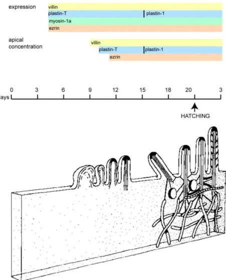
Figure 10 : Brush border morphogenesis during embryo development ... 39
Figure 11 : Brush border morphology in absence of the major microvillar proteins ... 48
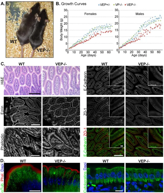
Figure 12 : The VEP−/− mice have growth defects, but the morphology of their intestinal epithelium looks normal. ... 50
Figure 13 : Microvilli still form in the VEP−/− mice ... 52
Figure 14 : The organization of the actin bundle is affected in the VEP−/− microvilli...56
Figure 15 : Distribution of potential actors of microvilli morphogenesis identified by proteomics analysis………....57
Figure 16 : Not all polarity markers are affected in VEP-/- mice ... 59
Figure 17 : Apical enzyme localisation in EP-/- and VP-/- enterocytes ... 59
Figure 18 : VEP−/− mice show apical localization defects of major digestive and absorptive brush border components ... 60
Figure 19 : Unaffected apical trafficking markers in VEP-/- mice ... 62
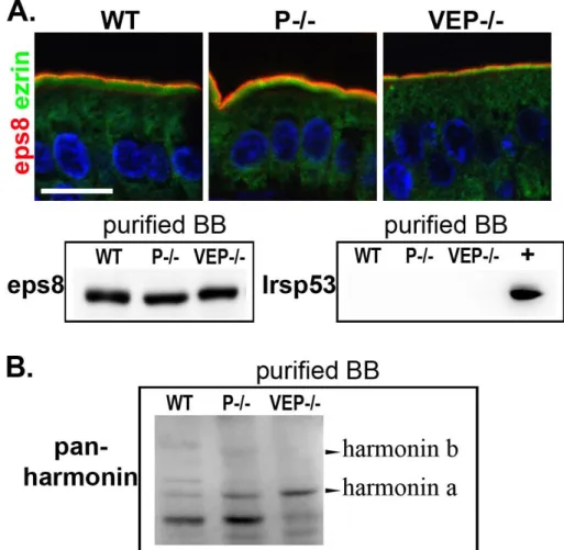
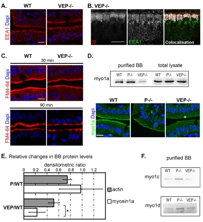
Figure 20 : The apical domain retention machinery is affected in VEP−/− mice ... 63
Figure 21 : Vesicular membrane extrusion is preserved in brush borders of VEP-/- mice ... 64
Figure 22 : Structure of the myosin-1a / calmodulin cross-bridges. ... 74
Figure 23 : Identification of two actin nucleators in the brush border fraction……….….75
Figure 24 : Calcium concentration regulates villin activities ... 78
Figure 25 : Cell migration is a stepwise process ... 83
Figure 26 : Model of villin regulation in actin remodeling at the membrane... 86
Figure 27 : Schematic representation of the transgenesis plasmids ... 96
Figure 28 : The transgenic mouse models recapitulate endogenous villin expression and distribution 97 Figure 29 : Transgenic villin proteins are functional and villin severing activity is required for brush border dissasembly upon carbachol treatment in vivo ... 99
Figure 30 : Efficient colonic wound healing requires villin through its actin severing property ... 100
Figure 31 : Villin positively regulates cell migration through its actin severing property ... 101
Figure 32 : VillinΔsev protein retains its microvillar distribution and its morphogenetic effect ... 103
Figure 33 : Microvilli disassemble upon cell migration via villin severing activity ... 105
Figure 34 : Microvillar actin is rapidly integrated at the lamellipodium following microvillus disassembly ... 107
Figure 35 : Villin is essential for brush border disassembly upon cell migration in vivo. ... 109
Introduction
In multicellular organisms, numerous vital functions are rationally compartmentalized in specialized units made up of specialized cells. Cell specialization enhances the efficiency of these biological functions and reduces global energy consumption. Cells achieve specialization by acquiring a differentiated state, which is defined by the establishment of adapted structures associated to the synthesis of suited proteins. In complex organisms such as mammals, specialized cells executing a common function are usually grouped together to form a tissue. Tissues can furthermore be assembled into an organ to perform more complex functions. Due to the broad variety of functions achieved by organs, specialized cells present an immense diversity of expression profiles and structural properties. Such differentiated state is beautifully exemplified in the cells that constitute the model we used in this study: the intestine, the organ dedicated to digestion and absorption. Before describing our working model, I will give a description of the cytoskeleton and define its role in the establishment of epithelial polarity. Epithelium and polarity are indeed two notions that characterize the intestinal tissue.
A. The cytoskeleton: the building block of cell specializations
A fundamental aspect of differentiation resides in the design of specialized cellular domains within the cell. This compartmentalization into organelles segregates biochemical reactions and increases locally the concentration of molecules, thereby promoting cellular processes. Their efficiency can be further enhanced by a structural specialization of some organelles, such as those found on the apex of intestinal cells. Structural specializations are typically shaped by the cytoskeleton, which brings mechanical support to cellular components. The cytoskeleton consists on an organized and intricate network of cytoplasmic fibrilar proteins present in the cytoplasm. This key element of cell architecture is additionally critical to elaborate the proper intracellular organization. In eukaryotic cells, three main types of fibers compose the cytoskeleton: the actin filaments (or micro-filaments), the intermediate filaments and the microtubules.
Microtubules are long tube-like polymers made up of ubiquitous and conserved globular proteins: and tubulin. These tubulin heterodimers assemble to form the protofilaments, the building block of the microtubules. Microtubules are polarized fibers; they present two distinct extremities, termed + and -, on which different subunits of the dimer are exposed. Elongation of microtubules essentially occurs at the + end (Figure 1). Even if exceptions do exist, microtubules typically arise from the microtubule-organizing center where they are nucleated. Among the diverse functions carried out by microtubules in the cell, they play major role in intracellular trafficking. Two
families of specialized
molecular motors moving
vesicular cargoes along
microtubules have been
characterized: dyneins and kinesins which differ in their polarity of displacement.
Indeed, dyneins move
preferentially towards the + ends of microtubule whereas kinesins are - end directed
motors. In addition to
molecular motors, various proteins regulate microtubule assembly or disassembly,
thereby conferring a
dynamic property to this cytoskeletal element.
Figure 1: The three cytoskeletal filamentous elements of the cell
A. Immunostainings against the different fibers that constitute the cytoskeleton. Left micrograph: actin filaments
(red) and microtubules (green); nuclei are shown in blue. Right image: Cytokeratins (blue) and thee nucleus specific
intermediate filament protein Lamin A (green). Micrographs obtained from:
http://www.bscb.org/softcell/images/mp_tripple.gif and http://www.piercenet.com/media/000022-ICC-cytokeratin-DyLight-405-lamin-488.jpg. B. The highly conserved proteins tubulins and actin form two types of polarized filaments: microtubules and actin filaments. Intermediate filaments arise from multimerisation of intermediate filament monomers. Images obtained from Wikipedia.
In te rm ed ia te fi la m en t Microtubule
In contrast to microtubules, intermediate filaments do not present any polarity and are composed by molecules encoded by a very diverse array of genes. This heterogeneity is mainly based on the high divergence of the N- and C- terminal domains. The central domain, more conserved among the intermediate filaments molecules, allows their dimerization (Figure 1). Assembly of these dimers creates stable and deformable fibers. The high number of different intermediate filaments molecules reflects their diverse intracellular functions and the different cell types from where they originate. For instance, lamins form a meshwork in the inner layer of nuclei. Keratins are found in epithelial cells whereas vimentin is characteristic of mesenchymal cells (Figure 1). The intermediate filaments do not seem to have any function in intracellular trafficking. Their primary role seems rather to confer mechanical resistance to the cell owing to their elastic properties.
The last element that constitutes the cytoskeleton, the actin filaments, or microfilaments, shares two important characteristics with microtubules. Both of them are polarized fibers and made of polymers of globular proteins ubiquitous among different cell types (Figure 1). Actin filaments also play a major role in intracellular trafficking in concert with molecular motors: the myosins.
Actin: a highly conserved protein.
Actin filaments are constituted by monomers of actin – or G-actin -, a globular protein of around 42 kDa found in virtually all eukaryotic cells. Its considerable importance is illustrated by a highly conserved sequence among different species. Structurally, G-actin is composed of four domains that appear as two lobes organized around a central cleft. The cleft binds ATP or ADP and a divalent cation, calcium or magnesium, which results in ADP- and ATP-bound monomers of different conformations (Figure 2A). These actin monomers can assemble into filament composed of two intertwined helices. The actin filament – F-actin – exhibits a polar organization owing to the intrinsic horizontal asymmetry of globular actin. Their polarity can be revealed experimentally by the ability of the head domain – S1 - of myosin II to bind specifically microfilaments. When bound to an actin filament the S1 fragments, which appear as arrowheads at the ultra-structural level, uniformly point towards one extremity: the pointed end. The additional extremity is usually named the barbed end. Besides this discernible polar structure revealed by myosin S1 decoration, polarity is functional, as the two extremities present different biochemical properties (Figure 2) (Pollard and Borisy, 2003).
Generation of actin filaments.
Actin monomers spontaneously assemble, or nucleate, into filaments in vitro upon addition of salts in the solution. The inherent instability of actin dimers and trimers makes however this process unfavorable, resulting in a latency period observed when measuring polymerization rate
in vitro. Once this step is overcome, actin polymerizes rapidly until reaching a plateau
corresponding to a rate limiting availability of G-actin. Concentration of actin monomers is thus a critical parameter influencing actin polymerization.
Assembly of monomers into microfilaments obeys to a critical concentration, above which all G-actin will polymerize. For Mg-ATP-actin, the critical concentration is lower at the barbed end than at the pointed end, whereas they are identical for ADP bound actin (Figure 2B). Indeed, albeit dispensable for polymerization, Mg-ATP bound to the cleft stabilizes the molecule and favors its addition to an existing filament. Consequently, at steady state, addition of ATP linked monomers is favored at the barbed ends, or growing ends. Upon ATP-actin insertion into a filament, actin starts undergoing ATP hydrolysis. As mentioned earlier, ADP-actin monomers are less stabilized within the filament and thus progressively dissociate from the filament. Thereby, ATP hydrolysis appears to act as an internal clock that determines ageing of actin molecules within a filament by inducing dissociation of “aged” molecules. This intrinsic property of actin biochemistry gives rise to the treadmilling process, in which ATP-bound monomers addition at the barbed end is compensated by dissociation of ADP-bound monomers at the pointed ends. Nevertheless, F-actin treadmilling is a relatively slow process that cannot account for the explosive bursts of actin polymerization found in highly dynamic structures such
Figure 2: Actin filament assembly
A. Structural model of an actin monomer and an
actin filament. The actin monomer is constituted by two lobes separated by the central cleft where a cation and ATP or ADP bind. Actin monomers assemble into polarized filaments. Note the intrinsic rotation due to the helical arrangement of the filament. Images are taken from Wikipedia. B.
Characteristics of the dynamics of
polymerization/depolymerization of an actin filament. The critical concentrations (Cc) from filament extremities for ATP bound actin differ at the barbed and pointed ends, giving rise to a slow treadmilling. ATP or ADP bound monomers are indicated with T or D, respectively. Modified from Pollard and Borisy, 2003.
as lamellipodia or filopodia of migrating cells. Likewise, the design of specialized cellular domains requires generation of elaborate actin networks. Therefore, cells express a cohort of actin binding proteins which exploit diverse aspects of actin biochemistry to control actin assembly and disassembly (Pollard and Borisy, 2003).
Cells regulate the availabilities of actin monomers and filament growing ends to control the polymerization rate.
The actin binding protein profilin catalyzes the ADP nucleotide exchange for ATP thus increasing the pool of monomers ready to polymerize. Following nucleotide exchange, profilin stays bound to actin monomer and does not impede its association to existing filaments. In contrast, thymosin B4 which also binds monomers, blocks their nucleation and association. Thus competition between these small actin sequestering proteins regulates the quantity of monomers available for polymerization (Figure 3) (Pollard and Borisy, 2003).
Another level of regulation is provided by capping proteins which bind to barbed ends and terminate their growth (Figure 3). Capping prevents rapid depletion of available monomers that would occur in case of uncontrolled growth of barbed ends. Therefore, capping of growing filaments counterintuitively allows bursts of high actin polymerization. Indeed, the free actin monomers are directed to the few barbed ends that remain uncapped, in a “funneling” effect (Cooper and Schafer, 2000; Le Clainche and Carlier, 2008).
Finally, proteins that accelerate actin disassembly from filaments to exceed the natural dissociation by ATP hydrolysis replenish the actin monomers pool. The most studied member is certainly cofilin, from the actin depolymerizing factor – ADF – family. ADF/cofilin is thought to promote monomer disassembly by two mechanisms. ADF/cofilin binds preferentially to ADP bound monomers and in vitro studies have shown that cofilin enhances monomers off rate – or dissociation – from pointed ends. ADF/cofilin also cuts – or severs – actin filaments, leading to their depolymerization in coordination with capping proteins which stop actin assembly. In fact, recent experimental data suggest that ADF/cofilin replenishes the monomer pool through severing rather than by facilitating dissociation (Figure 3) (Kiuchi et al., 2007). In contrast to ADF/cofilin, proteins from the gelsolin family harbor a capping activity in addition to their F-actin severing property. These proteins are therefore sufficient to increase the quantity of monomeric actin available for polymerization without the support of additional capping proteins.
Facilitated initiation of new actin filaments.
The initiation of new actin filaments is an unfavorable event. Nevertheless, a prompt generation of a large number of new filaments, required in many cellular processes, is achieved by several actin binding proteins that trigger nucleation of new filaments. So far two sub-classes of actin nucleators have been described. They are characterized by the architecture of the actin network which results from nuclation: branched or unbranched.
Responsible for branched nucleation, the Arp2/3 complex binds to a pre-existing filament and initiates the formation of a newborn filament at a 70° angle from the mother filament. Arp2/3 mediated nucleation thus results in a dendritic array of actin filaments. Nucleation occurs as Arp2 and Arp3 subunits structurally resemble monomeric actin and mimic the first monomers of the filament. Originally in an inactive state, Arp2/3 is activated by upstream nucleating promoting factors. Depending on the biological process it is involved in, different factors activate Arp2/3. As an example, the Wiskott-Aldrich symptom protein – WASp – triggers Arp2/3 activation during cellular motility whereas the related protein WASH is rather involved during vesicular fission (Yarar et al., 1999; Derivery et al., 2009). This discrepancy illustrates the versatility and importance of the Arp2/3 complex to initiate two major cellular events.
Figure 3: A cohort of actin binding proteins regulates actin dynamics and organization
Actin depolymerization at the pointed end is favored by proteins of the ADF/cofilin family which sever filaments and/or favor disassembly. Actin assembly at the barbed end is regulated by profilin, thymosin B4 and capping proteins. Actin dynamics is additionally regulated by actin severing proteins which create barbed and pointed ends. The actin network can be subsequently structured and stabilized by actin cross-linking and bundling proteins. Adapted from Revenu et al., 2004.
Proteins of the formin family, which are characterized by the presence of a formin homology domain - FH -, rather induce the formation of linear unbranched filaments. In contrast to Arp2/3, they do not possess any actin-like domain. How formins mechanistically induce the formation of new actin filaments remains unclear, but in vitro studies suggest that they interact with and stabilize actin dimers and trimers through a dimerization via their FH domains (Zigmond et al., 2003). Another intriguing property of formins is their ability to remain associated – or to undergo rapid cycles of association and dissociation – with the barbed end of the growing filament while promoting rapid monomer incorporation (Zigmond et al., 2003; Kozlov and Bershadsky, 2004). In other words, formins act as a processive elongator of actin filaments additionally to their nucleation property. Formins might be of high importance in cell processes based on the formation of long unbranched filaments. Besides formins, other nucleators of unbranched filaments, such as cordon-bleu or spire, exist but are recently identified and relatively less studied (Renault et al., 2008).
B. Elaboration of complex specialized domains
As discussed earlier, the intracellular compartmentalization of different functionally specialized domains is crucial to maximize their efficiency. Many of these specialized structures are designed by networks of actin filaments, which exert mechanical force on the membrane.
A few biophysical parameters on the actin cystoskeleton.
Diverse cellular processes – e.g. motility, membrane deformation and scission - rely, for a large part, on the ability of actin cytoskeleton to generate mechanical forces coupled with a surface. Early experimental studies have proposed that mechanical forces generated by actin polymerization could account for the formation of the acrosomal process, a long linear cell extension emanating from spermatozoids of certain ectoderms during fertilization (Tilney et al., 1973). The formal demonstration that actin polymerization alone could provide forces capable of triggering a cellular process came from the understanding of the actin-based propulsion of enteropathogens (Listeria monocytogenes and Shigella flexneri), and subsequent reconstitution of actin-based motility in a cell-free system (Theriot et al., 1992; Loisel et al., 1999).
An order of magnitude of the force yielded by actin polymerization can be estimated from measurements in vitro which indicate that the formin-induced insertion of one actin monomer at the barbed end produces a force around 1 pN (Kovar and Pollard, 2004; Berro et al., 2007). For actin filament polymerization to exert a force on a load, a mechanical coupling between these two parameters is required. From this statement, two important points can be raised: How can filament elongate if butting a surface? In addition, polymerizing filament would require to be anchored to a substratum; otherwise polymerization force would result in a pushing of the filament rearward. An elegant theoretical answer for these problems consists on the elastic Brownian ratchet model in which the actin filament, as well as the load, is considered to undergo constant oscillations due to thermal energy (Peskin et al., 1993; Mogilner and Oster, 1996). When filament intermittently bends, free space is made available for a monomer to insert thereby lengthening the filament. The restoring force of the filament then pushes the load forward, converting actin polymerization into net movement. However, actin polymerization alone is not sufficient to account for the diversity of actin-based cellular processes, each of them requiring different levels of stabilization and regulation. For instance, this is the case for membrane deformations which require very stiff counterpoises to overcome the high resistance of the membrane. Mechanical properties allow the cytoskeleton to generate scaffolds supporting specialized actin-based structures. Their analysis reveals two main types of actin organization.
Dendritic arrays of actin filaments: the example of the lamellipodium.
Lamellipodia at the front of migrating cells is the main illustration of a specialized domain based on an extensive branched organization. This structure consists on a large membrane extension driven by an intense actin polymerization (Figure 4) (Wang, 1985; Svitkina et al., 1997). The lamellipodium plays major role in cell migration, even though its absence appears to be compensated by other migratory structures (Suraneni et al., 2012; Wu et al., 2012). Electron microcopy studies revealed the elaborate network of actin filaments, constituted by two areas showing different actin organizations. Close to the leading edge (1-3 µm), a preponderant intricate meshwork of short branched actin filaments generates a dendritic-like organization (Svitkina et al., 1997; Svitkina and Borisy, 1999). These filaments are of uniform polarity with barbed ends facing the migrating edge (Svitkina et al., 1997).The existence of branched actin filaments within lamellipodia has been subject to intense debates, but is now universally accepted (Small et al., 1994; Resch et al., 2002; Koestler et al., 2008; Urban et al., 2010; Vinzenz
et al., 2012). A few micrometers away from the leading edge, a less dense organization in long linear filaments predominates (Svitkina et al., 1997). Experiments revealed that the lamellipodium is indeed constituted by two distinct cytoskeletal networks differing in their dynamics: an actin network showing a rapid actin retrograde flow close to the membrane and a more stable network behind (Ponti et al., 2004). The dendritic organization that prevails in lamellipodia suggests that it arises from an Arp2/3-mediated actin nucleation. Accordingly, lamellipodial structure entirely depends on the presence of the nucleator Arp2/3 which initiates and maintains the structure in a branched organization (Suraneni et al., 2012; Vinzenz et al., 2012; Wu et al., 2012). The meshwork of actin branches is furthermore stabilized by filamin which binds high angle orthogonal filaments (Flanagan et al., 2001; Stossel et al., 2001). Proteins from the tropomyosin family also indirectly stabilize actin filaments by preventing binding of several actin regulatory proteins.
Figure 4: The different types of actin networks building specialized cellular domains
Two types of actin architecture support specialized membrane domains: The bundled network (a: bristles, b: intestinal microvilli, and c: stereocilia) and branched network (e: lamellipodia). Filopodia (d) are supported by an actin bundle but seem to arise from a branched network. The main proteins organizing the actin networks are indicated in the red boxes. Bristles: A micrograph obtained by scanning electron microscopy and a phalloidin staining are shown. Intestinal microvilli: Micrographs from scanning and transmission electron microscopy. Stereocilia: A micrograph obtained by scanning electron microscopy and a phalloidin staining are shown. Filopodia and lamellipodia: micrographs from transmission electron microscopy are shown. Adapted from Revenu et al., 2004.
To explain lamellipodium generation and maintenance, a model proposed by Pollard and Borisy is shown in Figure 5. In this model, Arp2/3 activation close to the membrane nucleates branched filaments that push against the membrane. The dendritic network of actin filaments originates as a result of Arp2/3 mediated nucleation. Capping proteins stop barbed ends elongation and prevent uncontrolled growth. Replenishment of the pool of monomers in ensured by ATP hydrolysis altogether with ADF/cofilin which induces dissociation of “aged” monomers and/or severs filaments. The coordinated action of profilin and thymosin 4 furthermore regulates the rate of monomers ready to elongate uncapped barbed ends.
The lamellipodium is certainly the most widely studied organelle showing a dendritic organization, which seems also to be found in the immunological synapse, a large structure between an antigen presenting cell and a T lymphocyte (Billadeau and Burkhardt, 2006).
Figure 5: Model for nucleation and treadmilling at the lamellipodium
1-6: Activation of Arp2/3 at the membrane leads to the generation of a dendritic network of actin filaments, which
pushes the membrane and drives the membrane extension. 7: Capping proteins prevent the uncontrolled elongation of the filaments. 8-9: The rear of the network disassembles efficiently due to natural “ageing” of filaments and severing and/or facilitated depolymerization by ADF/cofilin. 10-11: Actin depolymerization releases numerous actin monomers. Profilin catalyzes ADP to ATP exchange thus regulating the pool of monomers ready to “feed” actin assembly. Adapted from Pollard and Borisy, 2003.
Networks of parallel filaments.
At the leading edge of motile cells, a second motility organelle is often generated: the filopodium. Filopodia also extend from the growth cone of developing neurons. Filopodia are highly dynamic finger-like membrane protrusions of several micrometers long emerging from the lamellipodium sheet. They are thought to function as directional sensors but experimental proof is still lacking. These projections contain long linear filaments of uniform polarity; their growing ends oriented towards the structure tip (Figure 4) (Lewis and Bridgman, 1992; Svitkina et al., 1997). Since EM analyses showed that filopodia from motile cells systematically arise from the lamellipodial dendritic network, an attracting hypothesis concerning their initiation proposes that reorganization of the lamellipodial network allows the emergence of filopodia (Svitkina et al., 2003; Vignjevic et al., 2003). This possibility could be experimentally demonstrated in vitro: a reduction of capping proteins concentration leads to the formation of filopodia-like structures from a dendritic network (Vignjevic et al., 2003). Formins, in particular mDia2, localized at the filopodia tips may drive their initiation from the lamellipodia by protecting barbed ends from capping proteins and/or through nucleation and processive elongation (Zigmond et al., 2003; Yang et al., 2007). Also localized at the filopodia tips, proteins from the Ena/VASP might favor elongation owing to their ability to compete with capping proteins for barbed end binding (Bachmann et al., 1999). The importance of lamellipodia / filopodia interface highlights the possibility of intermediate state between purely dendritic and parallel filaments. Nevertheless, this model cannot be taken as a general rule for filopodial initiation. In this regard, fibroblasts devoid of Arp2/3, which cannot form a lamellipodium, present an increase in number of filopodia that seems to compensate for the loss of lamellipodium to ensure motility (Suraneni et al., 2012; Wu et al., 2012).
In filopodia as well as other types of slender actin-based membrane protrusions, the long linear filaments of uniform polarity are tightly packed together by diverse actin bundling proteins which organize the filaments in a bundle. Biophysical studies have proposed that such bundled organization provides stiffness to the actin network to avoid its buckling due to the opposite membrane tension (Mogilner and Rubinstein, 2005; Atilgan et al., 2006; Claessens et al., 2006; Bathe et al., 2008). The actin bundling proteins are a subset of actin cross-linking proteins, which bind several filaments to create an undefined network. The actin-bundling proteins additionally organize filaments in a regular, tight, parallel array to form a bundle; the latter can only be demonstrated by electron microscopy. The ability to bundle can be conferred by the presence of two F-actin binding motifs or by dimerization of a protein containing a unique F-actin binding
region. Actin filaments within filopodia are bundled by the actin binding proteins fascin, and T-plastin (Figure 4). Fascin depletion results in a dramatic decrease in the number of filopodia, highlighting the importance of the bundled organization to initiate and sustain membrane protrusions (Kureishy et al., 2002; Vignjevic et al., 2006).
Another example of long membrane protrusion is the bristle from drosophila. Contrarily to the other membrane protrusion, each drosophila cell extends only one bristle. Bristles are long membrane deformations of around 400 µm and serve as sensory organelles (Figure 4). A bristle is composed by long 11 actin bundles that are constituted by overlapping short actin filaments packed by the actin bundling proteins forked and fascin (Tilney et al., 1995; Guild et al., 2003).
Stereocilia are shorter apical membrane protrusions. Stereocilia, present on the apex of inner ear cells, allow mechanosensory transduction elicited by sound pressure. They display an organization that resembles a staircase of three rows (Figure 4) (Tilney et al., 1992). Each stereocilium of 1.5 – 5.5 µm long contains numerous actin filaments – until 900 – bundled by the actin bundling proteins T-plastin and espin (Tilney et al., 1988; Zheng et al., 2000). Their characteristic differential length renders stereocilia an excellent system to study the contribution of actin regulatory proteins to shape determination. In particular, several actin binding that regulate stereocilia length have been identified (Boëda et al., 2002; Belyantseva et al., 2005). They are differentially expressed between stereocilia rows but all localize to the stereocilia tips, illustrating the importance of this structure in morphogenesis of actin-based membrane protrusions. Nevertheless, a recent study following protein turnover in stereocilia revealed that these organelles might be much less dynamic than previously thought, as no actin treadmilling could be detected in the core of the protrusion (Zhang et al., 2012).
Microvilli are another typical example of short membrane protrusions supported by a central core of actin filaments. These structures are present on the surface of most of the cells, irrespective of their origin. Nevertheless, they are generally relatively short (<500 nm) and highly unstable (Gorelik et al., 2003), thus preventing any specialization. The inherent dynamics of these short-lived membrane projections combined to their ability to generate structures of higher order, suggest that they act as “elementary blocks” to assemble highly specialized structures on the cell surface (Gorelik et al., 2003). Indeed, various stable specialized domains seems to initially form by the aggregation of tiny membrane protrusions resembling microvilli; this is the case for the inner ear stereocilia and the drosophila bristle (DeRosier and Tilney, 2000). During the first stage of bristle formation, actin bundles are initiated from microvillus
tip-like structures at the cell surface. Their subsequent aggregation and organization by the actin bundling proteins forked and fascin generate a mature bristle (Tilney et al., 1996, 1998). Early in development, the surface of inner ear cells is covered by microvilli (Tilney and DeRosier, 1986). The stereocilia develop by the elongation and lateral addition of actin bundles from this microvilli precursors (Tilney and DeRosier, 1986). On the intestinal epithelium these short-lived structures also aggregate into stable highly ordered structures (Figure 4). Thus, as proposed Tilney and DeRosier, microvilli might represent the “archetypal factory of F-actin bundles” (DeRosier and Tilney, 2000).
C. The epithelia: a polarized interface
The cytoskeleton has a central role in a broad range of cellular processes, which allow the generation of elaborated structures like the one found in epithelial cells. Epithelia ensure protection and concomitantly perform numerous exchanges with the external milieu. Accordingly, epithelial cells are designed to efficiently accomplish these functions (Figure 6). Epithelial cells are polarized; they display two distinct membrane domains: the apical pole facing the lumen and the basolateral pole in contact with the neighboring cells and with the basal lamina. Each domain presents a proper protein and lipid composition. This property results in the formation of two functionally distinct membrane domains. For instance, on the absorptive intestinal cells, the apical pole is highly enriched in numerous digestive enzymes and peptides transporters, whereas their basolateral surface contains mainly canals to allow nutrients passage in the blood. Lipid composition also varies in epithelial cells, the epithelial pole being enriched in cholesterol and sphingolipids whereas the basolateral pole is rather composed of phosphatydilcholine (van Meer and Simons, 1988). In addition to the functional discrepancies between the membrane domains, polarity can also be structural as exemplified in the apical pole of absorptive epithelia covered with numerous microscopic membrane extensions: the microvilli. In vertebrates, the apical and basal poles are physically separated by a molecular complex: the tight junctions, or zonula occludens, a type of highly adhesive cell-cell junction essentially impermeable to fluid. In addition, these intimate cell-cell contacts link together the cytoskeleton of adjacent cells. Tight junctions thus guarantee the cohesion of the tissue, ensure the barrier function of tight epithelia and help to maintain cellular polarity. Located in a more basal manner, adherens junctions and desmosomes represent the anchoring junctional complexes. These types
of junctions are important for transmission of mechanical forces along the epithelium; they interconnect cytoskeletal elements of the neighboring cells. Indeed adherens junctions are tethered to apical cortical actin, a belt of actin filaments oriented in a parallel fashion to the apical pole. Desmosomes indirectly link together intermediate filaments from the neighboring cells (Figure 6).
Polarized cells are endowed with the characteristics required to perform the epithelial functions; generation of an appropriate polarity is hence fundamental to epithelia. How polarity is established has fascinated numerous scientists since decades and is still not fully understood. It is however currently established that it requires the concerted action of the cytoskeleton, vesicular sorting, specific lipids synthesis, signaling molecules and polarity complexes.
Figure 6: Intracellular organization of an epithelial cell
A. Schematic illustration of the major characteristics of an epithelial cell with a focus on the apical pole. The
cytoskeletal elements actin, microtubules and intermediate filaments are depicted in red, light blue and light green, respectively. Polarities of actin filaments and microtubules are indicated. Tight junctions (TJ) seal the epithelium. Note the transversal actin belt emanating from the adherens junctions (AJ). Intermediate filaments are linked to the desmosomes (D). Adapted from Ubelmann et al., 2011. B. Detailed organization of the elements of the junctional complexes. From top to bottom: tight junctions, adherens junctions and desmosomes are depicted. Adapted from Wikipedia (http://www.wikipedia.org).
Organization of polarized trafficking in epithelia.
Establishment of polarity results in an asymmetrical distribution of proteins and lipids on the apical and basolateral domains. This discrepancy is maintained by specific sorting of cargoes at the level of the Golgi apparatus or later on in post-Golgi endocytic compartments. How is polarized trafficking organized in epithelia?
Polarized trafficking includes sorting of proteins towards the apical or the basolateral membranes via vesicular or tubular structures and their final insertion at the membrane of destination by vesicle fusion. Polarized trafficking has been conceptually divided in different routes. In the biosynthetic route, the newly synthesized proteins can be directly delivered to their final destination - apical or basolateral - by early sorting. Alternatively, certain proteins follow an indirect pathway, or transcytosis; in other words, they first reach the basolateral membrane and are subsequently re-addressed to the apical pole, with or without endocytic intermediate. Polarized trafficking also relies for an important part on endocytic routes which allow protein recycling and exchanges between apical and basal poles (Figure 7).
Figure 7: Vesicular trafficking in epithelia
Scheme depicting the main biosynthetic trafficking routes in epithelia. Post-Golgi vesicles are sorted and follow direct routes or transcystosis (red arrows). Vesicles movement is shown in blue arrows. Vesicles either reach directly their destination or traverse endocytic intermediates such as the common recycling endosome – CRE –. Some proteins are sorted apically accordingly to their lipid raft affinity. Fusion at the membrane usually involves SNAREs proteins. Adapted from Weisz and Rodriguez-Boulan, 2009.
The fate of delivery is governed by an important diversity of signals; I will try to summarize the most relevant for epithelial intestinal cells. The sorting machinery recognizes and interacts with these signals, which are generally harbored by the proteins.
Basolateral and apical sorting signals.
Glycosylation and GPI anchoring represent the two most well understood apical targeting determinants. The earliest identified apical sorting signal is certainly the addition of a glycosylphosphatidylinositol – GPI – lipid to a newly synthesized protein as a post-translational modification (Lisanti et al., 1989). Indeed, recombinant addition of a GPI anchor to a normally basolateral destined protein leads to its readdressing at the apical pole (Brown et al., 1989; Lisanti et al., 1989). Glycosylation also plays important role in polarized trafficking: on certain apical proteins, proper insertion of N- and O- glycans are essential for apical targeting (Scheiffele et al., 1995; Yeaman et al., 1997). Nevertheless N- and O- glycosylation of basolateral proteins is also relatively common, indicating that N- and O- linked glycans signals might be recessive relative to basolateral determinants (Rodriguez-Boulan et al., 2005).
The proposal of the existence of lipid rafts represents an important breakthrough in the field of polarized trafficking, as they have been proposed to function as a major sorting platform for apical exocytosis (van Meer and Simons, 1988; Brown and Rose, 1992; Simons and Ikonen, 1997). Albeit still controversial, the lipid raft hypothesis is strongly supported by a whole set of experimental data (Munro, 2003; Rodriguez-Boulan et al., 2005). Lipid rafts are membrane-associated cholesterol- and sphyngomyelin- rich microdomains which are initially assembled in the Golgi complex. They are biochemically defined as membrane resistant to detergent extraction (Brown and Rose, 1992). The lipid raft hypothesis states that raft-dependent proteins are sorted apically due to their affinity for lipid microdomains generated at the Golgi complex. The lipid microdomains and their associated proteins are subsequently delivered to the apical surface through the apical transport machinery (Figure 7). The raft-proteins complex might be further stabilized by lectin proteins while reaching the apical membrane. In particular, galectin 4 has a prominent role in the apical delivery of several raft associated glycoproteins, likely by its virtue to form stable lattices constituted of glycolipids and glycoproteins (Braccia et al., 2003; Stechly et al., 2009).
Critical role of the cytoskeleton and regulators of vesicular trafficking.
Sorting and trafficking involve a coordinated action between the cytoskeleton and organizers of membrane trafficking.
The integrity of microtubule and actin networks is of prime importance to direct intracellular trafficking. For instance, cargoes exit the trans-Golgi network as long tubular structures or vesicles, both of them moving along microtubules (Kreitzer et al., 2003; Polishchuk et al., 2004). Interfering with the microtubule molecular motors dyneins causes a failure for proteins to reach the apical pole (Tai et al., 1999; Noda et al., 2001). This result altogether with dynamic imaging of vesicular movement demonstrate that microtubules are used as tracks for protein delivery (Toomre et al., 1999; Kreitzer et al., 2000). In enterocytes, a selective chemical perturbation of microtubules abrogates apical delivery of membrane proteins. Interestingly, enterocyte apico-basal polarity is also abolished following such treatment (Pavelka et al., 1983; Achler et al., 1989). An intact microtubule network is thus required to establish and maintain the polarity itself; presumably through delivery of polarity determinants. The actin cytoskeleton is also critical at different steps of polarized trafficking. Perturbing actin cytoskeleton generally disrupts proper targeting of basolateral and apical proteins. Actin cytoskeleton reorganization at the Golgi membrane provides the force to allow the budding of vesicular structures (Almeida et al., 2011). F-actin is additionally used as tracks for myosin motors-based movement of vesicular structures as revealed by time-lapse imaging (Jacob et al., 2003). Since the participation of the actin cytoskeleton to apical trafficking in enterocyte is an important part of this study, this specific aspect will be further discussed later on.
Vesicular trafficking is overall coordinated by a large family of small GTPases: the rab proteins. They are critical throughout the whole trafficking pathway, from vesicle budding, to vesicular movement and fusion at the membrane. For instance, rab6 mediates intra-Golgi trafficking (Olkkonen and Stenmark, 1997). Rab8 is another example of rab protein of high importance during polarized trafficking. Rab8 genetic ablation in mouse leads to a global impairment of apical trafficking in intestinal cells. Interestingly, this trafficking failure is coupled with a severe defect in apical membrane functional and structural specialization, providing an additional illustration of the tight interplay between membrane trafficking and polarization (Sato et al., 2007). The case of rab8 furthermore exemplifies the difficulty to define the functions of trafficking regulators among experimental models as it was previously shown to regulate basolateral trafficking in cultured cells from kidney epithelium (Huber et al., 1993).
Following intracellular movement, vesicles fuse at the membrane. An important mediator of this process is represented by the SNARE proteins. SNARE proteins are localized at the vesicle or at the target membrane, and accordingly named v-SNARE or t-SNARE. Interaction between v- and t-SNARE promotes the vesicle docking at the membrane (Figure 7). Specific t-SNAREs are localized at each poles, but both apical and basolateral vesicle fusions involve SNARE proteins (Nejsum and Nelson, 2009).
D. The intestine: a specialized organ
Cellular specialization achieved by polarity is beautifully exemplified in the model we used in this study: the intestine, an organ dedicated to digestion and absorption. These two complementary processes ensure the nutritive function. Following ingestion of aliments, they are reduced – or digested - in simpler elements that can readily be assimilated by the organism after their absorption. Digestion of food is achieved by mechanical and enzymatic processes whereas absorption relies on cellular transport. In most of the animals, the nutritive function is performed in the digestive tract. In mammals, this tract extends from the stomach to the anus. The upper part of the tract is composed of the esophagus and the stomach. The lower part comprehends the small and large intestines.
The stomach fulfills an important part of the digestion by mechanical and enzymatic actions. It mashes the masticated food and secretes abundant quantities of digestive enzymes. The digestive process is further pursued in the small intestine, organ constituted by highly specialized intestinal cells, which harbor and secrete digestive enzymes. The vast majority of intestinal cells is represented by the enterocytes, a cell type specialized in absorption. This cell type absorbs peptides, amino acids and nutrients either passively through diffusion or actively via the expression of channels and transporters. The last part of the lower digestive tract, the colon, allows water re-absorption. Since these tissues are continuously exposed to ingested food, they are often confronted to noxious agents. To avoid deleterious effects to the organism, intestinal cells display a high level of cellular plasticity. In this PhD work, we investigated two important aspects of the biology of the cells that constitutes the intestines: how their functionality is established and how they rapidly adapt in a stress context.
Histo-Anatomical considerations on the large and small intestines.
As an epithelium, the intestinal mucosa separates the organism from the external milieu and ensures the integrity of the organism while performing numerous exchanges with the environment. The two latter processes are achieved by the formation of a virtually impermeable tissue and a spatial segregation of specific functions within the intestinal cell. These properties are characteristic from polarized epithelial cells. The intestine is constituted by a tubular structure covered by a simple polarized columnar mucosal epithelium organized around a lumen. The small intestine is sub-divided in three portions sharing relatively similar anatomical and gross functional properties: the duodenum, jejunum and ileum. Nevertheless, the protein composition slightly differs in these three parts, according to their respective specializations. Epithelial cells from the duodenum are particularly enriched in digestive enzymes. The jejunum is more specialized in absorption of luminal nutrients; jejunal cells express particularly high levels of transporters. Ileum preferentially absorbs vitamins and bile salts and accordingly ileal cells express high level of vitamins transporters.
Histologically, the intestinal tissue, made up of epithelial cells, lays on top of a connective tissue, the lamina propria. An organization around multiple levels of folds is a histological feature characteristic of the intestinal epithelium. The tissue itself is arranged in large folds, which themselves are formed by numerous finger-like extensions of the mucosa called the villi. An obvious advantage conferred by this architecture is the maximization of the surface contact with the lumen, a particularity well adapted to the specialization of this organ (Figure 8). The epithelial tissue is constituted by several cell types which arise from the stem cells and differentiate. A cell compartmentation exists along the villi axis: the differentiated cells lining the villi are luminally exposed whereas the stem cells located at the inter-villi invaginations – the crypts - are relatively hidden and protected (Figure 8). On the other hand, the global organization of the colonic tissue is slightly different as no villi structure exists. Instead the tissue is shaped around larger folds.
The intestinal mucosa is a highly dynamic tissue. Indeed the entire epithelium is renewed each three to five days due to the fast and continuous cycling of the stem cells which reside in the crypts. As a result, cells originating from the crypt continuously flow toward the villi. While reaching the crypt/villi frontier, cells engage their differentiation programs. Finally, differentiated cells undergo apoptosis when reaching the tips of the villi and are subsequently expelled in the lumen (Figure 8).
The different cell types that constitute the intestinal epithelium
Stem cells: The stem cells, “buried” deeply in the crypts and continuously dividing, ensure the rapid renewal of the epithelium. Their “stemness” property has been assessed by the capacity of their progeny to differentiate into the different cell types and by their ability to retain DNA labeling. Moreover, these stem cells are able to generate organoids in vitro that closely resemble the structure of the organ (Sato et al., 2009). Up to now, two populations of stem cells characterized by expression of signatures genes have been identified. The Lgr5 positive stem cells were originally identified by the group of Hans Clevers, and the Bmi1 positive cells in the laboratory of Mario Capecchi (Barker et al., 2007; Sangiorgi and Capecchi, 2008). Lgr5 and Bmi1 cells present different spatial distribution in the crypt: cells positive for Lgr5 lay at the base of the crypts and Bmi1 cells localize in a slightly upper position. A hierarchy between stem
Figure 8: Organization of the intestinal epithelium
Organization of the intestinal epithelium around the crypt-villus axis. Daughter cells arising from the stem cells migrate towards the villus and differentiate into five differentiated cell types belonging to the secretory and absorptive lineages: Paneth cells, enteroendocrine cells, goblet cells, tuft cells (not represented) and enterocytes. Adapted from Crosnier et al., 2006.
cells populations seems to exist, illustrated by the fact that Bmi1 positive cells might give rise to Lgr5 positive cells. The presence of two pools of potential stem cells could be of high importance to compensate the loss of one population in case of injury (Tian et al., 2011).
The differentiated intestinal cell types: The immediate progeny of the stem cells, the transit-amplifying cells, undergoes a limited round of divisions and, while migrating in the villus, differentiates in two lineages: absorptive and secretory. The lineages are constituted by five types of differentiated cell, identified so far.
The secretory lineage comprises the Paneth cells, the enteroendocrine cells, the goblet cells and the recently characterized tuft cells (Figure 8). As the functions of these cells do not directly relate with this study, only a short description will be provided. The enteroendocrine cells locate in the intestinal villi and secrete various hormones which mostly regulate energy homeostasis. The Goblet cells are also present in villi and secrete mucus that covers the epithelium surface. Mucus provides a physical barrier, adding another level of protection against deleterious luminal agents. The tuft cells are under-represented in intestinal villi. Their exact functions remain less clear as they were recently characterized (Gerbe et al., 2011). They secrete opioids, synthesize prostaglandins and trefoil peptides. Interestingly, trefoil peptides have been shown to be essential for injury repair in the colon (Mashimo et al., 1996). In contrast, the Paneth cells reside in the crypts where they intercalate between stem cells. Functionally, they release several anti-microbial compounds in the lumen. They also participate in generating the appropriate microenvironment – or niche – for stem cells: a set of secreted molecules that ensures stem cells homeostasis.
Finally the absorptive lineage is represented by the enterocytes (Figure 8). Enterocytes are, by far, the most abundant cell type in the intestinal epithelium, representing more than 80% of the total cell population. The enterocyte is a polarized cell highly specialized in absorption, orienting its apical pole towards the lumen. On the apex, the functional specialization is provided by the membrane composition combined with a specific structural organization, which further enhances the efficiency of this epithelium. Indeed the enterocyte apex is covered by an organized array of regular microvilli increasing cell size by 30-40 fold: the brush border (Brown, 1962). Microvilli are highly enriched in numerous digestive enzymes, channels and peptide transporters promoting the absorptive capacity. In human, the brush border covering each intestinal enterocytes consists of around 1700 microvilli measuring 1-2 m in length and 0.1 m in diameter (Brown 1962). Each microvillus is supported by 20-30 uniformly polarized actin