HAL Id: hal-03038187
https://hal.archives-ouvertes.fr/hal-03038187
Submitted on 15 Dec 2020
HAL is a multi-disciplinary open access
archive for the deposit and dissemination of
sci-entific research documents, whether they are
pub-lished or not. The documents may come from
teaching and research institutions in France or
abroad, or from public or private research centers.
L’archive ouverte pluridisciplinaire HAL, est
destinée au dépôt et à la diffusion de documents
scientifiques de niveau recherche, publiés ou non,
émanant des établissements d’enseignement et de
recherche français ou étrangers, des laboratoires
publics ou privés.
translation in the germ plasm
Anne Ramat, Maria-Rosa Garcia-Silva, Camille Jahan, Rima Naït-Saïdi,
Jérémy Dufourt, Céline Garret, Aymeric Chartier, Julie Cremaschi, Vipul
Patel, Mathilde Decourcelle, et al.
To cite this version:
Anne Ramat, Maria-Rosa Garcia-Silva, Camille Jahan, Rima Naït-Saïdi, Jérémy Dufourt, et al.. The
PIWI protein Aubergine recruits eIF3 to activate translation in the germ plasm. Cell Research, Nature
Publishing Group, 2020, 30 (5), pp.421-435. �10.1038/s41422-020-0294-9�. �hal-03038187�
ARTICLE
OPEN
The PIWI protein Aubergine recruits eIF3 to activate
translation in the germ plasm
Anne Ramat1, Maria-Rosa Garcia-Silva1, Camille Jahan1, Rima Naït-Saïdi1, Jérémy Dufourt 1,5, Céline Garret1, Aymeric Chartier1,
Julie Cremaschi1, Vipul Patel1, Mathilde Decourcelle 2, Amandine Bastide3, François Juge 4and Martine Simonelig 1
Piwi-interacting RNAs (piRNAs) and PIWI proteins are essential in germ cells to repress transposons and regulate mRNAs. In Drosophila, piRNAs bound to the PIWI protein Aubergine (Aub) are transferred maternally to the embryo and regulate maternal mRNA stability through two opposite roles. They target mRNAs by incomplete base pairing, leading to their destabilization in the soma and stabilization in the germ plasm. Here, we report a function of Aub in translation. Aub is required for translational activation of nanos mRNA, a key determinant of the germ plasm. Aub physically interacts with the poly(A)-binding protein (PABP) and the translation initiation factor eIF3. Polysome gradient profiling reveals the role of Aub at the initiation step of translation. In the germ plasm, PABP and eIF3d assemble in foci that surround Aub-containing germ granules, and Aub acts with eIF3d to promote nanos translation. These results identify translational activation as a new mode of mRNA regulation by Aub, highlighting the versatility of PIWI proteins in mRNA regulation.
Cell Research (2020) 30:421–435; https://doi.org/10.1038/s41422-020-0294-9
INTRODUCTION
Translational control is a widespread mechanism to regulate gene expression in many biological contexts. This regulation has an essential role during early embryogenesis, before transcrip-tion of the zygotic genome has actually started. In Drosophila, embryonic patterning depends on the translational control of a
small number of maternal mRNAs.1 Among them, nanos (nos)
mRNA encodes a key posterior determinant required for
abdominal segmentation and development of the germline.2
nos mRNA is present in the whole embryo, but a small proportion accumulates at the posterior pole in the germ plasm, a specialized cytoplasm in which the germline
devel-ops.3,4 Localization and translational control of nos mRNA are
linked, such that the pool of nos mRNA present in the bulk of the embryo is translationally repressed, whereas the pool of nos mRNA localized in the germ plasm is translationally activated to produce a Nos protein gradient from the posterior pole.3,5,6Both repression of nos mRNA translation in the bulk of the embryo and activation in the germ plasm are required for embryonic development.
The coupling between mRNA localization and translational control depends in part on the implication of the same factors in
both processes. The Smaug (Smg) RNA binding protein specifically
recognizes nos mRNA through binding to two Smaug recognition elements (SRE) in its 3′UTR.7,8Smg is both a translational repressor
of nos, and a localization factor through its role in mRNA deadenylation and decay in the bulk of the embryo, by
recruitment of the CCR4-NOT deadenylation complex.7–9 Smg
directly interacts with the Oskar (Osk) protein that is specifically synthesized at the posterior pole of oocytes and embryos and
drives germ plasm assembly.7,10 Smg interaction with Osk
prevents Smg binding to nos mRNA, thus contributing to relieving both Smg-dependent translational repression and mRNA decay in the germ plasm.7,9,11Osk is therefore a key player in the switch of
nos and other germ cell mRNA regulation between soma and germ plasm of the embryo.
More recently, we have demonstrated the role of Aubergine (Aub) in the localization of germ cell mRNAs to the germ
plasm.12,13 Aub is one of the three PIWI proteins in Drosophila.
PIWI proteins belong to a specific clade of Argonaute proteins that
bind 23–30 nucleotides (nt)-long small RNAs referred to as
Piwi-interacting RNAs (piRNAs).14,15piRNAs and PIWI proteins have an
established role in the repression of transposable elements in the germline of animals. piRNAs target transposable element mRNAs through complementarity and guide interaction with PIWI proteins that, in turn, cleave targeted mRNAs through their endonucleolytic activity. In addition to this role, piRNAs have a conserved function in the regulation of cellular mRNAs in various
biological contexts.16In the Drosophila embryo, Aub loaded with
piRNAs produced in the female germline is present both at low levels in the bulk of the embryo and at higher levels in the germ
plasm.17,18 Aub binds maternal germ cell mRNAs through
incomplete base pairing with piRNAs.12,18,19Aub binding to these
mRNAs induces their decay in the bulk of the embryo, either by direct cleavage or recruitment together with Smg of the
CCR4-NOT deadenylation complex.12,18In contrast, in the germ plasm
Received: 29 November 2019 Accepted: 11 February 2020 Published online: 4 March 2020
1
mRNA Regulation and Development, Institute of Human Genetics, UMR9002 CNRS-Univ Montpellier, 141 rue de la Cardonille, 34396 Montpellier Cedex 5, France;2BioCampus Montpellier, CNRS, INSERM, Univ Montpellier, Montpellier, France;3
IGF, Univ Montpellier, CNRS, INSERM, Montpellier, France and4
Institut de Génétique Moléculaire de Montpellier, Univ Montpellier, CNRS, Montpellier, France
Correspondence: Martine Simonelig (Martine.Simonelig@igh.cnrs.fr) 5
Present address: Institut de Génétique Moléculaire de Montpellier, Univ Montpellier, CNRS, Montpellier, France These authors contributed equally: Anne Ramat, Maria-Rosa Garcia-Silva
www.cell-research.com
1234567
Aub recruits Wispy, the germline-specific cytoplasmic poly(A) polymerase, leading to poly(A) tail elongation and stabilization of
Aub-bound mRNAs.13Thus, Aub and piRNAs play a central role in
the localization of germ cell mRNAs through two opposite functions in mRNA stability: mRNA destabilization in the bulk of
the embryo and stabilization in the germ plasm. The role of piRNAs and PIWI proteins in cellular mRNA regulation in other contexts, including mouse spermiogenesis and sex determination in Bombyx, also depends on their function in the regulation of mRNA stability.20–24
Size ( m)
c
osk-OE : UASp-osk/+; nos-Gal4/+
osk-OE; aub-/-: UASp-osk, aubHN2/aubQC42; nos-Gal4/+
osk-OE; armi-/-: UASp-osk/+; armi72..1 nos-Gal4/armi1 WT osk-OE osk-OE; armi -/-osk-OE osk-OE; aub -/-nos mRNA level normalized to RpL32 ns ns ns
osk-OE osk-OE; aub
-/-a
WT Nos GFP-Aub WT osk-OE osk-OE; aub-/-Fluorescence intensity (a.u.)
**** ****
b
Tubulin Nos
Osk
WT osk-OE osk-OE; aub WT
-/-osk-OE osk-OE; armi
-/-WT Nanos WT Oskar osk-OE; armi -/-osk-OE osk-OE; aub -/-osk-OE; armi -/-osk-OE osk-OE; aub -/-**** ns * **** ns No rmali ze d p ro tein lev el Nanos
e
Wild type Reduced size Reduced intensity No proteinh
g
f
Reduced size Nos Reduced intensity Nos Nos Wild type Reduced size nos nosd
Aub crosslink sites nos 1 kb piRNA (roo)UUCAUUUCAACACACAUAUCUAUAUAUAUAUAUAUAUAUAUAUAUAUAUAUAUAUAUAUAUAUGUUAUAUAUUUAUUCAAUUUUGUUUACCAUUGAUCAAUUUUUCACACAUGAAACAACCGCCAGCAUUAUAUAAUUUUUUUAUUUUUUUAAAAAAUGUGUACAC 3’AGGUUUGUGUGUAUAUAUAUAUUUAU5’ 3’AGCCCGACUGUAUAUAAAUAAGUUAA5’ piRNA (412) Posterior nos mRNA level (%) WT
nos pi nos( piroo pi412)/+; nosBN/BNx 20 10 30 40 50 60 70 80 90 100 0 n=300 n=377
Posterior Nos level (%)
20 10 30 40 50 60 70 80 90 100 0 n=306 n=312
i
j
Fluorescence intenisty (a.u.) 0
400 800 1200 1600 20 40 60 80 100 Size ( m) 0 400 800 1200 1600 0 20 40 60 80 100 120 140 Wild type nos pi nos pi WT wild type nos pi 2120 2285 **** **** 120
Posterior nos mRNA Posterior Nos protein
Here, we describe translational activation as a new mechanism of mRNA regulation by piRNAs and PIWI proteins. Using ectopic expression of Osk in the whole embryo to mimic the germ plasm, we show that Aub and piRNAs are required for nos mRNA translation. Mass spectrometry analysis of Aub interactors in early
embryos identifies several components of the translation
machin-ery, including translation initiation factors. We find that Aub
physically interacts with the poly(A)-binding protein (PABP) and several subunits of the translation initiation complex eIF3. Furthermore, PABP and eIF3d accumulate in foci that assemble around and partially overlap with Aub-containing germ granules in the germ plasm. Polysome gradient analysis indicates that Aub activates translation at the initiation step. Finally, functional experiments involving the concomitant decrease of Aub and eIF3d show that both proteins act together in nos mRNA translation in the germ plasm. These results identify translational activation as a new level of mRNA regulation by PIWI proteins. Moreover, they expand the role of the general eIF3 translation initiation complex in translation regulatory mechanisms required for developmental processes.
RESULTS
Aub is required for nos mRNA translation
Only a low amount (4%) of nos mRNA is localized to the germ
plasm and actually translated.3,4 nos mRNA stabilization and
translation in the germ plasm depend on the presence of Osk. Therefore, we ectopically expressed Osk in the whole embryo
using UASp-osk25 and the germline-specific driver nos-Gal4, to
increase translated nos mRNA levels and address the mechanisms of translational activation. Osk overexpression (osk-OE) in embryos from UASp-osk/+; nos-Gal4/+ females led to increased and ectopic
Nos protein synthesis in whole embryo (Fig.1a). Quantification of
Nos protein levels in osk-OE embryos, either following Nos visualization using immunostaining or western blot, revealed a
2-fold increase compared to wild-type (WT) embryos (Fig.1a, b). In
contrast, nos mRNA levels quantified using RT-qPCR were similar
in osk-OE and WT embryos (Fig.1c). This is consistent with the
presence of high amounts of nos mRNA in the bulk of embryos, and nos spatial regulation depending mostly on translational
control at these stages (0–2 h embryos). Therefore, Osk
over-expression in 0–2 h embryos led to ectopic translational activation
of nos mRNA without changes in nos mRNA levels.
Aub protein is present at low levels in the bulk of WT embryos
and highly accumulates in the germ plasm.18Ectopic expression of
Osk led to an homogeneous redistribution of Aub in the embryo (Fig.1a). Strikingly, the lack of Aub in osk-OE embryos resulted in
the lack of Nos protein synthesis (Fig.1a, b), although nos mRNA
levels were not decreased (Fig.1c). This result suggested that Aub
was required for nos mRNA translational activation in the presence
of Osk. Importantly, the level of Osk protein was not significantly
affected by aub mutation, indicating that the lack of Nos protein did not result from the lack of Osk (Fig.1b). Of note, the UASp-osk transgene almost exclusively overexpressed the long Osk isoform of the two isoforms, Short-Osk and Long-Osk (Supplementary information, Fig. S1a). Long-Osk can induce germ plasm assembly
when overexpressed although less actively than Short-Osk.26
Long-Osk levels were poorly affected by aub mutations, making this UASp-osk transgene a useful tool to address direct nos mRNA
regulation by Aub and piRNAs (Fig.1b; Supplementary
informa-tion, Fig. S1a).
We analyzed the role of Armitage (Armi), another component of
the piRNA pathway with a prominent role in piRNA biogenesis,27
in nos mRNA translational activation. Nos protein levels were
strongly reduced in osk-OE; armi−/−embryos, compared to osk-OE
embryos, although again, the levels of Osk protein were not
significantly decreased (Fig.1b). nos mRNA levels quantified using
RT-qPCR remained unaffected by armi mutation (Fig.1c), revealing
a role of Armi in nos mRNA translational control. Armi does not
localize to the germ plasm.13 Instead the defect in nos mRNA
translational activation in armi mutant might depend on highly
reduced piRNA levels in this mutant,27 suggesting that piRNAs
were required in Aub binding to nos mRNA for its role in translational activation. This is consistent with Aub iCLIP assays showing that an Aub double point mutant in the PAZ domain,
AubAA that is unable to load piRNAs, was also unable to bind
mRNAs.12 To confirm the role of piRNAs in Aub-dependent
translational activation of nos, we took advantage of the nos (ΔpirooΔpi412) transgene, in which two piRNA target sites (from roo and 412 transposable elements) in close proximity to a
prominent Aub-binding site in nos 3′UTR have been deleted12,18
(Fig. 1d). We have shown before that deletion of these piRNA
target sites affected nos mRNA localization to the germ plasm,
without affecting its level.13 Using single molecule fluorescence
in situ hybridization (smFISH), we confirmed the posterior
localization defect of nos mRNA from this transgene: 36% of embryos showed a reduced domain of posterior localization
(Fig. 1e, g, i). As previously reported, this defect was moderate
because several Aub-binding sites remained unaffected in the nos
(ΔpirooΔpi412) transgene (Fig. 1d).13 Recording nos mRNA
translation in nos(ΔpirooΔpi412)/+; nosBN/BNx embryos using
immunostaining showed a similar percentage of embryos (38%)
with reduced protein accumulation (Fig.1f, h, j). However, protein
synthesis appeared to be more affected than mRNA localization in these embryos since 3.8% of them did not produce any Nos protein, a defect (no localization) that did not occur with nos
mRNA (Fig. 1e, g). In addition, when taking into account all
embryos, immunofluorescence intensity was reduced in nos
Fig. 1 Aub and Armi are required for nos mRNA translation. a Immunostaining of WT, osk-OE and osk-OE; aub−/−embryos with anti-Nos
antibody (top panels). The genotypes are indicated. Fluorescence quantification was performed using the ImageJ software with 5–6 embryos per genotype. Error bars represent SEM. ****P < 0.0001 using the unpaired Student’s t-test. Immunostaining of UASp-GFP-Aub nos-Gal4 and UASp-osk/+; UASp-GFP-Aub nos-Gal4/+ embryos with anti-GFP antibody, showing the distribution of Aub protein (bottom panels). Posterior is to the right. b Western blots of WT, osk-OE, osk-OE; aub−/−and osk-OE; armi−/−embryos revealed with anti-Nos, anti-Osk and anti-α-Tubulin antibodies.α-Tubulin was used as a loading control. Quantification was performed using the ImageJ software with 3–6 biological replicates. Error bars represent SEM. ****P < 0.0001, **P < 0.01, *P < 0.05, ns not significant, using the unpaired Student’s t-test. c Quantification of nos mRNA using RT-qPCR in WT, osk-OE, osk-OE; aub−/− and osk-OE; armi−/− embryos. mRNA levels were normalized with RpL32 mRNA. Quantification of 4–8 biological replicates. Error bars represent SEM. ns not significant, using the unpaired Student’s t-test. d Schematic representation of nos mRNA and 3′UTR targeting with piRNAs. Thin boxes are 5′UTR and 3′UTR, lines are introns and thick boxes are exons. Clusters of Aub crosslink sites are indicated in red.12The sequence of the region with the strongest crosslink sites and base pairing with representative roo and 412 piRNAs are shown. The deletions overlapping with the piRNA target sites in the nos(ΔpirooΔpi412) transgene are boxed.18 Aub crosslinked nt are in red. e, f nos smFISH (e) and immunostaining with anti-Nos antibody (f) of wild-type and nos (ΔpirooΔpi412)/+; nosBN/BNxembryos. Posterior of embryos with the three types of staining: wild type, reduced size or reduced intensity, are
shown. Scale bars, 20μm. g–j Quantification of posterior staining shown in e and f using the ImageJ software. For each genotype, the percentage of embryos with each staining category was recorded for nos mRNA (g) and Nos protein (h). ****P < 0.0001 using theχ2test. Scatter plots of size andfluorescence intensity of posterior staining for each embryo, for nos mRNA (i) and Nos protein (j). Two-way ANOVA showed significant difference (P < 0.001) in fluorescence intensity of posterior staining for nos mRNA and Nos protein between genotypes.
nos GFP-Aub GFP-Aub Nos Merge GFP-Aub Osk
c’’
b’
b
b’’
a
a’
a’’
c’
c
Merge MergeUASp-osk/+; UASp-GFP-Aub nos-Gal4/+, soma
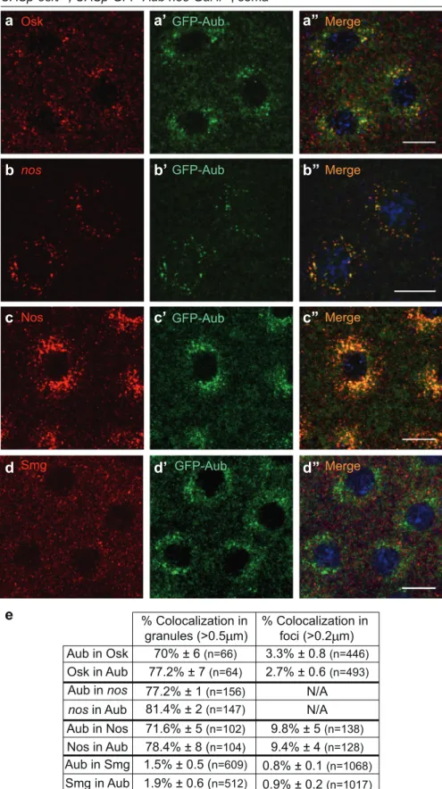
e
Aub in Osk Osk in Aub Aub in nos nos in Aub Aub in Nos Nos in Aub % Colocalization in foci (>0.2 m) % Colocalization in granules (>0.5 m) 70% ± 6 (n=66) 77.2% ± 7 (n=64) 81.4% ± 2 (n=147) 77.2% ± 1 (n=156) 78.4% ± 8 (n=104) 71.6% ± 5 (n=102) 9.8% ± 5 (n=138) 9.4% ± 4 (n=128) N/A N/A 3.3% ± 0.8 (n=446) 2.7% ± 0.6 (n=493) Aub in Smg Smg in Aub GFP-Aub Smgd’
d’’
d
Merge 1.5% ± 0.5 (n=609) 1.9% ± 0.6 (n=512) 0.8% ± 0.1 (n=1068) 0.9% ± 0.2 (n=1017)Fig. 2 Ectopic expression of Osk nucleates RNA granules related to germ granules in the soma. a–d” Immunostaining of osk/+; UASp-GFP-Aub nos-Gal4/+ embryos with anti-Osk (red) and anti-GFP (green) to visualize Aub (a–a”); anti-Nos (red) and anti-GFP (green) (c–c”); and anti-Smg (red) and anti-GFP (green) (d–d”); and smFISH of embryos with the same genotype revealing nos mRNA and GFP-Aub through GFP fluorescence (b–b”). DNA was visualized using DAPI. Scale bars, 10 μm. e Quantification of colocalization of immunostaining and smFISH shown in a–d”, using the Imaris software. Granules (> 0.5 μm) and foci (> 0.2 μm) were quantified around nuclei and in the cytoplasm between nuclei, respectively.
(ΔpirooΔpi412)/+; nosBN/BNx embryos compared to WT, again a
defect that did not occur with smFISH (Fig. 1i, j). Therefore,
deletion of piRNA target sites in nos mRNA affected Nos protein synthesis, in addition to reducing mRNA localization.
These data are consistent with a direct role of Aub and piRNAs in nos mRNA translation through piRNA-guided binding of Aub to
nos. In this hypothesis, a piRNA pathway component specifically
involved in transposable element regulation should not interfere with nos mRNA translational control. We used Panoramix (Panx), a key factor in Piwi-dependent transcriptional silencing of transpo-sable elements, which acts downstream of Piwi and has no
function in piRNA biogenesis.28,29panx mutants had no effect on
Aub Tud PABP eIF3d eIF4E eIF3k eIF3b Rin Nop56 Smn FibSF2 Egl BicC Nop5 Rump Imp Larp CG7878 Csul Tral Vret Bel Cup Aub Tud Aub Tud PABP eIF3d eIF3k eIF4E eIF3b eIF2 Aub Tud Tral Vret Csul Cup Bel CG7878 Larp Rump Nop5 BicC Nop56 Su(f) Imp Fib Nup98-96 RanGAP Upf1
f
FLAG Initiation Factors
Transfection into S2R+ cells FLAG Immunoprecipitation Measure FFL and RL activities
IP/Input 0.0 0.5 1.0 1.5 IP ef ficiency (%): RL (IP/Input)/FFL(IP/Input) IP Control ns ns ns ns ns ns *** *** *** *** *** ** Aub/P ABP
Aub/eIF3dAub/eIF3fAub/eIF3gAub/eIF3hAub/eIF3kAub/eIF4AAub/eIF4E Aub/Sd Aub/Cherry Cherry/Cherry Aub/eIF3b eIF3 Controls + FFL Aub RL IF
c
d
e
b
Aub interactors GO terms (104 genes) Biological process Cytoplasmic translation TranslationPeptide Biosynthesis process
p-value Matches 5.168318e-68 1.644336e-35 2.109374e-34 51 60 60
a
WT WT osk -/-osk -/-425Nos protein levels in osk-OE embryos, consistent with a role of Aub and piRNAs in nos mRNA translation, independent of their role in transposable element regulation (Supplementary information, Fig. S1b).
Finally, most aub mutant embryos fail to develop, although they are fertilized.12,30 To address whether the lack of Nos protein in
osk-OE; aub−/− embryos could result from their arrest of
embryonic development, we quantified Nos protein levels in
osk-OE unfertilized eggs that are activated by egg laying but do not develop. Nos levels were similar in osk-OE unfertilized eggs and embryos, demonstrating that the defect in Nos protein
synthesis in osk-OE; aub−/−embryos did not result from their lack
of embryonic development (Supplementary information, Fig. S1c). Together, these results show that piRNA-guided Aub binding to nos mRNA plays a direct role in translational activation in the presence of Osk.
Ectopic expression of Osk leads to the formation of granules related to germ granules in the soma
In the germ plasm, Osk leads to the assembly of germ granules that are large ribonucleoprotein particles containing mRNAs
required for germ cell specification and development.4,31 In
addition to Osk, Aub is a core component of germ granules.32
We asked whether Osk ectopic expression in the somatic part of the embryo could lead to the formation of RNA granules related to germ granules, containing Aub and nos mRNA. Immunostaining of osk-OE embryos also expressing GFP-Aub revealed that Osk was present in the bulk of the embryo where it accumulated in
cytoplasmic foci that became larger around nuclei (Fig.2a).
GFP-Aub was also present in cytoplasmic foci in the bulk of osk-OE embryos and in larger foci around nuclei. Small foci of either Osk or GFP-Aub were dispersed in the cytoplasm and did not colocalize. However, Osk and GFP-Aub colocalized in larger foci that surrounded nuclei, indicating a different composition of these
large foci (Fig.2a–a”, e). smFISH of nos mRNA in embryos of the
same genotype showed that nos mRNA accumulated in larger foci
around nuclei where it colocalized with GFP-Aub (Fig.2b–b”, e).
Strikingly, Nos protein also accumulated around nuclei and partially colocalized with GFP-Aub in large foci, suggesting that nos mRNA translation occurred in the vicinity of these granules
(Fig. 2c–c”, e). In contrast, in osk-OE embryos, Smg protein was
present in foci that did not concentrate around nuclei and did not colocalize with large GFP-Aub foci (Fig.2d–d”, e). This result was consistent with the reorganization of Smg into small foci in the germ plasm as compared to the somatic region in WT embryos, which suggested that Smg interaction with Osk did not take place
within germ granules.13
We conclude that the presence of Osk in the somatic part of osk-OE embryos induces the formation of RNA granules that share functional similarities with germ granules, in which Aub and nos mRNA accumulate and at the proximity of which nos mRNA is translated.
Aub interacts with translation initiation factors
To further decipher the function of Aub, we identified Aub interactors in embryos. GFP-Aub was immunoprecipitated from
UASp-GFP-Aub nos-Gal4 0–2 h embryos and the coprecipitated proteins were analyzed using mass spectrometry. Embryos expres-sing GFP alone were used as negative controls (Supplementary
information, Fig. S2a). 107 proteins were significantly enriched in
GFP-Aub immunoprecipitation (IP) (P < 0.05) (Supplementary
infor-mation, Table S1). Known Aub interactors were identified, including
Tudor (Tud) that is restricted to the germ plasm and required for
Aub accumulation in the germ plasm,33,34three components of the
nos translation repressor complex, Trailer hitch (Tral), Belle (Bel) and
Cup,35and Capsuleen/PRMT5 (Csul), the methyltransferase
respon-sible for Aub arginine dimethylation36 (Fig. 3a). Several
RNA-binding proteins were also found in GFP-Aub IP (Fig. 3a).
Importantly, six translation initiation factors were identified as
Aub interactors, among which are PABP, three subunits of eIF3 (eIF3d, eIF3k and eIF3b), and eIF4E, another component of nos translation repressor complex35(Fig.3b). In addition, 48 ribosomal proteins coprecipitated with Aub (Supplementary information, Fig. S2b). Gene Ontology (GO) term enrichment analysis using
FlyMine (http://www.flymine.org) identified “Translation” as the
most enriched term among Aub interactors (Fig. 3e). We also
analyzed Aub interactors in osk54mutant embryos that do not form
germ plasm, with the aim of identifying specific Aub interactors in
the germ plasm, which might be lost in osk mutant embryos.
However, mass spectrometry of GFP-Aub IP from osk54 mutant
embryos identified a very similar set of proteins to that identified in
osk+ embryos (Fig. 3c, d; Supplementary information, Fig. S2c,
Table S1). These data suggested that Osk might not affect Aub interaction with most of its protein interactors, but rather their activity. Indeed, PABP and eIF4E are found in the nos translational repressor complex, although they do not activate translation in this
complex.35eIF3 subunits were found to be in complex with Aub in
the absence of Osk, suggesting that they might also be present in the nos repressed mRNP. The presence of Osk, by remodeling the mRNP, would allow to switch on their activity in translational activation.
We used quantitative luminescence-based coIP (LUMIER) assays
to validate Aub interactions with translation initiation factors.37
Aub was fused to FLAG-tagged Firefly luciferase (FFL), whereas potential interactors were fused to Renilla luciferase (RL).
Following transient expression in Drosophila S2R+ cells, Aub was
immunoprecipitated with FLAG antibodies, or without
anti-bodies as negative control, and interactor coIP was quantified by
recording Renilla and Firefly luciferase activities (Fig. 3f). PABP, four subunits of eIF3 (eIF3b, eIF3d, eIF3g and eIF3h) among six
tested subunits, and eIF4E were found to significantly
coprecipi-tate with Aub in these assays. Thus, although eIF3k that was
identified as an Aub interactor by mass spectrometry could not be
confirmed with the LUMIER assay, eIF3d and eIF3b interaction with
Aub was confirmed, and two other eIF3 subunits, eIF3g and eIF3h
were found to be in complex with Aub. Differences in the interaction between Aub and eIF3 individual subunits between
embryos and S2R+ cells likely resulted from differences in these
two experimental systems.
These results reveal that Aub physically interacts with the translation machinery and are consistent with a direct function of Aub in translation regulation.
Fig. 3 Identification of Aub-interacting partners. a–d Volcano plots showing the mass spectrometry analysis of GFP-Aub immunoprecipita-tion from 0–2 h embryos. Embryos expressing cytoplasmic GFP were used as control. UASp-GFP-Aub nos-Gal4 embryos (a, b); osk54; UASp-GFP-Aub/nos-Gal4embryos (c, d). The analysis was based on four biological replicates. The red line indicates the significance threshold (P = 0.05). Known Aub interactors and RNA-binding proteins are indicated in red and purple, respectively (a, c); translation initiation factors are indicated in blue (b, d). e GO analysis of proteins identified as Aub interactors by mass spectrometry. f Validation of Aub interactors using the LUMIER assay. Left: schematic representation of the assay (FFL Firefly luciferase; RL Renilla luciferase). Right: graph plotting the IP efficiency of the indicated proteins. The values are IP efficiencies of the coprecipitation of the RL fusion proteins (IP/Input) normalized by the IP/Input values for FLAG-FFL-Aub. Error bars represent SD. Stars indicate values significantly greater than six times the mean value obtained in the control IPs without anti-FLAG antibody (Control). Scalloped (Sd) and Cherry proteins were used as negative controls. ***P < 0.001, **P < 0.01, ns not significant, using the Z-test.
Aub interaction with PABP and eIF3d
Because PABP and eIF3d showed the strongest association with Aub in the mass spectrometry analysis, and have key roles in translation initiation, we further investigated their interaction with Aub. We used coIP to address Aub physical interaction with
PABP in embryos. PABP coprecipitated with GFP-Aub in 0–2 h
embryos; however, this coprecipitation was strongly reduced in
the presence of RNase (Fig.4a). In the reverse experiment, PABP
was also able to coprecipitate Aub, but this coprecipitation was
abolished in the presence of RNase (Fig.4b). These results could
indicate either that Aub and PABP did not interact directly and
coprecipitated through their binding to the same mRNAs, or that Aub direct interaction with PABP was stabilized by mRNA in a tripartite association. To address this question, we analyzed direct interaction between Aub and PABP using GST pull-down assays. Aub has three domains characteristic of Argonaute proteins (PAZ, MID and PIWI) and was separated into two parts,
Aub (1–482) that contains the N-terminal and PAZ domains, and
Aub (476–866) that contains the MID and PIWI domains (Fig.4c).
PABP is composed of four RNA recognition motifs (RRM1-4), a proline-rich linker region and a PABP C-terminal (PABC)
domain.38 Each RRM and the PABC domain were fused
WT GFP-Aub WT GFP-Aub WT GFP-Aub
a input GFP IP no RNase + RNase PABP GFP-Aub input PAB P IP mock IP PAB P I P mock IP b Aub PABP no RNase + RNase input GFP IP no RNase + RNase
HA-eiF3d HA-eiF3d GFP-Aub HA-eiF3d HA-eiF3d GFP-Aub HA-eiF3d HA-eiF3d GFP-Aub GFP-Aub
HA-eiF3d
input HA IP
no RNase + RNase
HA-eiF3d HA-eiF3d HA-eiF3d
Aub HA-eiF3d WT WT WT f g MID PIWI PA Z 1 866 1-482 476-866 + -PABP Aub RRM1 RRM2 RRM3 RRM4 1 634 1-89 81-180 168-284 260-367 526-634 c HA-Aub 1-482 HA-Aub 476-866 Input GST GST -RRM1 GST -RRM2 GST -RRM3 GST -RRM4 GS T-P ABC
GFP-Aub HA-eIF3d Merge
h h’ h’’
i
j j’ j’’
i’ i’’
GFP-Aub PABP Merge
d d’ d’’
e e’ e’’
PABC
separately to GST. In vitro-translated HA-tagged Aub(1–482) bound to recombinant GST-RRM1, but not to the other PABP domains fused to GST or GST alone. In addition, HA-Aub
(476–866) did not bind to any PABP domain (Fig.4c). These data
revealed direct interaction between RRM1 of PABP and the N-terminal half of Aub. They were consistent with the model in which Aub directly interacted with PABP and mRNA stabilized this interaction in embryos.
We then analyzed potential colocalization of Aub and PABP. Co-immunostaining of GFP-Aub-expressing embryos with anti-PABP and anti-GFP antibodies showed that PABP was distributed in the whole embryo and specifically accumulated in the germ plasm (Fig.4d–d”). Strikingly, PABP was present in foci, and in the germ plasm a large proportion of Aub-containing germ granules (79.4%, Supplementary information, Fig. S3a) either colocalized with, or were in close proximity to and surrounded by PABP foci, with a
partial overlap of both proteins (Fig. 4e–e”; Supplementary
information, Fig. S3a).
eIF3 is composed of twelve subunits and one associated
factor, and coordinates several steps of translation initiation.39
Interestingly, in addition to this role in basal translation, eIF3
plays regulatory roles in the translation of specific mRNAs. eIF3d
appears to be a major actor in eIF3 regulatory functions, either
through its binding to 5′UTR of specific mRNAs, leading to
cap-independent translation, or directly through its interaction with
the cap structure.40,41Aub interaction with eIF3d was analyzed
in embryos using coIP. GFP-Aub was able to coprecipitate HA-eIF3d in 0–2 h embryos, and this coprecipitation was maintained
in the presence of RNase (Fig.4f). Conversely, HA-eIF3d was able
to coprecipitate Aub in 0–2 h embryos, and although less
efficient, this coprecipitation remained in the presence of RNase
(Fig. 4g). Colocalization of Aub and eIF3d was analyzed in
embryos expressing both GFP-Aub and HA-eIF3d. eIF3d was present in the whole embryo with a slight accumulation in the
germ plasm (Fig.4h–i”; Supplementary information, Fig. S3b, c).
Similarly to PABP, eIF3d formed foci, and in the germ plasm most Aub-containing germ granules (83.2%, Supplementary information, Fig. S3d) colocalized with or were surrounded by eIF3d foci, with a partial colocalization of both proteins at the
edge of the granules (Fig. 4j–j”; Supplementary information,
Fig. S3d).
Taken together, these results show that Aub is in complex with the translation initiation factors PABP and eIF3d. In the germ
plasm, PABP and eIF3d have a specific organization around germ
granules and colocalize with Aub at the periphery of the granules, suggesting that translation might take place at the edge of germ granules.
Mechanism of Aub-dependent translational activation
Aub association with translation initiation factors suggested that Aub might activate nos mRNA translation at the level of initiation.
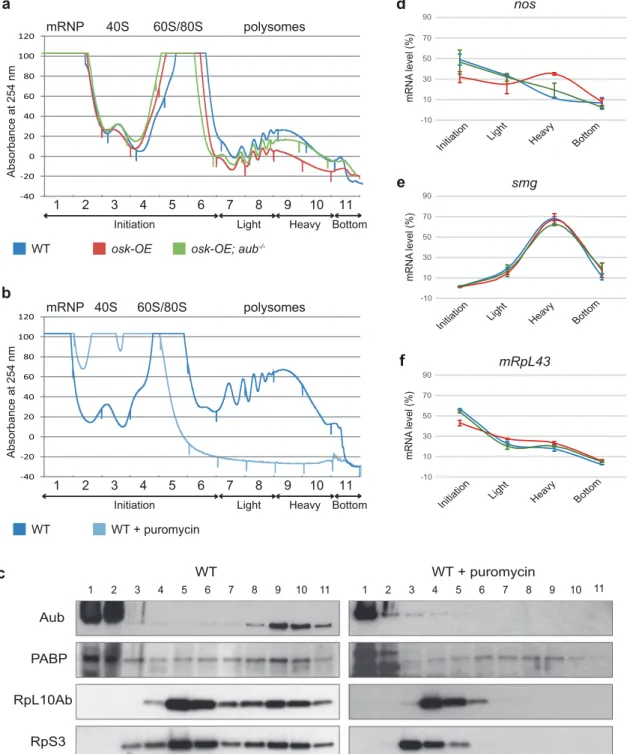
We directly addressed this question using polysome profiling in
which mRNA-protein complexes are separated by fractionation
through linear sucrose gradients.42 mRNA localization within the
sucrose gradient reflects its translation status: migration in the
light RNP or monosomal fractions of the gradient indicates a lack of translation, whereas migration in the heavy polysomal fractions indicates active translation. Polysome profiling was performed
with 0–2 h WT, osk-OE and osk-OE; aub−/− embryos. The
abundance of polysomes was reduced in osk-OE embryos compared to WT, indicating that ectopic expression of Osk in
the whole embryo affected basal translation (Fig.5a). In contrast,
polysome abundance was partially restored in osk-OE; aub−/−
embryos, revealing that translation was active in these embryos
(Fig. 5a). Thus, the level of basal translation was affected
oppositely to the level of Nos protein. This is consistent with Aub being involved in a regulatory mode of translation occurring
on specific mRNAs. To confirm this point, we used smg mRNA as a
control, since it is highly translated in the whole embryo upon egg
activation.43,44Smg protein levels were not decreased in osk-OE;
aub−/− embryos compared to osk-OE embryos, confirming the
specificity of Aub-dependent translational activation
(Supplemen-tary information, Fig. S4a). Western blot analysis of the gradient fractions revealed co-sedimentation of Aub with actively translat-ing mRNAs in the heavy polysomal fractions, and the presence of
PABP in these fractions (Fig. 5b, c). To confirm Aub association
with actively translating mRNAs, we treated embryo lysates with puromycin that causes premature termination of elongating
ribosomes. Puromycin treatment efficiency was validated by the
complete disassembly of polysomes visualized by absorbance measurement of OD at 254 nm, and the shift of ribosomal proteins to monosomal and lighter fractions containing 60S and 40S
ribosomal subunits (Fig. 5b, c). Aub shifted to the light mRNP
fractions in the presence of puromycin, indicating its bona fide
association with translating mRNAs. In contrast, although PABP was shifted towards lighter fractions of the gradients in the presence of puromycin, a certain amount remained present in most fractions, suggesting the presence of heavy RNA complexes
containing PABP in Drosophila embryos (Fig.5c). This is consistent
with the presence of mRNAs in heavy fractions of sucrose gradients independently of translation, in polysome gradients
from early embryos.45 We then quantified mRNA through
polysome gradients using RT-qPCR. nos mRNA was mostly present in initiation and light polysomal fractions in WT embryos, in agreement with a low amount of nos mRNA being actively
Fig. 4 Aub physical interaction with PABP and eIF3d. a, b CoIP of PABP with GFP-Aub (a) and of Aub with PABP (b) in 0–2 h embryos. WT (mock IP) or nos-Gal4 UASp-GFP-Aub (GFP IP) embryo extracts were immunoprecipitated with anti-GFP, either in the absence or the presence of RNase A. Western blots were revealed with anti-GFP and anti-PABP (a). WT embryo extracts were immunoprecipitated with anti-PABP (PABP IP) or rabbit serum (mock IP), either in the absence or the presence of RNase A. Western blots were revealed with anti-PABP and anti-Aub (b). Inputs are extracts before IP in a and b. c GST pull-down assays between GST-PABP and HA-Aub. Constructs and interactions are shown in the table. HA-tagged Aub fragments were revealed using western blot with anti-HA. Inputs correspond to 1/10 of in vitro synthesized HA-Aub fragments before pull-down. GST alone was used as negative control. GST and GST-recombinant proteins used in each pull-down are shown in the bottom gel. d–e” Immunostaining of UASp-GFP-Aub nos-Gal4 embryos with anti-GFP (green) to visualize Aub and anti-PABP (red). Posterior of embryos are shown. Higher magnification showing the distribution of Aub-containing germ granules and PABP foci (e–e”). Colocalization and overlap between Aub and PABP staining are quantified in Supplementary information, Fig. S3a. The white arrowhead shows PABP foci surrounding a germ granule. Scale bars, 20μm in d and 5 μm in e. f, g CoIP of HA-eIF3d with GFP-Aub (f) and of Aub with HA-eIF3d (g) in 0–2 h embryos. UASp-HA-eIF3d/+; nos-Gal4/+ (mock IP) or UASp-HA-eIF3d/+; UASp-GFP-Aub nos-Gal4/+ (GFP IP) embryo extracts were immunoprecipitated with GFP, either in the absence or the presence of RNase A. Western blots were revealed with GFP and anti-HA (f). WT (mock IP) or UASp-anti-HA-eIF3d/+; nos-Gal4/+ (HA IP) embryo extracts were immunoprecipitated with anti-HA, either in the absence or the presence of RNase A. Western blots were revealed with anti-HA and anti-Aub (g). Inputs are extracts before IP in f and g. h–j” Immunostaining of UASp-HA-eIF3d/+; UASp-GFP-Aub nos-Gal4/+ embryos with anti-GFP (green) to visualize Aub and anti-HA (red) to visualize eIF3d. Posterior of embryos are shown. Higher magnification showing the slight accumulation of HA-eIF3d at the posterior pole (i–i”), and the distribution of Aub-containing germ granules and eIF3d foci (j–j”). Colocalization and overlap between Aub and eIF3d staining are quantified in Supplementary information, Fig. S3d. The white arrowhead shows eIF3d foci surrounding a germ granule. Scale bars, 20μm in h, 10 μm in i and 5μm in j.
translated (Fig. 5d). In osk-OE embryos, the level of nos mRNA decreased in the initiation fractions whereas it increased in the heavy polysomal fractions, consistent with the 2-fold increase of Nos protein levels in these embryos (Figs.1a, b,5d). Quantification
of nos mRNA through the gradient in the presence of puromycin confirmed that the pool of nos present in the heavy fractions was
indeed associated with actively translating polysomes
(Supple-mentary information, Fig. S4b–d). Interestingly, in osk-OE; aub−/−
embryos, the distribution of nos mRNA was similar to that in WT embryos, with higher amounts of mRNA in initiation fractions and
lower amounts in heavy polysomal fractions (Fig.5d). These results
suggested the role of Aub at the initiation step of translation. To
a
d
-10 10 30 50 70 90Initiation Heavy Bottom
Light mRNA level (%) nos
e
-10 10 30 50 70 90Initiation Light Heavy Bottom
mRNA level (%) smg -10 10 30 50 70 90
Initiation Heavy Bottom Light mRNA level (%) mRpL43
f
Absorbance at 254 nm 1 2 3 4 5 6 7 8 9 10 11 mRNP 40S 60S/80S polysomesInitiation Light Heavy Bottom
WT osk-OE osk-OE; aub -/--40 -20 0 20 40 60 80 100 120
b
Absorbance at 254 nm 1 2 3 4 5 6 7 8 9 10 11 mRNP 40S 60S/80S polysomesInitiation Light Heavy Bottom
WT WT + puromycin -40 -20 0 20 40 60 80 100 120 WT WT + puromycin RpL10Ab RpS3 Aub PABP 1 2 3 4 5 6 7 8 9 10 11 1 2 3 4 5 6 7 8 9 10 11
c
Fig. 5 Aub acts at the level of translation initiation. a Profile of absorbance at 254 nm for 0–2 h WT (blue), osk-OE (red) and osk-OE; aub–/–
(green) embryo extracts fractionated into 10%–50% sucrose gradients. Complete genotypes are as in Fig. 1. Fractions were pooled as indicated on the graph into: initiation (fractions 1–6), light polysomes (fractions 7 and 8), heavy polysomes (fractions 9 and 10) and bottom (fraction 11). b Profile of absorbance at 254 nm for 0–2 h WT embryos treated (light blue), or not (dark blue) with puromycin, fractionated into 10%–50% sucrose gradients. c Western blot showing the distribution of Aub and PABP through the gradient from WT embryos treated or not with puromycin. Two ribosomal proteins, RpL10Ab (60S ribosome subunit) and RpS3 (40S ribosome subunit) were used to record puromycin treatment efficacy. d–f Quantification of nos (d), smg (e) and mRpL43 (f) mRNAs using RT-qPCR in the different fractions of the gradients for WT (blue), osk-OE (red) and osk-OE; aub–/–(green) embryos. mRNA levels are indicated in percentage of total mRNA in all the fractions. Mean of two biological replicates, quantified in triplicate. Error bars represent SEM. smg and mRpL43 were used as control mRNAs.
further confirm the role of Aub in translational activation of specific mRNAs, we quantified smg and mRpL43 mRNAs through the polysome gradients. Consistent with smg active translation in early embryos, most smg mRNA was present in heavy polysomal
fractions, and this profile was not affected in osk-OE and osk-OE;
aub−/−embryos, indicating that smg translation was independent
of both Osk and Aub (Fig. 5e). mRpL43 was used as a control
mRNA that is not bound by Aub12 and similarly, its distribution
through the gradient was not strongly affected in OE and osk-OE; aub−/−embryos (Fig.5f).
These results show that Aub plays a role in the translation of
specific mRNAs and are consistent with Aub acting at the level of
translation initiation.
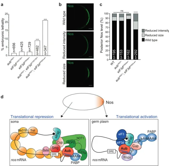
eIF3d plays a role in Aub-dependent translational activation To address the biological relevance of Aub/eIF3d physical interaction, we analyzed the effect of the concomitant reduction of aub and eIF3d gene dosage by half. Although single aub or eiF3d heterozygous mutant embryos showed a low level of
lethality (2%–3%), embryonic lethality significantly increased up to 21% in double heterozygous mutants, suggesting that Aub and
eIF3d act together in embryonic development (Fig.6a). nos mRNA
translation was then recorded in these embryos using
immunos-taining. The Nos protein level visualized by immunofluorescence
at the posterior cortex was quantified. In WT, 85% of embryos
showed a full accumulation of Nos protein at the posterior pole,
whereas 15% had a reduced accumulation (Fig. 6b, c). Nos
accumulation in heterozygous aub or eIF3d mutant embryos was
similar to that in WT. In contrast, in aub–/+; eIF3d–/+ double
heterozygous mutants, the percentage of embryos with reduced
Nos accumulation significantly increased to 34% (Fig. 6b, c;
Supplementary information, Fig. S5a). This reduction of Nos accumulation in double heterozygous mutants did not correlate with reduced Osk accumulation or reduced nos mRNA localization
at the posterior pole (Supplementary information, Fig. S5b–e),
indicating a direct defect in nos mRNA translation. We conclude that Aub/eIF3d physical interaction is required for nos mRNA translational activation. a % embryonic lethality *** *** aub HN2/+ aub HN2/+ ; eIF3d EY05735/+ aub HN2/+ ; eIF3d EP654-a/+ eIF3d EY05735/+ eIF3d EP654a/+ n=656 n=425 n=729 n=482 n=347 0 5 10 15 20 25 b e p yt dli W e zi s d e c u d e R Nos Nos Nos yti s n et ni d e c u d e R c
Posterior Nos level (%)
WT n=195 n=153 n=162 n=292 0 20 10 30 40 50 60 70 80 90 100 **** ns aub HN2/+ aub HN2/+ ; eIF3d EP654-a/+ eIF3d EP654a/+ d Translational repression eIF3 eIF3d TraI Me31B Smg Cup Belle POP2 NOT1 nos mRNA soma AAAAAAA A nos Aub Wispy AAAA PABP SRE Aub Cup eIF4E A PABP eIF4E CCR4 Wild type Reduced size Reduced intensity Translational activation Nos eIF3 eIF3d AAAAAAA A AAAAAA mRNA germ plasm Aub SRE Wispy Cup eIF4E A
Fig. 6 Aub and eIF3d functionally interact for nos mRNA translation. a Percentage of embryonic lethality of single or double aub and eIF3d heterozygous mutants. The genotypes are indicated. ***P < 0.001, using theχ2test. b Immunostaining of single and double aub and eIF3d
heterozygous mutant embryos with anti-Nos antibody. Posterior of embryos with the three types of staining: wild type, reduced size or reduced intensity, are shown. Scale bars, 20μm. c Quantification of posterior staining shown in b using the ImageJ software. For each genotype, the percentage of embryos with each staining category was recorded. ****P < 0.0001, ns not significant, using the χ2test. d Model of Aub-dependent translational activation. In the somatic part of the embryo, nos mRNA translation is repressed by two mechanisms: a cap-dependent mechanism that involves Cup binding to eIF4E, and a cap-incap-dependent mechanism that involves the coating of the mRNA by Me31B and Tral. Both mechanisms might depend on the CCR4-NOT complex recruited by Smg and Aub. In the germ plasm, Smg binding to Osk precludes its interaction with nos mRNA, leading to depletion of CCR4-NOT and remodeling of the mRNP. This would lead to the dissociation of Me31B/Tral from the mRNA. Aub interaction with PABP and eIF3 subunits would allow unconventional translation, bypassing eIF4E requirement. The recruitment of Wispy poly(A) polymerase by Aub leading to polyadenylation is likely to also contribute to translation activation. Note that eIF3 might be present in the repressor complex in the soma, since eIF3 was found as Aub interactor in osk mutant embryos; however, its activity in translation activation would be repressed.
DISCUSSION
Several studies have reported the role of PIWI proteins in cellular mRNA regulation at the level of stability. piRNA-dependent binding of mRNAs by PIWI proteins leads to their decay in different biological
systems.16In addition, in Drosophila embryos, mRNA binding by the
PIWI protein Aub also leads to their stabilization in a spatially
regulated manner.13 Here, we report a novel function of Aub in
direct translational control of mRNAs. Using nos mRNA as a paradigm, we show that Aub is required for nos mRNA translation. Nos protein levels are also strongly reduced in armi mutant, in which
piRNA biogenesis is massively affected,27 suggesting that Aub
loading with piRNAs is necessary for its function in translational
activation. Consistent with this, wefind that deletion of two piRNA
target sites in nos mRNA decreases its translation. Importantly, Nos levels are not affected in a panx mutant background. Panx is a piRNA factor required for transcriptional repression of transposable elements, but has no function in piRNA biogenesis.28,29In addition,
as is the case for aub and armi mutants, panx mutant embryos do not develop.28,29Finally, Nos levels are similar in unfertilized eggs
and embryos overexpressing Osk, demonstrating that Nos protein synthesis is independent of embryonic development. Together, these results strongly argue for a direct role of Aub and piRNAs in nos mRNA translational control, independently of their role in transposable element regulation or developmental defects in piRNA pathway mutants.
Mass spectrometry analysis of Aub interactors points to a strong link with the translation machinery. In addition, polysome gradient analyses reveal Aub association with actively translated mRNAs in polysomal fractions. A link has been reported previously between the PIWI proteins Miwi and Mili and the translation machinery in mouse testes, where Miwi and Mili were found to associate with
the cap-binding complex.46,47However, the role of Miwi and Mili
in translational control has not been characterized. We now decipher the molecular mechanisms of Aub function in transla-tional activation of germ cell mRNAs in the Drosophila embryo. We demonstrate a physical interaction between Aub and the translation initiation factors PABP, eIF4E and subunits of the eIF3 complex. These interactions are in agreement with polysome gradient analyses in WT and aub mutant backgrounds that indicate a role of Aub in translation initiation.
Recent data have identified specific roles of eIF3 in the
regulation of translation. eIF3 is the most elaborate of translation initiation factors containing twelve subunits and an associated factor, eIF3j. This complex promotes all steps of translational initiation and does so in part through direct association with other translation initiation factors, contributing to their functional
conformations on the small ribosomal subunit surface.39 In
addition to this role in basal translation, the eIF3a, b, d and g subunits were shown to directly bind 5′UTR of specific mRNAs,
leading to cap-dependent translation activation or repression.40
The eIF3d subunit that attaches to the edge of the complex appears to play an especially important role in various modes of eIF3-dependent translational control: (1) eIF3d is involved in the translational repression of Drosophila sex-lethal mRNA through binding to its 5′UTR.48(2) eIF3d was reported to directly bind the
cap structure of specific mRNAs in mammalian cells, thus
bypassing the requirement of eIF4E binding to the cap for translation initiation.41(3) In the same line, eIF3d was involved in
cap-dependent translational activation of specific mRNAs for
neuronal remodeling in Drosophila larvae, in a context where
eIF4E is blocked by 4E-binding protein (4E-BP).49 Other studies
have reported the role of eIF3 in promoting cap-independent translation, thus highlighting eIF3 functional versatility in the control of translation. eIF3 was shown to directly bind methylated
adenosine m6A, in mRNA 5′UTRs to induce cap-independent
translation under stress conditions.50Furthermore, PABP bound to
the poly(A) tail was also shown to cooperate with eIF3 for its
binding to mRNA 5′UTR triggering cap-independent translation.51
Here, we described a new mode of eIF3-dependent translational activation through its recruitment by the PIWI protein Aub. Based on previous information on the nos translation repressor complex and data presented here on translational activation, we propose
the following model (Fig.6d). nos mRNA translation is repressed in
the somatic part of the embryo by two mechanisms.11,35First, the
4E-BP protein Cup in complex with Smg binds to eIF4E and
prevents eIF4G recruitment and cap-dependent translation.11,52
The detailed mechanism of Cup recruitment to the repressor
complex has not been clarified, but Cup was shown to directly
associate with the Not1 subunit of the CCR4-NOT complex and this interaction might stabilize Cup association with eIF4E.53
CCR4-NOT itself is recruited to nos mRNA by Smg and Aub.9,18Second,
two translational repressors, the RNA helicase Me31B (Drosophila DDX6) and its partner Tral coat the length of nos mRNA and
prevent translation through a cap-independent mechanism.35
Again the mode of Me31B/Tral specific recruitment to nos mRNA
has not been determined, but the CCR4-NOT complex might also be involved since DDX6 directly binds the Not1 subunit of
CCR4-NOT.54,55 Aub coprecipitation with components of the nos
translational repressor complex is consistent with its association
with the CCR4-NOT complex in the soma18and suggests that Aub
might be involved in translational repression, in addition to mRNA decay. In the germ plasm, Osk interaction with Smg prevents Smg
binding to nos mRNA9 and this contributes to CCR4-NOT
displacement from the mRNP complex. Consistent with this,
CCR4 is depleted in the germ plasm.13The lack of CCR4-NOT on
nos mRNA might preclude the recruitment of Me31B/Tral and relieve the cap-independent mechanism of translational
repres-sion (Fig.6d). Wefind that Aub physically interacts with PABP and
several subunits of eIF3. We propose that these associations would lead to translational activation independently of eIF4E through binding of eIF3 to nos 5′UTR, followed by direct recruitment of the 40 S ribosome by eIF3 and PABP, as previously reported for
translation of XIAP mRNA.51Alternatively, eIF3 might act through
direct binding of eIF3d to the cap structure; however, we do not favor this hypothesis. Indeed, if eIF3d interaction with the cap was
involved, overexpression of the point mutant eIF3dhelix11that is
unable to bind the cap,41would be expected to induce negative
dominant defects, due to the lack of translation mediated by this interaction.49However, overexpression of eIF3dhelix11with the nos-Gal4 driver did not induce any defects in embryonic development or Nos protein synthesis (Supplementary information, Fig. S6).
Germ granules coordinate germ cell mRNA regulation with piRNA inheritance through the role of PIWI proteins in both processes. Recent studies in C. elegans have shown that piRNA/ PRG1-dependent mRNA accumulation in germ granules prevent their silencing, strengthening the function of piRNAs in germ
granules for mRNA storage and surveillance.56,57 In Drosophila,
Aub mediates the link between piRNAs and mRNA regulation in germ granules since Aub localization to germ granules depends
on its loading with piRNAs12and Aub/piRNAs play a general role
in the localization and stabilization of germ cell mRNAs in germ
granules.13,19How do germ granules accommodate translational
control has remained more elusive. In Drosophila embryos, germ
granules contain mRNAs that are translated sequentially.58 We
demonstrate a direct role of Aub in translational activation. Strikingly, PABP and eIF3d tend to colocalize with Aub at the periphery of germ granules. This is reminiscent of a study analyzing translational control in relation to RNA granules in Drosophila oocytes, in which translational repressors such as Me31B were found to concentrate in the granule core with repressed mRNAs, whereas the translational activator Orb was localized at the edge of the granules where mRNAs docked for
translation.59 Similarly, germ granules in embryos might be
partitioned into functional subdomains involved in various steps of mRNA regulation, including storage (in an internal region of granules) and translational activation (at the granule periphery).
Our work reveals the central role of Aub in activation of translation. Future studies will undoubtedly address the complex-ity of mRNA regulation by PIWI proteins in relation with germ granules.
While this manuscript was under review, a role of Miwi and piRNAs in translational activation during mouse spermiogenesis
has been demonstrated.60Miwi was shown to be in complex with
PABP and several subunits of eIF3 for its function in translational activation, which is required for spermatid development. This reveals a striking evolutionary conservation of PIWI protein function in translational control for key developmental processes.
MATERIALS AND METHODS Drosophila lines
w1118was used as a control. Mutant alleles and transgenic lines
were aubQC42 cn1bw1/CyO and aubHN2cn1bw1/CyO,61
nos-Gal4-VP16,62UASp-osk-K10,25 panxM1and panxM4,29 armi1,63 armi72.1,64
nosBNx,65 nos(ΔpirooΔpi412); nosBN/TM3 Sb,13 w; osk54
nos-Gal4-VP16/TM3 Sb and yw; osk54e UASp-GFP-Aub/TM3 Sb,12
UASp-GFP-Aub,66 UAS-GFP cytoplasmic (gift from J.M. Dura), eIF3dEY05735
(#20072) and eIF3dEP-654a(#43437) (Bloomington Drosophila Stock
Center). The UASp-HA-eIF3d and UASp-HA-eIF3dhelix11 lines were
generated in this study by insertion of PhiC31 recombination into
attP40 site (BestGene). The genotypes of embryos (aged 0–2 h)
indicated throughout were the genotypes of mothers. Females of the indicated genotypes were crossed with WT males.
S2R+ cells
S2R+ cells (Gift from G. Cavalli) were cultivated at 25 °C in Schneider medium complemented with 10% fetal bovine serum (Gibco) and 1% penicillin-streptomycin (Gibco).
Immunostaining and image analysis
0–2 h embryos were collected in a basket from plates, washed in
tap water and dechorionated using commercial bleach for 2 min,
rinsed and dried. Embryos were thenfixed at the interface of a
1:1 solution of 36% formaldehyde:100% heptane for 5 min, followed by 100% methanol devitellinization. Embryos were re-hydrated, blocked in 1% BSA for 1 h and incubated overnight with primary antibodies. Secondary antibody incubation, after washes in PBS, 0.1% Tween, was performed for 1 h at room temperature. Embryos were mounted in Vectashield (Vector Laboratories) for imaging. Antibodies used: rabbit anti-Osk (1:1000, gift from P. Lasko), rabbit Nos (1:1000, gift from A. Nakamura), rabbit
anti-PABP (1:500, gift from A. Vincent), rabbit anti-Smg (1:2000),67
mouse anti-HA (1:2000, ascites produced from clone 12CA5), rabbit anti-GFP (1:1000, Invitrogen), mouse anti-GFP (1:1000, Roche), goat anti-mouse IgG Cy3 (1:1000, Jackson ImmunoRe-search), goat anti-rabbit IgG Alexa-488 (1:800, Invitrogen) and donkey anti-rabbit Cy3 (1:1000, Jackson ImmunoResearch). Micro-scopy was performed using a Leica SP8 confocal scanning microscope. Data were processed and analyzed using the ImageJ software.
smFISH
Dechorionated embryos were fixed at the interface of a
1:1 solution of 10% formaldehyde:100% heptane for 20 min, followed by 100% methanol devitellinization. After permeabiliza-tion in ethanol, embryos were washed 4 times for 15 min in PBT and then once for 20 min in Wash Buffer (10% 20× SCC, 10% formamide). They were then incubated overnight at 37 °C in Hybridization Buffer (10% formamide, 10% 20× SSC, 400 µg/mL tRNA, 5% Dextran sulfate, 1% VRC (Vanadyl Ribonucleoside Complexes, Sigma)) with anti-nos probes (Supplementary informa-tion, Table S2) coupled to CAL Fluor Red 590 (Stellaris). Embryos were washed in Wash Buffer at 37 °C and then in 2× SCC, 0.1% Tween at room temperature before mounting (Pro-Long Gold
antifade reagent, Invitrogen). Microscopy was performed using a Leica SP8 confocal scanning microscope. Data were processed and analyzed using the ImageJ software.
RNA extraction and RT-qPCR
Total RNA was prepared from 30 embryos using Trizol (Invitrogen) following recommendations from the manufacturer. For RT-qPCR,
1μg of total RNA was reverse transcribed using Superscript III
(Invitrogen) and random hexamers. Quantitative PCR (qPCR) was performed on a LightCycler LC480 (Roche) with Lightcycler 480 SYBR green master (Roche) and primers are listed in Supplemen-tary information, Table S3. Quantifications were performed in triplicate.
Coimmunoprecipitation and western blot
GFP immunoprecipitations for mass spectrometry were performed
as follows: 0.5 g of 0–2 h-dechorionated embryos were crushed in
DXB buffer (25 mM HEPES, 250 mM sucrose, 1 mM MgCl2, 1 mM
DTT, 150 mM NaCl, protease inhibitor) with 0.1% Triton X-100 and RNasin and incubated on ice for 30 min. Lysates were centrifuged for 10 min and the supernatant was transferred to a new tube. Lysates were incubated on equilibrated GFP-trap beads (Chromo-tek) overnight at 4 °C on a wheel. Beads were washed seven times in DXB buffer complemented with 1% Triton X-100 and RNasin. Beads were suspended in 2× NuPAGE Blue supplemented with 50 mM DTT and incubated for 10 min at 95 °C. The quality of the samples was assessed by silver staining (SilverQuest, Invitrogen).
For coIP experiments, 0.15–0.18 mg of 0–2 h embryos were
crushed in IP buffer (20 mM Tris, pH 7.5, 150 mM NaCl, 0.2% NP-40, 1.5 mM DTT, 10 mM EGTA, protease inhibitor) with either 40 U/ µL RNase A or 100 U/µL RNase inhibitor. Extracts were centrifuged at 10,000× g for 10 min at 4 °C and incubated on pre-equilibrated magnetic beads with anti-GFP (Chromotek) or anti-HA (Pierce) antibody for 2.5 h at 4 °C. After incubation, the beads were washed five times with IP buffer and immunoprecipitated proteins were eluted from beads by incubation with 2× Laemmli buffer
supplemented with 10% β-mercaptoethanol for 5 min at 95 °C.
Samples were then analyzed by western blot. For western blot analysis, protein extracts obtained from 30 embryos crushed in
30 µL of 2× Laemmli buffer supplemented with 10%
β-mercaptoethanol were boiled for 5 min at 95 °C. Samples were then loaded onto 10% SDS-PAGE gels before transfer to a nitrocellulose membrane. The membrane was blocked for 1 h in 5% milk diluted in 1× PBS, 0.1% Tween 20 before proceeding to primary antibody incubation (overnight, 4 °C on a rotating plate). Antibodies and dilutions for western blot were: rabbit anti-Nos (1:1000, gift from A. Nakamura), rabbit anti-Osk (1:2000, gift from
P. Lasko), mouse anti-Aub (4D10, 1:5000),68 guinea pig anti-Smg
(1:2000, gift from C. Smibert), rabbit anti-PABP (1:500, gift from A. Vincent), rabbit anti-GFP (1:1000, Invitrogen), anti-HA (1:1000, Covance) and mouse anti-α-Tubulin (1:5000, Sigma). After washes in 1× PBS, 0.1% Tween 20, the membrane was incubated for 1 h at room temperature with secondary antibody coupled with HRP (Jackson ImmunoResearch). After washes, HRP-conjugated sec-ondary antibodies were revealed by chemiluminescent detection
(Pierce). Quantifications were performed with the ImageJ software
using the Gels tool. Mass spectrometry
Total protein elute was loaded on 10% SDS-PAGE gels (Mini-Protean TGX Precast gels, Bio-Rad). For each sample, one band was cut after stacking migration. Gel pieces were destained with three washes in 50% acetonitrile and 50 mM TEABC (trimethy ammonium bicarbonate buffer). After protein reduction (10 mM DTT in 50 mM TEABC at 60 °C for 30 min) and alkylation (55 mM iodoacetamide in TEABC at room temperature in the dark for 30 min), proteins were in-gel digested using 1 µg Trypsin (Trypsin Gold, Promega). Digested products were dehydrated in a vacuum 432
centrifuge. Obtained peptides were analyzed online using
Q-Exactive Plus mass spectrometer (Thermo Fisher Scientific)
interfaced with a nano-flow HPLC (RSLC U3000, Thermo Fisher
Scientific). Samples were loaded onto a 15 cm reverse phase
column (Acclaim Pepmap 100, NanoViper, Thermo Fisher
Scien-tific) and separated using a 103-min gradient of 2%–40% of buffer
B (80% acetonitrile, 0.1% formic acid) at aflow rate of 300 nL/min.
MS/MS analyses were performed in a data-dependant mode
(Xcalibur software 4.1, Thermo Fisher Scientific). Full scans
(375–1500 m/z) were acquired in the Orbitrap mass analyzer with
a 70,000 resolution at 200 m/z. The twelve most intense ions
(charge states≥ 2) were sequentially isolated and fragmented by
HCD (high-energy collisional dissociation) in the collision cell and detected at 17,500 resolution. The spectral data were analyzed
using the Maxquant software (v1.5.5.1) with default settings.69All
MS/MS spectra were searched by the Andromeda search engine against a decoy database consisting of a combination of
Drosophila melanogaster entries from Reference Proteome
(UP000000803, release 2018_02, https://www.uniprot.org/),
iso-form C sequence of Aub protein and classical contaminants, containing forward and reverse entries. Default search parameters were used; Oxidation (Met) and Acetylation (N-term) as variable
modifications and Carbamidomethyl (Cys) as fixed modification
were applied. FDR was set to 1% for peptides and proteins. A representative protein ID in each protein group was automatically selected using in-house bioinformatics tool (Leading_v3.2). First,
proteins with the most numerous identified peptides are isolated
in a“match group” (proteins from the “Protein IDs” column with
the maximum number of“peptide counts”). For the match groups
where more than one protein ID are present after filtering, the
best annotated protein in UniProtKB, release 2019_01 (reviewed entries rather than automatic ones, highest evidence for protein existence) is defined as the “leading” protein. Label free
quantification (MaxQuant LFQ) was used to identify differential
proteins between samples. LUMIER assays
S2R+ cells (250,000) were transfected using Effectene transfection
reagent (Qiagen) and incubated for 48 h at 25 °C. Cells were lysed in
HNTG buffer (20 mM HEPES, 150 mM NaCl, 1 mM MgCl2, 1 mM
EGTA, 1% Triton, 10% glycerol) complemented with protease inhibitor (cOmpleteTMEDTA-free Protease Inhibitor Cocktail, Roche). On a plate (LUMITRAC 600 96 W Microplate High Binding, Greiner) pre-coated with anti-FLAG antibody (Sigma, F1804) and blocked for 1 h with a blocking solution (3% BSA, 5% sucrose and 0.5% Tween 20), lysates were incubated for 3 h on ice. After washes with HNTG, luminescence was revealed using Dual-Luciferase Reporter Assay System (Promega, E1910) and read on a luminometer Tristar LB941. Transfections were repeated 8–32 times.
Polysome profiling
For lysis of embryos, we used either of two methods that produce
similar results. Either 0.2 g of fresh 0–2 h embryos were
homo-genized in lysis buffer composed of 20 mM Tris-HCl, 140 mM KCl,
5 mM MgCl2, 0.5 mM DTT, 1% Triton X-100, 0.02 U/µL RNasin, 1×
protease inhibitor and 0.1 mg/mL cycloheximide, or 0.2 g of frozen
0–2 h embryos were homogenized in lysis buffer composed of
30 mM Tris-HCl, 100 mM NaCl, 10 mM MgCl2, 0.5 mM DTT, 1% Triton
X-100, 0.02 U/µL RNasin, 1× protease inhibitor and 0.1 mg/mL cycloheximide. Embryo extracts were incubated for 30 min on ice. Homogenates were cleared by full speed centrifugation for 30 min at 4 °C. For treatment with puromycin, fresh embryos were homo-genized in lysis buffer composed of 20 mM Tris-HCl, 140 mM KCl, 0.5 mM DTT, 1% Triton X-100, 0.02 U/µL RNasin, 1× protease inhibitor and 2 mM puromycin. Extracts were incubated for 20 min on ice followed by 20 min at 37 °C and cleared as before. The amount of RNA in the extracts was quantified using Nanodrop. Volumes of extract containing equal amounts of RNA were loaded on top of
10%–50% sucrose gradient containing cycloheximide except for
puromycin-treated samples. Gradients were centrifuged for 2 h at 34,000 rpm in a SW41 rotor, with no brake. 1 mL fractions were collected using a ISCO gradient collector. 900 µL of each fraction were used for RNA extraction and 100 µL for protein precipitation. For RNA extraction, 500 pg of luciferase RNA was added to 900 µL of fraction to control RNA extraction. 0.5% SDS and 10 mM EDTA was then added to each fraction, followed by a 5 min incubation. Then, RNAs were prepared using acid phenol-chloroform. One volume of acid phenol-chloroform was added, and the mixture was vortexed and centrifuged at 12,000× g for 15 min. The aqueous phase was put in a new tube with 2 µL of glycoblue and 1 volume of isopropanol, incubated for 15 min and centrifuged for 15 min at 12,000× g. The RNA pellet was washed twice with 1 volume of 75% ethanol. The RNA pellet was dried for 5 min and resuspended in 20 µL of
RNase-free H2O. 5 µL of each RNA sample were used for cDNA synthesis
using superscript III and random primers. cDNAs were diluted at 1:10 for qPCR reactions that were performed using LightCycler 480 (Roche) and the primers are listed in Supplementary information,
Table S3. Data were analyzed using theΔCp method.70For protein
preparation, 400 µL of methanol and 100 µL of chloroform were added to 100 µL of fraction and the mixture was vortexed. The
mixture was then added with 300 µL of H2O, vortexed and
centrifuged at full speed for 5 min. The upper phase was discarded, 435 µL of methanol were added and the mixture was centrifuged at
full speed for 5 min. The protein pellet was dried for 1–2 min and
resuspended in 100 µL of 2× Laemmli buffer supplemented with
10% β-mercaptoethanol. Proteins were analyzed by western blot;
antibodies and dilutions were mouse anti-Aub (4D10, 1:5000),68
rabbit anti-PABP (1:1000, gift from A. Vincent), rabbit anti-RpL10Ab (1:5000)71and rabbit anti-RpS3 (1:1000).71
Cloning and recombineering
To produce the UASp-HA-eIF3d and UASp-HA-eIF3dhelix11
trans-genes, eIF3d and eIF3dhelix11coding sequences were amplified by
PCR from clones provided by S. Rumpf.49 PCR fragments were
cloned into pENTR by directional Topo cloning (Invitrogen). The generated plasmids were used in gateway cloning to insert the
sequences into pPHW (UASp-HA-attR1-ccdB-attR2-SV40 3′UTR) in
which an attB (pPHW-attB) site has been inserted. The resulting fragments were then inserted in the Drosophila genome by PhiC31 recombination into the attP40 site (BestGene).
To produce FLAG-FFL-Aub, Aub-coding sequence was PCR
amplified from the p8161 plasmid66 and cloned into pENTR by
directional Topo cloning (Invitrogen). The resulting plasmid was used in gateway cloning to insert Aub sequence into
pAct-FLAG-Firefly-RfA72(pAFW (DGRC) in which the FFL-coding sequence has
been added). To produce HA-RL tagged versions of eIF3b (DGRC,
FI08008), eIF3d,49 eIF3f (DGRC, LD47792), eIF3k (DGRC, LD03569)
and eIF4E (from E. Wahle), the coding sequences were amplified by PCR and cloned into pENTR by directional Topo cloning. The resulting plasmids were used in gateway cloning to insert the
coding sequences into pAct-HA-Renilla-RfA72 (pAHW (DGRC) in
which the RL-coding sequence has been added). For the HA-RL tagged versions of PABP (DNASU, DmCD00772781), eIF3g (DNASU, DmCD00766429), eIF3h (DNASU, DmCD00764259) and eIF4a (DNASU, DmCD00764657), plasmids were directly used in gateway cloning to insert the sequence into pAct-HA-Renilla-RfA.
FLAG-FFL-Cherry, HA-RL-Cherry and Sd-RL-HA72 were used as
negative controls. To produce GST-PABP clones, the coding
sequences offive pAbp domains, RRM1, RRM2, RRM3, RRM4 and
PABC were amplified by PCR from a plasmid provided by E. Wahle.
A stop codon (TAA) was added at the end of each domain. The different fragments were cloned into pGEX-4T-1 (Sigma) digested with EcoRI and XhoI. The plasmids containing HA-Aub(1-482) and
HA-Aub(476-866) fragments were generated previously.13 The
primers used to generate the constructs and the constructs are listed in Supplementary information, Tables S3 and 4, respectively.