Development and Application of Chemical Tools for Investigating Dynamic Processes in Cell Migration
by
MASSACHUSETTS I OF TECHNOL Brenda Nicole Goguen
JUNO0 7 2
B.S., Chemistry and B.A., BiologyUniversity of Virginia, 2005 LIBRARI
NSTITlTE 1
)GY
O11
ES
Submitted to the Department of Chemistry in Partial Fulfillment of the Requirements for the
Degree of Doctor of Philosophy
at the
Massachusetts Institute of Technology
June 2011
@ 2011 Massachusetts Institute of Technology
All rights reserved
ARCHNES
Signature of Author:. Department of Chemistry May 17, 2011 Certified By: Accepted By: Barbara Imperiali Class of 1922 Professor of Chemistry and Professor of BiologyThesis Supervisor
Robert W. Field Haslam and Dewey Professor of Chemistry Chairman, Departmental Committee on Graduate Students
This doctoral thesis has been examined by a committee of the Department of Chemistry as
follows:
JoAnne Stubbe Chair Novartis Professor of Chemistry and Professor of Biology Massachusetts Institute of Technology
Barbara Imperiali Thesis Supervisor Class of 1922 Professor of Chemistry and Professor of Biology Massachusetts Institute of Technology
Moh ad Movassaghi I/fessor of Chemistry Massach (Institute of Technology
Development and Application of Chemical Tools for Investigating Dynamic Processes in Cell Migration
by
Brenda Nicole Goguen
Submitted to the Department of Chemistry on May 17, 2011 in Partial Fulfillment of the Requirements for the Degree of Doctor of Philosophy
in Organic Chemistry
ABSTRACT
Cell migration is a dynamic process essential for many fundamental physiological functions, including wound repair and the immune response. Migration relies on precisely orchestrated events that are regulated in a spatially and temporally controlled manner. Most traditional approaches for studying migration, such as genetic methods or the use of chemical inhibitors, do not offer insight into these important components of protein function. However, chemical tools, which function on a more rapid timescale and in localized regions of the cell, are capable of providing real-time information about protein activity. Herein, the development and application of chemical approaches to investigate proteins central to cell migration are presented.
Myosin II, an ATPase motor protein required for cell motility, is activated by phosphorylation of the associated myosin regulatory light chain (mRLC) protein at Serl9. To generate a photoactivated mRLC variant that offers control over the timing and localization of myosin activity, the mRLC has been prepared by expressed protein ligation for the site-specific incorporation of 1-(2-nitrophenyl)ethyl (NPE)-caged phosphoserine at position 19. The NPE caging group masks the phosphate functionality and inhibits protein function until irradiation at
365 nm releases the native phospho-mRLC to restore myosin activity. Introduction of the caged mRLC into cells enables interrogation of the role of myosin in coordinating cell contractility.
To expand the scope of the caging approaches, the NPE caging group has been applied in concert with the [7-(diethylamino)coumarin-4-yl]methyl (DEACM) group, which is released by irradiation at 420 nm, to enable two different phosphopeptides to be sequentially released within one system. Preparation of DEACM-caged phosphoamino acid building blocks for solid phase peptide synthesis enables convenient incorporation of these residues into peptides and proteins. This sequential uncaging approach has been exploited to initiate and subsequently inhibit a biochemical reaction in an enzyme-independent fashion using two wavelengths of light.
Finally, a fluorogenic sensor to monitor the real-time activity of the GTPase Cdc42, an essential regulator of migratory processes, has been developed. The solvatochromic fluorophore 4-N,N-dimethylamino-1,8-naphthalimide has been incorporated into a protein fragment that binds only the activated conformation of Cdc42. This sensor reports Cdc42 activation through significant increases in fluorescence and has been applied in a cellular context to monitor endogenous Cdc42 activity. This fluorogenic sensor and the caging approaches together demonstrate the power of chemical tools for interrogating diverse aspects of cell migration.
Thesis Supervisor: Barbara Imperiali
Acknowledgements
Graduate school at MIT, and particularly in the Imperiali Lab, has been a truly wonderful experience. I am happy to now have the opportunity to thank those who have contributed so much to my time here. First, I thank my advisor Barbara Imperiali for her constant support and careful guidance. Barbara, you have maintained the fine balance of promoting my independence as a scientist while offering thoughtful direction. Thank you for providing so many wonderful opportunities throughout my graduate career through collaborations and scientific conferences. I admire your courage in tackling challenging scientific problems that span chemistry and biology, and I am so grateful to have had the opportunity to learn from you. I also thank Professor JoAnne Stubbe, my thesis chair, for enlightening scientific discussions and valuable advice, and I thank Professor Mohammad Movassaghi and Professor Sarah O'Connor for support and guidance throughout the years.
Much of the work presented in this thesis is the result of collaborations with colleagues in the Imperiali lab and in labs outside MIT. I thank Professor Martin Schwartz and Dr. Brenton Hoffman for their dedication to the cellular myosin studies and for welcoming me into their lab on numerous occasions. Brent, those visits were intense, but thank you for your patience in explaining microscopy and cell culture and for remembering my unique taste in food. Dr. Jim Sellers, thank you for your generosity in inviting me to perform the sliding filament assays in your lab and for sharing your boundless knowledge of myosin. I thank Dr. Andreas
Aemissegger for synthesizing most of the amino acid building blocks I used in this work and for his meticulous record-keeping and organization. Dr. Galen Loving was instrumental in establishing the Cdc42 sensor project. Galen, you have been a wonderful collaborator, always excited to discuss the project and offer thoughtful insight. In addition, I thank Professor Gaudenz Danuser and Dr. Jessica Tytell for supporting our collaboration with the Cdc42 sensor. Thank you for providing me with the unique opportunity to perform the microscopy experiments, and thanks to Dr. Jennifer Waters and Wendy Salmon of the Nikon Imaging Center at Harvard Medical School for helpful advice during these studies.
To all the lab members I have had the honor of working with, thank you for being wonderful friends and making the lab such a joy to work in. First, to my classmates, Meredith Hartley, Angelyn Larkin, and Wendy Iskenderian-Epps, I'm so happy we went through this journey together - and we made it! Meredith, we've shared many lunchtime moments ranging from the weekly "lunch disaster" to many fun lunch adventures, and, of course, long discussions over "The Cookie." I've enjoyed our many long emails (especially during my internship), joint parties, and beef nights. Angelyn, visits to your pod were always filled with interesting discussions - scientific and otherwise - and lots of laughter. Thanks for your constant encouragement! Wendy, I'll never forget the fun times we had first year as 2/3 of Wenda, from being the last ones to finish p-sets to the weekly Friday night dinners and dancing in Boston. To Elvedin, Galen, and Matthieu, I have looked up to you guys from the beginning. You made the lab such a fun place to be (especially at night), and I have learned so much from you (and on some rare occasions, even more than I needed to know). Dr. Elvedin Lukovid, Ph. D., I have always been inspired by your humor, work ethic, and determination. And for all the times you guilted me into staying out longer, thank you - those were some of the best! Galen Loving, I admire your curiosity and thorough understanding of science, and I always think of you when I see a Jablonski diagram! Matthieu Sainlos, it was always enlightening to discuss my project
with you, and thanks for encouraging me through challenges along the way. Beth Vogel-Taylor, thank you for welcoming me into the lab. You are a wonderful teacher. Nono, every time I think of you, I smile. You are among the most genuine people I know. Langdon Martin, minimeeting just wasn't the same without your clever puns and the matching LBT shirts. I thank James Morrison for being a great late night lab buddy, Mike Morrison for our many attempts to fix the mass spec, and Mark Chen for career advice and for introducing me to your friends, including Alex, William, and Thomas. Jay Troutman, I could always relate to your outlook on people, life, and science, and thanks for keeping in touch. Michelle Chang, I appreciate your calm and careful attitude to research. Vinita Lukose, hearing your contagious laughter in the next pod always brightened my day. Marcie Jaffe, I enjoyed joining in on your lively debates and discussions. Andrew Krueger, thanks for increasing my knowledge of pop culture and for your determination in solving my many problems from peptide synthesizer repair to assigning NMR spectra. To Professor Cliff Stains, discussions with you always helped to resolve my scientific dilemmas - thanks for always being so willing to listen. Nelson Olivier, it's always fun to hear about your exploits. Many thanks go to Susana Gordo and Melanie Bonnekessel, who were great partners on the caging project. Elke Socher, thank you for teaching me German (ich mag lila) and for your fun attitude towards life and science. Philipp Schneggenburger, thanks for showing me some awesome dance moves and for lots of advice about thesis writing
-UnbeLIEVable. Austin Travis - good luck in graduate school. Also, many thanks go to Elizabeth Fong, who ensured that the lab was running smoothly.
I thank all the teachers who have enabled me to reach this point. I thank those from
Aquinas School, who offered so much patience and support, and those from TJHSST, and in particular Mr. Lampazzi, who helped me further develop my passion for science. I thank Professor Patrick Gillevet for giving me the valuable opportunity to perform research early on and Professor Cassandra Fraser for providing incredible guidance and for encouraging me to apply to graduate school.
I also thank my classmates at MIT who have been a great source of support and who
maintained the Friday night dinner tradition for many years: Omar Ahmad, Peter Bernhardt, Scott Geyer, Wendy Iskenderian-Epps, Wenhao Liu, Pam Lundin, Lindsey McQuade, Montana Petersen, and Nancy Yerkes. Also, to Chia-Hung Wu, you have been a great lab-neighbor and friend. Thank you for your special home- and lab-cooked foods.
Mohammad Seyedsayamdost, thank you for providing unwavering support throughout the past five years. You taught me to be confident and happy, and you believed in me even when
I didn't. Thank you for sharing your knowledge of biochemistry and kinetics, no matter how
many times I asked you the same questions. And most importantly, I thank you for all the happiness you've brought to my life during the past five years.
I thank my parents, to whom I attribute any and all successes, for their constant love and
encouragement and for providing such a supportive and stimulating environment throughout my life. You have both, in your own ways, been models of strength, courage, and confidence. To my mom, you have taught me to think independently and rationally, and I strive to face challenges with the same grace, fearlessness, and efficiency that you demonstrate. To my dad, even from the times in middle school when I forced you to quiz me on my notes, you have been my biggest supporter, and I thank you for your enthusiasm and interest in my work.
Table of Contents
Abstract... 3
Acknow ledgem ents ... 5
Table of Contents ... 7
List of Abbreviations ... 9
List of Figures...12
List of Schem es...15
List of Tables ... 16
Chapter 1 : Exploiting Chemical Biology for Interrogating the Details of Cell Migration. 17 Introduction ... 18
1.1 Cell M igration... 19
1.2 Rho GTPases... 23
1.3 Light-Activated Proteins ... 25
1.4 Chem ical Inducers of D im erization ... 40
1.5 Fluorescent Sensors of Protein Activity ... 44
Conclusions ... 51
Thesis Outlook ... 52
Acknowledgem nents ... 53
References ... 54
Chapter 2: Light-Triggered Myosin Activation for Probing Dynamic Cellular Processes 61 Introduction ... 62
Results and D iscussion... 65
2.1 Protein Sem isynthesis ... 65
2.2 Sem isynthetic m RLC Regulation of M yosin Activity... 75
2.3 HM M ATPase Activity with Sem isynthetic m RLC ... 84
2.4 Sliding Filam ent A ssays ... 86
Conclusions ... 90
Acknowledgem ents ... 90
M ethods ... 92
References ... 112
Chapter 3 : Cellular Studies of Photoactivated Myosin Regulatory Light Chain...117
Introduction ... 118
Results and D iscussion... 122
3.1 Incorporation of the m RLC into Cellular M yosin ... 122
3.2 Uncaging Studies ... 129
Conclusions ... 140
Acknowledgem ents ... 141
M ethods ... 142
References ... 147
Chapter 4: Sequential Modulation of Protein Function Using Spectrally Differentiated Caged Phosphoamino Acids...149
Introduction ... 150
Results and D iscussion... 154
4.1 Synthesis of DEACM-Caged Phosphorylated Amino Acid Building Blocks for Fm oc-Based Solid Phase Peptide Synthesis ... 154
4.2 Sequential Uncaging of the Myosin Regulatory Light Chain... 156
4.3 Sequential Uncaging of Wip1 Phosphatase Substrate and Inhibitor Peptides... 162
Conclusions ... 170
Acknowledgements ... 170
M ethods ... 171
NM R Characterization ... 187
References ... 194
Chapter 5 : Development of a 4-DMN-Based Sensor for Cdc42... 197
Introduction ... 198
Results and D iscussion... 202
5.1 Sensor Design and Synthesis ... 202
5.2 Fluorescence Properties of the Sensor ... 206
5.3 U se of the Sensor as a Probe of Cdc42 Activity ... 209
5.4 Future D irections: FRET Capture... 212
5.5 Cellular Studies ... 216
Conclusions ... 222
Acknowledgem ents ... 222
M ethods... 224
References ... 237
A ppendix: O ptim ization of Paxillin Expression... 240
Introduction ... 241
Results and D iscussion... 247
Conclusions ... 248
M ethods ... 250
List of Abbreviations
Standard 3-letter and 1-letter codes are used for the natural amino acids. Standard 1-letter codes are used for the nucleotides.
4-DMN 4-NN-dimethylamino-1,8-naphthalimide
4-DMNA 4-NN-dimethylamino-1,8-naphthalimido alanine
AF Alexa Fluor
Alloc allyloxycarbonyl
ANOVA analysis of variance
ATP adenosine triphosphate
BME -mercaptoethanol
Boc tert-butyloxycarbonyl
BSA bovine serum albumin
CaM calmodulin
cAMP cyclic adenosine monophosphate
CCD charge-coupled device
CFP cyan fluorescent protein
CID chemical inducer of dimerization
CIP calf intestine alkaline phosphatase
CNB carboxy-nitrobenzyl
cpSer caged phosphoserine
cpThr caged phosphothreonine
cpTyr caged phosphotyrosine
CRIB Cdc42/Rac interactive binding
DCM dichloromethane
DEACM [7-(diethylamino)coumarin-4-yl]methyl
DEANS 5-((2-aminoethyl)amino)naphthalene-1-sulfonic acid
DIC differential interference contrast
DIPEA NN-diisopropylethylamine
DMEM Dulbecco's Modified Eagle Media
DMF NN-dimethylformamide
DMSO dimethylsulfoxide
DTT dithiothreitol
EDTA ethylenediaminetetraacetic acid
EGTA glycol-bis(2-aminoethylether)-N,N,N',N'-tetraacetic acid
EM-CCD electron-multiplying digital charge-coupled device EPL expressed protein ligation
ERK extracellular signal-regulated kinase
ESI-MS electrospray ionization mass spectrometry
FBS FMN Fmoc FRET GAP GB1 GDI GDP GEF GFP GIT1 GST GTP HATU HBTU HEPES HMM HOAt HOBt HPG IPTG JNK LB LOV MALDI-MS MAP MEF MESNa MLCK MOPS mRLC MWCO NB NBD NCL NonP NMR NPE NTA OD
fetal bovine serum flavin mononucleotide 9-fluorenylmethoxycarbonyl
fluorescence resonance energy transfer GTPase-activating protein
immunoglobulin binding domain of Streptococcal protein G guanine nucleotide dissociation inhibitor
guanosine diphosphate
guanine nucleotide exchange factor green fluorescent protein
G-protein coupled receptor kinase interacting protein 1 glutathione-S-transferase
guanosine triphosphate
O-(7-azabenzotriazole-1-yl)-N,N,N',N'-tetramethyluronium hexafluorophosphate
O-benzotriazole-1-yl-N,N,N',N'-tetramethyluronium hexafluorophosphate 4-(2-hydroxyethyl)- 1 -piperazineethanesulfonic acid
heavy meromyosin
1 -hydroxy-7-azabenzotriazole
1 -hydroxy-benzotriazole
homopropargylglycine
isopropyl- 1 -thio-fp-D-galactopyranoside Jun N-terminal kinase
Luria-Bertani
light-oxygen-voltage
matrix-assisted laser desorption ionization mitogen activated protein
mouse embryonic fibroblast sodium 2-mercaptoethanesulfonate myosin light chain kinase
3-(N-morpholino)propanesulfonic acid myosin regulatory light chain
molecular weight cut off nitrobenzyl
nitrobenzoxadiazole native chemical ligation nonphosphorylated
nuclear magnetic resonance 1-(2-nitrophenyl)ethyl nitrilotriacetic acid optical density
P PBS PCB PCR PDGF PKA pSer pThr pTyr PyBOP ROCK RP-HPLC SD SDS PAGE SEM SH2 siRNA SPPS TBS TBS-T TCEP TEV TFA THF TNBS Tris TRITC tRNA
UV
UV-vis VSMC WASP Wip 1 YFP phosphorylated phosphate-buffered saline phycocyanobilinpolymerase chain reaction platelet derived growth factor cAMP-dependent protein kinase phosphoserine
phosphothreonine phosphotyrosine
benzotriazole-1-yl-oxy-tris-pyrrolidino-phosphonium hexafluorophosphate Rho-associated protein kinase
reversed phase high performance liquid chromatography standard deviation
sodium dodecyl sulfate polyacrylamide gel electrophoresis standard deviation of the mean
Src homology 2
short interfering ribonucleic acid solid phase peptide synthesis tris-buffered saline
tris-buffered saline with Tween-20 tris(2-carboxyethyl)phosphine tobacco etch virus
trifluoroacetic acid tetrahydrofuran
2,4,6-trinitrobenzene sulfonic acid tris(hydroxymethyl)aminomethane tetramethyl rhodamine isothiocyanate transfer ribonucleic acid
ultraviolet
ultraviolet-visible
Vacular smooth muscle cells Wiskott-Aldrich Syndrome Protein wild-type p53-induced phosphatase yellow fluorescent protein
List of Figures Chapter 1 Figure 1-1. Figure 1-2. Figure 1-3. Figure 1-4. Figure 1-5. Figure 1-6. Figure 1-7. Figure 1-8. Figure 1-9. Figure 1-10. Chapter 2 Figure 2-1. Figure 2-2. Figure 2-3. Figure 2-4. Figure Figure Figure Figure Figure Figure Figure Figure Figure Figure Figure Figure 2-5. 2-6. 2-7. 2-8. 2-9. 2-10. 2-11. 2-12. 2-13. 2-14. 2-15. 2-16.
Steps of cell m igration... 20
Actin dynamics at the leading edge of the cell... 22
GTPase regulation by GEFs and GAPs... 24
General caging strategy ... 27
Caged peptides and proteins for studying migration... 29
Mechanism of native chemical ligation... 32
Light-activated Rac1 based on plant photoreceptors ... 36
Chemical inducers of dimerization... 42
Fluorescent sensors for GTPase activation ... 46
Fluorogenic sensor for Cdc42. ... 49
M echanism of NPE uncaging... 64
Photoactivated m R LC ... 65
Synthesis of the mRLC by native chemical ligation to incorporate phosphorylated or caged phosphorylated residues at Thr18 or Ser19... 66
Structures of caged phosphoamino acid building blocks for Fmoc-based solid phase peptide synthesis. ... 70
Semisynthesis of the full-length mRLC ... 75
12% SDS PAGE gel of representative myosin and HMM exchanges ... 76
Results of the myosin ATPase assays ... 77
10% Glycerol-urea gel of of nonphosphorylated and phosphorylated mRLC... 78
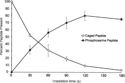
Uncaging time course of the NPE-caged pSerl9 mRLC peptide ... 79
Uncaging of NPE-caged pSerl9 mRLC (2-5) verified by Western blotting ... 79
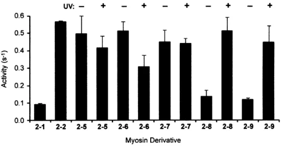
Myosin ATPase activity with caged semisynthetic mRLC derivatives before (- UV) and after (+ UV) irradiation... 81
Uncaging time course of the double NPE-caged pSerl9 peptide at 365 nm. ... 82
Results of HMM ATPase assays with native HMM and HMM exchanged with sem isynthetic derivatives ... 85
Results of HMM ATPase assays with caged semisynthetic mRLCs... 86
M yosin sliding filam ent assays ... 88
Results of in vitro sliding filament assays with caged mRLC variants... 89
Chapter 3 Figure Figure Figure 3-1. 3-2. 3-3. Figure 3-4. mRLC phosphorylation within the cell ... 121
Cells injected with Oregon Green 488-labeled double NPE-cpSerl9 mRLC... 125
NIH 3T3 cells injected with nonphosphorylated or phosphorylated Oregon G reen 488-labeled m RLC ... 126
Figure 3-5. Figure 3-6. Figure 3-7. Figure Figure Figure Figure Figure Figure 3-8. 3-9. 3-10. 3-11. 3-12. 3-13.
Methods to incorporate the semisynthetic mRLC into cells ... 129
Uncaging of mRLC 2-5 in VSMC ... 131
Comparison of stress fibers and Ser19 mRLC phosphorylation in COS-7 and rat V SM C ... 132
Thiophosphate resistance to cellular phosphatases ... 133
Comparison of antibodies specific for pSer19 mRLC ... 135
Uncaging of mRLC 2-6 in COS-7 cells ... 137
Anti-pSer19 mRLC antibody signals after uncaging ... 138
Localized uncaging within the cell... 139
Preliminary studies on the effect of uncaging NPE-c(S)pSerl9 mRLC 2-6 on focal adhesion form ation ... 140
Chapter 4 Figure Figure Figure Figure Figure Figure Figure Figure Figure Figure Figure Figure Figure Figure Figure Figure Figure 4-1. 4-2. 4-3. 4-4. 4-5. 4-6. 4-7. 4-8. 4-9. 4-10. 4-11. 4-12. 4-13. 4-14. 4-15. 4-16. 4-17. Sequential release of caged phosphopeptides ... UV-Vis spectra of DEACM- and NPE-caged peptides... Proposed mechanism of DEACM uncaging. ... DEACM-caged phosphoserine, threonine, and tyrosine building blocks ... HPLC uncaging time course of DEACM- and NPE-caged peptides at 410 nm.. Sequential uncaging of the m RLC ... HMM ATPase activity with NPE-cpThrl8 DEACM-cpSer19 mRLC... Sequential uncaging of the mRLC - Approach 2... HMM ATPase activity with DEACM-cpThrl8 NPE-cpSerl9 mRLC... Sequential uncaging to control activity of Wipl phosphatase ... Uncaging time course of DEACM-caged substrate and NPE-caged inhibitor peptides at 420 nm ... Uncaging time course of the NPE-caged inhibitor at 365 nm... Wip 1 activity with DEACM-caged and native substrate peptides... Effect of the NPE-caged inhibitor peptide on WipI activity ... Effect of the NPE-caged inhibitor on Wip 1 activity after irradiation ... Effect of 365 nm irradiation on Wip1 activity ... W ipl phosphatase activity... 152 153 154 155 157 158 159 160 161 162 164 165 166 167 168 168 169 Chapter 5 Figure 5-1. Figure Figure Figure Figure Figure Figure Figure Figure 5-2. 5-3. 5-4. 5-5. 5-6. 5-7. 5-8. 5-9. Structures of the environment sensitive fluorophores NBD, EDANS, m erocyanine, and 4-D M N ... 199
4-DMN-Based sensor for active GTP-bound Cdc42 ... 202
Generation of the 4-DMN-labeled WASP protein by cysteine labeling ... 205
Semisynthesis of the 4-DMN-modified sensor ... 206
Fluorescence properties of the 4-DMN-based sensors for activated Cdc42... 207
Fluorescence of a non-binding mutant sensor (5-14)... 209
Application of the sensor to monitor Cdc42 nucleotide exchange... 210
Tim e course of Cdc42 GTPase activity... 211
Determination of the dissociation constant between 4-DMN-modified sensor 5-3 and C dc42(G TP-yS)... 212
Figure Figure Figure Figure Figure 5-10. 5-11. 5-12. 5-13. 5-14. Appendix Figure A-1. Figure A-2. Figure A-3. Figure A-4. Figure A-5. FR E T C apture... 2 14
Proposed FRET Capture for the Cdc42 sensor ... 215
Scheme for the generation of the FRET Capture Cdc42 sensor... 216
Images of cells injected with sensor 5-3 and mutant sensor 5-14... 220
Comparison of the 4-DMN/Alexa Fluor 594 fluorescence intensity ratios resulting from sensor 5-3 and mutant sensor 5-14 ... 221
Representation of the domains of paxillin... 242
Semisynthesis of caged paxillin by protein semisynthesis... 243
Sequence of the paxillin gene... 246
Purification of GST-Paxillin(38 - 557)-FLAG ... 247
List of Schemes
Chapter 2 Scheme 2-1. Scheme 2-2. Scheme 2-3.
Synthesis of the C-terminal mRLC thioester... 69
Synthetic route to the double NPE-caged pSer19 peptide thioester (2-23)... 71 Synthesis of a guanidinium-modified CNB-caged peptide thioester. ... 73
Chapter 3
Scheme 3-1. Synthesis of the Oregon Green 488-labeled peptide thioester. ... 124
Chapter 4
Scheme 4-1. Synthesis of DEACM-caged phosphoserine. ... 155
Chapter 5 Scheme 5-1. Scheme 5-2.
Approaches for the synthesis of 4-DMN-labeled Cdc42 sensors... 204 Synthesis of a m utant Cdc42 sensor ... 208
List of Tables
Chapter 1
Table 1-1. Summary of protein targets discussed in this chapter... 25
Table 1-2. Comparison of methods for light-mediated protein function. ... 39
Chapter 2
Table 2-1. Semisynthetic mRLC Derivatives. ... 68 Table 2-2. Reaction Conditions for Peptide Thioesterification... 70
Chapter 1 : Exploiting Chemical Biology for
Interrogating the Details of Cell Migration
Introduction
Cell migration is a complex process essential for many fundamental functions including
wound repair, the immune response, and embryogenesis. In addition to supporting normal
physiology, cell migration contributes to pathological processes including cancer metastasis,
vascular disease, and inflammatory conditions such as rheumatoid arthritis.1 Migration is a
highly dynamic process, governed by precisely coordinated protein interactions in specific
regions of the cell. In addition, the timing of gene transcription and protein activation and
2
deactivation is tightly regulated at defined points of the migration cycle. Techniques to study
migration, including genetic approaches such as gene deletions, siRNA-knockdown of gene
expression, and site-directed mutagenesis, have revealed many details about the mechanisms
involved.3 However, migration and associated processes occur on the seconds to minutes
timescale, and genetic techniques, which act on the order of hours to days, preclude detailed
study of the temporal component of protein activity. The application of small molecule
inhibitors has also been used to provide information about protein function,4 but effects from
such inhibitors are global and cannot be localized to a specific region of the cell. In contrast to
these more conventional techniques, chemical approaches can be applied more rapidly and with
tunable effects.5 These technologies provide precise control over the location and timing of
protein activation, enabling elucidation of the spatial and temporal roles of the protein within a
complex network of interactions. Thus, methods drawing on chemical biology are poised to
make substantial contributions to the study of cell migration.
This chapter describes the steps involved in cell migration and highlights approaches that
Techniques that will be addressed include light-activated proteins, chemical genetics methods,
and fluorescent sensors of protein activity.
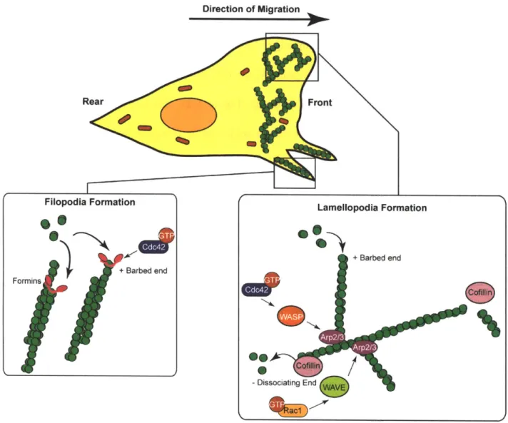
1.1 Cell Migration
Cell migration is an iterative process, characterized through five primary stages (Figure
1-1).1 First, upon exposure to a migration-inducing signal, the cell becomes polarized with a
distinct front (leading edge) and rear (trailing edge) (1). The cell then extends protrusions in the
direction of movement (2) and forms transient adhesions with the extracellular matrix (3)
through interactions between integrins and the actin network. The adhesions provide traction as
the cell body translocates forward (4). Finally, the adhesions at the rear of the cell are
1. Polarization
Adhesions
2. Extension
3. Adhesion Formation
4. Cell Body Translocation
5. Dissociation of the Rear
Figure 1-1. Steps of cell migration. Migration begins with polarization of the cell. The cell
then extends protrusions in the direction of migration. Adhesions to the extracellular matrix provide the stabilizing forces for the cell to propel forward. The final step of the migration cycle involves disassembly of adhesions at the rear of the cell.
Cell polarization is the first step in migration and results in an asymmetric distribution of
proteins to create distinct leading and trailing edges and to define the direction of migration.
Integrin receptors, the signaling molecule phosphatidylinositol (3,4,5)-triphosphate (PIP3), and
actin-regulating proteins, such as the GTPases Cdc42 and Rac, concentrate at the leading edge,
Additionally, the microtubule-organizing center is positioned in front of the nucleus, enabling
microtubules to grow toward the leading edge.7
Following the establishment of polarity, the cell extends protrusions at the leading edge
through formation of lamellopodia and filopodia (Figure 1-2). These protrusions are driven by
the actin network, which undergoes constant reorganization as migration progresses., 8 Thus,
this protrusive machinery is tightly regulated by actin-binding proteins, including the Arp2/3
complex, formins, and cofilin. Arp2/3, which is modulated by the small GTPases Rac and
Cdc42, binds along the actin filaments, thereby nucleating the growth of new branching
filaments and creating a broad network that comprises the lamellopodia.9 Alternatively, the cell can extend finger-like filopodial extensions, mediated by parallel bundles of actin. Formins,
which are regulated by Cdc42, generate these structures by promoting polymerization at the
rapidly growing, or barbed, ends of actin filaments. In addition, cofilin contributes to actin
polymerization by severing the filaments to provide new sites of nucleation. This protein also
promotes dissociation of actin monomers at the slow-growing end to increase the pool of actin
Direction of Migration
Figure 1-2. Actin dynamics at the leading edge of the cell. Filopodia formation is mediated by
Cdc42, while lamellopodia formation is controlled by Cdc42 and Rac1 through the Arp2/3 complex.
As the migrating cell extends protrusions, adhesions to the substrate are established.
Integrin receptors cluster into focal complexes by binding the extracellular matrix or other cells
and interacting with intracellular adaptor proteins, such as paxillin and talin, which bind actin
filaments within the cell. The integrin-mediated adhesions provide the mechanical support
necessary for the cell to propel forward.10 They function in concert with the ATPase motor
fibers.'1 Importantly, the adhesions also serve as signaling foci to facilitate communication between the extracellular matrix and the cytoplasmic region of the cell. While the signaling
cascades at these hubs regulate migration, they also control cell proliferation, apoptosis, and
differentiation.10 The final step in migration, adhesion disassembly and rear retraction, is controlled by multiple mechanisms, including mechanical contraction from myosin II motor
activity,'2 phosphorylation by focal adhesion kinase (FAK) and Src, and proteolytic degradation
of proteins in the adhesion.'3
1.2 Rho GTPases
In the development of tools to investigate migration, particular focus has been devoted to
examining the Rho-family GTPases, including RhoA, Rac1, and Cdc42, because of the
fundamental role that they play in orchestrating the process. Cycling between an inactive
GDP-bound state and an active GTP-GDP-bound state, these proteins function as molecular switches and
together regulate many aspects of the actin cytoskeleton.9 All three GTPases can be localized to
the membrane through prenylation of the cysteine residue within the C-terminal CAAX
sequence, where C is cysteine, A is an aliphatic residue, and X is any residue. Although these
GTPases can also be localized to the cytosol and to other organelles, in migration they are
activated at the membrane and exert effects there.'4 In addition to this spatially controlled
regulation, the activities of these GTPases are highly coordinated through the action of many
accessory proteins (Figure 1-3). Guanine nucleotide exchange factors (GEFs) activate the
GTPases by promoting exchange of GDP for GTP, while GTPase activating proteins (GAPs)
accelerate the rate of intrinsic GTP hydrolysis to deactivate them. A third regulatory mechanism
activation by preventing nucleotide exchange and by sequestering the proteins in the
cytoplasm.'5
GDP
Figure 1-3. GTPase regulation by GEFs and GAPs. GEFs exchange GDP for GTP to activate the GTPase, while GAPs accelerate the rate of intrinsic GTP hydrolysis. GDI sequesters the GTPase in the cytoplasm to prevent activation.
The Rho GTPases in turn mediate cellular functions through interactions with over 60
known effector proteins, including kinases and scaffold proteins.'5 Both Cdc42 and Rac activate
the actin-regulating Arp2/3 complex to induce the assembly of branched actin filaments at the
leading edge of the cell. Additionally, Cdc42 induces formation of filopodia, whereas Rac
activation causes lamellopodial protrusions (Figure 1-2).9 RhoA activation is involved in
membrane protrusions,1 but it also leads to the formation of stress fibers and induces cell
contractility. In addition to these roles, the GTPases participate in signaling pathways and
control gene expression and cell cycle progression." 4
Although much is known about cell migration, recent advances in chemical biology have
enabled investigations of protein dynamics with greater resolution. A summary of the protein
targets and corresponding methods of study described in this chapter are provided in Table 1-1.
Table 1-1. Summary of protein targets discussed in this chapter.
Protein Approach for Study
FAK Caged peptide
FKBP12-FRB system
Cofilin Caged protein
PKA Caged protein
mRLC Caged phosphoprotein
Paxillin Caged phosphoprotein
Raci Photoactivation
* LOV domain fusion * FKF1 / GI system
* PhyB / PIF3 system FKBP12-FRB system Bimolecular FRET
Cdc42 Photoactivation: PhyB / PIF3
Biosensor designs
* Intramolecular FRET
* Merocyanine environment sensitive fluorophore * 4-DMN environment sensitive fluorophore
Vinculin Intramolecular FRET
1.3 Light-Activated Proteins
Approaches in chemical biology have proven to be particularly valuable for gaining
insight into the transient protein-protein interactions that mediate migration. Many of these
techniques rely on the application of light-sensitive compounds to systematically control or
perturb cellular processes. The use of light activation provides unique advantages for
photoactivatable molecule in the cell. The light intensity and wavelength can be readily
controlled to enable dose-dependent activation and fine-tuning of the light-mediated event. The
localization of irradiation can be defined through the use of lasers or directed light sources to
produce activation at a particular region of a cell, allowing investigation of the spatial component
of protein function.17 However, it must be noted that the effects of localized photoactivation may not be apparent due to rapid diffusion and dilution of the activated molecule.18 Thus, in some cases, anchoring the light-sensitive protein at the plasma membrane or at an organelle can
better reveal the consequences of spatially defined uncaging. Light-activated proteins can be
chemically derived by appending an organic photolabile protecting group onto an essential
functionality of the protein,19 or the protein can be fused to a naturally occurring photoreceptor domain.17 Both techniques have been successfully applied to interrogate processes in cell
migration.
1.3.1 Caged Peptides and Proteins
Caged peptides and proteins exploit the versatility of chemical synthesis for the study of
biological processes. With the caging technique, the functional portion of a signaling molecule
or the catalytic residue of a protein, for instance, is prepared with a covalently bound, photolabile
protecting group. This caging group renders the molecule inactive within the biological system.
However, upon irradiation, the mask is removed and the native active species is immediately
2
released, enabling the downstream effects of the molecule to be observed in real-time. 0 Figure
1-4 depicts an example of caging in which a phosphate is the key determinant for function, 2but
this technique can be applied to other functionalities, such as the side chain of cysteine,2 2
23 o 24 on
The most commonly used caging groups are derived from the ortho-nitrobenzyl family.
Due to synthetic tractability, many analogs with variable photophysical properties have been
described,26 but the 1-(2-nitrophenyl)ethyl (NPE) group is particularly useful because it is
released at wavelengths that are compatible with cellular studies (365 nm). In addition, the
photobyproduct, nitrosoacetophenone, is less reactive than the nitrosobenzaldehyde released
upon photolysis of nitrobenzyl groups.2 7 More recently, caging groups from the coumarinly
family28, 29 and derivatives such as the nitrodibenzofuran chromophore30 for two-photon
uncaging have been developed for cellular applications. Caged peptides have been successfully
applied for the study of myosin light chain kinase (MLCK)31 and FAK,3 2 while caged proteins,
including cofilin, PKA,33 myosin regulatory light chain,34 and paxillin,3 5 have been developed
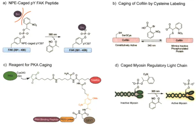
to probe the cellular functions of these proteins.
02N
O=P-O
O=P-0-0 365 nm 0
Protein ~ NO O rti
Inactive Active
Figure 1-4. General caging strategy. Caging of an important functionality of a protein renders
the protein inactive within the biological system. Irradiation at 365 nm releases the caging group to generate the active protein that can exert native effects within the system.
Caged Peptides
Caged peptides have been applied for cellular studies because they combine specificity
with synthetic accessibility. Peptide inhibitors of calmodulin and MLCK have been synthesized
phosphorylation during the migration of eosinophil (white blood) cells.? Irradiation of motile
eosinophils that had been microinjected with the caged peptides inhibited further migration,
implicating calmodulin-dependent MLCK and myosin II as essential components for
locomotion.
Another study used caged peptides for investigation of focal adhesion signaling.32 Focal
adhesions connect the cell to the extracellular matrix and enable signaling between external
factors and cytoplasmic proteins.13 A central component of these structures is the non-receptor
tyrosine kinase FAK. Integrin clustering induces the autophosphorylation of FAK at Tyr397,
which recruits other proteins to create a signaling complex. FAK-mediated signal transduction
not only governs migratory processes but also influences cell growth and apoptosis.'0
The role of FAK phosphorylation was investigated by the microinjection of caged
peptides based on the FAK autophosphorylation sequence.3 2 NPE-caged phosphotyrosine397
was incorporated into the peptides by Fmoc-based solid phase peptide synthesis using the
corresponding caged phosphorylated amino acid building block.36 Microinjection and uncaging of these peptides within cells temporarily inhibited lamellar protrusions by competitively binding
FAK effectors such as Src and PI3K, which are necessary for proper migration (Figure 1-5a).
Subsequent dephosphorylation of the uncaged peptides by cellular phosphatases reversed the
effects of irradiation. This study illustrates an advantage of the caging technology because
although injection of the phosphorylated peptide induced an immediate response, the caged
peptides were inactive until irradiation and offered precise temporal control over phosphopeptide
b) Caging of Cofilin by Cysteine Labeling
a) NPE-Caged pY FAK Peptide
1NO2 I 365nm -0 I NO O NPE-caged pY397 FAK_(391 408)
c) Reagent for PKA Caging
Cys343 0 H2N 0 0 -SH___/ ti 0 H ome SH Ser3Cys Contittvely Active 0 HO Br N02 340 nm S NO2 Mimcs inactive Phosphorylated Proain
d) Caged Myosin Regulatory Light Chain 02N 0 0 -O- - - --- O 0o ~~ 365 nni NOO 0
Inactive Myosin & I, Active Myosin
Figure 1-5. Caged peptides and proteins for studying migration. a) NPE-caged pTyr397
peptide based on the FAK autophosphorylation sequence. Upon irradiation and uncaging, Src and other effectors can bind the peptide and disrupt migration. b) Constitutively active cofilin can be caged by reaction with a-bromo-(2-nitrophenyl)acetic acid. The caged protein mimics the inactive phosphorylated protein. c) The multi-functional reagent for caging PKA. The PKA binding peptide targets the reagent to PKA and positions the maleimide near Cys343 for labeling. The QSY7 chromophore quenches fluorescence of the TAMRA fluorophore. Uncaging releases the PKA inhibitor peptide and the quencher to restore PKA activity and to generate a fluorescence signal. d) Incorporation of the caged mRLC into myosin eliminates activity. Uncaging releases the native phosphoprotein to restore myosin function.
While the application of caged peptides has enabled the study of FAK phosphorylation
and MLCK activity during migration, this approach is limited by several drawbacks. With the
FAK study, for instance, the short peptides may not faithfully represent the specificity of the
corresponding protein in a cellular context and may interact with non-cognate binding partners.
Second, following irradiation, caged peptides generally function as inhibitors of the target
protein rather than as activators. Although this is useful in many situations, a more
O=P-0-pY397
straightforward approach to examine the function of a protein of interest is to cage the full-length
protein itself. This allows irradiation to directly activate the protein and induce the native
downstream effects. Additionally, application of the full-length protein ensures that other
binding sites or signaling interactions are maintained. Caging of full-length proteins has been
achieved through site-specific cysteine labeling, though a more general approach exploits protein
semisynthesis.
Caged Proteins by Cysteine Labeling
Cysteine-targeted caging has been successfully employed to cage the actin-regulating
protein cofilin.2 Cofilin is involved in the reorganization of actin networks, primarily by
severing actin filaments and creating new sites of nucleation for actin polymerization. As the
protein is deactivated by phosphorylation at Ser3, a photoactivated variant of this protein was
generated by selectively labeling a Ser3Cys mutant with a-bromo-(2-nitrophenyl)acetic acid
(Figure 1-5b). While the negatively charged caging group mimicked the phosphorylated residue
and rendered the protein inactive, irradiation released the constitutively active Ser3Cys protein.
Microinjection of the protein into cells and uncaging induced actin polymerization and cell
protrusions and, in contrast to previous studies, suggested that active cofilin is required for
migration.
The cysteine labeling methodology has recently been extended to cAMP-dependent
protein kinase (PKA), which contains multiple reactive residues.3 3 This kinase is involved in
signaling cascades governing many aspects of migration, including actin rearrangements, Rho
GTPases, and mRLC phosphorylation.37 The photoactivatable PKA was modified with a
concomitantly generate a fluorescent signal to report successful photolysis (Figure 1-5c). With
this strategy, a short inhibitory peptide sequence targeted to PKA was elaborated with a caged
linker, a fluorophore-quencher pair, and an electrophilic maleimide. Binding of the peptide to
the enzyme positioned the maleimide near reactive Cys343 to promote alkylation with the caging
agent, thereby covalently tethering the peptide inhibitor to the protein. Microinjection of caged
PKA into cells and uncaging generated a fluorescent signal, released the inhibitor, and,
consistent with the role of PKA in cytoskeletal rearrangements, induced stress fiber degradation.
Caged Proteins by Semisynthesis
While cysteine labeling represents an efficient and direct approach for the attachment of
caging groups onto a protein, this strategy is not feasible when the essential residue cannot be
selectively targeted for reaction. In these cases, protein semisynthesis and native chemical
ligation can by exploited for the site-specific incorporation of unnatural residues into full-length
proteins (Figure 1-6).38 With this technique, a C-terminal peptide thioester reacts with a peptide
containing an N-terminal cysteine to generate a native amide bond between the two fragments.39
The portion of the protein containing the unnatural residue is prepared through solid phase
peptide synthesis, while the remainder of the protein is recombinantly expressed. Methods to
generate the N-terminal cysteine and C-terminal thioester by peptide synthesis or through protein
expression have been developed, enabling convenient incorporation of unnatural residues into
full-length proteins. However, as robust peptide synthesis is limited to chains of 40 to 50
residues, this approach is most convenient when the unnatural residue is incorporated near either
S + HNTransthioesterification
H2N f P
Excess thiols, pH 8 C-terminal thioester N-terminal cysteine
O SH
-H ~eptide S to N Acyl Shift
H2 H
Figure 1-6. Mechanism of native chemical ligation. A C-terminal peptide thioester is reacted
with a peptide containing an N-terminal cysteine. Transthioesterification occurs until the N-terminal cysteine reacts. A spontaneous S to N acyl shift generates the native amide bond between the two fragments to yield the full-length protein. The unnatural residue can be incorporated into either peptide fragment.
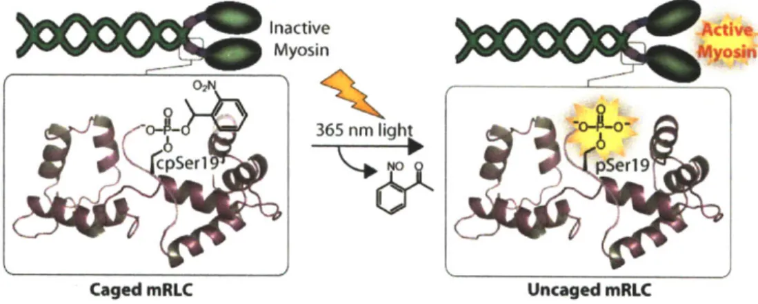
In this thesis, we have applied this semisynthetic technique to prepare a photoactivated
myosin (Figure 1-5d). Myosin II is an ATPase motor protein responsible for generating the
contractile forces necessary to initiate focal complex formation at the leading edge of a migrating
cell as well as to release adhesions at the rear. The protein complex is activated upon
phosphorylation of the associated myosin regulatory light chain (mRLC) protein at Serl9. To
directly probe the effects of myosin on cellular actin dynamics, a photoactivated variant of the
mRLC was generated through protein semisynthesis to install NPE-caged phosphoserine at
position 19.3 Following incorporation of this caged protein into the myosin complex, myosin
was inactive in the caged state, but activity was restored through irradiation and release of the
native phospho-mRLC (Figure 1-5d). When introduced into a cell, this protein tool can be used
to activate myosin to probe the localized effects of myosin contractility in real-time. These
studies are further detailed in Chapters 2 and 3 of this thesis.
Another attractive target for caging is paxillin, which is a multi-domain protein that
survival, and proliferation. Phosphorylation occurs throughout the molecule and creates docking
sites for other adaptor and regulatory proteins. For instance, phosphorylation at Tyr31 stimulates
the Rho GTPases Cdc42 and Rac1 through binding of GTPase regulatory proteins. These
GTPases in turn promote the formation of membrane protrusions and control focal adhesion
turnover.40 To interrogate the role of this protein in migration, and particularly the effect of
Tyr31 phosphorylation, paxillin was synthesized by native chemical ligation to incorporate
NPE-caged phosphotyrosine at position 31.35 The semisynthetic protein was tested to ensure that
binding to native protein partners was intact and that it remained a substrate for known kinases.
Irradiation released phospho-paxillin, validating the potential of the tool for unraveling signaling
pathways at focal adhesions. To further facilitate the semisynthesis, examinations into
heterologous expression of paxillin in E. coli are described in Appendix 1 of this thesis.
While protein semisynthesis has efficiently yielded caged proteins for probing various
aspects of migration, an alternative to this approach relies on in vivo amber suppression methods
41
using evolved orthogonal tRNA/tRNA synthetase pairs specific for the unnatural amino acid.
Recent studies have shown that this system can be applied in mammalian cells to produce caged
proteins in situ.4 2
, 43 While caged derivatives of lysine have been successfully incorporated in
HEK293 cells, the tRNA/tRNA synthetase pairs for caged serine and caged tyrosine44 have
been established for use in bacterial systems. This technology complements semisynthetic
approaches by providing an alternative route for the preparation and delivery of the caged
proteins. Specifically, a three-segment semisynthesis is required if the unnatural residue must be
incorporated in the middle of a protein because solid phase peptide synthesis is limited to
peptides of about 50 residues.3 8 In contrast, with amber suppression, the unnatural residue may
introduced into cells through microinjection, which requires specialized equipment and restricts
the number of cells that can be analyzed, whereas the amber suppression technology eliminates
the need for external delivery methods. Despite these advantages, application of this technique
to any new unnatural amino acid requires evolution of a new tRNA/tRNA synthestase pair that is
compatible with mammalian expression. In addition, the amino acid must be non-toxic and
efficiently taken up into the cell.41 Finally, incomplete suppression and the formation of
truncated protein may introduce compounding effects. Nonetheless, this method, in combination
with semisynthetic approaches, offers flexibility for the preparation of protein tools for
investigating cell migration.
1.3.2 Photoreceptor Fusions
As a complementary approach to caging, plant photoreceptors have been adapted to
provide genetically encoded proteins that can be activated by light.17 Proteins from the phytochrome and phototropin classes respond to light through bound chromophores and undergo
reversible conformational changes upon irradiation. In plants, these proteins are involved in
mediating phototropism, the process of directional growth in response to light. While
phytochromes respond to red light through a bilin chromophore, phototropins bind flavins and
absorb blue light.45 Both families of protein domains have been engineered to enable
photoactivation of the Rho GTPases.
LOV Domain Fusions
Phototropins react to light through light-oxygen-voltage (LOV) domains, which bind
flavin mononucleotide and undergo a characteristic photocycle in which the protein forms a
formation of a covalent adduct through a cysteine residue of the LOV domain and the FMN
molecule. The bond formation induces a concomitant conformational change, in which an
associated a-helix, termed the Ja helix, is released and unwinds.46 Fusion of the LOV domain to
a protein at an appropriate position generates photo-dependent allosteric inhibition and can
render protein-protein interactions or activity sensitive to light. This approach has been
successfully applied for the generation of a photoactivated Rac 1.
The photoactivated Racl was created by fusing a LOV domain to a constitutively active
mutant of Rac1 (Figure 1-7a).47 In the dark state, the LOV domain sterically inhibits interactions
between Rac1 and effector proteins. However, upon irradiation at 458 or 473 nm, unwinding of
the Ja helix relieves inhibition and enables the active Racl to interact with native effectors.
When expressed in mammalian cells, brief laser pulses localized to specific regions at the edge
of the cell initiated protrusions and ruffling events, which were reversible and dependent on the
intensity of irradiation. In subsequent studies, this tool was applied to examine the role of Racl
during migration in more complex systems. In zebrafish neutrophils, localized Rac1 activation
48
enabled interactions with regulatory proteins to be examined, while application of the protein in
the Drosophila egg chamber allowed interrogation of the influence of the GTPase on coordinated
migration.49 These studies together demonstrate that this tool can be exploited to extract detailed information about Racl in live cells. Further investigation of the light-dependent dynamics of
the LOV domains has revealed mutations that can modulate the properties of the domains for
a) LOV Domain S4Lamellopodia formation b F S b) EKEI/GI System Lamellopodia formation 0 0 HO OH OH 0
Flavin mononucleotide cofactor for the LOV domain and
FKF1/GI systems
c) PhyB/PIF System
650 nm ) CAAX
GTP Hydrolysist Rac Activation
750 nm
Lamellopodia formation
Figure 1-7. Light-activated Raci based on plant photoreceptors. a) Photoactivated Rac1 was
generated by fusing constitutively active Rac1 to the LOV domain. Irradiation causes the Ja helix to unravel and allows effectors to bind Rac1. b) In the FKF1/GI system, irradiation at 450 nm recruits the FKF1-Racl construct to the membrane-bound GI protein. Because Rac1 is active at the membrane, irradiation induces lamellopodia formation. c) The PhyB/PIF system enables reversible activation of endogenous Rac1. Upon irradiation, the PJF-Tiam construct binds to membrane-bound PhyB. This recruitment leads to Rac1 activation. Irradiation at
In addition to the LOV domain fusion, two other photoreceptor systems, the flavin
binding, Kelch repeat, F-box1 (FKFl) / GIGANTEA (GI) system5' and the phytochrome B (PhyB) / phytochrome interaction factor 3 (PIF3) system,5 2 have been engineered for
applications to control protein activity. For both protein pairs, irradiation induces binding
between the two partners: FKF1 and GI or PhyB and PIF3. Thus, fusion of these photoreceptor
domains to two proteins of interest enables irradiation to drive dimerization. Both systems have
been adapted for investigating the function of the GTPase Rac1 at the cell membrane. In these
studies, the photoreceptor domain is expressed with a C-terminal CAAX sequence to anchor the
module to the membrane, and the GTPase or a GTPase activating protein is fused to the
corresponding photoreceptor binding partner. This construct remains cytosolic until irradiation
promotes binding between the light-sensitive proteins and localizes the GTPase or activator to
the membrane. Because Rac1 is active primarily at the plasma membrane, light-mediated
recruitment stimulates lamellopodia formation.
FKF1/GI System
In the case of FKF1 and GI from Arabidopsis thaliana, exposure to blue light induces
dimerization (Figure 1-7b).5 FKF1 contains a LOV domain, and when irradiated, Cys91 forms
a covalent bond with FMN. The resulting conformational change enables the protein to bind the
nuclear protein GI. To create a tool for the study of Racl, the FKF1 and GI proteins were
engineered to remain cytosolic and to only dimerize when exposed to light, even if expressed at
high concentrations. Rac1, without the native C-terminal CAAX box, was expressed as a fusion
to FKFl, and a GI-mCherry-CAAX construct was co-expressed within the cell. Irradiation of
cytoplasmic FKFl-Racl to the membrane via the GI-mCherry-CAAX protein, thereby inducing
lamellipodia formation. Due to the long half-life of the FMN-FKF1 complex, sustained
interactions were achieved, though extended irradiation for 5 min was necessary. This strategy is
promising, but further improvements in the association and dissociation kinetics of the two
binding partners will create a system that can be more generally applied. Additionally, because
this approach relies on the interaction between two protein constructs, the expression levels must
be carefully controlled to ensure proper responses.
Phytochromes
In contrast to the LOV domains, phytochromes respond to red light when the associated
bilin chromophore undergoes a cis-to-trans isomerization. PhyB from A. thaliana covalently
binds the tetrapyrrole chromophore phycocyanobilin (PCB), which undergoes reversible
photoisomerization upon irradiation with red or near-infared light and mediates the transition of
PhyB between the red-absorbing (Pr) state and the far-red-absorbing (Pfr) state. The PIF3 protein binds PhyB only after it has absorbed 650 nm light and exists in the Pfr state, and in
plants, this heterodimer translocates from the cytoplasm to the nucleus to mediate gene
transcription. Spontaneous dissociation occurs over hours, but irradiation with infrared light
(750 nm) immediately reverses binding.45
The ability of the PhyB/PIF3 system to control protein-protein interactions was first
validated through an in vitro study using the Cdc42 GTPase and an effector binding domain.54 In subsequent studies, the PhyB/PIF system was adapted for application in mammalian cells and
was optimized for reversible association on the seconds timescale.5 2 The protein binding pair
PhyB domain was expressed with a CAAX box, and the catalytic domain of the Rac GEF Tiam
was fused to the phytochrome-binding domain of PIF6 (Figure 1-7c). Cells were treated with
PCB and a patterned light source, which transmitted the deactivating infrared light to the entire
sample while focusing a red laser (-3 [tm) to a localized area of the cell. This patterning ensured
that the activated protein was present only in the desired region by deactivating molecules that
diffused away from that area. The 650-nm red laser initiated the PhyB(Prf)-PIF6 interaction to
recruit the Rac GEF to the membrane, thereby activating endogenous Racl to induce
lamellipodia formation. While this system offers the advantages of rapid interaction kinetics and
reversibility, the PCB cofactor, which is not produced in mammalian cells, must be isolated from
natural sources and added to the culture. Additionally, like the FKFl-GI system, the PhyB and
PIF6 fusions must be expressed at similar levels for robust responses.
Table 1-2. Comparison of methods for light-mediated protein function.
Approach Molecular Weight Reversible?
Caging Group 150 - 300 Da No
LOV Domain 16,000 Da Yes
FKF1/GI FKF1: 68,000 Da; GI: 129,000 Da Slowly
PhyB/PIF PhyB: 100,000 Da; PIF: 11,000 Da Immediately with infared light
Table 1-2 summarizes the approaches that have been described in the preceding sections.
Compared to small molecule caging techniques, the light-sensitive protein domains are
advantageous because they are genetically encoded and do not require complex delivery
methods. Second, unlike small molecule caged compounds that are irreversibly activated upon