Optimized EPA/DHA 6/1 formulation prevents Angiotensin-II induced hypertension and endothelial dysfunction in rats
256
0
0
Texte intégral
(2) UNIVERSITE DE STRASBOURG. Ecole Doctorale des Sciences de laVie et de la Santé. THÈSE présentée pour obtenir le grade de Docteur de l’Université de Strasbourg Discipline : Sciences du Vivant Domaine : Pharmacologie. Optimized EPA: DHA 6:1 formulation prevents Angiotensin-II induced hypertension and endothelial dysfunction in rats Par. Zahid Rasul NIAZI Soutenue le 06/07/2016 devant la commission d’examen : Professeur Dominique DELMAS. Rapporteur externe. Professeur Isabelle LARTAUD. Rapporteur externe. Professeur Dominique STEPHAN. Examinateur. Professeur Catherine MARCHAND-LEROUX. Examinateur. Professeur Valérie B. SCHINI-KERTH. Directeur de these. Docteur Cyril AUGER. Co-directeur de these.
(3) Dedication. This thesis is dedicated to my beloved father, late mother, my lovely wife and children for their love, patience and understanding.. -1-.
(4) Acknowledgements I would like to thank all the people who contributed in some way to the work described in this thesis. First and foremost, I would like to express my sincere gratitude to my supervisor, Professor Valérie Schini-Kerth for accepting me into her group and my co-supervisor Dr Cyril Auger. During my thesis, they contributed to the success of this work by giving me intellectual freedom in my work, supporting my attendance at various conferences, engaging me in new ideas, and demanding a high quality of work in all my endeavors. Additionally, I would like to thank all my committee members Professor M.D.DELMAS, Professor Isabelle LARTAUD, Professor M.D. STEPHAN and Professor C. MARCHAND-LEROUX for their interest in my work. Special thank goes to Dr Malak Abbas, Thaise Proto, Dr Sherzad Khorsheed Rasheed, Waseem Ashraf and to Ikram Ullah Khan for their help and support during my thesis. Every result described in this thesis was accomplished with the help and support of fellow lab mates Chabert Philippe, Antonio josé Léon Gonzaléz, Sonia khemais, , Said Amissi, Nguyen Phuong Nga, , Mohamad kassem and special thank for Brigitte Pollet and Evelyne Lacoffrette- Cronier for their full support and assistance, thank you All. I am also thankful to Hira Hassan, Abdul Wahid and Mohammad Akmal Farooq who supported me a lot during my thesis writing.. -2-.
(5) Table of Contents Dedication .......................................................................................................................................- 1 List of publications .........................................................................................................................- 5 List of Abbreviations ......................................................................................................................- 7 List of Figures and Tables ............................................................................................................. - 10 Tables .......................................................................................................................................... - 10 Abstract ....................................................................................................................................... - 11 Résumé ....................................................................................................................................... - 15 1. Chapter 1 ............................................................................................................................. - 20 -. Physiology of the endothelium ..................................................................................................... - 20 -. 1.1 1.2 1.3. Cardiovascular diseases.............................................................................................. - 21 Vascular endothelium ................................................................................................. - 22 Endothelial regulation of vascular tone ...................................................................... - 23 -. 1.3.1. The endothelium-derived vasorelaxing factors.................................................................. - 25 -. 1.3.1.1 Nitric oxide (NO) ................................................................................................................. - 25 1.3.1.2 Endothelium-derived hyperpolarization (EDH) .................................................................. - 32 1.3.1.3 Prostacyclin ........................................................................................................................ - 34 1.3.2. The endothelium-derived vasocontracting factors ............................................................ - 36 -. 1.3.2.1 Angiotensin II ...................................................................................................................... - 36 1.3.2.2 Endothelin-1 ....................................................................................................................... - 38 1.3.2.3 Thromboxane A2 & Prostacyclin I2 (TxA2 & PGI2) ............................................................. - 40 1.3.2.4 Oxidative stress and reactive oxygen species (ROS) .......................................................... - 42 -. 1.4 1.5. Endothelial dysfunction.............................................................................................. - 43 Endothelial dysfunction and hypertension ................................................................. - 45 -. 1.5.1. Models of Hypertension ..................................................................................................... - 47 -. 1.5.2. Excessive Reactive Oxygen Species Production in Hypertension ....................................... - 48 -. 1.5.2.1 Inflammatory Regulation of Hypertension-Associated Endothelial Dysfunction .............. - 49 1.5.3. 1.6 2. Antihypertensive treatments ............................................................................................. - 50 -. Endothelial Dysfunction and Diabetes ....................................................................... - 52 -. Chapter 2 Omega-3 and cardiovascular effects ...................................................................... - 54 -. 2. Lipids ....................................................................................................................................... - 55 -. 2.1 2.2 2.3. Saturated fatty acids (SFA) ........................................................................................ - 56 Monounsaturated fatty acids (MUFA) ....................................................................... - 57 Polyunsaturated fatty acids (PUFA) ........................................................................... - 58 -. 2.3.1. Trans fatty acids (TFA) ........................................................................................................ - 59 -3-.
(6) 2.3.2. n-3 polyunsaturated fatty acids (n-3 PUFAs)...................................................................... - 59 -. 2.3.3. n-6 polyunsaturated fatty acids ......................................................................................... - 62 -. 2.4 2.5 2.6. The importance of the ratio of omega-6/omega-3 essential fatty acids ..................... - 62 Metabolim of PUFA ................................................................................................... - 64 Health benefits of n-3 polyunsaturated fatty acids ..................................................... - 67 -. 2.6.1. n-3 polyunsaturated fatty acids and diabetes.................................................................... - 67 -. 2.6.2. n-3 polyunsaturated fatty acids and cardiovascular diseases ............................................ - 67 -. 2.6.2.1 Lipid Profile ......................................................................................................................... - 69 2.6.2.2 Inflammation and endothelial function ............................................................................. - 69 2.6.2.3 Atherosclerosis ................................................................................................................... - 70 2.6.2.4 Platelets aggregation .......................................................................................................... - 70 -. 2.7 3. Clinical implications of omega-3 fatty acids.............................................................. - 71 -. RESULTS ............................................................................................................................... - 75 -. GENERAL DISCUSSION AND PERSPECTIVES.................................................................................. - 117 DISCUSSION ET PERSPECTIVES .................................................................................................... - 129 REFERENCES .............................................................................................................................. - 130 -. -4-.
(7) List of publications 1. Publications: Rasul Zahid Niazi, Silva G.C, Porto Ribeiro T, Zgheel F, Auger C, Schini-Kerth V. Choronic oral Intake of the omega 3 formulations EPA:DHA 6:1 prevents the angiotensin II-induced hypertension and endothelial dysfunction in rats (submitted to the British Journal of Pharmacology). Mohamad Kassem, Zahid Rasul Niazi , Malak Abbas , Ali El Habhab, Guillaume Kreutter , Sonia Khemais-Benkhiat, Cyril Auger, Maria-Cristina Antal, Valerie B. Schini-Kerth, Florence Toti, Laurence Kessler.The Endocrine Pancreas, an Early Sensor of Senescence in Middle-aged Rats with Still Normal Vascular Function. (Under revision) Alain N’guessan Yao, Zahid Rasul Niazi, Iveta Najmanová, Mamadou Kamagaté, Amissi Said, Henri Die-Kacou, Cyril Auger, Philippe Chabert, Valérie Schini-Kerth. Chronic treatment with an aqueous extract of Phyllanthus amarus prevents hypertension and endothelial dysfunction in Docasalt rats. (paper in preparation) Amissi Said, Zahid Rasul Niazi, Mélanie Burban, Romain Kessler, Mathieu Canuet, Florence Toti, Laurent Monassier, Nelly Boehm, Cyril Auger, Ferhat Meziani and Valérie B. Schini-Kerth. The omega-3 EPA:DHA 6:1 superior formulation prevents the monocrotaline-induced pulmonary hypertension, endothelial dysfunction and vascular remodeling, and right ventricular failure in rats. (paper in preparation) 2. Poster Communications: 1) Niazi Z, Silva G.C, Porto Ribeiro T, Zgheel F, Auger C, Schini-Kerth V. Choronic oral Intake of the omega 3 formulation EPA:DHA 6:1 prevents the angiotensin II-induced hypertension and endothelial dysfunction in rats, JCI (Campus Illkirch day) 13-14 April Illkirch France 2015. 2) Niazi Z, Silva G.C, Porto Ribeiro T, Zgheel F, Auger C, Schini-Kerth V. Choronic oral Intake of the omega 3 formulation EPA:DHA 6:1 prevents the angiotensin II-induced hypertension and endothelial dysfunction in rats, CPBI (Congrès de la société de physiologie et de biologie intégrative) 4-6 May Strasbourg-France 2015. 3) Niazi Z, Silva G.C, Porto Ribeiro T, Zgheel F, Auger C, Schini-Kerth V. Choronic oral Intake of the omega 3 formulation EPA:DHA 6:1 prevents the angiotensin II-induced hypertension and endothelial dysfunction in rats, FMTS (Fédération de médecine translationnelle de Strasbourg) 16-17 April, Strasbourg-France 2015. 4) Kassem M, Niazi Z, Abbas M, Samama B, Auger C, Constantinescu A, Vauchelles R, Lessinger JM, BOEHM N, Schini-kerth V, Toti F, Kessler L. Morphological and functional abnormalities of the endocrine pancreas in middle age rat: Impact for transplantation, CPBI (Congrès de la Société de Physiologie et de Biologie Intégrative) 4-6 may, Strasbourg-France. La résumé est publié au journal Acta Physiologica Volume 214, Issue Supplement S700, Pages 1-92, May 2015. -5-.
(8) 5) Niazi Z, Silva G.C, Porto Ribeiro T, Zgheel F, Auger C, Schini-Kerth V. Choronic oral Intake of the omega 3 formulation EPA:DHA 6:1 prevents the angiotensin II-induced hypertension and endothelial dysfunction in rats, (Printemps de la cardiologie) 2-3 April Toulouse-France 2015. 6) Niazi Z, Silva G.C, Porto Ribeiro T, Zgheel F, Auger C, Schini-Kerth V. Choronic oral Intake of the omega 3 formulation EPA:DHA 6:1 prevents the angiotensin II-induced hypertension and endothelial dysfunction in rats, ESC (European society of cardiology) 23-26 May Seville-Spain 2015. 7) Niazi Z, Silva G.C, Porto Ribeiro T, Zgheel F, Auger C, Schini-Kerth V. Choronic oral Intake of the omega 3 formulation EPA:DHA 6:1 prevents the angiotensin II-induced hypertension and endothelial dysfunction in rats, CIC (French society of pharmacology and therapeutics) 21-23 April Caen-France 2015. 3. Oral Communications 1) Niazi Z, Silva G.C, Porto Ribeiro T, Zgheel F, Auger C, Schini-Kerth V. Choronic oral Intake of the omega 3 formulation EPA:DHA 6:1 prevents the angiotensin II-induced hypertension and endothelial dysfunction in rats, CIC (French society of pharmacology and therapeutics) 21-23 April Caen-France 2015. 2) Niazi Z, Silva G.C, Porto Ribeiro T, Zgheel F, Auger C, Schini-Kerth V. Choronic oral Intake of the omega 3 formulation EPA:DHA 6:1 prevents the angiotensin II-induced hypertension and endothelial dysfunction in rats, JCI (journées scientifiques du campus universitaire d'Illkirch) 2113 May Strasbourg-France 2016. 3) Kassem M, Niazi Z, Abbas M, Vauchelles R, Auger C, Antal MC, Schini-kerth V, Toti F, Kessler L. The endocrine pancreas an early sensor of senescence in middle-aged rats: Impact for transplantation, ADA (American Diabetes Association) 10-14 June New Orleans, LA- USA 2016.. -6-.
(9) List of Abbreviations. AA AC ACE Ach ADM ADMA ADP ALA ANOVA ARAs ARBs AT1R AT2R ATP BH4 BP BW CAD cAMP CBDL cGMP CHD CHF COX CRP CVD Cx37 CYP DHA DM DPA EC EDCF EDH EDHF EDRF EETs EFA eNOS. Arachidonic Acid Adenylyl cyclase Angiotensin Converting Enzyme Acetylcholine Adenosine Monophosphate Asymmetric Dimethylarginine Adenosine Di-Phosphate Alpha-Linolenic Acid Analysis of Variance Angiotensin Receptor Antagonists Angiotensin Receptor Blockers Angiotensin II Type 1 Receptor Angiotensin II Type 2 Receptor Adenosine Tri-Phosphate Tetrahydrobiopterin Blood Pressure Bodyweight Coronary Artery Disease Cyclic Adenosine-3’,5’-Mono-Phosphate Common Bile Duct Ligation Cyclic Guanosine 3'-5' Monophosphate Coronary Heart Disease Congestive Heart Failure Cyclooxygenases C-reactive protein Cardiovascular Disease Connexin 37 Cytochrome Docosahexaenoic Acid Diabetes Mellitus Docosapentaenoic Acid Endothelial Cell Endothelium-Derived Contracting Factor Endothelium-Dependent Hyperpolarization Endothelium-Derived Hyperpolarizing Factor Endothelium-Derived Relaxing Factor Epoxyeicosatrienoic Acids Essential Fatty Acids Endothelial Nitric Oxide Synthase -7-.
(10) EPA EPCs ER FAME FAs GAPDH GC/MS GLA GLC GPx GTP H2O2 HDL HETEs HO-1 HSP-90 IKCa IL-6 Inos JAKs LA LDL Lev L-NA L-NAME LOX LPC LPS LTs MAPKs MCP-1 MGJ MMPs MnTMPyP MUFA NADH NADPH nNOS NO NOS O2OA. Eicosapentaenoic Acid Endothelial Progenitor Cells Estrogen Receptor Fatty Acid Methyl-Esters Fatty Acids Glyceraldehyde-3-Phosphate Dehydrogenase Gas Chromatography/Mass Spectrometry Gamma-Linolenic Acid Gas Liquid Chromatography Glutathione Peroxidase Guanosine Triphosphate Hydrogen Peroxide High Density Lipoprotein Hydroxyeicosatetraeneoic Acids Hemeoxygenase-1 Heat Shock Protein 90 Intermediate conductance calcium-activated potassium channel Interleukin-6 Inducible Nitric Oxide Synthase Janus kinases Linoleic Acid Low Density Lipoprotein Levcromakalim Nω-Nitro-L-arginine Nω-Nitro-L-Arginine Methyl Ester Lipoxygenases Lysophosphatidylcholine Lipopolysaccaride Leukotrienes Mitogen activated protein kinases Monocyte Chemoattractant Protein-1 Myoendothelial Gap Junction Matrix Metalloproteinase Mn (III) Tetrakis(1-methyl-4 pyridyl) porphyrin, Superoxide Dismutase Mimetic Mono-Unsaturated Fatty Acids Nicotinamide Adenine Dinucleotide Nicotinamide Adenine Dinucleotide Phosphate Neuronal Nitric Oxide Synthase Nitric Oxide Nitric Oxide Synthase Superoxide Anions Oleic Acid -8-.
(11) OH. ONOOPBS PC PDGF PEG-SOD PGE PGG2 PGH2 PGI2 PI3K PKA PKC PKG PLA-2 PLD PUFA RAAS RAS RIPA RLP ROS SEM SFA Sgc SHR Skca SNP SOD TGF-B1 TNF-α TXA2 VCAM VEGF-A VEGFR VLDL VSMC WHO. Hydroxyl Groups Peroxynitrite Phosphate buffer saline Phosphatidylcholine Platelet-Derived Growth Factor Polyethylene Glycol-Superoxide Dismutase Prostaglandins E2 Prostaglandin G2 Prostaglandins H2 Prostacyclin Phosphoinositide 3-Kinase Protein Kinase A Protein Kinase C Protein Kinase G Phospholipase A-2 Phospholipase D Poly-Unsaturated Fatty Acids Renin-Angiotensin-Aldosterone-System Renin-Angiotensin System Radioimmunoprecipitation assay Remnant lipoprotein Reactive Oxygen Species Standard error mean Saturated fatty acids Soluble Guanylyl Cyclase Spontaneously hypertensive rat Small conductance calcium-activated potassium channel Sodium nitroprusside Superoxide Dismutase Transforming Growth Factor-B1 Tumor Necrosis Factor-alpha Thromboxane A2 Vascular Cell Adhesion Molecule-1 Vascular Endothelial Growth Factor A Vascular Endothelial Growth Factor Receptor Very Low Density Lipoprotein Vascular Smooth Muscle Cell World Health Organization. -9-.
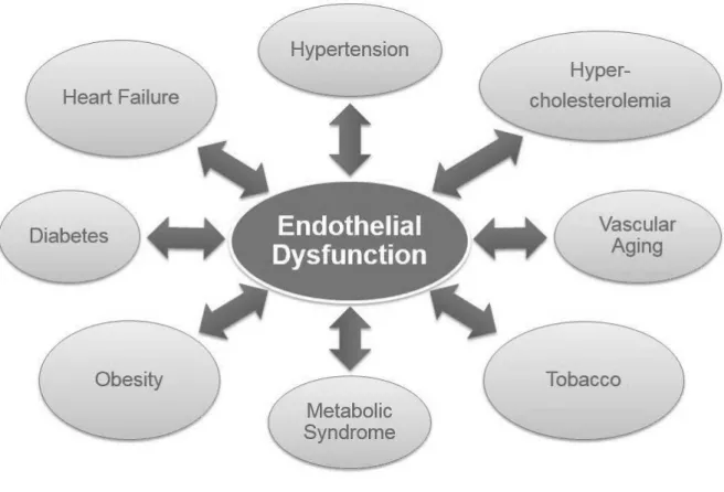
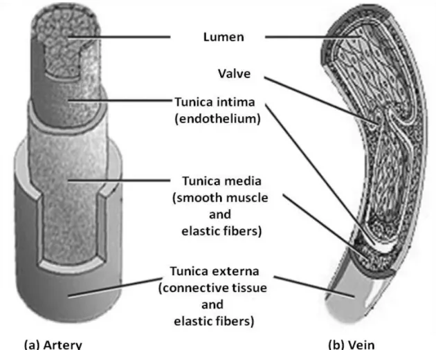
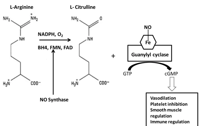
(12) List of Figures and Tables Figure 1. Endothelial dysfunction; A hallmark of major cardiovascular and other diseases .................. - 21 Figure 2. Structure of the blood vessel (adapted from Boston University School of Public Health). ..... - 23 Figure 3. Endothelium-dependent release of vasocontracting and vasorelaxing factors. ..................... - 25 Figure 4. NO generation from L-Arginine and its functional properties (Ghalayini 2004) ..................... - 26 Figure 5. Nitric oxide synthesis pathway in the endothelial cell and its actions in the vascular smooth muscle cell............................................................................................................................................... - 27 Figure 6. The pleiotropic effects of NO. .................................................................................................. - 28 Figure 7. The activation and inhibition sites of eNOS. ............................................................................ - 30 Figure 8. Regulation of eNOS activity. .................................................................................................... - 31 Figure 9. Hypothesis describing the endothelium-derived hyperpolarizing pathway............................ - 33 Figure 10. Production and action of prostaglandins (Araujo, Soeiro et al. 2005). ................................. - 35 Figure 11. Summary of acute and chronic stimulation of angiotensin II receptors (Dasgupta and Zhang 2011). ...................................................................................................................................................... - 37 Figure 12. The biological effect of endothelin-1 on arteries in physiological and pathophysiological conditions................................................................................................................................................ - 39 Figure 13. Prostanoids biosynthesis and response pathways (Zhang, Gong et al. 2010). ...................... - 41 Figure 14. The effects of vascular endothelial factors on the function of vascular smooth cells in healthy and pathological conditions. ................................................................................................................... - 44 Figure 15. Mechanisms implicated in essential hypertension-associated endothelial dysfunction ...... - 46 Figure 16. Progression of endothelial dysfunction in relation to the progression of insulin resistance (Cosentino and Lüscher 1997). ............................................................................................................... - 53 Figure 17. Monounsaturated fatty acids MUFAs .................................................................................... - 57 Figure 18. Major Polyunsaturated fatty acids (PUFA). (Nair, Leitch et al. 1997). ................................... - 58 Figure 19. Structures of dietary ω 3 and ω 6 polyunsaturated fatty acids. A: C18 ω 3 and ω 6 PUFA. B: C20–22 ω 3 and ω 6 PUFA (Jump, Depner et al. 2012)........................................................................... - 61 Figure 20. Importance of omega-6/omega-3 ratio. ................................................................................ - 64 Figure 21. Synthesis pathway of omega-6 and omega-3 fatty acids in mammals. ................................ - 66 Figure 22. Physiological effects of n-3 PUFA that might influence CVD Risk (Mozaffarian and Wu 2011). .. 68 Figure 23. Omega-3 EPA:DHA 6:1 prevents Ang II-induced endothelial dysfunction and hypertension in rats. ....................................................................................................................................................... - 123 Figure 24. Perspectives in animal models. ............................................................................................ - 127 Figure 25. Clinical perspective. ............................................................................................................. - 128 -. Tables Table 1. Structure and taxonomy of fatty acids (Simopoulos 1998) ...................................................... - 55 Table 2. Structure of different unbranched fatty acids with a methyl end and a carboxyl (acidic) end.- 56 -. - 10 -.
(13) Abstract Cardiovascular diseases are the leading cause of death worldwide, both in developed and developing countries. The incidence of cardiovascular diseases is linked to several risk factors that are either non-modifiable (age, gender, genetic background) or modifiable. The modifiable risk factors include lifestyle (western diet, tobacco use, alcohol abuse, physical inactivity) and treatable diseases such as hypertension, dyslipidemia, obesity, and diabetes. Many preclinical and clinical studies have shown that development of cardiovascular diseases and risk factors are associated early with an endothelial dysfunction. The endothelium, the monocellular layer lining all blood vessels, represents the largest cell compartment in contact with blood flow. Endothelial cells contribute to the vascular tone and constitute a protective surface with anti-thrombotic properties, mainly through the release of potent vasoprotective factors such as nitric oxide (NO). In cardiovascular diseases and also during physiological ageing, the endothelial dysfunction is characterized by a decreased formation of vasoprotective factors and an increased formation of vasoconstricting factors, resulting in an imbalance leading towards the accelerated development of vascular pathologies. Endothelial dysfunction is involved in atherosclerotic lesion formation by the promotion of both the early and late mechanisms of atherosclerosis including up-regulation of adhesion molecules, increased chemokine secretion and leukocyte adherence, increased cell permeability, enhanced low-density lipoprotein oxidation, platelet activation, cytokine elaboration, and vascular smooth muscle cell proliferation and migration. In addition, several studies have demonstrated that endothelial dysfunction and cardiovascular diseases development are also associated with an increased vascular oxidative stress and an upregulation of the local angiotensin system. Angiotensin II (Ang II) contributes to the pathophysiology of atherosclerosis and vascular diseases not only via its role in hypertension but also via its direct effects on vascular cell growth and migration. Ang II also contributes to the development of cardiovascular diseases through the induction of oxidative stress by up-regulating NADPH oxidase, the main producer of reactive oxygen species (ROS) in the vascular wall.. - 11 -.
(14) Diets could play a role in the development of cardiovascular diseases. Indeed, while western diet rich in saturated fatty acids has been associated with an increased risk of cardiovascular diseases, other diets such as the Mediterranean diet rich in unsaturated fatty acids have been associated with a reduced incidence of cardiovascular diseases up to 30 %. Several epidemiological studies and clinical trials have shown that dietary intake of fish, fish oil or omega-3 polyunsaturated fatty acids (n-3 PUFAs) have beneficial effects against coronary heart disease, stroke, and hypertension. Moreover, the dietary consumption of the major n-3 PUFAs, namely eicosapentaenoic acid (EPA) and docosahexaenoic acid (DHA), has been related to a reduced risk of cardiovascular disease morbidity/mortality. The exact mechanism by which n-3 PUFAs inhibit atherosclerosis is still unclear, but it may relate to the modulation of lipid metabolism, decrease in pro-inflammatory cytokine production, and inhibition of inflammatory processes. It has been demonstrated in endothelial cells that EPA stimulates the endothelial nitric oxide synthase (eNOS) activation by inducing its detachment from the inhibitory supportive protein caveolin, while DHA stimulates eNOS activity by increasing the interaction between eNOS and heat shock protein 90 (HSP-90) which activates PKB/Akt pathway resulting in eNOS phosphorylation and activation. In conditions like hypertension or renal failure, n-3 PUFA could reduce the increased level of asymmetric circulating dimethylarginine (ADMA, an endogenous inhibitor of eNOS) resulting in an increase of eNOS activity. Moreover, our research team recently demonstrated that the stimulation of the endothelial function by n-3 PUFAs is dependent on both the purity and the ratio of EPA and DHA. Indeed, an optimized EPA:DHA 6:1 formulation is a potent stimulator of the endothelial formation of NO, and to a lesser extent, of an increased endothelium-dependent hyperpolarisation (EDH) response. The induction of the endothelial formation of NO by omega-3 fatty acids is mediated by redox-sensitive activation of the Src/PI3-kinase/Akt and MAPKs pathways leading to eNOS activation, which is dependent on the ratio and amount of the EPA:DHA in the formulation. In humans, a direct vasodilatory effect has been demonstrated following intake of DHA, which inhibits the vasoconstrictor response produced by angiotensin and norepinephrine. A large body of studies demonstrated that n-3 PUFA are able to reduce systemic blood pressure and a recent. - 12 -.
(15) meta-analysis confirmed that a consumption of more than 2 g/d of EPA + DHA can reduce systolic and diastolic blood pressure in humans. The aim of the present study was to determine whether chronic oral intake of the optimized EPA:DHA 6:1 formulation is able to prevent the hypertension and endothelial dysfunction induced by Ang II in rats. Male Wistar rats received by daily gavage 500 mg/kg BW of either corn oil (control) or the EPA:DHA 6:1 formulation. After one week, the rats underwent either sham surgery or implantation of an osmotic mini-pump infusing 0.4 mg/kg/day of angiotensin II. Systolic blood pressure was measured twice weekly using the tail cuff sphingomanometry method. After 4 weeks of gavage, the animals were euthanized and the organ was collected. The secondary branch of mesenteric artery were used for vascular reactivity studies using a wire myograph, for immunofluorescence and fluorescence histochemistry studies on frozen section, and for western blot analysis of protein expression. The major results of our study show that infusion of Ang II (0.4 mg/kg/day) caused a significant increased in systolic blood pressure, which was reaching 194±5.9 mmHg compared to 120±5.6 mmHg in control rats. Oral intake of EPA:DHA 6:1 (500 mg/kg/day) significantly prevented the Ang II-induced hypertension (147±5.9 mmHg), while having no effect on basal systolic blood pressure (110±3.9 mmHg). The chronic intake of the optimized EPA:DHA 6:1 formulation is associated with significantly increased plasmatic presence in omega-3 fatty acids, mainly as EPA, DHA and the intermediate elongated metabolite of EPA, the docosapentaenoic acid (DPA), resulting in a decreased omega6/omega-3 ratio. The reduction of this ratio have been associated with a shift towards beneficial health effects of omega-3, including reduced cardiovascular and cancer risk, whereas increased ratios such as the Western diet has been associated with increased prevalence of cardiovascular and chronic diseases Vascular reactivity studies in the secondary branch of mesenteric artery indicate that Ang II induced an endothelial dysfunction characterized by reduced relaxations in response to acetylcholine affecting both the NO- and EDH-mediated component, and increased formation of endothelium-derived contractile factors (EDCFs) in response to acetylcholine. The - 13 -.
(16) chronic intake of EPA:DHA 6:1 normalized both the NO, EDH and EDCF responses in secondary branch of mesenteric artery (Simopoulos 2002). To better characterize the molecular mechanisms involved in the protective effects of EPA:DHA 6:1 intake, we performed quantitative analysis of protein expression in the secondary branch of the mesenteric artery by immunofluorescence. Firstly, we studied the expression of eNOS, arginase-1, SKCa and Cx37 (EDH component of relaxation), and cyclooxygenases (COXs, involved in EDCFs). Compared to the controls, Ang II significantly up-regulate the expression of eNOS, arginase 1, COX-1 and COX-2, the inducible isoform of COXs, while down-regulating the expression of SKCa and Cx37. The intake of EPA:DHA 6:1 also normalized the expression levels of eNOS, arginase 1, SKCa, Cx37, COX-1 and COX-2. As endothelial dysfunction is associated with a vascular oxidative stress, we measured the level of oxidative stress in the vascular wall of the secondary branch mesenteric artery using the redoxsensitive fluorescent probe dihydroethidium (DHE). Ang II induced a significant increases of DHE fluorescence throughout the vascular wall as compared to control rats, which was significantly prevented by the EPA:DHA 6:1 intake. As the increased vascular oxidative stress in Ang IIinduced hypertension has been attributed, at least in part, to the up-regulation of NADPH oxidase expression through the activation of the Ang II type 1 receptor (AT1R), we then determined that expression levels of both AT1R and NADPH oxidase sub-units p22phox and p47phox. Compared to control rats, the secondary branch of the mesenteric artery of rats infused with Ang II exhibit a significantly increased expression level of AT1R, p22phox and p47phox. The EPA:DHA 6:1 treatment significantly improves the Ang II-induced vascular oxidative stress, up-regulation of AT1R and NADPH oxidase. To confirm the results obtained by immunofluorescence in the secondary branch of the mesenteric artery, we performed Western blot analysis of the expression levels of eNOS, COX-2, and the NADPH oxidase subunit p22phox in the main mesenteric artery. The Ang II group presented a significantly increased expression of eNOS, COX-2, and the NADPH oxidase subunit p22phox, that was prevented by the chronic oral intake of EPA:DHA 6:1. - 14 -.
(17) Altogether, the present findings indicate that chronic intake of the optimized EPA:DHA 6:1 formulation prevented the development of hypertension and endothelial dysfunction induced by the infusion of Ang II in rats. The Ang II-induced endothelial dysfunction is associated to an upregulation of the local angiotensin system and an increased vascular oxidative stress. The beneficial effect of EPA:DHA 6:1 is mediated by an improvement of both the NO- and the EDHmediated relaxations and a reduction of endothelium-dependent contractile response, most likely by preventing the oxidative stress induced by the up-regulation of the local angiotensin system.. Résumé Les maladies cardiovasculaires représentent la première cause de mortalité dans le monde, que cela soit dans les pays développés ou ceux en cours de développement. L’incidence des maladies cardiovasculaires est associée à de nombreux facteurs de risques pouvant être non-modifiables (âge, sexe, patrimoine génétique …) ou modifiables. Parmi ces derniers, on trouve des facteurs liés au style de vie (régime occidental, tabagisme, alcoolisme, sédentarité) ou des pathologies pouvant être traitées comme l’hypertension, les dyslipidémies, l’obésité ou les diabètes. De plus, de nombreuses études précliniques et cliniques ont montré que les maladies cardiovasculaires sont précocement associées à une dysfonction endothéliale. L’endothélium, la monocouche cellulaire tapissant l’intérieur des vaisseaux sanguins, est le plus grand organe en contact direct avec le flux sanguin. Les cellules endothéliales ont un rôle clé dans le maintien du tonus vasculaire et constituent une couche protective exerçant des effets antithrombotiques, principalement grâce à la formation et libération de puissants facteurs vasoprotecteurs tels que le monoxyde d’azote (NO). Dans les maladies cardiovasculaires ou au cours du vieillissement physiologique, apparait une dysfonction endothéliale caractérisée par une diminution de la formation des facteurs protecteurs et une augmentation de la formation des facteurs vasoconstricteurs, le tout engendrant un déséquilibre menant au développement accéléré des pathologies vasculaires. La dysfonction endothéliale est impliquée dans la formation de lésions athéromateuses en favorisant les mécanismes précoces et tardifs du développement de l’athérosclérose dont l’augmentation de l’expression des molécules d’adhésion, de la sécrétion de - 15 -.
(18) chimiokines, de l’adhésion des leucocytes, de l’oxydation des LDL, de l’activation plaquettaires, et de la prolifération et de la migration des cellules musculaires lisses vasculaires. De plus, plusieurs études ont montré que la dysfonction endothéliale et le développement des maladies cardiovasculaires sont associés à une augmentation du stress oxydant vasculaire et à une surexpression du système angiotensine local. L’angiotensine II (Ang II) participe à la physiopathologie de l’athérosclérose et des maladies vasculaires non seulement de par son rôle dans l’hypertension, mais aussi de par son effet direct sur la prolifération et la migration des cellules vasculaires. L’Ang II contribue aussi au développement des maladies cardiovasculaires de par l’augmentation du stress oxydant vasculaire induit par la surexpression de la NAPDH oxydase, la principale source des espèces réactives de l’oxygène dans la paroi vasculaire. L’alimentation peut jouer un rôle dans le développement des maladies cardiovasculaires. Ainsi, alors que le régime occidental riche en graisses saturées a été associé à une augmentation du risque de maladies cardiovasculaires, d’autres types d’alimentation tels que le régime Méditerranéen riche en graisse non-saturées ont montrées une réduction de l’incidence de maladies cardiovasculaires allant jusqu’à 30 % de réduction. De nombreuses études épidémiologiques ou d’intervention ont montré que la consommation alimentaire de poisson, d’huile de poisson ou d’acides gras polyinsaturés omega-3 (n-3 PUFAs) exerçait des effets bénéfiques vis-à-vis de la maladie coronarienne, des accidents vasculaires cérébraux et de l’hypertension. De plus, la consommation des acides gras n-3 PUFAs majeurs, à savoir l’acide eicosapentaénoïque (EPA) et l’acide docosahexaénoïque (DHA), est associée à une réduction de la morbi-mortalité cardiovasculaire. Les mécanismes par lesquels les n-3 PUFAs inhibent le développement de l’athérosclérose restent à éclaircir, mais pourraient être dû, au moins partiellement, à une modulation des métabolites lipidiques, une diminution de la production de cytokines proinflammatoires et une réduction des processus inflammatoires. Il a été montré que dans les cellules endothéliales, l’EPA stimule l’activation de la NO synthase endothéliale (eNOS) en induisant sa dissociation d’avec la protéine inhibitrice cavèoline, alors que le DHA stimule la formation endothéliale de NO en augmentant les interactions entre la eNOS et la protéine chaperonne HSP90 qui active la voie PKB/Akt conduisant à la phosphorylation activatrice de la eNOS. Dans les situations physiopathologiques comme l’hypertension ou l’insuffisance rénale, les n-3 PUFAs. - 16 -.
(19) peuvent réduire l’augmentation des niveaux circulants de diméthylarginine (ADMA, un inhibiteur endogène de la eNOS), ce qui induit une augmentation de l’activité de la eNOS. De plus, notre équipe de recherche a récemment démontré que la stimulation de la fonction endothéliale par les n-3 PUFAs dépend à la fois du ratio et du degré de pureté de la formulation en EPA et DHA. En effet, la formulation optimisée EPA:DHA 6:1 est un puissant activateur de la formation endothéliale de NO, et de façon moindre de l’augmentation de la réponse d’hyperpolarisation dépendante de l’endothélium (EDH). L’induction par les n-3 PUFAs de la formation endothéliale de NO due à l’activation de la eNOS via les voies de signalisation redoxsensibles Src/PI3-kinase/Akt et MAPKs, est dépendante du ratio et de la quantité de EPA et DHA dans la formulation. Un grand nombre d’études clinique montre que la consommation des n-3 PUFAs réduit la pression artérielle systolique chez l’homme, et une récente méta-analyses a confirmé que la consommation de EPA plus DHA supérieure à 2 g/j pouvait réduire les pressions artérielles systolique et diastolique. L’objectif de la présente étude est de déterminer si la consommation chronique de la formulation optimisée EPA:DHA 6:1 est capable de prévenir l’hypertension et la dysfonction endothéliale induites par l’Ang II chez le rat. Des rats Wistar males ont reçu quotidiennement par gavage 500 mg/kg soit d’huile de maïs (contrôle) soit de la formulation EPA:DHA 6:1. Après une semaine, les rats subissent soit une procédure simulée, soit l’implantation d’une mini-pompe osmotique infusant 0,4 mg/kg/j d’Ang II. La pression artérielle est mesurée deux fois par semaine pendant l’ensemble de la procédure expérimentale à l’aide de la méthode de sphyngomanométrie par brassard caudal. Après 4 semaines de gavage, les animaux sont euthanasiés et les organes sont prélevés. Les branches secondaires de l’artère mésentérique sont utilisées pour l’étude de la réactivité vasculaire à l’aide d’un myographe à fil, pour des études en immunofluorescence et histochimie fluorescente sur coupes congelées, et pour des analyses en Western blot de l’expression de protéines. Les principaux résultats de notre étude indiquent que l’infusion d’Ang II (0,4 mg/kg/j) à des rats induit une augmentation significative de la pression artérielle systolique, qui atteint 194±5,9 mmHg par rapport au 120±5,6 mmHg chez les rats contrôles. La consommation orale de - 17 -.
(20) EPA:DHA 6:1 (500 mg/kg/j) prévient significativement l’hypertension induite par l’Ang II (147±5,9 mmHg) mais n’a aucun effet sur la tension artérielle normale (110±3,9 mmHg). Les études de réactivité vasculaire dans les branches secondaires de l’artère mésentérique montrent que l’Ang II induit une dysfonction endothéliale caractérisée à la fois par une diminution des relaxations en réponse à l’acétylcholine affectant les composantes NO et EDH de la relaxation, et par une augmentation de la formation des facteurs constricteurs dérivés de l’endothélium (EDCFs) en réponse à l’acétylcholine. La consommation chronique de EPA:DHA 6:1 normalise les réponses NO, EDH et EDCFs dans les branches secondaires de l’artère mésentérique. Afin de mieux caractériser les mécanismes moléculaires impliqués dans l’effet protecteur de EPA:DHA 6:1, des analyses quantitatives des niveaux d’expression de protéines ont été effectués dans les branches secondaires de l’artère mésentérique par immunofluorescence sur coupes congelées. Dans un premier temps, nous avons étudié l’expression de la eNOS et de l’arginase 1 (composante NO de la relaxation), de SKCa et Cx37 (composante EDH de la relaxation), et de cyclooxygénases (COXs, impliquées dans les réponse EDCFs). Par rapport aux animaux contrôles, l’Ang II induit une augmentation significative de l’expression de la eNOS, d’arginase 1, et de COX-1 et COX-2, la forme inductible des COXs, et une diminution significative de l’expression de SKCa et Cx37. La prise chronique de EPA:DHA 6:1 prévient significativement les effets de l’Ang II sur l’expression des protéines cibles. Comme la dysfonction endothéliale est associée à un stress oxydant vasculaire, nous avons évalué le niveau de stress oxydant dans les branches secondaire d’artère mésentérique à l’aide de la sonde fluorescente redox-sensible dihydroethidium (DHE). L’Ang II induit une augmentation significative de la fluorescence dans l’ensemble de la paroi vasculaire en comparaison des rats contrôles, qui est significativement prévenue par la prise chronique de EPA:DHA 6:1. Du fait que l’augmentation de stress oxydant dans l’hypertension induite par l’Ang II est due, du moins partiellement, à une surexpression de la NAPDH oxydase liée à l’activation du récepteur de l’angiotensine II de type 1 (AT1R), les niveaux d’expression d’AT1R et des sous-unités p22phox et p47phox de la NADPH oxydase ont été déterminés. En comparaison des branches secondaires - 18 -.
(21) d’artère mésentérique de rat contrôles, les rats recevant l’Ang II montrent une augmentation significative des niveaux d’expression d’AT1R, p22phox et p47phox. Le traitement avec EPA:DHA 6:1 prévient significativement cette augmentation de stress oxydant vasculaire et de l’expression d’AT1R et des deux sous-unités de la NADPH oxydase. Afin de confirmer ces résultats obtenus par immunofluorescence et histochimie fluorescente sur les branches secondaires d’artère mésentérique, l’analyse par Western blot de l’expression des protéines eNOS, COX-2, et la sous-unité p22phox de la NADPH oxydase a été réalisée dans l’artère mésentérique principale. L’Ang II induit une augmentation significative de l’expression de eNOS, COX-2, et p22phox, et cette surexpression est significativement prévenue par la prise de EPA:DHA 6:1. L’ensemble des résultats obtenus lors de la présente étude indique que la prise chronique de la formulation optimisée EPA:DHA 6:1 prévient le développement de l’hypertension et de la dysfonction endothéliale induites par l’infusion d’Ang II chez le rat. La dysfonction endothéliale induite par l’Ang II est associée à une régulation positive du système angiotensine local et une augmentation du stress oxydant vasculaire. Les effets bénéfique de la consommation chronique de EPA:DHA 6:1 impliquent une amélioration des composantes de relaxations NO et EDH, et à une diminution des réponses contractiles dépendantes de l’endothélium, probablement via la prévention du stress oxydant vasculaire induit par la régulation positive du système angiotensine local.. - 19 -.
(22) 1 Chapter 1 Physiology of the endothelium. - 20 -.
(23) 1.1 Cardiovascular diseases Cardiovascular diseases are a group of several pathologies including coronary heart diseases (CHD) such as myocardial infarction, cerebrovascular diseases (stroke), hypertension, peripheral artery diseases, rheumatic heart diseases, congenital heart diseases and heart failure (Cheng, Austin et al. 2002). Cardiovascular diseases are the leading cause of mortality and morbidity worldwide, with an estimated 17 million annual deaths (WHO 2011) (Towfighi and Saver 2011). Amongst cardiovascular diseases, CHD accounts for 7.2 million deaths and 5.7 million are due to stroke (Lloyd-Jones, Adams et al. 2009). It is now well established that endothelial dysfunction is an early hallmark of major cardiovascular and other diseases (Figure 1), which is thought to contribute to the initiation and the development of these diseases (Austin, Lentz et al. 2004).. Figure 1. Endothelial dysfunction: A hallmark of major cardiovascular and other related diseases.. - 21 -.
(24) 1.2 Vascular endothelium All blood vessels including arteries, arterioles, capillaries, veins and venules are the part of the circulatory system. The wall of the blood vessels can be divided into three distinguished layers which are the intima, the media and the adventitia (Pugsley and Tabrizchi 2000) (Figure 2). The intima (tunica interna/intima) is the innermost monocellular layer called endothelium which is supported by connective tissue and the internal elastic lamina (Krstic 2013). The endothelium is located at the interface between the blood flow and the vessel wall. The media (tunica media) is mainly buildup of smooth muscle cells, collagen fibers and elastic lamina. (Severs and Robenek 1992). The adventitia (tunica externa or tunica adventitia) is the outermost layer comprising of collagen, elastin, fibroblasts, macrophages, vasa vasorum, nerve endings and fibers for the protection of the blood vessels (Mulvany 1990). The importance of each layer depends on the size and location of the arteries. In large conducting arteries, a high number of elastic fibers are present in the media. Muscular arteries contain more smooth muscles cells while only few smooth muscle cells along the internal elastic lamina are found in arterioles. Capillaries only contain endothelial cell and the basement membrane with connective tissues (Pais, Meiselman et al. 2010). Endothelial cells line the whole circulatory system starting from large arteries arising from the heart to the capillaries and veins. They regulate the flow of nutrient substances and blood cells (Mangge, Becker et al. 2014) and act as a selective barrier between the lumen of blood vessel and surrounding tissues (Galvão, Araújo et al. 2006). Covering a largest surface area, the endothelium plays an important role in the regulation of blood flow and is a chief regulator of body homeostasis through the synthesis and secretion of various active molecules, including vasodilatating factors and vasoconstricting factors finely controlling vascular tone. Endothelial cells also regulate smooth muscle cells (SMC) proliferation, exchanges of molecules between the plasma and the interstitial fluid. They also play a vital role in the balance between pro- and anticoagulant mechanisms and in immunity (Klein 2013).. - 22 -.
(25) Figure 2. Structure of the blood vessel (adapted from Boston University School of Public Health).. 1.3. Endothelial regulation of vascular tone The vascular endothelium plays a major role in the regulation of vascular tone through a. variety of mechanisms. Several mediators which can modify vascular tone are derived from the endothelium (Schalkwijk and Stehouwer 2005; Klein 2013). In 1980, Furchgott and Zawadzki demonstrated the phenomenon of endothelium-dependent arterial relaxation. Acetyle choline induces relaxation in arterial rings by releasing endothelium-derived relaxing factor (EDRF) which stimulates soluble guanylyl cyclase responsible for the conversion of GTP to cyclic GMP. Later on, EDRF was identified as the radical gas nitric oxide (NO) (Arnal, Dinh-Xuan et al. 1999). NO diffuses from the endothelium to the underlying smooth muscle where it activates soluble guanylyl cyclase to cause a rise in intracellular cyclic GMP and relaxation of the vessel wall (Gryglewski, Palmer et al. 1986; Rubanyi and Vanhoutte 1986). - 23 -.
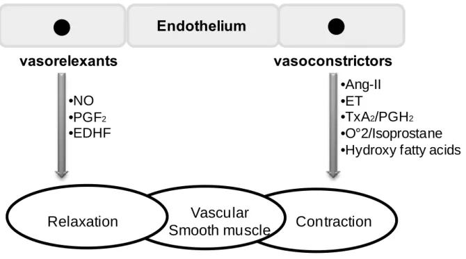
(26) The endothelium has the capacity to regulate the local vascular homeostasis by maintaining the balance between vasodilation and vasoconstriction, by controlling vascular smooth muscle cell (VSMC) proliferation and migration, and by acting on thrombosis and fibrinolysis via the release of various factors (Davignon and Ganz 2004). Imbalance of these different mechanisms promotes endothelial dysfunction, which may lead to serious cardiovascular diseases such as hypertension, a major CVD risk factors. In response to physical and chemical stimuli such as changes in pressure, shear stress, and pH as well as to substances released by autonomic and sensory nerves and circulating hormones, autacoids, and cytokines, the vascular endothelium synthesizes relaxing and contractile factors responsible for the modulation of VSMC tone. Relaxation factors include nitric oxide (NO), prostacyclin (PGI2), endothelium-derived hyperpolarization (EDH), and contractile factors includes thromboxane A2, isoprostanes, superoxide anions (ROS), endothelin-1, and angiotensinII factors (Figure 3) (Mombouli and Vanhoutte 1999; Feletou and Vanhoutte 2006).. - 24 -.
(27) Endothelium vasorelexants. vasoconstrictors •Ang-II •ET •TxA2/PGH2 •O°2/Isoprostane •Hydroxy fatty acids. •NO •PGF2 •EDHF. Relaxation. Vascular Smooth muscle. Contraction. Figure 3. Endothelium-dependent release of vasocontracting and vasorelaxing factors. NO, nitric oxide; PGI2, prostacyclin; EDHF, endothelium-derived hyperpolarizing factor; A-II, angiotensin II; ET, endothelin; TxA2/PGH2, thromboxaneA2/prostaglandin H2O2, hydrogen peroxide (Abeywardena and Head 2001).. 1.3.1 The endothelium-derived vasorelaxing factors 1.3.1.1. Nitric oxide (NO) NO is a key cellular signaling molecule involved in a number of physiological and. pathological processes. In the beginning, it was identified as a factor capable of activating soluble guanylyl cyclase responsible for the relaxation of vascular smooth muscle cells (Katsuki, Arnold et al. 1977). In 1980, Furchgott and Zawadski revealed that the endothelium causes vasorelaxation by the production of endothelium-derived relaxing factor (EDRF) which was later identified as NO (Furchgott and Zawadzki 1980; Palmer, Ferrige et al. 1987; Palmer, Ashton et al. 1988; Palmer and Moncada 1989). Besides its role in regulation of vascular tone, NO inhibits leukocytes adhesion, platelet aggregation, and has anti-apoptotic and antithrombotic effects. Moreover, NO is an important factor of endothelium viability, longevity and cardiovascular health (Morello, Perino et al. 2009).. - 25 -.
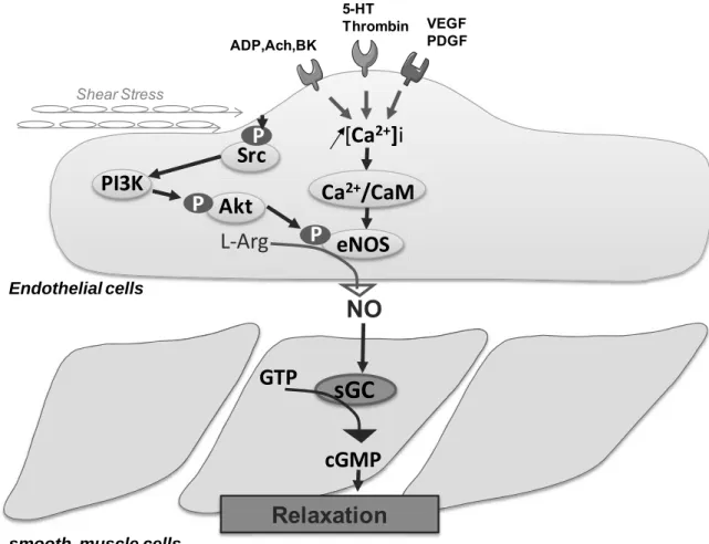
(28) The endothelium-derived NO is produced by the endothelial NO synthase (eNOS) from L-arginine and plays a critical role in normal vascular biology and pathophysiology (Cai and Harrison 2000) (Figure 4).. L-Arginine. L- Citrulline. NO NADPH, O2 Fe. BH4, FMN, FAD. +. Guanylyl cyclase GTP. cGMP. Vasodilation Platelet inhibition Smooth muscle regulation Immune regulation. NO Synthase. Figure 4. NO generation from L-Arginine and its functional properties (Ghalayini 2004). There are three different isoforms of NO synthase; the neuronal NOS (NOS1 or nNOS), the inducible NOS (NOS2 or iNOS) and the endothelial NOS (NOS3 or eNOS)(Weiming, Liu et al. 2002). iNOS is an inducible isoform whereas nNOS and eNOS are being consititutively expressed (Mombouli and Vanhoutte 1999; Stuehr 1999). Under normal conditions, the majority of eNOS is bound to the protein caveolin-1, which inactivates eNOS, and this complex is located in micro domains in the cell membrane named caveolae (Michel, Feron et al. 1997; Bucci, Gratton et al. 2000). eNOS can be activated by Ca2+ dependent and independent pathways. eNOS can be activated by substituting caveolin-1 by Ca2+/CaM in response to Ca2+-mobilizing agonists. When - 26 -.
(29) intracellular Ca2+ levels increase, calmodulin detaches eNOS from caveolin-1 thus permitting the enzyme to become active. Furthermore, eNOS has been shown to be regulated by the interaction with positive and negative protein modulators such as heat shock protein 90 (Ju, Zou et al. 1997; Garcia-Cardena, Fan et al. 1998; Pritchard, Ackerman et al. 2001) (Figure 5).. 5-HT Thrombin ADP,Ach,BK. VEGF PDGF. Shear Stress. [Ca2+]i. P Src PI3K P. Ca2+/CaM. P Akt. P eNOS. L-Arg. Endothelial othelial cells. NO GTP. sGC GTP P cGMP. Relaxation smooth muscle cells. Figure 5. Nitric oxide synthesis pathway in the endothelial cell and its actions in the vascular smooth muscle cell. ACh, acetylcholine; BK, bradykinin; ADP, adenosine diphosphate; 5-HT, serotonin; VEGF, vascular endothelial growth factor; PDGF, platelet-derived growth factor; Src, Sarcoma-family kinases; PI3/kinase, phosphoinositide 3kinase; Akt, Protein kinase B; Ca2+ / CaM, calcium calmodulin; L-Arg, L-arginine; eNOS, endothelial Nitric Oxide Synthase; NO, nitric oxide; sGC, soluble guanylyl cyclase; GTP, Guanosine 5'-Triphosphate; cGMP, cyclic Guanosine 3'-5' monophosphate. - 27 -.
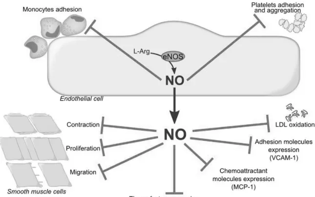
(30) NO freely diffuses to the underlying VSMC where it activates the soluble guanylyl cyclase converting GTP into cyclic guanosine 3’-5’monophosphate (cGMP), which leads to vascular smooth muscle cells relaxation. In addition to vasorelaxation, NO exerts several vasoprotective and anti-atherogenic effects including inhibition of platelets aggregation, monocyte adhesion, vascular smooth muscle cell migration and proliferation, oxidation of LDL, and of the expression of pro-inflammatory and proatherothrombotic mediators such as monocyte chemoattractant protein-1 (MCP-1), adhesion molecules and tissue factor (Tsao, Buitrago et al. 1996; Dimmeler, Haendeler et al. 1997; Hermann, Zeiher et al. 1997). (Figure 6). Figure 6. The pleiotropic effects of NO. VCAM-1; Vascular cell adhesion molecule-1, MCP-1; Monocyte chemoattractant protein-1, LDL; Low density lipoproteins.. - 28 -.
(31) eNOS can also be regulated in endothelial cells at a post-translational level primarily through multisite phosphorylations and protein/protein interactions. Residues Ser1177 and Ser 615 are the activation sites and the residues Thr495 and Ser114 are the inhibition sites on eNOS (Dimmeler, Haendeler et al. 1997; Bohm, Ahlborg et al. 2002; Bauer, Fulton et al. 2003; Fleming 2010) (Figure 7). In response to several physiological stimuli, phosphorylation of eNOS across key regulatory sites plays an important a role in the regulation of the enzymatic activity (Ju, Zou et al. 1997; Newby, Hess et al. 2012). Phosphorylation of eNOS at Ser1177 is associated with an increased enzyme activity (Gallis, Corthals et al. 1999; McCabe, Fulton et al. 2000). Akt, one of the major regulatory targets of PI3-kinase, has been shown to directly phosphorylate eNOS at Ser117 and activate the enzyme in response to vascular endothelial growth factor (VEGF), sphingosine-1-phosphate, and estrogen (Dimmeler, Haendeler et al. 1997; Fulton, Gratton et al. 1999). Furthermore, eNOS can be also activated by phosphorylation on Ser1177 by AMP-activated protein kinase, protein kinase A (PKA), and protein kinase G (PKG) (Busse, Edwards et al. 2002; Flemming and Wingender 2010).. - 29 -.
(32) VEGF Hypoxia Adiponectin Fluid shear stress VEGF Bradykinin. O2 OX-LDL constitutive. Fluid shear stress Insulin Angiotensin II. Constitutive Acetylcholine Angiopoletin ATP BK Estrogen S1P Thapsigergin VEGF1. VEGF Insulin Estrogen Fluid shear stress. Activity Bradykinin [Ca2+ ]. Figure 7. The activation and inhibition sites of eNOS. Green arrows for activation, red arrows for inhibition, black arrow for no direct effect on enzyme activity. The numbers refer to the human sequence (Fleming 2010).. There are various assumed phosphorylation sites, but the most extensively studied eNOS residues, are serine residue in the reductase domain (human eNOS sequence: Ser1177; bovine sequence Ser1179), which positively regulates NO production, and a threonine residue within the CaMbinding domain (human eNOS sequence: Thr495; bovine sequence Thr497) (Boo, Hwang et al. 2002). Ischemia-reperfusion injury is another eNOS regulator which leads to the eNOS phosphorylation at Ser1177 and Ser 633 through the activation of PKA pathway (Li, Yang et al. 2010). Furthermore, there are numerous kinases reported to be involved in the phosphorylation of eNOS following cell activation by different stimuli such as shear stress, vascular endothelial growth factor (Butt, Bernhardt et al. 2000), hypoxia (Michell, Griffiths et al. 1999; Chen, Liu et al. 2008), including extracellular signal-regulated kinase 1/2 which alters eNOS protein expression. - 30 -.
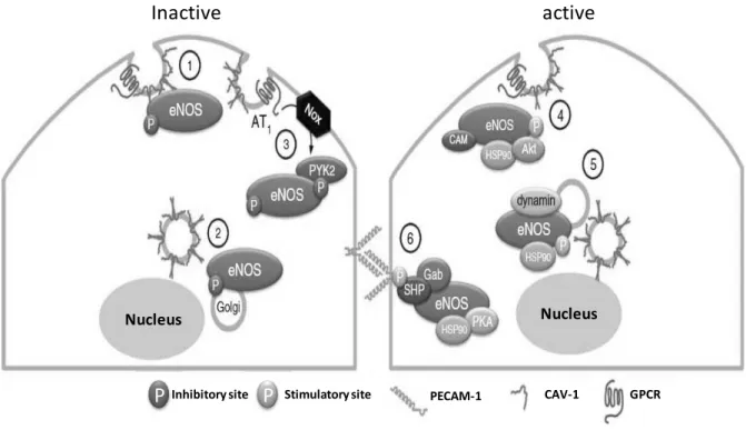
(33) and activity (Ramasamy, Parthasarathy et al. 1998). eNOS can be phosphorylated on serine, tyrosine, and threonine residues leading to eNOS activation or inactivation (Figure 8).. Inactive. active. Nucleus. Nucleus. P Inhibitory site P. Stimulatory site. PECAM-1. CAV-1. GPCR. Figure 8. Regulation of eNOS activity. (1) At rest, the eNOS is coupled to cav-1 (caveolin-1, a structural protein of caveolae) that decreases its activity. (2) eNOS is constitutively phosphorylated at Thr 495 preventing its activation by the Ca 2+/CaM. (3) eNOS may be inhibited in response to oxidative stress by tyrosine phosphorylation by PYK2 (proline-rich tyrosine kinase). (4) eNOS can be activated by both Ca2+/CaM (calcium / calmodulin) and phosphorylation of Ser1177. Hsp90 (heat shock protein) facilitates the recruitment of Akt responsible for the phosphorylation of eNOS (Fleming 2010).. - 31 -.
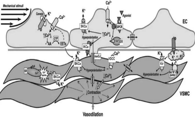
(34) 1.3.1.2 Endothelium-derived hyperpolarization (EDH) EDH is defined as a hyperpolarization of endothelial origin that is transmitted to the vascular smooth muscle leading to its relaxation. The EDH was formerly known as the Endothelium-Derived Hyperpolarizing Factor (EDHF). Beside NO and prostacyclin, the EDH mediated component of relaxation plays a major role in endothelium-dependent relaxation in most of the medium to small calibre resistance arteries, small arteries and arterioles such as second and third-branch mesenteric artery as well as in coronary arteries (Feletou and Vanhoutte 1996; Shimokawa, Yasutake et al. 1996). The role of the EDH component of relaxation is more important in resistance blood vessels as compare to that of NO and prostacyclin, including in humans (Nakashima, Mombouli et al. 1993; Shimokawa, Yasutake et al. 1996). The EDH component of the relaxation is evaluated in the presence of the combination of inhibitors of eNOS like L-NAME and of COXs like indomethacin (Gerber, Anwar et al. 1998). In the EDH mediated response, SKCa and IKCa are activated so that potassium ions move from the intracellular compartment to the extracellular space of endothelial cells, which leads to their hyperpolarization (Figure 9). This higher concentrations of potassium ions in the extracellular space can activate inwardly rectifying K+ (KIR) channels and Na+/K+-ATPase to cause potassium ions efflux from VSMC leading to hyperpolarization and hence, relaxation (Edwards, Dora et al. 1998; Félétou and Vanhoutte 2006). In 1998, Edwards et al reported that hyperpolarization can also be transferred from endothelial cells to VSMC via myo-endothelial gap junctions. Myo-endothelial gap junctions are intracellular channels which can transfer signals from the endothelial cells to the underling vascular smooth muscle cells (Sandoo, van Zanten et al. 2010). Hyperpolarization of VSMC leads to the reduction in cytosolic calcium concentration following closure of voltage-activated calcium channels leading to relaxation. Endothelial hyperpolarization can also be mediated by hydrogen peroxide (H2O2) (Matoba, Shimokawa et al. 2002) or arachidonic acid-derived metabolites including epoxyeicosatrienoic acids (Quilley and McGiff 2000).. - 32 -.
(35) Figure 9. Hypothesis describing the endothelium-derived hyperpolarizing pathway. AA, arachidonic acid; ACh, acetylcholine, [Ca2+]i, intracellular calcium concentration; CYP, cytochrome P450 epoxygenase; EC, endothelial cell; EETs, epoxyeicosatrienoic acids; ER, endoplasmic reticulum; GPCR, G proteincoupled receptor; BKCa, large conductance Ca2+-activated K+ channel; SKCa, small-conductance Ca2+-activated K+ channel subtype 3; IKCa, intermediate-conductance Ca2+ -activated K+ channel; Kir, inwardly rectifying K+ channel; meGJ, myo-endothelial gap-junction; RyR, ryanodine receptor; SR, sarcoplasmic reticulum; VDCC, voltagedependent Ca2+ channel; VSMC, vascular smooth muscle cell (Grgic, Kaistha et al. 2009).. - 33 -.
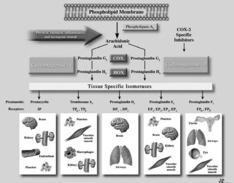
(36) 1.3.1.3 Prostacyclin Prostacyclin (PGI2) is the major product of cyclooxygenase (COX) catalyzed metabolism of arachidonic acid in the endothelium (Cheng, Austin et al. 2002). Prostacyclin is produced from prostaglandin under the action of the enzyme prostacyclin synthase (Dogne, de Leval et al. 2004). PGI2, PGG2 and PGH2 are major products of vascular cyclooxygenase (COX). There are two isoforms of COX encoded by two separate genes. COX-1 is constitutively expressed and is present in many tissues, including endothelial cells (Gryglewski, Uracz et al. 2002). COX-2 is not constitutively expressed, but can be induced rapidly and transiently in many cells, including vascular endothelial cells and smooth muscle cells, under the effect of physical stimuli and proinflammatory agents. PGI2 stimulates smooth muscle relaxation by stimulating adenylyl cyclase and formation of cyclic adenosine -3', 5'- monophosphate (cAMP) that activates protein kinase A, which reduces intracellular Ca2+ by decreasing Ca2+ release from the endoplasmic reticulum and by stimulating its uptake by it. The vasodilator activity of PGI2 is determined by the expression of specific receptors which are prostaglandin I2 receptors of the G-protein coupled receptor family in vascular smooth muscle cells. PGI2 is a potent vasodilator, and an effective endogenous inhibitor of platelet aggregation (Coleman, Smith et al. 1994). In addition, PGI2 facilitates the release of NO by endothelial cells (Shimokawa, Flavahan et al. 1988) and in turn, the action of PGI2 in vascular smooth muscle cells and platelets is potentiated by NO (Delpy, Coste et al. 1996). PGI2 synthase preferentially couples with COX-2 rather than COX-1 in coexpression systems (Figure 10) (Ueno, Takegoshi et al. 2005).. - 34 -.
(37) Figure 10. Production and action of prostaglandins (Araujo, Soeiro et al. 2005).. - 35 -.
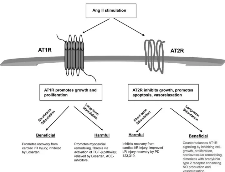
(38) 1.3.2. The endothelium-derived vasocontracting factors. 1.3.2.1 Angiotensin II The octapeptide Angiotensin II (Ang II) is a potent vasoconstrictor hormone of the reninangiotensin system (RAS) that is formed following the conversion of Ang I into Ang II by Angiotensin I Converting Enzyme (ACE) (Baker, Chernin et al. 1990). Angiotensin II (Ang II) is a multifunctional peptide hormone that regulates blood pressure (BP), plasma volume, as well as cardiac, renal and neuronal function, and controls thirst responses. This peptide is of central importance in hypertension and myocardial remodeling, and is the main effector of the reninangiotensin system (RAS) (Weber and Brilla 1991). Taken as a whole, the RAS is involved in different cardiovascular pathologies such as left ventricular hypertrophy, post-infarct remodeling, or neointima formation (Li, McTiernan et al. 2000). The classical effects of Ang II on its target organs are mostly mediated by two membrane receptors, the Ang II type 1 receptors (AT1R) and type 2 receptors (AT2R), which mediate tissue-specific functions (Horiuchi, Akishita et al. 1999). AT1R and AT2R are a G-protein coupled receptors involved in the regulation of vascular cell proliferation and cell death (Kaschina and Unger 2003). AT1R are expressed in all organs, including heart, kidney, liver, adrenal glands, brain, lung and in all cells of the cardiovascular system, namely endothelial cells, smooth muscle cells, fibroblasts, monocytes, macrophages and cardiac myocytes and, thus, is important in cardiovascular pathobiology. While AT2R is highly expressed in fetal heart and fetal aorta, lung and liver (Dasgupta and Zhang 2011), AT2R expression declines fast after birth, but can be induced later in adult life under pathological conditions (Figure 11). The acute vasoconstrictor function of Ang II is primarily mediated through AT1R by classical G-protein-dependent signaling mechanisms. Depending on cell types, Ang II activates AT1R that can in turn activate at least four different effector, namely voltage-gated Ca2+ channels, phospholipase C, phospholipase D and phospholipase A-2 (PLA-2) and can inhibit adenylyl cyclase (Greco 2007). In addition, Ang II stimulation of AT1R activates the extracellular-signalregulated kinase (ERK) cascade, platelet-derived growth factor, epidermal growth factor receptor (EGFR), insulin receptor pathways and non-receptor tyrosine kinases belonging to the c-Src family,. - 36 -.
(39) proline-rich tyrosine kinase 2, focal adhesion kinase and janus kinases (JAKs) (Berry, Touyz et al. 2001). Unlike AT1R, the AT2R contributes to the maintenance of blood pressure by controlling the vascular tone through vasodilatation (Dasgupta and Zhang 2011). AT2R stimulation by Ang II leads to an increase in cGMP levels through a mechanism involving bradykinin B2 receptor, causing endothelial formation of NO (Abadir, Periasamy et al. 2006). Although AT2R expression decreases after birth, it can increase again in some pathophysiological conditions. Stimulation of de novo AT2R expression may inhibit neointima formation, cell proliferation, and inflammation in vascular injury, myocardial infarction and ischemic diseases, suggesting its protective role (Ichiki, Takeda et al. 2001).. Figure 11. Summary of acute and chronic stimulation of angiotensin II receptors (Dasgupta and Zhang 2011).. - 37 -.
(40) 1.3.2.2 Endothelin-1 Endothelin (ET)-1 is a potent vasoconstrictor peptide originally isolated from endothelial cells. There are three structurally different ET isoforms (i.e. ET-1, ET-2, ET-3) as well as a vasoactive intestinal constrictor (Böhm and Pernow 2007). Amongst the three ET isopeptides, the 21-amino acid peptide ET-1 is regarded as the most prominent isoform in the cardiovascular system, accounting for the majority of pathological effects exerted by ETs (Barton, Traupe et al. 2003). Under physiological conditions, ET-1 is produced in small amounts mainly in endothelial cells, primarily acting as an autocrine/paracrine mediator (Pernow, Shemyakin et al. 2012). Under pathophysiological conditions, however, the production is stimulated in a large number of different cell types, including endothelial cells, vascular smooth muscle cells, cardiac myocytes, and inflammatory cells such as macrophages and leukocytes (Grieve, Byrne et al. 2004). The biological effects of ET-1 are transduced by two distinguishable receptor subtypes, ETA and ETB receptors, respectively (Hunley and Kon 2001). In the vasculature, the ETA receptor is mainly located on vascular smooth muscle cells and mediates potent vasoconstriction. ET-1 may also induce indirect vasoconstrictor effects due to the generation of endothelium-derived thromboxane A2 (Marasciulo, Montagnani et al. 2006). The ETB receptor is primarily located on endothelial cells, but may also be present on vascular smooth muscle cells (Schneider, Boesen et al. 2007). Stimulation of the endothelial ETB receptor results in release of NO and prostacyclin which cause vasodilatation, whereas stimulation of the vascular smooth muscle cell ETB receptor results in vasoconstriction (Seo, Oemar et al. 1994). Thus, the net effect produced by ET-1 is determined on the receptor localization and the balance between ETA and ETB receptors (Davie, Haleen et al. 2002) (Figure 12).. - 38 -.
(41) Figure 12. The biological effect of endothelin-1 on arteries in physiological and pathophysiological conditions. In healthy arteries the production of ET-1 is small and the bioavailability of NO is preserved. In endothelial dysfunction there is increased expression of ET-1 in smooth muscle cells and macrophages (MØ). Both the ETA and the ETB receptor on smooth muscle cells may mediate formation of superoxide (O2−). Collectively the balance of effects is shifted towards more vasoconstriction, inflammation and oxidative stress in endothelial dysfunction (Böhm and Pernow 2007).. - 39 -.
(42) 1.3.2.3 Thromboxane A2 & Prostacyclin I2 (TxA2 & PGI2) The prostanoids prostacyclin (PGI2) and thromboxane A2 (TXA2) play an essential role in the maintenance of vascular homeostasis. PGI2 is a vasodilator and an inhibitor of platelet aggregation, whereas TXA2 is a vasoconstrictor and a promoter of platelet aggregation (Gamble, James et al. 2001). PGI2 and TXA2 are products of arachidonic acid (AA) metabolism by cyclooxygenase (COX), followed by metabolism of the COX product, PGH2, by the terminal synthase enzymes, prostacyclin or TX synthase, respectively (Ruan, So et al. 2011). Two isoforms of COX have been identified: COX-1 is expressed constitutively in most cell types, whereas COX-2 is induced by inflammatory stimuli such as bacterial endotoxin and cytokines (Caughey, Cleland et al. 2001). It is considered that PGI2 is the main prostanoid synthesized by vascular endothelium and TXA2 is the main prostanoid produced by platelets (Smith, Borgeat et al. 1991). However, the endothelium has been reported to synthesize TXA2 in addition to PGI2, and both COXs isoforms have been observed, with only COX-1 being detectable in unstimulated cells (Morteau 2001). Endothelial COX-2 can be up-regulated in vitro by inflammatory stimuli and shear stress (Brown and DuBois 2005). Because the balance between PGI2 and TXA2 production is central in the maintenance of vascular tone and platelet aggregation (Konturek and Pawlik 1986; Sobrino, Oviedo et al. 2010), determination of the roles of endothelial COX isozymes, particularly with regard to the contribution of COX-2 in the regulation of prostanoids biosynthesis by the endothelium, is important (Figure 13) (Caughey, Cleland et al. 2001).. - 40 -.
(43) Figure 13. Prostanoids biosynthesis and response pathways (Zhang, Gong et al. 2010).. - 41 -.
(44) 1.3.2.4 Oxidative stress and reactive oxygen species (ROS) Reactive oxygen species (ROS) are recognized as important signaling molecules in the cardiovascular system and are released by vascular cells during pathophysiological conditions like hypertension, diabetes mellitus, atherosclerosis and in acute and chronic inflammatory diseases (Eisenberg and Ghigliotti 1999). NO, (O2●-) the hydroxyl radical (·OH), H2O2, and peroxynitrite (ONOO−·) are produced in the vasculature under both normal and stress conditions such as inflammation or injury. Superoxide anions (O2●-) can be generated by different enzymes (e.g., NADPH oxidase, xanthine oxidase, cyclooxygenases, NO synthases, cytochrome P450 monooxygenases, and enzymes of the mitochondrial respiratory chain) in virtually all cell types, including vascular smooth muscle and endothelial cells (Félétou and Vanhoutte 2006). ROS and in particular superoxide anions can also act directly or indirectly as potent contracting agents via the reduction of the NO bioavailability or by activating COXS in vascular smooth muscle cells (Hibino, Okumura et al. 1999), leading to attenuated endothelium-dependent relaxations (Aubin, Carrier et al. 2006; Liu, You et al. 2007). Moreover, ROS can also impair EDH-mediated endotheliumdependent relaxations through the reduction of calcium-activated potassium channels activity (Kusama, Kajikuri et al. 2005) or by modifying the transmission of the hyperpolarization from endothelial cells to the underlying smooth muscle cells through myoendothelial gap junctions (Griffith, Chaytor et al. 2005). Several studies have shown the beneficial effects of antioxidants on the deleterious effect of oxidative stress on the endothelial function (Kanani, Sinkey et al. 1999; Aubin, Carrier et al. 2006; Liu, You et al. 2007).. - 42 -.
(45) 1.4 Endothelial dysfunction Endothelial dysfunction is a broad term which implies dysregulation of endothelial cell functions, including impairment of the barrier functions of endothelial cells, vasodilation, disturbances in proliferative capacities, migratory as well as tube formation properties, angiogenic properties, attenuation of synthetic function, and deterrence of white blood cells from adhesion and diapedesis. Several factors contribute to endothelial dysfunction including smoking, high blood pressure, diabetes, high cholesterol levels, obesity, hyperglycemia, advance glycation end products (AGEs), and genetic factors. Endothelial dysfunction has been associated with an impairment of endothelium-dependent relaxations involving a reduced bioavailability of NO in major CV diseases such as hypertension, atherosclerosis, chronic renal failure, and diabetes (Griendling and FitzGerald 2003; Rush, Denniss et al. 2005). The mechanism underlying endothelial dysfunction has been linked to increased oxidative stress which is associated with a reduced NO bioavailability and the formation of inflammatory mediators such as vascular cell adhesion molecule-1 (VCAM1) expression (Figure 14) (Khan, Harrison et al. 1996; Libby 2002). In addition, different enzymes have been involved in the arterial oxidative stress involving NADPH oxidases, xanthine oxidases, COX-1 and COX-2, cytochrome P450 monooxygenases, enzymes of the mitochondrial respiratory chain, and uncoupled eNOS. Superoxide anion can react with NO to form the radical peroxynitrite (Koppenol, Moreno et al. 1992), leading to the oxidation of the eNOS cofactor tetrahydrobiopterin (BH4) and the subsequent uncoupling of eNOS, thereby further promoting oxidative stress (Cai and Harrison 2000).. - 43 -.
Figure
+7
Documents relatifs
[r]
Zone herbeuse dominant la grève du lac de Bret (de 549269/152103, alt. 676 m): Acer pseudoplatanus Alliaria petiolata Alnus glutinosa Arenaria serpyllifolia Galium uliginosum
De même, pour la corrélation négative entre les liens faits avec le cours et l’intérêt porté à la tâche (plus les élèves font des liens avec le cours,
The main findings were the following: 12 hours after LPS injection (0.5 mg/kg), a marked reduction in myocar- dial contractility was observed in the isolated perfused senes- cent
The present study, performed in mouse model of MI-induced CHF, shows that selective deletion of PTP1B in the endothe- lium is not only associated with markedly reduced endothelial
Our findings suggest that VSL#3 treatment prevents bacterial translocation since the plasma concentrations of pro-inflammatory cytokines were reduced in CBDL rats treated
The objectives of this study were: (1) to report on the breeding and selection of genotypes representing novel species hybrids of shrub willow and their biomass growth potential on
Effectuez chaque expression à l’aide de l’ordre correct des opérations.. Priorité des Opérations sur les
