https://doi.org/10.1007/s12576-018-0607-7 ORIGINAL PAPER
Isoflurane anesthesia does not affect spinal cord neurovascular
coupling: evidence from decerebrated rats
Thierry Paquette1,2,3 · Hugues Leblond2,3 · Mathieu Piché1,2
Received: 15 January 2018 / Accepted: 22 March 2018 / Published online: 29 March 2018 © The Physiological Society of Japan and Springer Japan KK, part of Springer Nature 2018
Abstract
Neurological examination remains the primary clinical investigation in patients with spinal cord injury. However, neuroim-aging methods such as functional magnetic resonance imneuroim-aging (fMRI) are promising tools for following functional changes in the course of injury, disease and rehabilitation. However, the relationship between neuronal activity and blood flow in the spinal cord on which fMRI relies has been largely overlooked. The objective of this study was to examine neurovascular coupling in the spinal cord of decerebrated rats during electrical stimulation of the sciatic nerve with and without isoflurane anesthesia (1.2%). Local field potentials (LFP) and spinal cord blood flow (SCBF) were recorded simultaneously in the lumbosacral enlargement. Isoflurane did not significantly alter LFP (p = 0.53) and SCBF (p = 0.57) amplitude. Accordingly, neurovascular coupling remained comparable with or without isoflurane anesthesia (p = 0.39). These results support the use of isoflurane in rodents to investigate nociceptive functions of the spinal cord using fMRI.
Keywords Spinal cord · Neurovascular coupling · Pain · Nociception · Blood flow · Local field potential
Introduction
The assessment of spinal cord functions and integrity is of critical importance in several clinical conditions, including spinal cord injury and sensorimotor dysfunction. Neurologi-cal examination remains the primary cliniNeurologi-cal investigation in these patients. However, non-invasive neuroimaging meth-ods have been developed recently and may bring comple-mentary information to standard neurological examination [1]. For instance, functional magnetic resonance imaging (fMRI) is a promising tool for following functional changes in the course of injury, disease and rehabilitation.
The most common functional magnetic resonance imag-ing (fMRI) methods for the spinal cord rely on the blood oxygen level-dependent (BOLD) signal [2]. Using these methods, neuronal activity can be inferred from hemody-namic changes and oxygen metabolism, based on the so-called neurovascular coupling. Thus, characterization of neurovascular coupling in various conditions is of critical importance to obtain valid results and interpret fMRI data correctly. Surprisingly, in spite of several spinal fMRI stud-ies, only one study investigated the relationship between neuronal activity and hemodynamic changes in the spinal cord [3]. In this animal study, tight neurovascular coupling was observed during limb stimulation, in spite of large systemic blood pressure fluctuations associated with the stimulus. One limitation of this study was the use of iso-flurane anesthesia, which may alter the results and prevent their extrapolation to human spinal fMRI. Isoflurane is commonly used in animals to conduct spinal fMRI stud-ies for its fast action and recovery time and the possibility of fine tuning the depth of anesthesia [4, 5]. However, it also has some disadvantages. Indeed, isoflurane has vasoac-tive properties, which interferes with hemodynamic func-tions and neurovascular coupling [6, 7], disturbs blood flow autoregulation [8, 9] and reduces neural metabolism [8, 10]. Furthermore, isoflurane may produce dissociative effects on * Mathieu Piché
[email protected] http://www.uqtr.ca/cognac
1 Department of Chiropractic, Université du Québec à
Trois-Rivières, 3351 Boul. Des Forges, C.P. 500, Trois-Trois-Rivières, QC, Canada G9A 5H7
2 CogNAC Research Group, Université du Québec
à Trois-Rivières, 3351 Boul. Des Forges, C.P. 500, Trois-Rivières, QC, Canada G9A 5H7
3 Department of Anatomy, Université du Québec à
Trois-Rivières, 3351 Boul. Des Forges, C.P. 500, Trois-Rivières, QC, Canada G9A 5H7
cortical neurovascular coupling, where cerebral blood flow increases dose-dependently while no effect is observed on local field potentials (LFP) [11]. Whether spinal neurovas-cular coupling is altered in a similar way, however, remains unclear. Considering that isoflurane anesthesia alters spinal cord function, including the suppression of presynaptic inhi-bition [12, 13], it is crucial to examine its effect on spinal neurovascular coupling. Decerebrated rat preparations allow the investigation of spinal functions without anesthesia [14,
15]. Thus, the aim of the present study was to determine the effects of isoflurane anesthesia on the relationship between neuronal activity (LFP) and spinal cord blood flow (SCBF) in decerebrated rats. Considering that systemic mean arte-rial pressure (MAP) changes may affect regional blood flow changes when nociceptive stimuli are applied [16, 17], we also examined whether systemic MAP changes may affect spinal neurovascular coupling in non-anesthetized rats.
Based on previous studies on cerebral neurovascular cou-pling [11], we hypothesized that decerebrated rats without anesthesia would show different SCBF responses to sciatic nerve stimulation compared with decerebrated isoflurane-anesthetized rats, while LFP amplitude would be comparable between conditions. Accordingly, we expected a difference in neurovascular coupling between anesthetized and non-anesthetized conditions. Based on our previous study [3], we also anticipated no alteration of neurovascular coupling by systemic MAP changes in either condition.
Methods
Ethical approval
Experiments were performed on six male Wistar rats (body weight 350–475 g; age 18–22 weeks; Charles River Labo-ratories, Saint-Constant, Québec, Canada). Animals were housed in the animal care unit of Université du Québec à Trois-Rivières. A light–dark cycle of 14–10 h was main-tained. Experimental procedures were conducted in accord-ance with the local animal care committee, following the guidelines of the Canadian Council on Animal Care.
Animals and surgical procedures
All animals were in good health on the day of the experi-ment. Surgical procedures were conducted under isoflurane (2.5%). Stable systemic MAP and the absence of withdrawal reflexes (paw pinching) were used to confirm adequate anesthesia during the entire experiment. Body temperature was monitored and was maintained at 37.5 ± 0.5 °C with a custom-made temperature control system. Artificial ventila-tion was performed using a tracheal cannula and end-tidal CO2 was maintained around 3.0% (CAPSTAR-100 Carbon
dioxide analyzer, CWE inc., Ardmore, PA, USA). Intrave-nous injections were administered through the catheterized right jugular vein. MAP was recorded continuously using an intra-arterial cannula inserted in the left carotid artery and connected to a pressure transducer (Harvard Appa-ratus, Holliston, MA, USA). The right carotid artery was occluded with a microvascular clip to reduce cerebral blood flow and avoid major hemorrhage during the decerebration procedures.
A stereotaxic frame was used to fix the animal’s head (Model 900, Kopf Instruments, Tujunga, CA, USA) and the spine was stabilized with two vertebral clamps (Model 986C, Kopf Instruments, Tujunga, CA, USA). Vertebral seg-ments between T12 and L2 were laminectomized to access the lumbar enlargement. Warm mineral oil was applied on the exposed spinal cord following a longitudinal incision of the dura mater. The left sciatic nerve was exposed for the application of electrical stimulation. To prevent tissue dam-age, a warm mineral oil pool made with skin flaps attached to a metal holder was placed over the limb.
Mechanical decerebration was performed as described by Dobson and Harris [15]. After scraping the pericranium, two holes of approximately 3 mm in diameter were drilled in the parietal bones. Craniotomy was then completed using bone forceps leaving the sagittal and transverse sutures intact. The remaining parietal bone was used to pass a thread at two dif-ferent points in order to block superior sagittal sinus blood flow completely. The cerebral cortex was carefully removed by aspiration until superior and inferior colliculi were vis-ible. After a transection at the rostral edge of the colliculi, all tissues anterior to this cut and lateral to the colliculi were removed. The empty space was then filled with hemostatic gelatin sponge (Gelfoam, Pfizer, Kirkland, QC, Canada) and cotton material. After completing these procedures, the microvascular clip applied on the right carotid was removed. The absence of any bleeding was confirmed and the animal recovered until the experiment began.
Thirty minutes before the stimulation protocol, the level of anesthesia was decreased to 1.2% isoflurane and the ani-mal was paralyzed with gallamine triethiodide (20 mg/kg, i.v.). When the animal completely recovered, spinal poly-synaptic responses were unaffected by gallamine [18] and stable responses could be evoked, physiological and electro-physiological data was recorded for approximately 2 h. After the first part of the experiment was completed (decerebrated anesthetized condition), isoflurane was discontinued, leaving the decerebrated animal in a non-anesthetized condition. A second dose of gallamine was then administered. The ani-mal’s condition and physiological parameters were always stable when the stimulation protocol resumed 30 min later. The isoflurane wash out was confirmed by a slight increase in baseline MAP after approximately 1 min. At the end of the experiment, rats were killed by increasing isoflurane to
5%. When blood pressure fell to 0 mmHg and no heartbeat was observed, the thoracic cage was opened and the heart was cut before interrupting anesthesia.
Local field potential recordings
A microelectrode was inserted in the left lumbar enlarge-ment using a micromanipulator (Model 960, David Kopf Instruments, Tujunga, CA, USA). The electrode consisted of a multi-electrode made of 8 parallel shafts each comprising 8 recording sites (Model A8X8-10 mm-200-200-177, Neu-ronexus Technologies Inc., Ann Arbour, Michigan, USA). Its anteroposterior position and depth was determined when electrical stimulation of the sciatic nerve produced multiu-nit responses in most of the superficial channels (Smart-box, Neuronexus Technologies Inc., Ann Arbour, Michi-gan, USA; band pass 300–3000 Hz, gain 192 V/V). The signal was then recorded for all channels with a broad band (1–10,000 Hz, gain 192 V/V) and sampled at 20 kHz (Smart-box, Neuronexus Technologies Inc., Ann Arbour, Michigan, USA), resampled (5 kHz) and filtered offline (1–300 Hz) to obtain LFPs. For each rat, the channel among superficial channels of similar depth that showed the largest LFP was selected for analyses [3].
Spinal cord blood flow recordings
Spinal cord blood flow was recorded with a laser-Doppler probe (Micro-needle probe TSD145, Biopac systems, Goleta, CA, USA). The probe was placed on the spinal cord surface, as close as possible to the multi-electrode, care-fully avoiding large blood vessels. SCBF was sampled at 100 Hz with a time constant of 3 s and was recorded for offline analyses (Power 1401 acquisition system, Cambridge Electronic Design, Cambridge, UK).
Electrical stimulation
Electrical stimulation was applied to the sciatic nerve using a custom-made bipolar hook electrode and a constant-cur-rent stimulator (Model DS7A, Digitimer Ltd, Welwyn Gar-den city, UK). Stimulation consisted of trains of 10 s with 1-ms pulses delivered at 5 Hz. The inter-train interval of 55 s allowed response recovery before the subsequent stimulus.
Experimental design
LFP and SCBF were recorded simultaneously, beginning after a stabilization period of 1 h. The stimulation protocol consisted of a series of 8 graded electrical stimuli ranging between 0.1 and 9.6 mA applied under decerebrated anesthe-tized and non-anestheanesthe-tized conditions. The order of stimulus intensities was randomized to avoid sequence order effects
but was the same for anesthetized and non-anesthetized con-ditions for each animal.
Data analyses
LFP, SCBF and MAP data were analyzed using Spike2 software (Cambridge Electronic Design, Cambridge, UK, version 6.15). For the quantification of LFP amplitude, the peak-to-peak value of averaged potentials (50 responses induced by the stimulus train of 10 s at 5 Hz) was extracted with a custom-made Spike2 script within a 50-ms window following stimulus onset. For SCBF and MAP changes, the onset-to-peak value was extracted for each stimulus intensity within a 30-s window, and response amplitude was calcu-lated as the change relative to the mean signal value for the 30-s artifact-free baseline preceding stimulus onset. MAP responses were calculated as the raw change (mmHg) while SCBF responses were calculated as percent change. The SCBF and LFP values were also standardized as T scores to calculate the neurovascular coupling indexed as the SCBF/ LFP ratio.
Statistical analyses
All results are expressed as means ± SEM. Statistical analy-ses were performed with Statistica (Dell Inc. 2016. Dell Statistica version 13) with a significance threshold of p ≤ 0.05. The effect size is reported as partial eta-squared ( 𝜂2
p ). MAP, SCBF, LFP amplitude and latencies as well as neurovascular coupling ratios were compared between con-ditions using Greenhouse-Geisser corrected two-way repeated-measures ANOVAs. The two repeated factors included intensity (8 levels), and anesthesia (with and with-out isoflurane). Significant effects were decomposed with planned contrasts. When needed, one-sample t-tests were used to determine the significance of changes in MAP, SCBF and LFP for the 0.1-mA intensity compared with 0.
Results
Mean arterial pressure
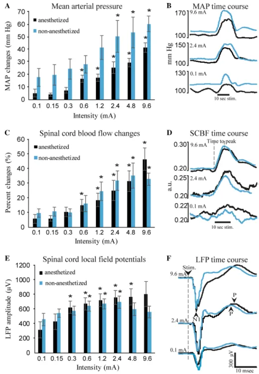
Results for MAP changes are presented in Fig. 1a. An indi-vidual example is also presented in Fig. 1b. Electrical stimu-lation induced intensity-dependent MAP changes (main effect F7,35 = 18.4, p < 0.001; 𝜂p2 = 0.79). As expected, MAP changes were decreased by isoflurane anesthesia, although this effect was marginal (main effect F1,5 = 5.6, p = 0.064; 𝜂2p = 0.53). However, isoflurane anesthesia did not produce significantly different changes in MAP between intensities
(interaction F7,35 = 0.65, p = 0.59; 𝜂2p = 0.12). For the inten-sity-dependent changes, one-sample t-tests revealed no sig-nificant response at the lowest intensity (0.1 mA) either in the anesthetized or non-anesthetized conditions (p = 0.17 and p = 0.057, respectively). Compared with these non-sig-nificant changes, planned contrasts revealed that MAP changes significantly increased from the 0.6- up to the 9.6-mA intensity under isoflurane anesthesia (all values of p < 0.05) and from the 2.4- up to the 9.6-mA intensity with-out anesthesia (all values of p < 0.05).
As expected, baseline MAP increased after isoflurane anesthesia was interrupted, going from 103.2 ± 3.1 to 120.7 ± 3.3 mmHg (F1,5 = 12.5, p = 0.02; 𝜂p2 = 0.71).
Spinal cord blood flow
Results for SCBF amplitude are presented in Fig. 1c. An individual example is also presented in Fig. 1d. Electrical stimulation produced intensity-dependent increases in SCBF amplitude (main effect F7,35 = 21.3, p < 0.001; 𝜂p2 = 0.80).
Fig. 1 Mean arterial pressure, spinal cord blood flow and local field potentials. a Sciatic nerve stimulation produced intensity-dependent increases in MAP. Without anesthesia, MAP changes tended to be smaller but this effect did not reach significance (p = 0.064). An individual example of SCBF changes in both conditions for maximal (9.6 mA), intermediate (2.4 mA) and minimal (0.1 mA) stimulus intensity is shown in b. c Sciatic nerve stimula-tion also produced intensity-dependent increases in SCBF. However, these responses were not affected by anesthesia. An individual example of SCBF changes in both conditions for maximal (9.6 mA), interme-diate (2.4 mA) and minimal (0.1 mA) stimulus intensity is shown in d. e LFP amplitude was larger at higher intensities compared with lower intensi-ties but changes were not linear. Amplitude stopped increasing and even tended to decrease at the highest intensities. An individual example of SCBF changes in both conditions for maximal (9.6 mA), intermediate (2.4 mA) and minimal (0.1 mA) stimulus intensity is shown in
f. Note the negative deflection
(N1) and the positive wave (P). Also note the latency shift due to anesthesia (see Table 2
for details). Error bars = SEM,
n = 6. *p < 0.05; **p < 0.01;
***p < 0.001 compared with the 0.1-mA intensity
Surprisingly, however, anesthesia did not affect the ampli-tude of SCBF changes (main effect F1,5 = 0.1, p = 0.74; 𝜂p2 = 0.02). Moreover, anesthesia did not significantly affect SCBF responses between intensities (interaction F7,35 = 2.9, p = 0.073; 𝜂2
p = 0.36). For the intensity-dependent changes, one-sample t-tests revealed no significant response at the lowest intensity (0.1 mA) under isoflurane but a significant response without anesthesia (p = 0.11 and p = 0.02, respec-tively). Compared with these changes, planned contrasts revealed that SCBF amplitude significantly increased from the 0.6- up to the 9.6-mA intensity under isoflurane (all val-ues of p < 0.05) and from the 1.2- up to the 9.6-mA intensity without anesthesia (all values of p < 0.05).
As for baseline SCBF, in spite of the increase in MAP reported above, it was stable after isoflurane anesthesia was interrupted, remaining identical (0.19 ± 0.03 vs 0.19 ± 0.03 a.u.; F1,5 = 0.0, p = 0.99; 𝜂2
p < 0.001).
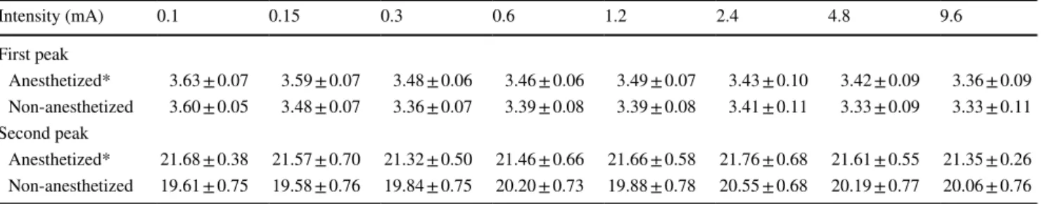
Peak latency of SCBF responses are presented in Table 1. Latency was significantly different for some intensities between anesthetized and non-anesthetized conditions (inter-action F7,35 = 4.5, p = 0.001; 𝜂2p = 0.47). Indeed, planned con-trasts revealed increased latencies for the 4.8- and 9.6-mA intensities in the anesthetized condition compared with the non-anesthetized condition (all values of p < 0.05).
Spinal local field potentials
Results for LFP amplitudes are presented in Fig. 1e. An indi-vidual example is also presented in Fig. 1f. Electrical stimu-lation produced robust LFPs and their amplitude was signifi-cantly affected by anesthesia between intensities (interaction F7,35 = 5.0, p = 0.02; 𝜂2
p = 0.50). For both the anesthetized and
non-anesthetized conditions, a significant LFP response was observed at 0.1 mA (p = 0.037 and p = 0.003, respectively). Compared with these changes, planned contrasts revealed that LPF amplitude significantly increased for the 0.3, 0.6, 1.2, 2.4 and 4.8 mA intensities under isoflurane anesthesia (all values of p < 0.05) and for the 0.6, 1.2 and 2.4 mA with-out anesthesia (all values of p < 0.05).
LFP peak latencies for the first negative peak (N1) and the positive peak (P-wave) are presented in Table 2. For the negative peak, latency was not significantly affected by anes-thesia between intensities (interaction F7,35 = 1.4, p = 0.24; 𝜂2p = 0.22) but was increased by anesthesia for all intensities confounded (main effect F1,5 = 12.7, p = 0.02; 𝜂p2 = 0.72). Latency also progressively decreased with intensity (main effect F7,35 = 3.1, p = 0.01; 𝜂p2 = 0.38). Similar effects were observed for the positive peak, the latency of which was not significantly affected by anesthesia between intensities (interaction F7,35 = 0.9, p = 0.5; 𝜂p2 = 0.16) but was increased by anesthesia for all intensities confounded (main effect F1,5 = 23.7, p = 0.005; 𝜂2
p = 0.83). However, the latency of the positive peak was not affected by stimulus intensity (main effect F7,35 = 1.1, p = 0.4; 𝜂p2 = 0.18).
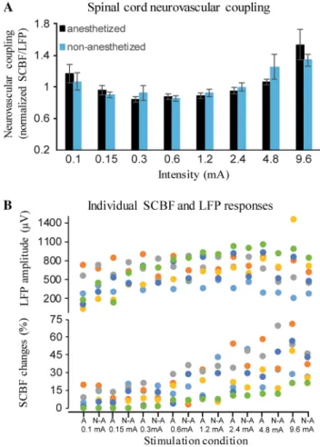
Neurovascular coupling
In order to examine the stability of neurovascular coupling across conditions, the normalized SCBF/LFP ratio [3, 16] was compared across stimulus intensities between anesthe-tized and non-anestheanesthe-tized conditions (see Fig. 2). Neuro-vascular coupling was significantly different between inten-sities (main effect F1,5 = 7.3, p = 0.03; 𝜂p2 = 0.59). However,
Table 1 Time to peak of SCBF responses (seconds)
*p < 0.05 compared with non-anesthetized condition
Intensity (mA) 0.1 0.15 0.3 0.6 1.2 2.4 4.8 9.6
Anesthetized 6.3 ± 0.7 8.1 ± 1.0 7.9 ± 1.0 7.0 ± 0.8 7.0 ± 0.8 7.5 ± 0.8 9.2 ± 0.7* 10.0 ± 1.0* Non-anesthetized 7.0 ± 0.8 7.5 ± 0.9 6.7 ± 1.0 7.3 ± 0.8 8.2 ± 0.5 8.1 ± 0.4 6.5 ± 0.7 6.6 ± 0.7
Table 2 LFP peak latencies (ms)
*p < 0.05, main effect of anesthesia
Intensity (mA) 0.1 0.15 0.3 0.6 1.2 2.4 4.8 9.6 First peak Anesthetized* 3.63 ± 0.07 3.59 ± 0.07 3.48 ± 0.06 3.46 ± 0.06 3.49 ± 0.07 3.43 ± 0.10 3.42 ± 0.09 3.36 ± 0.09 Non-anesthetized 3.60 ± 0.05 3.48 ± 0.07 3.36 ± 0.07 3.39 ± 0.08 3.39 ± 0.08 3.41 ± 0.11 3.33 ± 0.09 3.33 ± 0.11 Second peak Anesthetized* 21.68 ± 0.38 21.57 ± 0.70 21.32 ± 0.50 21.46 ± 0.66 21.66 ± 0.58 21.76 ± 0.68 21.61 ± 0.55 21.35 ± 0.26 Non-anesthetized 19.61 ± 0.75 19.58 ± 0.76 19.84 ± 0.75 20.20 ± 0.73 19.88 ± 0.78 20.55 ± 0.68 20.19 ± 0.77 20.06 ± 0.76
planned contrasts revealed no significant difference for any intensity from 0.15 to 9.6 mA compared with 0.1 mA (all values of p > 0.2). In addition, anesthesia did not affect neu-rovascular coupling either overall (main effect F1,5 = 0.1, p = 0.74; 𝜂2
p = 0.02) or across intensities (interaction F7,35 = 1.1, p = 0.39; 𝜂2
p = 0.18).
Discussion
The present study is the first to investigate spinal neurovas-cular coupling in a non-anesthetized decerebrated rat model. The novel finding of the study is that in spite of some effects of isoflurane anesthesia on MAP changes as well as on LFP and SCBF peak latencies, the amplitude of LFP and SCBF
changes was unaffected, resulting in comparable neurovascu-lar coupling with or without isoflurane anesthesia in a wide range of stimulus intensities. This has critical implications for spinal cord neuroimaging methods, such as fMRI, where reliability depends on tight coupling between neuronal activ-ity and hemodynamic changes.
Effects of isoflurane anesthesia on neurovascular coupling
The main objective of this study was to determine the effect of anesthesia on spinal cord neurovascular coupling. The present results indicate that isoflurane anesthesia at a con-centration of 1.2% does not affect neurovascular coupling, as calculated from the standardized amplitude of LFP and SCBF changes. Indeed, the amplitude of LFP and SCBF responses was not significantly different with or without isoflurane anesthesia in the present decerebrated rat model.
Spinal cord LFPs are evoked by cutaneous and muscle afferents when the sciatic nerve is stimulated directly with electrical pulses. In the present study, LFPs reflect synap-tic activity of dorsal horn interneurons receiving large fiber inputs [19]. This is observed as the first negative deflection (N1). The slow positive wave (P) following the N1 has a much longer latency and reflects synaptic activity of a dif-ferent population of interneurons, which evoke primary afferent depolarization (PAD) [19]. SCBF was recorded in order to measure regional blood flow changes related to these spinal processes. Indeed, laser Doppler flowmetry is limited in terms of recording depth, the laser beam reaching approximately 1000 µm [20, 21]. At L1, this covers approxi-mately layers I–V in the dorsal horn, where the interneurons of interest are located. Thus, neuronal and vascular signals reflect similar processes and are not contaminated by motor activity from deeper layers of the spinal cord.
Consistent with the present findings, it was reported that dorsal horn neurons’ activity evoked by noxious heat stimuli was not decreased at concentrations above 1 minimal alveo-lar anesthetic concentration (MAC), equivalent to approxi-mately 1.2% isoflurane, although the withdrawal reflex was reduced [22]. Also consistent with the present recordings from the dorsal horn of the spinal cord, it was suggested that neurons in the ventral horn of the spinal cord may be more sensitive to isoflurane anesthesia compared with neurons in the dorsal horn [22, 23]. In accordance with this, the present results apply to nociceptive stimulation and to neurovascu-lar coupling in the dorsal horn of the spinal cord only. This also implies that differential effects of a task between the dorsal and ventral horns observed with fMRI of the spinal cord in rats under isoflurane anesthesia may be confounded with the differential effects of isoflurane on neurovascular function. Indeed, we expect that different results may be obtained for motor tasks and neurovascular coupling in the
Fig. 2 Spinal cord neurovascular coupling. a Average spinal cord neurovascular coupling (normalized SCBF/LFP ratio) at different stimulus intensities for the anesthetized and non-anesthetized con-ditions. No significant difference was observed between the lowest stimulus and the succeeding intensities, although lowest and highest intensities tended to show a larger ratio. Also, isoflurane anesthesia did not affect neurovascular coupling. b Individual LFP and SCBF data for each stimulus intensity and each animal, in the anesthetized and non-anesthetized conditions
ventral horn of the spinal cord, so the present results should be interpreted with caution and they cannot be generalized to all experimental designs and conditions.
Another point that should be considered in the inter-pretation of the present findings is that neural activity was quantified with LFP amplitude. LFP amplitude is strongly associated with single-unit or multi-unit activity, although LFPs are driven by synaptic activity [24]. However, since excitatory and inhibitory synaptic currents contribute to LFPs, single- and multi-unit activity may be reduced in spite of stable or even increased synaptic activity, leading to dissociation between spiking activity and LFP amplitude. Since LFPs are more strongly related to BOLD responses in fMRI compared with other extracellular recording [25, 26], LFP recordings were more appropriate than measurements of single- or multi-unit activity in the present study.
A surprising result is the lack of effect of isoflurane on SCBF response amplitude. Indeed, isoflurane is known to produce vasodilatation in the brain and spinal cord [6,
7, 27–29]. Therefore, it would be expected that SCBF responses be altered. Generally, the present findings indi-cate that it is not the case, although some effects were noted for the two strongest stimulus intensities. Indeed, although SCBF amplitude was not affected by isoflurane, response latency was increased by approximately 3 s for the 4.8- and 9.6-mA intensities. This has implications for the use of spi-nal fMRI if strong intensities are needed for experimental purposes. Indeed, time-to-peak variations as small as 1 s in the BOLD response can lead to inaccurate interpretation of fMRI data [30]. Notwithstanding, the generally unaffected SCBF response under isoflurane anesthesia contrasts with significant changes in hemodynamic responses observed in awake vs isoflurane-anesthetized rodents in previous stud-ies [11, 31, 32]. This indicates that isoflurane differentially affects the brain and the spinal cord. However, species dif-ferences should be considered and the assessment of anes-thesia effects on neural responses should include response amplitude as well as overall time course. Indeed, a study in non-human primates indicates that although the general time course of the hemodynamic response was comparable between awake and anesthetized conditions, the amplitude of the response was much larger [33].
Effects of decerebration and implications for neurovascular coupling
In order to study spinal cord neurovascular coupling in the most physiologically relevant state, a decerebrated prepara-tion is an interesting model to avoid potentially confounding effects of anesthesia. However, decerebration is not with-out consequences and some implications should be consid-ered for the interpretation of the present results. Concern-ing spinal autoregulation, the interruption of descendConcern-ing
pathways from the brain does not seem to influence the intrinsic capacity of the spinal cord to regulate its blood flow [3, 34]. Accordingly, removing forebrain and midbrain structures as in the present preparation did not change the capacity of SCBF autoregulation, as indicated by the lack of effects of large MAP fluctuations induced by electrical stimulation. Indeed, sciatic nerve stimulation produced intensity-dependent MAP increases of up to 60 mmHg to reach over 160 mmHg on average (Fig. 1a). Nevertheless, these robust MAP changes did not seem to affect SCBF changes, as indicated by relatively constant neurovascu-lar coupling between intensities (see Fig. 1c, d). This is in accordance with results from our previous study, in which SCBF changes were mainly driven by local neuronal activity and not by MAP increases [3]. It is also consistent with stud-ies in rats, cats, dogs and rhesus monkeys showing that MAP fluctuation between 45 and 165 mmHg do not alter SCBF [35–39]. It should also be mentioned that basal MAP was not decreased by decerebration and that it was comparable to our previous study in intact anesthetized rats [3]. This is consistent with the well-known location of arterial blood pressure regulation centers in the brain stem, which remain intact in decerebrated animals.
Limitations and future directions
One limitation of the present study is that only isoflurane anesthesia was examined. Considering that several anesthet-ics are used in spinal fMRI and that each anesthetic relies on different mechanisms, the present findings cannot be extrapolated to other anesthetics. For example, isoflurane and halothane have different effects on spinal cord activ-ity [22]. Future studies should examine the effects of dif-ferent anesthetic regimens on neurovascular coupling. As mentioned above, the present results apply to nociceptive stimulation and to neurovascular coupling in the dorsal horn of the spinal cord only. Future studies are needed to assess neurovascular coupling in the ventral horn of the spi-nal cord in different tasks, including motor tasks. Also, the order of anesthesia conditions were not randomized. There-fore, sequence order effects may affect the results. However, considering the lack of difference between conditions, this is a minor issue for the present study. Finally, to confirm the extrapolation of the present findings to spinal fMRI, a combination of electrophysiological and fMRI methods is needed in future studies and other measures such as tis-sue oxygenation in addition to blood flow recordings are desirable.
In conclusion, the present results indicate that spinal hemodynamic changes reflect neuronal activity in decer-ebrated isoflurane anesthetized and non-anesthetized rats. Considering results from previous studies, these findings indicate that spinal and cerebral hemodynamic functions
are not affected equally by isoflurane anesthesia. Lastly, this study supports the use of isoflurane in spinal neurovas-cular studies in intact rats and suggests that isoflurane is an appropriate anesthetic for spinal fMRI studies investigating sensory functions.
Acknowledgements This project was funded by the Natural Sci-ence and Engineering Research Council (NSERC) of Canada (Grant Numbers: 402176 (MP); 05403 (HL)). TP was supported by scholar-ships from the NSERC, the department of anatomy (UQTR) and the “Fonds de Recherche du Québec en Santé” (FRQS). The contribution of Mathieu Piché was supported by the UQTR research chair in pain neurophysiology, the “Fondation de Recherche en Chiropratique du Québec” and the FRQS.
Author contribution TP contributed to all aspects of the research. HL contributed to all aspects of the research. MP contributed to all aspects of the research and obtained funding for the study.
Funding This study was supported by a grant from the Natural
Sci-ences and Engineering Research Council of Canada (Grant Numbers: 402176 (MP); 05403 (HL)).
Compliance with ethical standards
Conflict of interest Thierry Paquette reports no financial or other re-lationship that may lead to any conflict of interest. Hugues Leblond reports no financial or other relationship that may lead to any conflict of interest. Mathieu Piché reports no financial or other relationship that may lead to any conflict of interest.
Ethical approval All experiments followed “Guiding Principles for the Care and Use of Animals in the Field of Physiological Sciences” and were approved by the animal care committee of Université du Québec à Trois-Rivières, in accordance with the Canadian Council on Animal Care.
References
1. Wheeler-Kingshott CA, Stroman PW, Schwab JM, Bacon M, Bosma R, Brooks J, Cadotte DW, Carlstedt T, Ciccarelli O, Cohen-Adad J, Curt A, Evangelou N, Fehlings MG, Filippi M, Kelley BJ, Kollias S, Mackay A, Porro CA, Smith S, Strittmat-ter SM, Summers P, Thompson AJ, Tracey I (2014) The current state-of-the-art of spinal cord imaging: applications. Neuroimage 84:1082–1093
2. Ogawa S, Lee TM, Kay AR, Tank DW (1990) Brain magnetic resonance imaging with contrast dependent on blood oxygenation. Proc Natl Acad Sci USA 87(24):9868–9872
3. Piché M, Paquette T, Leblond H (2017) Tight neurovascular cou-pling in the spinal cord during nociceptive stimulation in intact and spinal rats. Neuroscience 355:1–8
4. Porszasz R, Beckmann N, Bruttel K, Urban L, Rudin M (1997) Signal changes in the spinal cord of the rat after injec-tion of formalin into the hindpaw: characterizainjec-tion using func-tional magnetic resonance imaging. Proc Natl Acad Sci USA 94(10):5034–5039
5. Yang P-F, Wang F, Chen LM (2015) Differential fMRI activation patterns to noxious heat and tactile stimuli in the primate spinal cord. J Neurosci 35(29):10493–10502
6. Iida H, Ohata H, Iida M, Watanabe Y, Dohi S (1998) Isoflurane and sevoflurane induce vasodilation of cerebral vessels via ATP-sensitive K+ channel activation. Anesthesiology 89(4):954–960
7. Farber NE, Harkin CP, Niedfeldt J, Hudetz AG, Kampine JP, Schmeling WT (1997) Region-specific and agent-specific dila-tion of intracerebral microvessels by volatile anesthetics in rat brain slices. Anesthesiology 87(5):1191–1198
8. Olsen KS, Henriksen L, Owen-Falkenberg A, Dige-Petersen H, Rosenorn J, Chraemmer-Jorgensen B (1994) Effect of 1 or 2 MAC isoflurane with or without ketanserin on cerebral blood flow autoregulation in man. Br J Anaesth 72(1):66–71 9. Tiecks FP, Lam AM, Aaslid R, Newell DW (1995) Comparison
of static and dynamic cerebral autoregulation measurements. Stroke 26(6):1014–1019
10. Newberg LA, Milde JH, Michenfelder JD (1983) The cer-ebral metabolic effects of isoflurane at and above concentra-tions that suppress cortical electrical activity. Anesthesiology 59(1):23–28
11. Masamoto K, Fukuda M, Vazquez A, Kim SG (2009) Dose-dependent effect of isoflurane on neurovascular coupling in rat cerebral cortex. Eur J Neurosci 30(2):242–250
12. Shimoji MDK, Fujiwara PDN, Fukuda MDS, Denda MDS, Takada MDT, Maruyama MDY (1990) Effects of isoflurane on spinal inhibitory potentials. Anesthesiology 72(5):851–857 13. Collins JG, Kendig JJ, Mason P (1995) Anesthetic actions
within the spinal cord: contributions to the state of general anesthesia. Trends Neurosci 18(12):549–553
14. Meehan CF, Mayr KA, Manuel M, Nakanishi ST, Whelan PJ (2017) Decerebrate mouse model for studies of the spinal cord circuits. Nat Protoc 12(4):732–747
15. Dobson KL, Harris J (2012) A detailed surgical method for mechanical decerebration of the rat. Exp Physiol 97(6):693–698 16. Jeffrey-Gauthier R, Guillemot JP, Piche M (2013) Neurovascular
coupling during nociceptive processing in the primary soma-tosensory cortex of the rat. Pain 154(8):1434–1441
17. Uchida S, Bois S, Guillemot J-P, Leblond H, Piché M (2017) Systemic blood pressure alters cortical blood flow and neuro-vascular coupling during nociceptive processing in the primary somatosensory cortex of the rat. Neuroscience 343:250–259 18. DeJong RH, Robles R, Morikawa KI (1968) Actions of
immobilizing drugs on synaptic transmission. Exp Neurol 21(2):213–218
19. Yates BJ, Thompson FJ, Mickle JP (1982) Origin and properties of spinal cord field potentials. Neurosurgery 11(3):439–450 20. Shih AY, Driscoll JD, Drew PJ, Nishimura N, Schaffer CB,
Klein-feld D (2012) Two-photon microscopy as a tool to study blood flow and neurovascular coupling in the rodent brain. J Cereb Blood Flow Metab 32(7):1277–1309
21. Fredriksson I, Larsson M, Stromberg T (2009) Measurement depth and volume in laser Doppler flowmetry. Microvasc Res 78(1):4–13
22. Jinks SL, Martin JT, Carstens E, Jung SW, Antognini JF (2003) Peri-MAC depression of a nociceptive withdrawal reflex is accom-panied by reduced dorsal horn activity with halothane but not isoflurane. Anesthesiology 98(5):1128–1138
23. Cuellar JM, Dutton RC, Antognini JF, Carstens E (2005) Differen-tial effects of halothane and isoflurane on lumbar dorsal horn neu-ronal windup and excitability. BJA Br J Anaesth 94(5):617–625 24. Buzsáki G, Anastassiou CA, Koch C (2012) The origin of
extra-cellular fields and currents—EEG, ECoG, LFP and spikes. Nat Rev Neurosci 13(6):407–420
25. Goense JB, Logothetis NK (2008) Neurophysiology of the BOLD fMRI signal in awake monkeys. Curr Biol 18(9):631–640 26. Logothetis NK, Pauls J, Augath M, Trinath T, Oeltermann A
(2001) Neurophysiological investigation of the basis of the fMRI signal. Nature 412(6843):150–157
27. Li C-X, Patel S, Wang DJJ, Zhang X (2014) Effect of high dose isoflurane on cerebral blood flow in macaque monkeys. Magn Reson Imaging 32(7):956–960
28. Matta BF, Heath KJ, Tipping K, Summors AC (1999) Direct cer-ebral vasodilatory effects of sevoflurane and isoflurane. Anesthe-siology 91(3):677–680
29. Hoffman WE, Edelman G, Kochs E, Werner C, Segil L, Albrecht RF (1991) Cerebral autoregulation in awake versus isoflurane-anesthetized rats. Anesth Analg 73(6):753–757
30. Handwerker DA, Ollinger JM, D’Esposito M (2004) Variation of BOLD hemodynamic responses across subjects and brain regions and their effects on statistical analyses. Neuroimage 21(4):1639–1651
31. Martin C, Martindale J, Berwick J, Mayhew J (2006) Investigating neural-hemodynamic coupling and the hemodynamic response function in the awake rat. Neuroimage 32(1):33–48
32. Pisauro MA, Dhruv NT, Carandini M, Benucci A (2013) Fast hemodynamic responses in the visual cortex of the awake mouse. J Neurosci 33(46):18343–18351
33. Shtoyerman E, Arieli A, Slovin H, Vanzetta I, Grinvald A (2000) Long-term optical imaging and spectroscopy reveal mechanisms underlying the intrinsic signal and stability of cortical maps in V1 of behaving monkeys. J Neurosci 20(21):8111–8121
34. Kobrine AI, Doyle TF, Newby N, Rizzoli HV (1976) Preserved autoregulation in the rhesus spinal cord after high cervical cord section. J Neurosurg 44(4):425–428
35. Kobrine AI, Doyle TF, Rizzoli HV (1976) Spinal cord blood flow as affected by changes in systemic arterial blood pressure. J Neu-rosurg 44(1):12–15
36. Marcus ML, Heistad DD, Ehrhardt JC, Abboud FM (1977) Regulation of total and regional spinal cord blood flow. Circ Res 41(1):128–134
37. Sato M, Pawlik G, Heiss WD (1984) Comparative studies of regional CNS blood flow autoregulation and responses to CO2
in the cat. Effects of altering arterial blood pressure and PaCO2
on rCBF of cerebrum, cerebellum, and spinal cord. Stroke 15(1):91–97
38. Hickey R, Albin MS, Bunegin L, Gelineau J (1986) Autoregula-tion of spinal cord blood flow: is the cord a microcosm of the brain? Stroke 17(6):1183–1189
39. Rubinstein A, Arbit E (1990) Spinal cord blood flow in the rat under normal physiological conditions. Neurosurgery 27(6):882–886