Publisher’s version / Version de l'éditeur:
Molecular Immunology, 43, April 10, pp. 1579-1586, 2006
READ THESE TERMS AND CONDITIONS CAREFULLY BEFORE USING THIS WEBSITE. https://nrc-publications.canada.ca/eng/copyright
Vous avez des questions? Nous pouvons vous aider. Pour communiquer directement avec un auteur, consultez la première page de la revue dans laquelle son article a été publié afin de trouver ses coordonnées. Si vous n’arrivez pas à les repérer, communiquez avec nous à PublicationsArchive-ArchivesPublications@nrc-cnrc.gc.ca.
Questions? Contact the NRC Publications Archive team at
PublicationsArchive-ArchivesPublications@nrc-cnrc.gc.ca. If you wish to email the authors directly, please see the first page of the publication for their contact information.
NRC Publications Archive
Archives des publications du CNRC
This publication could be one of several versions: author’s original, accepted manuscript or the publisher’s version. / La version de cette publication peut être l’une des suivantes : la version prépublication de l’auteur, la version acceptée du manuscrit ou la version de l’éditeur.
For the publisher’s version, please access the DOI link below./ Pour consulter la version de l’éditeur, utilisez le lien DOI ci-dessous.
https://doi.org/10.1016/j.molimm.2005.09.019
Access and use of this website and the material on it are subject to the Terms and Conditions set forth at
TLR3 activation inhibits human mast cell attachment to fibronectin and
vitronectin
Kulka, Marianna; Metcalfe, D. D.
https://publications-cnrc.canada.ca/fra/droits
L’accès à ce site Web et l’utilisation de son contenu sont assujettis aux conditions présentées dans le site LISEZ CES CONDITIONS ATTENTIVEMENT AVANT D’UTILISER CE SITE WEB.
NRC Publications Record / Notice d'Archives des publications de CNRC:
https://nrc-publications.canada.ca/eng/view/object/?id=4d1ff1a2-5501-4400-b17d-48612c59afd3 https://publications-cnrc.canada.ca/fra/voir/objet/?id=4d1ff1a2-5501-4400-b17d-48612c59afd3TLR3 activation inhibits human mast cell attachment
to fibronectin and vitronectin
夽
M. Kulka
a, D.D. Metcalfe
b,∗aAllergy-Immunology Division, Northwestern University Feinberg School of Medicine,
240 E Huron McGaw Pavilion #2305, Chicago, IL, USA
bLaboratory of Allergic Diseases, National Institute of Allergy and Infectious Diseases, National Institutes of Health,
Bldg 10, Room 11C205, 10 Center Drive, MSC 1881, Bethesda, MD 20892-1881, USA Received 28 April 2005; accepted 28 September 2005
Available online 8 November 2005
Abstract
Mast cells are involved in both the genesis of allergic inflammation and in host defense; and reside in tissues where their location and responsiveness is regulated in part by adhesion to extracellular matrix proteins (ECM). We have reported that human mast cells (huMC) express TLR1-7, and 9 and respond to toll-like receptors (TLR) ligands by releasing cytokines and leukotriene C4. To determine if TLR ligation could similarly affect mast cells via an influence on adhesion, we employed huMC; and as substrates, fibronectin (FN) and vitronectin (VN). huMC were thus treated with double-stranded RNA (dsRNA) and adhesion to ECM was quantified. FcRI dependent mast cell degranulation was assessed. Adhesion molecule expression and activation was measured by flow cytometry. Activation of huMC through TLR3 with increasing amounts of polyI:C inhibited mast cell adhesion in a dose-dependent manner. This decrease in adhesion was accompanied by a similar decrease in IgE-mediated mast cell degranulation. Activation of TLR3 on huMC resulted in a change in the conformation of CD29, the receptor for FN, to an inactive form. Thus, TLR3 activation decreases mast cell attachment to VN and FN through an active process and one, which would abrogate mast cell attachment dependent potentiation of IgE-mediated responses.
Published by Elsevier Ltd.
Keywords: Mast cells; Adhesion; Toll-like receptors; Integrins
1. Introduction
Mast cells have been reported to be recruited into innate immune responses in part through activation of mast cells via toll-like receptors (TLR) (Supajatura et al., 2002; Jawdat et al., 2004). Human mast cells (huMC), as an example, express TLR3 and release IFN-␣ in response to polyI:C, a double-stranded (dsRNA) ligand for TLR3 (Kulka et al., 2004). Ligation of TLR3 by polyI:C does not itself induce mast cell degranula-tion and polyI:C does not affect IgE-mediated cultured huMC
Abbreviations: dsRNA, double-stranded RNA; FN, fibronectin; HCMC, human cultured mast cell; LAD, laboratory of allergic disease mast cell line; LPS, lipopolysaccharide; MC, mast cell; PGN, peptidoglycan; polyI:C, polyinosine-polycytidylic acid; TLR, toll-like receptor; VN, vitronectin
夽 This work was supported by the intramural program at the National Institutes
for Health Research.
∗Corresponding author. Tel.: +1 301 496 2164; fax: +1 301 480 8384.
E-mail address:dmetcalfe@niaid.nih.gov (D.D. Metcalfe).
degranulation (Kulka et al., 2004). The lack of a direct polyI:C effect on mast cell degranulation has been confirmed in mouse bone marrow derived mast cells (Matsushima et al., 2004). BMMC treated with polyI:C or infected with virus upregulate costimulatory molecules, such as CD80 and CD28 and, inde-pendent of degranulation, release chemokines that recruit CD8+ T cells (Orinska et al., 2005). Similarly, polyI:C activates Th1 responses and reduces regulatory T cell development in a model of autoimmune gastritis (Kobayashi et al., 2004). Injection of mice with polyI:C suppresses both IgE responses and serum IgE levels, and stimulates an IgG2a response in an IFN␣/ depen-dent manner (Finkelman et al., 1991). These data suggest that TLR activation turns mast cell attention away from IgE/mast cell mediated inflammation and to responses beneficial to host viral defense.
Since adhesion to extracellular matrix (ECM) is known to potentiate mast cell IgE-mediated responses (Ra et al., 1994; Wyczolkowska et al., 1994) and integrin-mediated adhesion is important in this process (Edelson et al., 2004), we were
inter-0161-5890/$ – see front matter. Published by Elsevier Ltd. doi:10.1016/j.molimm.2005.09.019
1580 M. Kulka, D.D. Metcalfe / Molecular Immunology 43 (2006) 1579–1586
ested in determining if activation of mast cells through TLR3 would modify adhesion and as a consequence, influence IgE-mediated MC degranulation. As will be shown, ligation of TLR3 with dsRNA decreased huMC adhesion to fibronectin (FN) and vitronectin (VN). Attendant to this decrease in adhesion was a decrease in IgE-mediated mast cell degranulation. Thus, acti-vation of TLR3 not only leads to mast cell cytokine release, but also a second biologic effect, that of a decrease in attach-ment to matrix components. These data are consistent with the theme that TLR-mediated activation of mast cells focuses their responses to those that are beneficial to host defense.
2. Materials and methods
2.1. huMC culture
huMC (LAD1) (Kirshenbaum et al., 2003; Venkatesha et al., 2005) were cultured in serum free media (StemPro-34 SFM, Life Technologies) supplemented with 2 mM l-glutamine, 100 U/mL penicillin, 50 g/mL streptomycin and 100 ng/mL stem cell fac-tor (SCF). The cell suspensions were seeded at a density of 105cells/mL and maintained at 37◦C and 5% CO2.
Hemideple-tions were performed weekly with fresh media (Kirshenbaum et al., 2003).
2.2. Isolation of human lung and skin mast cells
Skin mast cells were isolated from human adult breast or infant foreskin samples (>1 g of sample) by gentle pro-tease digestion, enriched after being labeled with anti-FcRI-FITC, and incubated with anti-FITC microbeads using a mod-ified technique (Okayama et al., 1994). Briefly, skin samples were weighed and cut into small pieces. Skin pieces were then placed in RPMI (pH 7.4) containing 2 mM l-glutamine, 100 U/mL penicillin, 50 g/mL streptomycin, 20 mM HEPES, 2 mg/mL collagenase (Sigma), 1 mg/mL hyaluronidase (Sigma) and 2 mg/mL protease (Sigma), and incubated at 37◦for 24 h.
The cell suspension was next filtered through a 70 m Nylon cell strainer, labeled with FcRI-FITC, and incubated with anti-FITC microbeads as described (Okayama et al., 1994). Lung mast cells were similarly isolated from human lung samples (>1 g of sample, obtained from adult cadaveric tissue) by gentle protease digestion. Skin and lung mast cells were analyzed by toluidine blue staining, flow cytometry for granule content and FcRI and Kit expression. Only samples more than 95% pure were used for RNA analysis.
2.3. RNA isolation and quantitative polymerase-chain reaction (PCR)
RNA was purified using an RNeasy Mini Kit (Qiagen Inc., Valencia, CA). Genomic DNA was digested by incubating 10 g of total RNA with 2 U of DNAse (amplification grade; Life Technologies) in DNase buffer (200 mM Tris–HCl, 20 mM MgCl2, 500 mM KCl, pH 8.4; Life Technologies) and
RNase-free H2O for 10 min at room temperature. RNA was then
precip-itated with 3 M C2H2O2Na (pH 5.2; Sigma–Aldrich, St. Louis,
Table 1
Primers for the human toll-like receptors used for RT-PCR analysis
Gene Sequence Size Tm Cycles
TLR-1 tgaatatcagcaaggtcttgct 432 54 30 catctgtgtagtcatttcagct TLR-2 gagcatctgataatgacagagtta 773 60 40 gtgtcagtaagtatatttgaaga TLR-3 gtttggagcaccttaacatggaa 454 60 30 tgcttagatccagaatggtcaag TLR-4 gcatacttagactactacctcgat 342 60 35 aataacaccattgaagctcagatc TLR-5 acaccaatgtcactatagctg 645 50 30 tgtacaaagcctctgatggat TLR-6 cttggaaatgcctggtcagagt 544 60 35 atctgaaaacagagtcagtaagc TLR-7 gacctaagtggaaattgccct 538 60 35 ctcttgaatctcctgaaggtg TLR-8 aacagaatatcaccgttggtaaa 293 60 35 ttcagttccacttaacacttgag TLR-9 ggacctctggtactgcttcca 150 54 45 aagctcgttgtacacccagtct TLR-10 tgctcatctgcatctaaatactgt 671 60 35 agtctccagtttattgccattcaa gcatctgctggttgaagaatgc -actin atctggcaccacaccttctacaatgagctgcg 838 60 25 cgtcatactcctgcttgctgatccacatctgc
All sequences are in the 5′–3′orientation.
MO). Treated RNA (1 g) was reverse transcribed using 0.5 g oligo(dT), buffer (50 mM Tris–HCl, 75 mM KCl, 3 mM MgCl2,
pH 8.3, Life Technologies), 10 mM DTT, 10 mM of each dNTP, DEPC-treated water and 200 U M-MLV RT enzyme (all from Life Technologies) at 37◦C for 1 h.
Quantitative PCR was performed using Brilliant SYBR Green QPCR Master Mix (Stratagene, La Jolla, CA) according to manufacturer’s instructions. Briefly, 0.1 g of cDNA, 25 L of Master Mix, 50 nM of each primer and 0.75 L of ROX refer-ence dye was amplified for 45 cycles on an ABI PRISM 7700 at an annealing temperature of 50–60◦C. The sequences of each
primer are shown inTable 1and conditions for reaction were as described (Kulka et al., 2004). Data acquisition was done in both the annealing and extension step of each amplification cycle. Amplified products were also analyzed by 2% agarose gel electrophoresis and visualized by ethidium bromide staining to confirm a single product of correct size.
2.4. Adhesion assay
The 96-well Nunc Maxisorp plates (NUNC, Naperville, IL) were coated with 10 g/mL human FN or VN (Sigma) in phosphate-buffered saline (PBS) for 2 h at 37◦C or 16 h at
4◦C, washed three times with PBS, blocked with 3% bovine
serum albumin (BSA; Sigma) in HEPES buffer (10 mM HEPES, 137 mM NaCl, 2.7 mM KCl, 0.38 mM Na2HPO4·7H2O, 5.6 mM
Fig. 1. polyI:C inhibits resting huMC adhesion to VN and FN. (A) Adhesion of untreated (solid bars) or polyI:C treated (10 g/mL; open bars) mast cells to FN, VN, BSA and plastic in a 2 h adhesion assay. (B) Adhesion of mast cells to FN (solid bars) and VN (open bars) in the presence of increasing doses of polyI:C for 2 h. (C) Effect of time of polyI:C (10 g/mL) addition on adhesion when added after mast cells had been allowed to adhere. Mast cells were allowed to adhere to FN and VN for 0, 0.5, 1, 1.5 and 2 h at 37◦C and then polyI:C (10 g/mL) was added and the number of adherent mast cells was evaluated as described in Section2.
Background adhesion to BSA was 15%. (D) Effect of mast cell pre-treatment with polyI:C for indicated times. Cells were washed once with buffer, pre-treated with polyI:C (10 g/mL) for indicated times and allowed to adhere to FN or VN for 2 h. Asterisks show P < 0.01 significance when compared to control (n = 3 separate experiments, each performed in quadruplicate).
BSA, pH 7.4) for 1 h at 37◦C, then washed three times with
HEPES buffer. For stimulation with IgE or SCF, huMC were washed with HEPES buffer, resuspended at 1 × 10⁶cells/mL, and labeled with 5 M Calcein-AM (Molecular Probes, Eugene, OR) for 20 min at 37◦C. For stimulation via FcRI, huMC
were incubated with 1 g/mL IgE-biotin overnight at 37◦C in
medium, washed twice to remove unbound IgE, then resus-pended and labeled as described earlier. After labeling, cells were washed and resuspended at 1 × 10⁶cells/mL in HEPES buffer. Cell suspension (50 L) ± HEPES buffer containing IgE, SCF or streptavidin (Sigma) was added to each FN or VN-coated well and incubated at 37◦C for 2 h or the indicated times. In
Fig. 1C, mast cells were added to a 96 well plate coated with either FN or VN and incubated for 0, 0.5, 1, 1.5 and 2 h at 37◦C.
After incubation at the indicated time, polyI:C (10 g/mL) was added and adhesion was immediately assessed. In a second set of experiments, mast cells were added to a 96 well plate coated with either FN or VN and incubated for 2 h. After incubation, polyI:C (10 g/mL) was added and the plate was incubated for a further 2 h at which time adhesion was assessed. The degree of adhesion was quantitated at 485 nm excitation and 530 nm emission (HTS 7000 Bio Assay Reader, Perkin-Elmer, San Fran-cisco, CA) and is expressed as the percentage of fluorescence remaining in the wells after washing away unbound cells. Treat-ment of cells with inhibitors or antibodies was carried out by incubating Calcein-AM-labeled cells ± anti-TLR3 (10 g/mL; eBioscience) for 30 min at 37◦C prior to the addition of the cells
to the assay plate. A two-tailed paired Student’s t-test was used to determine statistical significance between adhesion values.
2.5. β-Hexosaminidase release assay
huMC were sensitized overnight with 1 g/mL of IgE-biotin. A total of 2 × 10⁵ cells were washed and resuspended
in HEPES buffer, then placed in FN, VN or BSA coated or uncoated wells. Cells were stimulated with streptavidin in the presence or absence of polyI:C (10 g/mL) and incubated at 37◦C for 1 h. The -hexosaminidase in the supernatants and
cell lysates was quantified by hydrolysis of p-nitrophenyl N-acetyl--d-glucosamide (Sigma–Aldrich) in 0.1 M sodium cit-rate buffer (pH 4.5) for 90 min at 37◦C. The percentage of
-hexosaminidase release was calculated as percent of total con-tent.
2.6. Fluorescence-activated cell sorter (FACS) analysis
huMC were washed and resuspended at 5 × 10⁵cells/mL in PBS/0.1% BSA and incubated with anti CD29-phycoerythrin (CD29-PE), CD31-PE, CD49b-FTIC, CD49c-PE, CD49d-PE and CD51/61-PE (all from BD Bioscience) for 30 min at 4◦C.
Cells were resuspended in PBS/0.1% BSA and analyzed using FACSCalibur (Becton Dickinson).
2.7. Statistical analysis
Each experiment was performed at least three separate times and in quadruplicate and values represent mean of
n= 3 ± standard error of the mean of the average of each exper-iment. P values were determined by Student t-test (between
1582 M. Kulka, D.D. Metcalfe / Molecular Immunology 43 (2006) 1579–1586
groups) or one-way ANOVA (comparing more than two groups).
3. Results
3.1. polyI:C inhibits huMC adhesion to FN and VN
SCF-dependent huMC adhesion to FN, as well as VN, was tested using a mast cell adhesion assay with or without polyI:C (10 g/mL). huMC spontaneously adhered to uncoated wells (plastic) and wells coated with BSA, FN and VN. polyI:C inhib-ited this adhesion to FN and VN but not to BSA or plastic (Fig. 1A) in a dose-dependent manner (Fig. 1B). Next, we deter-mined if polyI:C could reverse huMC adhesion. Thus, mast cells were allowed to adhere to FN and VN for 0, 0.5, 1, 1.5 and 2 h and polyI:C was added at different time points (Fig. 1C). Addition of polyI:C within the first hour significantly decreased huMC adhe-sion. However, addition of polyI:C 1.5 and 2 h after cells were allowed to adhere to FN and VN had no affect, suggesting that polyI:C blocked early adhesion events. Alternatively, polyI:C mediated inhibition of adhesion may require a longer period of time. To test this possibility, we incubated huMC on FN for 2 h, then added polyI:C and incubated for another 2 h. No affect of polyI:C on huMC was observed (data not shown). Finally, we determined if pre-treatment with polyI:C would block mast cell adhesion. Mast cells were treated with polyI:C for 1–30 min, washed once with buffer, then allowed to adhere to FN or VN (Fig. 1D). A 15–30 min pre-treatment with polyI:C inhibited huMC adhesion to FN and VN. Pre-treatment with polyI:C for less than 15 min did not significantly affect mast cell adhesion.
3.2. Characteristics of polyI:C effect
Since polyI:C is a synthetic dsRNA construct, we mea-sured huMC adhesion to FN and VN in the presence of other forms of dsRNA (polyG:C and polyA:U), ssRNA (polyC) and dsDNA (polydI:dC). Of all of the oligonucleotide constructs, only polyI:C, polyG:C and polyA:U inhibited mast cell adhe-sion to FN (Fig. 2A). polyI:C and polyG:C also significantly inhibited mast cell adhesion to VN. ssRNA and dsDNA did not significantly inhibit huMC adhesion to either FN or VN, sug-gesting that this effect is unique to dsRNA, the ligand for TLR3. To determine if the polyI:C effect was TLR3 mediated, huMC were pre-treated with anti-TLR3 (clone 3.7 (Matsumoto et al., 2002)) for 30 min and then allowed to adhere to FN or VN (Fig. 2B). Because TLR3 in part is located intracellularly, the use of blocking antibodies has limitations. That is, a partial block does not clarify if all effects are via TLR3 or if there is another intracellular mechanism of activation. In any case, anti-TLR3 had no affect on constitutive adhesion to FN or VN. However, anti-TLR3 abrogated the polyI:C inhibitory influence consistent with the conclusion that polyI:C inhibition of mast cell adhesion is at least in part TLR3 mediated. An isotype control antibody had no affect on polyI:C inhibition of adhesion (data not shown). To determine if polyI:C similarly inhibited the increase in adhesion known to occur in the presence of SCF following IgE-mediated activation, we measured huMC adhesion to FN or VN in the presence of SCF, IgE-biotin or IgE-biotin + streptavidin (Fig. 2C). SCF, as expected, increased mast cell adhesion to FN from 58.5 ± 2.6 to 95.2 ± 4.5%. Stimulation of mast cells with IgE/streptavidin modestly increased adhesion to 75.9 ± 3.8%
Fig. 2. Characterization of polyI:C effect. (A) Ligands for TLR-3 inhibit adhesion. (B) Anti-TLR3 antibody (10 g/mL) inhibits polyI:C effect on adhesion. (C) polyI:C inhibits huMC adhesion to FN induced by SCF, IgE and IgE + SA. Adhesion of untreated (white bars) or polyI:C treated (10 g/mL; black bars) huMC to FN in the presence of SCF (10 ng/mL), IgE (1 g/mL) or IgE + SA (100 ng/mL). (D) Effect of other TLR ligands on huMC adhesion. huMC (1 × 10⁶) were treated
with indicated concentrations of LPS (10 g/mL), PGN (10 g/mL), zymosan (10 g/mL), polyI:C (10 g/mL), CpG (10 g/mL) or flagellin (100 ng/mL) for 2 h on a FN or VN coated plate. Asterisks show P < 0.01 significance when compared to control (n = 3 separate experiments, each performed in quadruplicate).
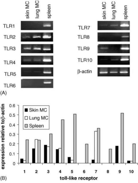
Fig. 3. Expression of TLR mRNA in mast cells isolated from human skin and lung. Mast cells were isolated from human lung and skin samples as described and RNA was isolated. (A) Semi-quantitative RT-PCR and real-time PCR. (B) Analysis of TLR expression in skin mast cells, lung mast cells and human spleen.
while IgE alone had no significant effect on adhesion (p < 0.01,
n= 3). polyI:C significantly inhibited adhesion of SCF and IgE/streptavidin stimulated mast cells by 40–60% (p < 0.01,
n= 3).
To determine if other TLR ligands were also able to inhibit mast cell adhesion, we measured huMC adhesion to FN or VN in the presence of lipopolysaccharide (LPS; ligand for TLR-4), peptidoglycan (PGN; ligand for TLR2), zymosan (TLR2), polyI:C (TLR3), CpG-A oligonucleotide (TLR9) and flagellin (TLR5). Only polyI:C and CpG-A oligonucleotide inhibited huMC adhesion to FN and VN (Fig. 2D). Therefore, it appears that only activation of huMC by TLR3 and TLR9 ligands mod-ifies huMC adhesion.
3.3. Human skin and lung mast cells express TLR3
Some reports have suggested that mast cells from skin or lung may respond differently to pathogens and may express different TLR. To determine whether human skin and lung mast cells sim-ilarly express TLR3, we isolated skin and lung MC from human tissues and analyzed purified mast cells by RT-PCR (Fig. 3A) and real-time PCR (Fig. 3B) for expression of TLR1-10. Results indicate that both skin and lung MC express TLR 2, 3 and 4. Compared to cultured huMC (Kulka et al., 2004), human skin and lung mast cells exhibited less (TLR5, TLR7, TLR9 and TLR10) or no (TLR1, TLR6 and TLR8) message. Some strik-ing differences in TLR expression were observed between skin
and lung mast cells. Skin mast cells expressed TLR9 but little TLR7 or TLR10, while lung mast cells expressed higher lev-els of TLR7 and TLR10 but no TLR9. Thus, among these mast cell types, and in huMC, TLR3 is among the most consistently expressed in huMC from separate sources and locations.
3.4. polyI:C induced mast cell adhesion is not inhibited by IFN-α
We have previously shown that huMC produce IFN-␣ when stimulated with polyI:C (Kulka et al., 2004). Mast cells treated with polyI:C produced IFN-␣ in a dose-dependent manner but did not produce TNF, IL-5 or IL-1. As IFN-␣ is not stored in huMC granules but must be synthesized de novo, it is thus not likely to influence the 2 h adhesion assay. However, in an in vivo context, it is possible that other mast cells (or other cell types) in the area of polyI:C (or viral) activation could synthesize IFN-␣ and modulate nearby mast cell adhesion. Therefore, we wanted to know whether IFN-␣ could modulate huMC adhesion. We thus measured mast cell adhesion in the presence of IFN-␣ and/or polyI:C. IFN-␣ treatment of huMC for 3 h had no affect on mast cell adhesion to either FN or VN alone or in combination with polyI:C (data not shown).
3.5. polyI:C decreases the expression of the active conformation of CD29
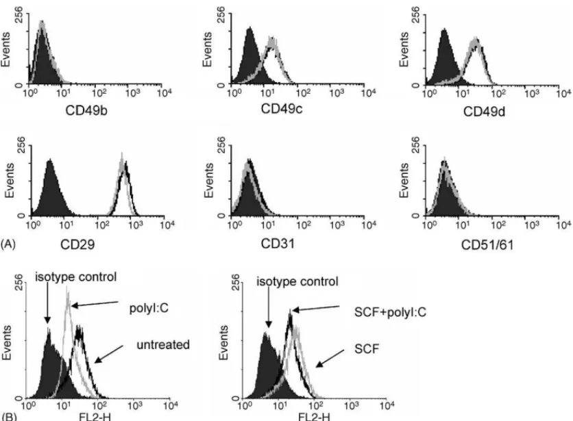
To determine if polyI:C was blocking adhesion by downregu-lating expression of adhesion molecules, we analyzed adhesion molecule expression following polyI:C treatment (Fig. 4A). huMC expressed CD29, the FN receptor, CD49c and CD49d. polyI:C treatment for 3 h (Fig. 4A) and 24 h (data not shown) did not modify expression of these adhesion molecules when compared to untreated cells.
Adhesion molecule affinity for their ligand is regulated by both expression levels of the adhesion molecule as well as con-formational changes due to inside-out signaling. To determine whether polyI:C was modifying the conformation of CD29, the FN receptor, we used an antibody 9EG7 which can recognize an activated epitope of CD29 (Bazzoni et al., 1995; Bodeau et al., 2001). Mast cells were untreated or treated with polyI:C in the presence or absence of SCF for 3 h and the expression of active CD29 was analyzed (Fig. 4B). Compared to untreated cells, flow cytometry shows that polyI:C decreased the expres-sion of the active conformation of CD29, even in the presence of SCF.
3.6. polyI:C inhibits huMC adhesion-dependent degranulation
The attachment of mast cells to substrate is known to enhance IgE-mediated degranulation. To determine if polyI:C affects huMC degranulation in the presence of FN, mast cells were sensitized with IgE-biotin overnight, then stimulated with strep-tavidin in FN coated wells for 1 h (Fig. 5A). Degranulation was determined by measuring -hex release. FN increased -hex release compared to BSA (Fig. 5A) or uncoated wells (data
1584 M. Kulka, D.D. Metcalfe / Molecular Immunology 43 (2006) 1579–1586
Fig. 4. polyI:C effect on adhesion molecule expression. (A) huMC were untreated (black line) or treated with polyI:C (10 g/mL; grey line) for 2 h and adhesion molecule expression was measured as compared to isotype control (filled in). (B) polyI:C modifies inside-out activation of CD29. huMC (1 × 10⁶) were treated with polyI:C (10 g/mL) for 2 h with or without SCF (100 ng/mL) and analyzed for expression of the active conformation of 1 integrin (CD29) using 9EG7 mAb.
Fig. 5. polyI:C inhibits adhesion dependent mast cell degranulation. (A) -Hex release from huMC stimulated via FcRI. Mast cells were allowed to adhere to FN or BSA (negative control) in the presence or absence of 10 g/mL polyI:C for 1 h. After 1 h, cells were stimulated via FcRI and -hex release was measured. (B) huMC were treated with polyI:C (10 g/mL; grey line) or untreated (dark line) and expression of TLR3, Kit and FcRI was assessed by flow cytometry. Isotype control is shown as filled in trace.
not shown) by approximately 20% (p < 0.01, n = 3). polyI:C did not affect mast cell -hex release in BSA coated wells and uncoated wells (data not shown). However, polyI:C inhibited the FN potentiation of -hex release (p < 0.01 when comparing fibronectin and fibronectin + polyI:C) so that the -hex released in the presence of polyI:C + FN was similar to that of BSA alone (Fig. 5A). To determine if this decrease in degranula-tion was because of a decrease in FcRI or Kit expression, huMC were treated with polyI:C (10 g/mL) for 12 h and expres-sion of TLR3, FcRI and Kit was measured (Fig. 5C). polyI:C did not affect FcRI and Kit expression but upregulated TLR3 expression. Thus, polyI:C decreases degranulation by decreas-ing adhesion, not by decreasdecreas-ing FcRI or Kit expression. 4. Discussion
In this study, we determined the effect of TLR ligation on huMC attachment to two extracellular matrix proteins (VN and FN) and on adhesion-dependent degranulation. We found that polyI:C, which is a ligand for TLR3, inhibited huMC adhesion to FN and VN in a dose-dependent manner when added to mast cells within the first 30 min of the adhesion assay (Fig. 1). This data is consistent with the conclusion that this effect is on the early events in mast cell adhesion to ECM. Similarly, only mast cells preincubated with polyI:C for at least 15 min showed inhi-bition in adhesion to FN. The polyI:C effect seems to occur within the period in which mast cells are forming adhesions to the ECM. IFN-␣, a product of polyI:C activated cells, does not inhibit huMC adhesion.
The observation that ligation of TLR3 inhibits mast cell adhe-sion may be explained by (1) changes in adheadhe-sion molecule expression, (2) cell death, (3) mast cell release of proteases and digestion of FN, (4) surface binding and steric hindrance of CD29 conformational changes and (5) alterations in inside-out signaling. We have shown that there are no changes in adhesion molecule expression (Fig. 4A) and cells are viable and able to degranulate normally following polyI:C treatment (Fig. 5A). polyI:C itself does not induce mast cell degranula-tion (Fig. 5A) and thus does not release granule proteases. It is possible that TLR3-independent binding of polyI:C to the cell surface and consequent steric hindrance of CD29 may be responsible for changes in adhesion. However, we have shown that other nucleotide constructs, such as polyC and polydI:dC do not block adhesion (Fig. 2A) and pre-treating with polyI:C for at least 15 min, then washing mast cells with buffer, still resulted in decreased adhesion (Fig. 1D). Furthermore, the majority of TLR3 is expressed intracellularly (Fig. 5C) and activation of TLR3 by polyI:C requires internalization.
Our data strongly suggests that polyI:C inhibition of huMC adhesion is dependent upon changes in inside-out signaling. 1 integrins (CD29) mediate adhesion to FN. Flow cytometric analysis using an antibody that specifically binds to the active conformation of CD29 showed that polyI:C activation alters the conformation of CD29 (Fig. 4B).
A direct consequence of TLR3 activation is the inhibitory affect on mast cell degranulation. polyI:C does not affect huMC degranulation when the mast cells are stimulated with IgE
cross-linking in the absence of an ECM. However, adhesion to FN potentiates mast cell degranulation in response to IgE cross-linking and polyI:C inhibits mast cell adhesion to FN, and there-fore abrogates this potentiating affect (Fig. 5A). A possible addi-tional consequence of the decrease in huMC adhesion caused by polyI:C could include a subsequent increase in cell mobil-ity, thereby allowing these cells to respond to nearby pathogens. Murine mast cells stimulated with polyI:C or infected with virus upregulate costimulatory molecules and secrete chemokines that recruit CD8+T cells (Orinska et al., 2005). Therefore, an accu-mulation of mast cells at sites rich in dsRNA may further recruit and activate cytotoxic T cells, which are necessary for antivi-ral host responses. polyI:C inhibition of mast cell adhesion may also reflect an “off” signal that resolves mast cell medi-ated inflammation during the course of an acute viral infection. Our studies further revealed that CpG oligonucleotides inhib-ited mast cell adhesion to FN and this supports a recent report that pre-treating mice with immunostimulatory CpG oligonu-cleotides inhibited the accumulation of peribronchial mast cells in a mouse model of ovalumin allergen induced chronic airway inflammation (Ikeda et al., 2003). Our PCR analysis indicated that although both human skin and lung mast cells express TLR3, only skin mast cells express TLR9. This would suggest that human skin mast cells are susceptible to the inhibitory effect of CpG oligonucleotides, whereas mucosal mast cells, such as those in the gut mucosa, may not be susceptible. However, dif-ferences in TLR expression between skin and lung mast cells in our study may also be dependent upon the source tissue, since lung mast cells were isolated from adult cadaveric tissue, whereas skin mast cells were obtained from infant foreskin sam-ples. Mast cells obtained from material several hours after death may have undergone changes in activation and/or undergone selection.
In total, the data presented demonstrate that polyI:C decreases mast cell attachment to both FN and VN. This in vitro demon-stration of polyI:C’s ability to inhibit mast cell adhesion offers one explanation of a decrease in accumulation of peribronchial mast cells following administration of CpG oligonucleotides to mice (Ikeda et al., 2003). The biologic consequences of these observations remain speculative. Certainly because mast cell attachment to substrate potentiates mast cell degranulation, release of dsRNA within tissues would decrease adhesion and indirectly mast cell degranulation at that site, consequences of which include an avoidance of potentiating IgE-mediated aller-gic responses during viral infection which would potentiate pathology. Alternatively, release of dsRNA may promote mast cell flux and migration to sites of viral infection, and thereby promote activation of adaptive immune responses.
References
Bazzoni, G., Shih, D.T., Buck, C.A., Hemler, M.E., 1995. Monoclonal anti-body 9EG7 defines a novel beta 1 integrin epitope induced by soluble ligand and manganese, but inhibited by calcium. J. Biol. Chem. 270, 25570–25577.
Bodeau, A.L., Berrier, A.L., Mastrangelo, A.M., Martinez, R., LaFlamme, S.E., 2001. A functional comparison of mutations in integrin beta cyto-plasmic domains: effects on the regulation of tyrosine phosphorylation,
1586 M. Kulka, D.D. Metcalfe / Molecular Immunology 43 (2006) 1579–1586 cell spreading, cell attachment and beta1 integrin conformation. J. Cell
Sci. 114, 2795–2807.
Edelson, B.T., Li, Z., Pappan, L.K., Zutter, M.M., 2004. Mast cell-mediated inflammatory responses require the alpha 2 beta 1 integrin. Blood 103, 2214–2220.
Finkelman, F.D., Svetic, A., Gresser, I., Snapper, C., Holmes, J., Trotta, P.P., Katona, I.M., Gause, W.C., 1991. Regulation by interferon alpha of immunoglobulin isotype selection and lymphokine production in mice. J. Exp. Med. 174, 1179–1188.
Ikeda, R.K., Miller, M., Nayar, J., Walker, L., Cho, J.Y., McElwain, K., McEl-wain, S., Raz, E., Broide, D.H., 2003. Accumulation of peribronchial mast cells in a mouse model of ovalbumin allergen induced chronic air-way inflammation: modulation by immunostimulatory DNA sequences. J. Immunol. 171, 4860–4867.
Jawdat, D.M., Albert, E.J., Rowden, G., Haidl, I.D., Marshall, J.S., 2004. IgE-mediated mast cell activation induces Langerhans cell migration in vivo. J. Immunol. 173, 5275–5282.
Kirshenbaum, A.S., Akin, C., Wu, Y., Rottem, M., Goff, J.P., Beaven, M.A., Rao, V.K., Metcalfe, D.D., 2003. Characterization of novel stem cell factor responsive human mast cell lines LAD 1 and 2 established from a patient with mast cell sarcoma/leukemia; activation following aggregation of FcepsilonRI or FcgammaRI. Leuk. Res. 27, 677–682.
Kobayashi, Y., Murakami, H., Akbar, S.M., Matsui, H., Onji, M., 2004. A novel and effective approach of developing aggressive experimental autoimmune gastritis in neonatal thymectomized BALB/c mouse by polyi-nosinic:polycytidylic acid. Clin. Exp. Immunol. 136, 423–431. Kulka, M., Alexopoulou, L., Flavell, R.A., Metcalfe, D.D., 2004. Activation
of mast cells by double-stranded RNA: evidence for activation through toll-like receptor 3. J. Allergy Clin. Immunol. 114, 174–182.
Matsumoto, M., Kikkawa, S., Kohase, M., Miyake, K., Seya, T., 2002. Estab-lishment of a monoclonal antibody against human toll-like receptor 3 that blocks double-stranded RNA-mediated signaling. Biochem. Biophys. Res. Commun. 293, 1364–1369.
Matsushima, H., Yamada, N., Matsue, H., Shimada, S., 2004. TLR3-, TLR7-, and TLR9-mediated production of proinflammatory cytokines and chemokines from murine connective tissue type skin-derived mast cells but not from bone marrow-derived mast cells. J. Immunol. 173, 531–541. Okayama, Y., Hunt, T.C., Kassel, O., Ashman, L.K., Church, M.K., 1994. Assessment of the anti-c-kit monoclonal antibody YB5.B8 in affinity mag-netic enrichment of human lung mast cells. J. Immunol. Methods 169, 153–161.
Orinska, Z., Bulanova, E., Budagian, V., Metz, M., Maurer, M., Bulfone-Paus, S., 2005. TLR3-induced activation of mast cells modulates CD8+
T cell-recruitment. Blood 106, 978–987.
Ra, C., Yasuda, M., Yagita, H., Okumura, K., 1994. Fibronectin receptor integrins are involved in mast cell activation. J. Allergy Clin. Immunol. 94, 625–628.
Supajatura, V., Ushio, H., Wada, A., Yahiro, K., Okumura, K., Ogawa, H., Hirayama, T., Ra, C., 2002. Cutting edge: VacA, a vacuolating cytotoxin of Helicobacter pylori, directly activates mast cells for migration and production of proinflammatory cytokines. J. Immunol. 168, 2603–2607. Venkatesha, R.T., Thangam, E.B., Zaidi, A.K., Ali, H., 2005. Distinct
regu-lation of C3a-induced MCP-1/CCL2 and RANTES/CCL5 production in human mast cells by extracellular signal regulated kinase and PI3 kinase. Mol. Immunol. 42, 581–587.
Wyczolkowska, J., Dastych, J., Slusarczyk, A., Kolago, B., 1994. Relations between Fc epsilon RI crosslinking-induced mast cell activation and adhe-sion to fibronectin. J. Physiol. Pharmacol. 45, 501–516.