HAL Id: tel-03156490
https://tel.archives-ouvertes.fr/tel-03156490
Submitted on 2 Mar 2021HAL is a multi-disciplinary open access archive for the deposit and dissemination of sci-entific research documents, whether they are pub-lished or not. The documents may come from teaching and research institutions in France or abroad, or from public or private research centers.
L’archive ouverte pluridisciplinaire HAL, est destinée au dépôt et à la diffusion de documents scientifiques de niveau recherche, publiés ou non, émanant des établissements d’enseignement et de recherche français ou étrangers, des laboratoires publics ou privés.
Dynamique des principales maladies transmises par les
moustiques en Indonésie
Triwibowo Ambar Garjito
To cite this version:
Triwibowo Ambar Garjito. Dynamique des principales maladies transmises par les moustiques en Indonésie. Sciences agricoles. Université Montpellier, 2020. Français. �NNT : 2020MONTT037�. �tel-03156490�
1
THÈSE POUR OBTENIR LE GRADE DE DOCTEUR
DE L’UNIVERSITÉ DE MONTPELLIER
Spécialité : Biologie SantéÉcole doctorale Sciences Chimiques et Biologiques pour la Santé (CBS2-ED168) Unité de recherche HSM-HydroSciences Montpellier (UMR 5569 IRD-CNRS-UM)
Unité de recherche Intertryp Montpellier (UMR 17 CIRAD-IRD)
Présentée par Triwibowo Ambar GARJITO
Soutenue le 16/12/2020 devant le jury composé de
Mme Indra Vythilingam, Professeure à l’Université Malaya Rapporteuse
M Christian Devaux, Directeur de Recherche au CNRS Rapporteur
M Mustofa, Professeur à l’Université Gadjah Mada Examinateur
M Tri Baskoro Tunggul Satoto, Professeur agrégé à l’Université Gadjah Mada Examinateur
M Ristiyanto, Directeur de Recherche à l’IVRCRD Examinateur
Mme Françoise Mathieu-Daudé, Chargée de Recherche à l’IRD Examinatrice Mme Sylvie Manguin, Directrice de Recherche à l’IRD Directrice de Thèse M Roger Frutos, Directeur de Recherche au CIRAD Directeur de Thèse
Dynamique des principales maladies tr ansmises par les
moustiques et de leurs vecteurs en I ndonésie
Dynamic of the main mosquito-borne diseases and their
vectors in Indonesia
2
Sommaire
Sommaire ... 2
Acknowledgements ... 4
List of abbreviations ... 6
List of Table and Figure ... 10
Résumé ... 12 General Introduction ... 20 Chapter 1. Background ... 25 Mosquito-borne diseases ... 25 Malaria ... 28 1. Malaria parasites ... 28
2. Life cycle of malaria ... 29
3. Plasmodium... 31
4. Malaria vectors ... 44
Arboviruses ... 70
5. Flaviviridae ... 72
6. Medically important mosquito-borne flaviviruses in Indonesia ... 81
7. Togaviridae ... 95
8. Medically important mosquito-borne Alphaviruses in Indonesia ... 103
9. Dengue and Chikungunya Vectors ... 114
Chapter 2. Epidemiology of Japanese Encephalitis and its vector distribution in Indonesia ... 118
Introduction ... 118
Articles displayed in Chapter 2 ... 119
Conclusions ... 132
Chapter 3. Anopheles species diversity and implications for malaria control interventions in Indonesia ... 134
Introduction ... 134
Article displayed in Chapter 3 ... 137
Conclusions ... 149
Chapter 4. Genetic diversity of dengue vector, vector surveillance methods and entomological indices to assess risk of dengue transmission in Indonesia ... 150
Introduction ... 150
Articles displayed in Chapter 4 ... 152
Conclusions ... 255
Chapter 5. The dynamic of Chikungunya virus in Indonesia ... 258
3
Article displayed in Chapter 5 ... 260
Conclusions ... 298
Chapter 6. Discussion and conclusions ... 299
Discussion... 299
Conclusions and perspectives... 306
Japanese encephalitis ... 307
4
Acknowledgements
First and foremost I would like to express my sincere gratitude to my two advisors, Prof. Sylvie Manguin and Prof. Roger Frutos for great support, patience, motivation, extensive knowledge and providing guidelines throughout the process of completing my PhD.
I would also like to thank to Dr. Françoise Mathieu-Daude, member of the doctoral steering committee and of my PhD defense committee. I would like to thank all other members of my PhD defense committee: Prof. Indra Vythilingam, Prof. Christian Devaux, Prof. Mustofa, Dr. Tri Baskoro Tunggul Satoto, and Dr. Ristiyanto for very useful and thoughtful questions. My sincere thanks also deliver to Dr. Michael J Bangs for very helpful discussions, great advices and data support. Of course, I also thanks to Dr. Laurent Gavotte for his great support and help on statistical analysis of several published papers. I would also thank Dr. Aneta Afelt, for her support and extraordinary discussions on geospatial analysis and maps in the first paper. I also thank Prof. Francisco Veas who helped providing molecular markers for arboviruses detection.
I also express my gratitude especially to Dr. Slamet, The Director General (DG) of National Institute of Health Research and Development (NIHRD), Ministry of Health (MoH) Republic of Indonesia; Dr. Siswanto and Prof. Tjandra Yoga Aditama , former DG of NIHRD, MoH Republic of Indonesia; Mr. Joko Waluyo, MScPH, Director of Institute for Vector and Reservoir Control Research and Development, NIHRD, MoH Republic of Indonesia; Prof. Mohammad Sudomo, Head of Commitee of Health Research Ethics, NIHRD, MoH Republic of Indonesia for their great official support.
I also would like to thank my beloved family. My utmost gratitude goes to my parents, Dad Elly Musiran and Mom Kastini, who have taught me from childhood about many things. All the things my parents have given me will never be forgotten. For my three siblings, Sulistyo Joko Purnomo, Sigit Setyawan Bomantoro and Cahya Daru Saputro. Thank you for the great support and prayers during my PhD study. I would
5 also like to express my upmost gratitude to my beloved wife, Wulan Amaliah who was very patient and very supportive while I was completing my PhD. Of course, I also thank my two beloved daughters, Aya and Noura, who were very understanding of the situation during my PhD. I love you all.
I would like also to thank everyone at the Virology, Mosquito rearing and mosquito systematics Labs in IVRCRD-NIHRD, especially Mrs. Widiarti APU, Arief Mulyono, Muhammad Choirul Hidajat, Siti Alfiah, Lulus Susanti, Evi Sulistyorini, Riyani Setyaningsih, Yusnita Mirna Anggraeni, Dhian Prastowo, Wigati, Mujiyono, Mega Tyas Prihatin, Ary Octsari Yanti, Lasmiati, Heru Priyanto, Kusno Barudin, Sekar Negari Kusumaningtyas, Rima Tunjungsari, Sapto Prihartono, Valentinus Widiratno, Arif Suryo Prasetyo, Fahmay Dwi Ayuningrum, Sidiq Setyo Nugroho, Mujiyanto, Dwi Susilo, Sri Wahyuni Handayani, Arum Tyas Wardhani, Marjiyanto, Hetty Nur Tri Utami, Warido, Sugiarto; zoonoses labs: Arum Sih Joharina, Ayu Pradipta Pratiwi, Farida Dwi Handayani, Dimas Bagus Wicaksono Putro, Dr. Wiwik Trapsilowati, Bernadus Yuliadi, Aryani Pujiyanti, Tika Fiona Sari, Esti Rahadyaningtyas, Aryo ardanto, Arief Nugroho, Wening Widjajanti, Revi Rosavika Kinanti, Wimbi Kartika Ratnasari, Rendro wiyanto, Nur hidayati, Restu Choirul Sa’ban, Aprilia safitri, Jeri cahyandaru, and for all support for fieldwork, laboratory work, pleasant scientific discussions, cooperation and for all the fun during the past four years.
6
List of abbreviations
ACT Artemisinin combination therapy ALF Alfuy virus
ARDS Acute respiratory distress syndrome AROA Aroa (a name of virus)
APOI Apoi (a name of virus) AURAV Aura virus
BAG Bagaza (a name of virus) BANV Banzi virus
BB Bukalasa bat (a name of virus) BC Batu cave (a name of virus) BEBV Bebaru virus
BFV Barmah forest virus
BI Breteau index
BOUV Bouboui virus
BSQ Bussuquara (a name of virus) CABV Cabassou virus
CFR Case-fatality rate CHIKV Chikungunya virus
CI Container index
CIV Carey islan virus
CM convoluted membrane
CoxI Cytochrome oxidase subunit I CPC Cacipacore (a name of virus) CR Cowbone ridge (a name of virus) CRP C-reactive protein
CSA Chondroitin sulfate
DB Dakar bat (a name of virus) DDT Dichlorodiphenyltrichloroethane DENV Dengue virus
DHF Dengue Haemorrhagic Fever dsRNA double-stranded RNA
DSS Dengue shock syndrome EB Entebbe bat (a name of virus) ECSA East Central and South African EEEV Eastern equine encephalitis virus EHV Edge Hill virus
ELISA Enzyme-linked immunosurbance assays EVEV Everglades virus
FETBE Far eastern tick borne encephalitis (a name of virus) FMV Fort morgan virus
GGY Gadgets gully (a name of virus)
HBI Human Blood Index
HI Hemagglutination inhibition
HI House index
HJV Highlands J virus HVR hypervariable region
7 IFA Indirect immunofluorescence assays
IFN-g Interferon-g
IGU Iguape (a name of virus) IL-10 Interleukin-10
ILH Ilheus (a name of virus) IOL Indian ocean lineage
IR Incidence rate
IRS Indoor residual spraying
ITV Israel Turkey meningoencephalomyelitis virus JE Japanese encephalitis
JEV Japanese encephalitis virus JUGV Jugra virus
JUT Jutiapa (a name of virus) KAD Kadam (a name of virus) KEDV Kedougou virus
KFD Kyasanur forest disease (a name of virus) KOK Kokobera (a name of virus)
KOU Koutango (a name of virus) KUNV Kunjin virus
LI Louping ill (a name of virus) LF Lymphatic Filariasis
LGT Langat (a name of virus)
LLINs Long-lasting insecticide-treated betnets MAYV Mayaro virus (a name of virus)
MDPV Mosso das pedras virus
MICA major histocompatibility complex class I chain-related protein A MICB major histocompatibility complex class I chain-related protein B MIDV Middleburg virus
MML Montana Myotis Leucoencephalitis (a name of virus) MNt Microneutralization
MOD Modoc (a name of virus) MoH Ministry of Health mtDNA mitochondrial DNA
MUCV Mucambo virus (a name of virus) MVE Murray Valley Encephalitis NDUV Ndumu virus (a name of virus) NEG Negishi (a name of virus) NHP Non-human primate NJL Naranjal (a name of virus) NTA Ntaya (a name of virus) NTD neglected tropical diseases
OHF Omsk haemorrhage fever (a name of virus) ONNV O’nyong-nyong virus
ORF Open reading frame
PBB Phnom Penh bat (a name of virus) Pf Plasmodium falciparum
PfAPI Plasmodium falciparum Annual Parasite Incidence PfEMP-1 Plasmodium falciparum erythrocyte protein-1 PfPR Plasmodium falciparum parasite rate
8 POTV Potiskum virus
POW Powassan (a name of virus)
Pv Plasmodium vivax
PvPR Plasmodium vivax parasite rate
RAPD Random amplification of polymorphic DNA RB Rio bravo (a name of virus)
RdRp RNA-dependent RNA polymerase RDT Rapid diagnostic tests
RF Royal farm (a name of virus)
RFLP Restriction fragment length polymorphism RIG-I retinoic acid-inducible gene
RLR RIG-I like receptors RNV Rio negro virus
ROC Rocio (a name of virus)
RR Ross River
RSSE Russian spring and summer encephalitis (a name of virus) RVE Rift Valley Encephalitis
SABV Saboya virus SEPV Sepik virus
SFV Semliki forest virus
SIN Sinbis virus
SLEV St Louis encephalitis virus SOF Sofjin (a name of virus) SOK Sokoluk (a name of virus) SP San perlita (a name of virus) SPDV Salmon pancreas disease virus SPO Spondweni (a name of virus) ss+RNA single-stranded positive-sense RNA ss-RNA single-stranded negative sense RNA SRE Saumarez reed (a name of virus) STR Stratford (a name of virus) SV Sal vieja (a name of virus) TGF- β transforming growth factor β TLR Toll-like receptor
TMU Tembusu (a name of virus) TNF-α tumor necrosis factor-α TONV Tonate virus
TROV Trocara virus
TYU Tyuleniy (a name of virus) UGSV Uganda S virus
UNAV Una virus
USU Usutu (a name of virus)
VCAM-1 Vascular cell adhesion molecule-1
Ve Vesicle membrane
VEEV Venezuelan equine encephalitis virus
WEE Western wquine encephalitis (a name of virus) WESSV Wesselsborn virus
WHAV Whataroa virus
WHO World Health Organization WNV West Nile Virus
9
Vp Vesicle packets
YAO Yaounde (a name of virus) YF Yellow fever (a name of virus) ZIKV Zika virus
10
List of Table and Figure
List of Table
Table 1. Classification of human Plasmodium... 32
Table 2. Plasmodium of humans, primates and other mammals ... 33 Table 3. Malaria vectors and their bionomics in Indonesia...59
List of Figure
Fig. 1. The life cycle of Plasmodium, malaria parasite ... 30 Fig. 2. Evolutionary relationship of Plasmodium spp. (P.) that infect gorillas (green),
chimpanzees (blue),and human (red). ... 34
Fig. 3. The map of endemicity of Plasmodium falciparum malaria PfPR2-10 ... 37 Fig. 4. The map of endemicity of Plasmodium vivax malaria PvPR1-99
(doi:10.1371/journal.pone.0037325.g001). Dark grey areas, where PvAPI≥0.1 per 1,000 pa was defined as stable limits of P. vivax malaria trasmission. While medium grey areas, where PvAPI <0.1 per 1,000 pa was defined as unstable areas. light grey, where PvAPI =0 defined as no risk of P. vivax malaria trasmission ... 38
Fig. 5. The distribution map of primary Anopheles malaria vectors in Indonesia ... 45 Fig. 6. Anopheles maculatus s.l. distribution in Indonesia. ... 52 Fig. 7. Changes in the incidence of malaria and various malaria control efforts in
Indonesia in the period of 2004 to 2017 ... 69
Fig. 8. Classification of arboviruses ... 71 Fig. 9. Arboviruses and virion schematic. Arboviruses related to human disease are
found within the Flaviviridae, Togaviridae and Bunyaviridae. ... 72
Fig.10. Organization of genome of the genus Flavivirus ... 73 Fig. 11. Reconstruction of three-dimensional cryo-electron of immature (left) and
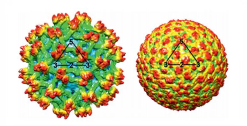
mature (right) particles of a dengue virus isolate. ... 74
Fig. 12. Flavivirus genome replication and assembly ... 78 Fig. 13. Phylogenetic tree of the genus Flavivirus showing the association of the groups
of related viruses with their invertebrate vectors, vertebrate hosts and geographic distribution. ... 80
11
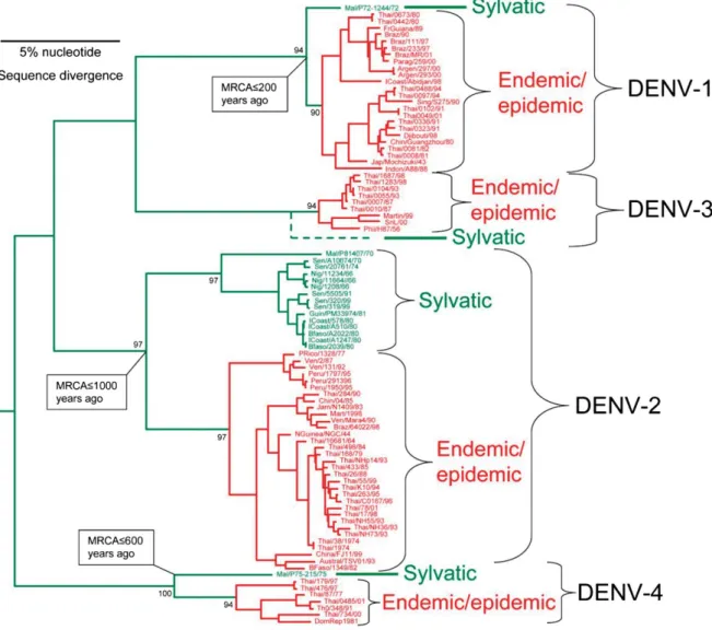
Fig. 14. Phylogenetic tree of DENV strains from four different serotypes derived from
complete open reading frames ... 89
Fig. 15. Schematic manifestations of dengue virus infection ... 91 Fig. 16. Phylogenetic tree of representative isolate of all alphavirus species constructed
from the E1 nucleotide sequences using the F48 algorithm of the neighbor-joining program ... 96
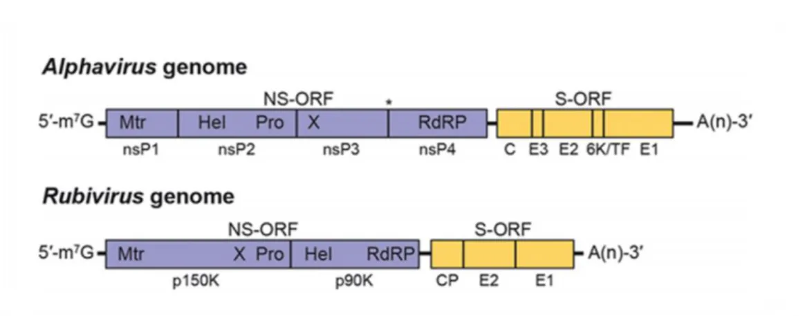
Fig. 17. Alphavirus and rubivirus genome organisation... 97 Fig. 18. Three-dimensional cryoelectron reconstruction of Chikungunya virus, a
member of Togaviridae family ... 98
Fig. 19. Alphavirus replication cycle ... 101 Fig. 20. Mid point rooted phylogenetic tree of representative isolates of all alphavirus
species generated from a conserved region of envelope protein gene nucleotide sequences (2184 nt) using the GTR+1+I substitution model and maximum likelihood methods ... 102
Fig. 21. Fluctuations of the number of Chikungunya cases and Chikungunya incidence
rate (per 100,000 population per year) reported by the Ministry of Health of Indonesia during the period of 2001 to 2016... 107
Fig. 22. Phylogenetic analysis of 99 CHIKV E1 sequences that demonstrate the main
genotypes and their lineages based on geographic distribution and time of outbreak. Numbers at nodes indicate bootstrap support of 1,000 replications ... 108
Fig. 23. The geographic distribution of East/Central/South Africa (ECSA)
Chikungunya virus and Asia Chikungunya virus genotypes in Africa and Asia in period of 2015-2014 ... 110
12
Résumé
L’Indonésie est un grand archipel situé entre les océans Indien et Pacifique. Il est le rendez-vous des deux zones biogéographiques que sont les régions asiatiques et australasiennes. En raison de sa géographie distincte et variée, ce pays est reconnu comme l’un des pays ayant la plus grande biodiversité dans le monde avec de nombreuses espèces endémiques, y compris parmi les moustiques. Au total, 457 des 3 567 espèces de moustiques Culicidae, répertoriées dans le monde, ont été identifiées en Indonésie. Certaines espèces sont responsables de la transmission de maladies, dont au moins 46 espèces de moustiques signalées comme étant des vecteurs d’agents pathogènes humains en Indonésie. Jusqu’à présent, 13 maladies transmissibles par les moustiques ont été signalées dans la population humaine de ce pays, dont le paludisme, la filariose lymphatique, la dengue (DEN), l’encéphalite japonaise (JE), l’encéphalite de la vallée de Murray (MVE), Zika (ZIK), Kunjin, le virus du Nil occidental (WNV), la Edge Hill, le chikungunya (CHIK), le Sinbis, le Getah et la Ross River. De toutes ces maladies transmises par les moustiques, la dengue, le paludisme, l’encéphalite japonaise et le chikungunya sont les plus importantes en Indonésie car leur impact sur la santé publique au cours des deux dernières décennies a été majeur. Les moustiques vecteurs des agents pathogènes concernés appartiennent principalement à trois genres, Anopheles, Aedes et Culex.
Dans le cadre de la réponse nationale visant à prévenir et à contrôler la propagation des principales maladies transmises par les moustiques, il est essentiel de comprendre la dynamique de transmission de ces maladies, qui doit être fondée sur des données mises à jour et précises. L’objectif de notre étude était de comprendre la dynamique des principales maladies transmises par les moustiques afin de renforcer et optimiser le système de surveillance en Indonésie. Nous avons également étudié la diversité et la phylogénie de certains vecteurs du paludisme et leur rôle dans la transmission de cette maladie. Les objectifs spécifiques de recherche étaient les suivants : 1) Mieux comprendre l’épidémiologie de l’encéphalite japonaise et sa transmission ; 2) Identifier les caractéristiques génétiques des flavivirus, en particulier ceux de l’encéphalite japonaise provenant de moustiques capturés sur le terrain ; 3) Décrire la diversité, la phylogénie et l’importance de certains vecteurs du paludisme ;
13 4) Evaluer la pertinence des indices entomologiques officiels pour et le risque de transmission de la dengue ; 5) Analyser l’efficacité des méthodes actuelles de surveillance vectorielle de la dengue en Indonésie ; 6) Décrire la variabilité génétique d’Aedes aegypti (Diptera : Culicinae), vecteur des virus de la dengue, du chikungunya et de la fièvre jaune ; et 7) Analyser l’épidémiologie moléculaire de la transmission du virus du chikungunya en Indonésie. Un certain nombre d’enquêtes sur le terrain et d’examens systématiques ont été utilisés pour atteindre ces objectifs.
Les études réalisées dans le cadre de cette thèse concernent la dynamique des principales maladies transmises par les moustiques en Indonésie, en mettant l’accent sur l’épidémiologie de l’encéphalite japonaise (JE) et la distribution de ses vecteurs (Chapitre 2). Le Chapitre 3 porte sur la diversité des espèces d’anophèles et les implications pour le contrôle du paludisme. Le Chapitre 4 décrit la diversité génétique des vecteurs de la dengue, les méthodes de surveillance vectorielle en Indonésie, et les indices entomologiques pour évaluer le risque de transmission.
L’encéphalite japonaise a été signalée comme une maladie importante transmise par les moustiques depuis qu’elle a été identifiée pour la première fois en Indonésie en 1960. Bien que JE puisse représenter une menace majeure pour la santé publique depuis longtemps en Indonésie, étonnamment, les études pour comprendre les facteurs qui jouent un rôle dans la transmission du virus et ses facteurs de risque, sont encore très limitées. Dans ce Chapitre 2, notre étude de JE fournit des informations sur l’épidémiologie de cette maladie en Indonésie. Notre étude a révélé que la JE a été détectée dans tout l’archipel indonésien avec des cas humains identifiés dans au moins 29 des 34 provinces. Des cas de JE chez des voyageurs venus en Indonésie ont également été signalés. Les facteurs de risque d’infection par le virus de JE (JEV) chez les voyageurs varient selon la destination, la durée du séjour, l’itinéraire, l’activité et l’hébergement. Une surveillance sentinelle et des activités de recherche ont été menées en Indonésie, mais des rapports réguliers sur la JE n’ont pas faits dans toutes les provinces. L’une des raisons de l’absence de rapports nationaux de surveillance de routine de la JE est la difficulté à effectuer un diagnostic des cas de JE au niveau hospitalier en raison du coût élevé de la logistique opérationnelle. Par conséquent, les données sur le nombre de cas et la charge de la maladie sur la population, comme base de mise en œuvre du programme de vaccination, ne peuvent pas être déterminées avec
14
précision au niveau national. Parmi les 9 espèces de moustiques qui ont été confirmées comme étant vecteurs de JEV, Culex tritaeniorhynchusest considéré comme le vecteur le plus important en Indonésie.Bien que les porcs aient servi d’amplificateur principal de JEV en Indonésie, d’autres vertébrés, tels quele bétail, les chèvres, les chevaux et les chiens étaient également positifs aux anticorps de JE par ELISA compétitive. Ainsi, le réservoir de JE être plus étudié de façon plus approfondie et le rôle des animaux d’élevage, autres que le porc, doit aussi être exploré afin de mieux comprendre la transmission de JEV et de mettre en place un contrôle approprié.
La seconde partie de notre étude sur JE montre la présence en Indonésie du génotype-1 de JEV chez une espèce de moustiques collectée sur le terrain (Culex
gelidus). Des études antérieures ont révélé que trois génotypes de JEV, le génotype II
(GII), le génotype III (GIII) et le génotype IV (GIV) ont été trouvés dans tout l’archipel indonésien de 1974 à 1987. Cependant, le génotype I (GI) et le génotype V (GV) n’ont jamais été signalés en Indonésie. Dans notre étude, le génotype I-a (GI-a) a été isolé pour la première fois en 2017 à partir d’un moustique de l’espèce Culex gelidus dans la province de Jambi, en Indonésie. L’analyse phylogénétique du gène E a indiqué que ce virus est étroitement lié à un isolat de GI de 1983 provenant de Thaïlande. GI remplace actuellement GIII en Asie. Ce génotype de virus n’a pas été trouvé dans le liquide céphalo-rachidien due à l’absence d’anticorps IgM spécifiques de JEV synthétisés avec du GIII-JEV. Cela peut causer un risque de faux négatif et de diagnostic erroné en présence de GI. Par ailleurs les vaccins actuels sont basés sur le GIII. D’autres études et le renforcement de la surveillance de JE devraient être mis en œuvre pour pouvoir déterminer la distribution précise du GI-JEV en Indonésie afin de faire face aux risques potentiels de transmission.Deux publications sur JE dont une revue de référence et un article de recherche original ont été publiés dans des revues internationales à comité de lecture.
Le Chapitre 3 porte sur l’importance de mieux comprendre la diversité des espèces d’Anopheles et leurs implications dans la transmission du paludisme pour la mise en place de méthodes de lutte antivectorielle appropriées. En Indonésie, l’étude des anophèles et les mesures de lutte, en particulier ciblant les espèces qui agissent comme d’importants vecteurs du paludisme, sont d’une grande importance dans l’optique de l’élimination du paludisme d’ici 2030. Cependant, les espèces d’Anopheles
15 en Indonésie s’avèrent être très complexes avec de nombreux vecteurs difficilement différentiables des espèces non-vectrices, associées à divers contextes épidémiologiques. Au moins 90 taxa d’Anopheles ont été identifiés en Indonésie dont 25 confirmés comme étant des vecteurs du paludisme. En outre, une compréhension globale de la dynamique de la transmission accompagnée d’efforts appropriés de lutte antivectorielle est assez compliquée à appréhender en raison de plusieurs facteurs, y compris les variations intraspécifiques des espèces liées aux changements écologiques et le statut vectoriel des espèces en fonction de leur distribution. La complexité et la diversité des espèces d’Anopheles pourraient être attribuées à la sélection naturelle, aux processus historiques, aux variations écologiques et aux flux génétiques. Cela a conduit à des divergences et à l’homogénéisation des variations à l’intérieur ou entre les espèces et pourrait être la clé pour comprendre la dynamique de la transmission du paludisme et la base d’une lutte antivectorielle appropriée. Ce chapitre présente l’homogénéité génétique d’Anopheles maculatus, l’un des vecteurs du paludisme les plus importants d’Indonésie. Ce taxon a été signalé comme étant un vecteur majeur du paludisme dans la région des Collines de Menoreh, à la frontière de la province centrale de Java et de la province de Jogjakarta. Il a également été confirmé en tant que vecteur important du paludisme dans le sud de Sumatra. Bien qu’An. maculatus soit largement réparti dans les principales îles de l’archipel indonésien, à l’exclusion des Moluques et de Papouasie, cette espèce n’a jamais été signalée comme vecteur du paludisme dans les îles de Bornéo, Célèbes, Bali et petites îles de la Sonde. Auparavant, cette espèce était considérée comme le seul membre du groupe Maculatus présent en Indonésie. Nous avons analysé la diversité et la phylogénie d’échantillons d’An. maculatus prélevés à plusieurs endroits à Java, aux petites îles de la Sonde, à Sumatra et à Kulon Progo (région des Collines de Menoreh). Des échantillons provenant d’une colonie maintenue en laboratoire depuis 30 ans et provenant de Kulon Progo ont également été inclus dans cette étude. Grâce aux outils d’identification moléculaire utilisant les marqueurs ITS2 (nucléaire) et cox1 (mitochondrial), deux espèces du groupe Maculatus ont pu être identifiées en Indonésie dont une nouvelle espèce provenant de Kulon Progo. Cette nouvelle espèce, génétiquement proche d’An. dispar présente uniquement aux Philippines, diffère de tous les autres membres connus du groupe Maculatus, y compris d’An. maculatus (s.s.). La population de Kulon Progo a été temporairement nommée An. maculatus var. menoreh. Ce résultat est important pour identifier et mettre en œuvre des stratégies ciblées et plus efficaces de lutte antivectorielle contre le paludisme. Dans
16 cette perspective, une meilleure connaissance de cette nouvelle espèce est maintenant nécessaire pour mieux définir sa distribution géographique et son rôle de vecteur du paludisme. Cette étude a été publiée dans une revues internationale à comité de lecture.
La dengue est un problème environnemental lié à plusieurs facteurs, tels que la croissance démographique, le mouvement de la population, le transport, l’approvisionnement en eau des ménages, les services d’assainissement et le comportement communautaire qui contribuent à créer les conditions optimales pour la reproduction des moustiques Aedes et pour la circulation du virus de la dengue (DENV). Des stratégies essentielles doivent être appliquées pour la prévention et la lutte contre la dengue, en particulier pour les cas de dengue (DF) et dengue hémorragique (DHF), la surveillance des vecteurs, la gestion des maladies, mais aussi le renforcement à la fois de la participation communautaire à la lutte contre la dengue et du réseau intersectoriel du gouvernement local et central. Le Chapitre 4 porte sur l’étude de la diversité génétique des vecteurs de la dengue, des méthodes de surveillance vectorielle et des indices entomologiques pour évaluer le risque de transmission de la dengue, afin de pouvoir mettre en place en Indonésie une lutte antivectorielle plus efficace.
Considérant le rôle d’Ae. aegypti en tant que vecteur majeur de la dengue dans les régions hyperendémiques d’Indonésie, l’étude des caractéristiques génétiques des populations d’Ae. aegypti est essentielle pour mieux comprendre leur variabilité génétique et les relations entre elles. Ces informations sont importantes pour identifier l’origine et la distribution de cette espèce qui peuvent être utilisées pour identifier la relation entre les populations d’Ae aegypti, et étudier leur compétence et capacité vectorielles, leur adaptation écologique, et leur résistance aux insecticides. L’étude des caractéristiques génétiques d’Ae. aegypti et Ae. albopictus a révélé un potentiel remplacement rapide des populations de ces deux espèces en Indonésie. Cette dynamique de remplacement représente une menace pour les stratégies massives de lutte antivectorielle contre la dengue. Une conséquence est que la lutte antivectorielle ne doit pas être basée sur la population. Que ce soient des populations d’Ae. aegypti et Ae. albopictus déjà établies ou invasives, elles devront se reproduire dans l’environnement humain et la meilleure façon d’empêcher toute population de vecteurs de prospérer est certainement de mettre en œuvre la lutte antivectorielle au niveau local,
17 au maximum au niveau communautaire, afin d’éliminer le plus possible les gîtes de reproduction en utilisant des moyens de contrôle très simples et abordables tels que l’élimination des conteneurs et des ordures. La stratégie de prévention de la transmission de la dengue par la participation communautaire est actuellement recommandée en Indonésie et la plus susceptible d’être le moyen le plus efficace.
Une analyse comparative des méthodes de collecte de moustiques a été effectuée dans le cadre de la thèse visant à évaluer l’efficacité relative de plusieurs méthodes, dont la collecte matinale des adultes à l’aide d’un aspirateur, la collection des nymphes, la capture sur animaux, la collecte sur appât humain durant une nuit entière (la loi indonésienne n’autorise pas cette activité dans la journée) et la collecte des larves pour la surveillance de la dengue, sont les thèmes discutés dans le Chapitre 4. Une étude a été menée dans 39 sites correspondant à 39 districts/municipalités de 15 provinces d’Indonésie, endémiques pour la dengue, d’Aceh aux Moluques du nord. En ce qui concerne le nombre d’échantillons prélevés, le plus grand nombre d’individus capturés a été obtenu lors des collectes de larves. Parmi les méthodes de collectes larvaires, celle dites des larves simples était la plus efficace en termes de nombre d’individus recueillis par rapport à la méthode d’élevage, aux collectes sur animaux, aux collectes sur appât humain durant la nuit et aux captures de faune résiduelle le matin. En ce qui concerne le nombre d’échantillons positifs pour la dengue, les résultats ont révélé que les larves de moustiques étaient la source presque exclusive du virus de la dengue (93,3 %), 70,8 % ayant été trouvé par la méthode des larves uniques et 22,5 % par la méthode d’élevage. Seulement 7,6 % des échantillons totaux prélevés sur les moustiques adultes étaient positifs au virus de la dengue. Parmi les collectes de moustiques adultes, 2,3 % des échantillons obtenus par captures nocturnes sur appât humain ont été trouvés positifs, comparativement à 4,4 % avec la méthode résiduelle du matin. En conclusion, il n’y avait pas de cohérence dans l’efficacité d’une méthode donnée de détection de la dengue. Par conséquent, des méthodes de surveillance vectorielle plus efficaces et plus appropriées sont nécessaires pour déterminer la distribution des vecteurs, leur densité, les habitats larvaires et les facteurs de risque liés à la transmission et à l’évaluation de des efforts de lutte antivectorielle. En outre, l’élaboration d’un nouvel ensemble d’indices est nécessaire comme outils efficaces pour gérer et anticiper le risque d’épidémies de dengue.
18 Une étude portant sur les indices Stegomyia et leur utilisation a également été réalisée pour analyser la relation entre les indices Stegomyia et le risque de transmission de la dengue sur une très grande zone couvrant 78 sites d’échantillonnage dans toute l’Indonésie, de Sumatra à la Papouasie. Les indices Stegomyia ont été élaborés en tant qu’indicateurs quantitatifs du risque de transmission et d’épidémie de dengue. Cette étude a été menée sur la base du fait que, conformément aux recommandations de l’OMS, l’Indonésie utilise ces indices Stegomyia pour l’analyse du risque de transmission de la dengue depuis plus de trois décennies. Les résultats de cette étude ont révélé qu’aucune corrélation n’existe entre l’incidence de la dengue et les indices Stegomyia. D’autres indices plus précis et plus sensibles, de nature sociétale et non entomologique, doivent être développés pour surveiller et prévoir plus efficacement et plus précisément le risque de transmission de la dengue en Indonésie.
Le Chapitre 5 traite de la dynamique des virus du Chikungunya (CHIKV) isolés de moustiques Ae. aegypti, Ae. albopictus et Ae. butleri capturé sur le terrain. L'étude a révélé que tous les CHIKV identifiés sur toute l'Indonésie dans cette étude étaient similaires à ceux isolés en Indonésie depuis 2000. Ces CHIKV appartiennent tous au génotype Asie-Pacifique, le nom du nouveau génotype CHIKV proposé dans cette étude, qui est différent du génotype Asiatique. Si tous les spécimens collectés d’Ae. aegypti appartiennent à la même population, ce n'est pas le cas pour les échantillons d’Ae. albopictus. Les individus positifs pour CHIKV et ceux négatifs pour CHIKV appartiennent à des groupes distincts. Cependant, la taille de l'échantillon est trop petite pour aboutir à une conclusion définitive et une étude plus large est nécessaire pour analyser correctement la structure de la population d'Ae. albopictus en relation avec la compétence vectorielle pour CHIKV. Les preuves du remplacement de la population de CHIKV et la faible diversité des Aedes vecteurs en Indonésie méritent une attention toute particulière afin de mettre en œuvre une gestion plus appropriée et efficace de prévention des épidémies potentielles par des actions locales de lutte contre les moustiques.
Cette thèse donne un aperçu de la dynamique actuelle et du risque de transmission des principales maladies transmises par les moustiques en Indonésie. En outre, l’évaluation des méthodes de collecte des moustiques pour la surveillance vectorielle est analysée dans cette thèse afin de soutenir la mise en œuvre de
19 programmes de surveillance et de contrôle des principales maladies transmises par les moustiques en Indonésie. Enfin, les conclusions de cette étude aideront le public et les autorités concernées à mettre en œuvre des programmes nationaux plus efficaces pour lutter contre les maladies à transmission vectorielle, en particulier le paludisme, l’encéphalite japonaise et la dengue.
20
General Introduction
Indonesia, the largest archipelagic country in the world, located between the Indian and Pacific oceans, has became the rendez-vous of two biogeographical zones: western Indonesia, which is more influenced by Asian organisms, and the east part, more influenced by Australian organisms. Due to its distinct and varied geography, this country contains many endemic and unique species of animals with various habitats and ecosystems, including mosquitoes. O’Connor and Sopa recorded a total of 457 species of mosquitoes from Indonesia out of 3,567 species of Culicidae listed worldwide (1,2). Certain species are responsible for important disease transmission, of which at least 46 species of mosquitoes have been reported as vectors of human pathogens in Indonesia (3–5).
Mosquito-borne diseases are illnesses caused by viruses or parasites transmitted by mosquitoes in human populations. In Indonesia, so far, 13 mosquito-borne diseases have been reported to infect humans, i.e. Malaria, Lympathic filariasis, Dengue (DEN), Japanese encephalitis (JE), Murray valley encephalitis (MVE), Zika, Kunjin, West Nile virus (WNV), Edge hill, Chikungunya (CHIK), Sinbis, Getah and Ross river (4,6– 9). Of all these, Dengue, Malaria, Japanese encephalitis, and Chikungunya, are the most important mosquito-borne diseases, for which a major impact on public health in the country has been recorded during the last two decades (3,4,9,10).
Dengue or Dengue Haemorrhagic Fever (DHF) is a benign to severe and even fatal syndrome caused by dengue viruses (DENV). Benign means asymptomatic or mild form with symptoms of undifferentiated fever, aches, pains, nausea, vomiting and rash. Meanwhile, severe dengue is a more serious form of clinical symptoms that can result in shock, internal bleeding complications, such as gingival bleeding, epistaxis, hematuria, gastrointestinal bleeding, menorrhagia, and even death (11). This disease is known as the most rapidly spreading mosquito-borne viral disease in the world. The World Health Organization (WHO) estimated that more than 2.5 billion people (over 40% of the world population) live in endemic countries in which more than 100 million dengue infections occur with 20,000 deaths worldwide every year. Indonesia is recognized as one of the highest dengue endemic countries in the world. Aedes aegypti
21 and Ae. albopictus are respectively the principal and secondary dengue vectors and breed extensively in all regions from western to eastern Indonesia (12). All four dengue serotypes (DENV1 to DENV4) are endemic in almost of the big cities of the country (13). In the past 45 years, annual DHF incidence increased significantly from 0.05/100,000 population (58 cases) in 1968 to 78.85/100,000 (204,171 cases) in 2016. By contrast, the fatality rate of DHF decreased considerably from 41% (24 deaths) in 1968 to 0.78% (1,598 deaths) in 2016. The areas affected by the disease in 2016 included 90.08% of the total 463 of districts/municipalities (14). To deal with this disease transmission, dengue control programs have been conducted since 1968 at the national level by the Ministry of Health (MoH) Indonesia that issued in 1992 a strategy concerning the national DF/DHF program. The critical strategies for the DF/DHF prevention and control include vector and human cases surveillance system, disease management, strengthening community participation in DF/DHF prevention and control activities, and cross-sectoral parthnership. The implementation of dengue control programs has also included health education at the community level, appropriate case management and vector control with focus on source reduction. Based on the community participation and intersectorial coordination, selected fogging (two cycles with weekly interval) of adult Aedes mosquitoes within 100 metres radius of reported DHF case house and mass larviciding were implemented (9). However, although dengue control efforts have been carried out continuously, the results is still not as expected. Dengue has spread in almost all regions of Indonesia with multiple co-circulating DENV serotypes (9,15,16). Moreover, major dengue outbreaks have been reported in the country over the past years (17–22).
The Japanese encephalitis virus (JEV) is another mosquito-borne flavivirus that has also became a public health threat in Indonesia. JEV is transmitted to humans through mosquito bites of Culex species from amplifier animals, especially pigs, as well as other vertebrate animals. This viral infection can cause severe central nervous system disorder with an estimated 68,000 cases every year and a case fatality rate (CFR) among patients ranging from 20% to 30% (23,24). The development of permanent neurological symptoms or psychiatric sequelae is estimated to occur in 30 to 50% of surviving patients (25–29). JEV was first isolated in the country from field-collected mosquitoes in Bekasi district, West Java and Kapuk sub-district, West Jakarta around 1972 (30). Since then, encephalitis cases have been reported in several big hospitals of Indonesia.
22 A total of 7,933 encephalitis cases were reported during 1979 to 1986 with 36.3% fatality (31). Further studies were then conducted in North Sumatera, West Kalimantan, North Sulawesi, South Sulawesi, East Nusa Tenggara, Papua, and Bali during 1993 to 2000. A total of 1,830 samples were collected among which 1,137 samples (62.13%) were reported as JE positive cases (32). Since then, several small-scale JEV studies have been conducted in Indonesia. In Bali, active surveillance of JEV was conducted in 10 hospitals during 2000 to 2002. A number of 33 positive cases of JEV infection were found among which 8.5% died (33). Although presumed to be endemic contrywide, the comprehensive national data on the current situation that describes the epidemiology of JE and its transmission patterns are still not available.
Beside dengue and Japanese encephalitis, Chikungunya is also an important arbovirus, which is a nationalwide public health problem in Indonesia. Chikungunya is caused by the chikungunya virus (CHIKV), a member of the genus Alphavirus belonging to the family Togaviridae and transmitted mainly by Ae. aegypti and Ae. albopictus mosquito species. The disease is a febrile illness characterized by high fever, arthralgia, myalgia, headache, skin rash and intense asthenia (34). Chikungunya was formally reported for the first time in Samarinda, East Kalimantan in 1973 (35,36). In the last 16 years, Indonesia has frequently experienced outbreaks of chikungunya fever caused by both the Asian and the East Central and South African (ECSA) lineages. Prior to these outbreaks, the incidence of Chikungunya was less than 10,000 cases/year. The massive nationwide outbreaks with 137,655 cases were reported during 2009 - 2010. Subsequent a smaller outbreak was also noted in 2013 with 15,324 cases. No death from chikungunya cases was reported during this outbreak. In 2009 and 2010, the incidence increased significantly to reach 83,756 and 53,899 cases, respectively (36– 38). In spite of many outbreaks that occurred in Indonesia, the data regarding the epidemiology, the magnitude of the disease, the role and capacities of Ae. aegypti and Ae. albopictus to transmit the virus and the dynamic of chikungunya transmission are still insufficient.
Malaria is still a prominent public health problem along the tropical belt, including Indonesia. According to a WHO report, there were about 228 million cases of malaria and an estimated 405,000 deaths in the world in 2018 (39). Indonesia is also an endemic malaria country and home to about 25 Anopheles species, which transmit
23 all five Plasmodium species that infect humans. In 2019, as many as 222,085 confirmed malaria cases with prevalence of 0.93 per 1,000 population, and 49 confirmed malaria deaths were reported (40). Malaria transmission occurs in 267 districts/municipalities in all of the provinces with highest risk of acquiring malaria in the eastern part of Indonesia. At present, Indonesia is heading towards the goal of malaria elimination. Comprehensive malaria control efforts continue to be made through strengthening the surveillance system, upscaling diagnostic and treatment interventions, and vector control not only in high-transmission districts, but also in low-transmission areas. Monitoring and evaluation efforts have been carried out to support the achievement of the target of malaria elimination by 2030 with the support of the national and local governments, national technical components (Directorate general of disease prevention and control-MoH and National Institute of Health Research and Development-MoH), donor agencies (Global Fund for Malaria, WHO, UNICEF), other governmental components, and private sectors. Several activities carried out include monitoring anti-malaria drug resistance; montoring the accuracy of diagnosis, both of rapid diagnostic test (RDT) and microscopy, monitoring the resistance of mosquito vectors to long lasting insecticide bednets (LLINs), mapping malaria receptivity, especially those areas that have been and will be eliminated, and monitoring behavior changes of malaria vectors (41).
As part of the national response to prevent and control the spread of main mosquito-borne disease in Indonesia, understanding epidemiology and transmission dynamics of these diseases is essential to provide up-to-date and accurate information on transmission pattern of these main mosquito-borne diseases in Indonesia.
Our objective was to understand the dynamics of the main mosquito-borne diseases to strengthen the surveillance system in Indonesia. We also investigated the diversity and phylogeny of malaria vectors and its roles in malaria transmission. The specific objectives of the research aims were:
1) To better understand the epidemiology of Japanese encephalitis and its transmission ecology in Indonesia;
2) To identify genetic characteristics of flaviruses, especially Japanese encephalitis from field-caught mosquitoes in study areas;
24 3) To describe the diversity and phylogeny of malaria vectors and their roles in
malaria transmission in Indonesia;
4) To identify the relationship between entomological indexes and the risk of arboviruses transmission in Indonesia;
5) To analyze the effectiveness of current dengue vector surveillance methods in Indonesia;
6) To describe the genetic variability of Aedes aegypti (Diptera: Culicinae), vector of dengue, chikungunya, and zika viruses in Indonesia;
7) To identify the dynamic of chikungunya virus in Indonesia.
A number of field surveys and systematic reviews were used to achieved these aims. All studies were published in international peer-reviewed journals, and submitted or in preparation. These articles are presented within chapters of this thesis.
25
Chapter 1. Background
Mosquito-borne diseases
Mosquitoes are insects belonging to the order of Diptera within the family of Culicidae. Currently, a total of 3,568 species of mosquitoes have been identified and classified into subfamilies and 113 genera (42). Mosquitoes are the most deadliest animals in the word. Females of many mosquito species are bloodsucking insects that have the ability to carry and spread pathogens (viruses, helminths, and protozoa) that causes mortality and morbidity within human population every year (42,43). Mosquito-borne diseases are the largest contributors of the vector-Mosquito-borne disease burden and important emerging diseases to human. According to the World Health Organization (WHO) report, malaria is the most important mosquito-borne parasite disease that caused in 2018 a total of 228 million human cases with 405,000 deaths worldwide (44). In addition, Dengue is the most important mosquito-borne virus that caused 4.2 million human cases in 2019. An estimated 500,000 cases annually had severe dengue requiring hospitalization, of which about 1 to 2.5% mortality (45,46). Mosquitoes also carry many other important human pathogens, such as viruses responsible for Japanese Encephalitis (JE), Yellow fever (YF), West Nile (WN), Muray Valley Encephalitis (MVE), Kunjin (KUN), Edge Hill (EH), Zika (ZIKV), Chikungunya (CHIKV), Getah (GET), Ross River (RR), Sinbis (SIN), Rift Valley Encephalitis (RVE) and parasites responsible for Lymphatic Filariasis (LF) (4,47,48). Mosquito vectors mostly belong to three genera, Anopheles, Aedes, and Culex.
Malaria parasites are transmitted to humans through the bite of female Anopheles mosquitoes. A total of 478 species as part of subfamily Anophelinae have been identified worldwide (1). At least 58 unknown member of the species complexes are also recognized based on biological and morphology of the Anopheles genus (1). The Anopheles species are distributed into eight subgenera, Anopheles (187 species), Baimaia (1 species), Cellia (233 species), Christya (2 species), Kerteszia (12 species), Lophopodomyia (6 species), Nyssorhynchus (42 species), and Stethomyia (3 species) (49). At least 70 species are showing vectorial capacity to transmit human malaria parasites and 41 species among them being considered as dominant malaria vector
26 species (50,51). Some Anopheles species are also known as important vectors of lymphatic filariae (48,52–54). Recent studies have revealed that at least 51 viruses have been identified and associated with Anopheles species. Many of these viruses have the potential to cause febrile disease and encephalitis in humans (55). Anopheles are nocturnal mosquitoes biting from sundown to sunset (6 pm to 6 am). They breed in a large variety of aquatic habitats, mostly natural, sometimes human made, with stagnant or slow running freshwater or brackish water, shaded or sunny, temporary or permanent, associated with sunlight or shade, water salinity, presence of floating or emergent vegetation, and turbidity (56–59). Anopheles mosquitoes colonize a large variety of environments from coastal to montainous areas, even caves. They are distributed worldwide, except the majority of the pacific islands (49).
Aedini (subfamily Culicinae) is the largest tribe of mosquitoes in the world. A total of 1,260 species within 10 genera have been recorded in this tribe. According to Wilkerson et al. (60), the genera of Aedini are as follows Zeugnomyia (4 species), Verallina (95 species), Udaya (3 species), Psorophora (49 species), Opifex (2 species), Heizmannia (39 species), Haemagogus (28 species), Eretmapodites (48 species), Armigeres (58 species), and traditional Aedes sensu (934 species). Several species of the genus Aedes, particularly Aedes (Stegomyia) aegypti and Ae. (Stegomyia) albopictus are known as principal vectors of several important arboviruses in the world, including dengue, yellow fever, chikungunya, and zika (12,61). Historically, Ae. aegypti originated from Egypt, Africa, as shown by its species name. Currently, the species has spread to all tropical and subtropical continents, and some temperate regions throughout the world. There are two different forms of Ae. aegypti according to geographic variations, behaviour, ecology and susceptibility to dengue virus, i.e. Ae. aegypti formosus and Ae. aegypti aegypti (62,63). Although morphologically difficult to distinguish, gene flow between them is restricted. Ae. aegypti formosus is involved in the dengue forest transmission in West Africa, while Ae. aegypti aegypti is the main dengue vector worldwide (64). Aedes aegypti formosus is also known as a less anthropophilic form. This mosquito is mostly reported to colonize natural breeding sites, whereas Ae. aegypti aegypti prefers to breeds in man-made water containers (62).
Aedes albopictus, also known as the Asian tiger mosquito, is native from temperate and tropical regions of Southeast Asia. Aedes albopictus is a diurnal outdoor
27 mosquito with a very broad range of hosts, including humans, reptiles, livestock, dogs and other mammals, ampibians and birds (65). The tiger mosquito is a competent vector for at least 22 arboviruses of human and animal related diseases that led to serious arboviruses outbreaks (66,67). This species is living at the edges of forests and breeds in various natural habitats such as bamboo stumps, tree holes, decaying leaves, and other small, restricted, shaded bodies of water surrounded by vegetation (68). However, Ae. albopictus has ecological flexibility to adapt well to many types of man-made sites, even very small water bodies (water storage containers, tires, bottles, cemetery urns, opened coconuts, etc) in suburban and urban environments. The flight range of adult Ae. albopictus is quite short (300 meters). Hence, the spread of this species has occurred due to passive transportation facilitated by human activities (boats, cars, planes, etc). The introduction of Ae. albopictus is mostly due to the easy transportation of dormant eggs that resist to dessiccation, especially through the trade of used tyres and lucky bamboos. As a consequence, the asian tiger mosquito has undergone a dramatic global expansion and colonized rapidly almost all continents (except Antartica) in the past 40 years. Competition between Ae. albopictus and Ae. aegypti has been recorded. In many places, distribution of both species partially overlap despite their occurrence in different biotopes. Currently, Ae. albopictus has been proven to have a competitive advantage over a number of other mosquito species, including Ae. aegypti (69,70).
The genus Culex is known as the most widespread mosquito across the globe (71,72). Culex contains 769 species belonging 26 subgenera distributed worldwide (73). Adult Culex species have high preference for biting humans and animals (74). Therefore, several species of Culex are vectors of many relevant animal and human pathogens including arboviruses responsible for Japanese encephalitis, Murray Valley Encephalitis, West Nile, Rift valley fever, St Louis Encephalitis and parasites such as Bancrofti lympatic filariae. The Culex pipiens complex is the most widely distributed and important one, which comprises 6 members, Cx. pipiens, Cx. molestus, Cx. globocoxitus, Cx. pallens, Cx. australicus, and C. quinquefasciatus (75,76). Cx. quinquefasciatus is known to have the major role in the transmission of several important vector-borne diseases. Cx. quinquefasciatus is a peridometic mosquito that breeds in various types of natural and man-made water containers, including permanent and temporary stagnant water bodies such as organic polluted breeding sites, septic tanks, drains, wet pit latrines, puddles, concrete tanks, vases, bottles, cans, skillet,
28 earthen plates, sewage drains, cesspools, etc (77,78). Culex quinquefasciatus has unique adaptation to various environments. Although the flight range of this mosquito is short (less than 600 meters), this species has spread throughout the world by commercial ships and aircraft (70,79,80).
Malaria
1. Malaria parasites
Malaria is an important vector-borne disease that causes death and illness in children, particularly those <5 years old, and adults in tropical and sub-tropical regions (81). Malaria is caused by single-cell protozoan parasites of the genus Plasmodium and transmitted to humans through bites from Anopheles species. There are five parasite species that infect and cause the disease in humans: Plasmodium falciparum, P. vivax, P. malariae, P. ovale (consist of 2 subspecies : P. ovale wallikeri and P. ovale curtisi), and P. knowlesi (82–84). Plasmodium falciparum is known as the deadliest form of human malaria, while P. vivax, P. malariae and P. ovale are milder forms and fatal cases rarely occur (85). Plasmodium falciparum is the most prevalent in sub-Saharan Africa, which represents 93% of all malaria cases worldwide in 2018 (44). Subsequently, Plasmodium vivax is predominant and widely distributed species compared to other human Plasmodium species (86,87). In the region of the Americas, P. vivax represents 64% of malaria cases, whereas in the Southeast Asia and the Eastern Mediterranean regions, 64% and above 30% of human malaria cases are caused by this species respectively (88). In the last decade, the fifth species of human malaria, P. knowlesi, has posed a threat to public health, especially in Southeast Asia. Plasmodium knowlesi is a simian malaria parasite species (89). It was experimentally transmitted to humans for the first time in the 1930s (90,91). The first case of natural infection in human was reported in Malaysia in 1965 (92). However, natural transmission to humans are considered rare and human are likely the accidental hosts. Plasmodium knowlesi received much attention after it was dicovered in the large number in human samples in the Kapit division of Sarawak, Malaysian Borneo (93). After that, subsequent studies showed that knowlesi malaria was also reported to infect humans in almost all regions of Southeast Asia. Currently, An. knowlesi is recognized as a cause
29 of potentially fatal human malaria, particularly in forest areas of Southeast Asia (93– 102).
Nearly half of the world population is at risk of malaria. A total of 91 countries are malaria endemic regions. Of which, most of the malaria cases were recorded from the African region (93%), followed by the Southeast Asian region (3.4%) and the Eastern Mediterranean region (2.1%) in 2018 (44). WHO estimated 228 million malaria cases with 405,000 deaths during 2019 (44).
2. Life cycle of malaria
All malaria parasites require two hosts to complete their life cycle (Fig. 1), i.e. the definitive host represented by a female Anopheles mosquito, in which reproduction occurs (sexual stages allow ookinete formation) and the intermediate host, which is a vertebrate host, including human (developement of asexual stages) (103,104). The cycle of malaria in humans is initiated by the inoculation of sporozoites into human blood vessel through the bite of a female Anopheles mosquito. Sporozoites, the infectious Plasmodium stage located in mosquito salivary glands, will then be transferred into the blood stream, and then enter the parenchyma cells of the liver. The parasite develops asexually, producing thousands of merozoites in the cell. This phase is known as exoerythrocytic schizogony or pre-erithrocytic schizogony (104).
The second phase is the parasite dispersal and invasion of the host target cells. This phase is called as erithrocytic schizogony (103). Merozoites come out from the liver parenchyma cells, gain the blood stream to invade red blood cells and initiate a series of asexual multiplication cycles from trophozoite, schizont and then produce 8 to 24 new infective merozoites per cell. At this point, the red blood cells burst and the infective cycle begins anew (105). The time length required for completion of the erythrocytic cycles varies, depending on the Plasmodium species. The length of the cycles ranges from approximately 24 hours (quotidian periodicity for P. knowlesi) to 72 hours (quartan periodicity for P. malariae). During this step, not all merozoites produced in the erithrocytic schizogony phase will re-infect red blood cells in the next cycles (106). Some merozoites will develop into immature gametocytes, which are precursors of male and female gametes (104).
30 Fig. 1. The life cycle of Plasmodium, malaria parasit (104)
During the sexual stage, when a female Anopheles bites an infected person, gametocytes are taken up together with the blood and they mature in the mosquito midgut. A mosquito blood meal is approximately 2 to 3 µl, and may contain at least one male (micro) and one female (macro) gametocytes (101). In the midgut or mesenteron, the mature microgametocytes and macrogametocytes shed their red cell envelopes and transform into the mature sexual forms, microgametes and macrogametes. Subsequently, the microgamete enters the macrogamete to form the zygote that develops into active moving ookinete. The ookinete then moves actively and forces its way into an epithelial cell of the Anopheles midgut, and becomes oocyst on the outside gut wall. Meiotic division occurs 48 hours after the blood meal during young oocyst phase (107). Afterwards, mitotic divisions take place to produce nuclei with the particular haploid number of chromosomes for the species. The number of nuclei increases when the oocyst grows. The cytoplasm splits into sporoblasts to be vacuoles. Afterwards, the sporozoite is formed. Each sporozoite contains a single nucleus and the number of sporozoites produced per oocyst has been estimated at around 10,000 (108).
(HUMAN
)
31 The sporozoites then migrate to all parts of the body. In mosquito vectors, some sporozoites will be able to enter the acinal cells of the salivary glands. Sporozoites enter the salivary duct when the mosquito begins to feed and enter to the blood stream of hosts (humans) (104). When mosquito vectors have sporozoites in their salivary glands, they may infect humans every two days for the rest of their life.
3. Plasmodium
3.1. Classification
The genus Plasmodium is the causative protozoan parasite of malaria, which belongs to the phylum Apicomplexa, a single-celled parasite with a unique form of apical secretory organelles that can help penetration into the host cell (109). Plasmodium is part of the sub-order Haemosporidae, member of apicomplexans that live in the blood cells. This genus also belongs to the family Plasmodiidae, which is characterized by three phases in the life cycle, i.e. 1. exoerythrocytic schizogony without pigmentation in monocytes of viscera and reticuloendothelial cells in hepatic cells of mammals and birds; 2. Schizogony with pigmentation in erythrocytes of the vertebrate hosts where periodic febrile coincide with the release of merozoites; 3. Sexual phase with pigment-producing gametocytes that emerge in the vertebrate host erythrocytes (110).
So far, fourtheen subgenera and nearly 200 species of Plasmodium have been identified on the basis of their morphology in blood smears of the infected hosts and their host range (111). The great majority of malarial parasites are transmitted by mosquitoes, for instance the parasites of humans are exclusively transmitted by species of the genus Anopheles. Human parasites are divided into two subgenera, Laverania and Plasmodium. The subgenus Laverania includes P. falciparum, the most pathogenic form of malaria, and the closely related species, P. reichenowi, a highly pathogenic form of primate parasites. The subgenus Plasmodium includes the remaining human parasites: P. vivax, P. malariae, P. ovale, and P. knowlesi, the fifth cause of human malaria. Parasites of the other mammals are included in two subgenera, namely Plasmodium and Vinckeia, whereas parasites in birds and reptiles are classified in the
32 subgenus Plasmodium (103). The complete classification of the human Plasmodium is showed in Table 1.
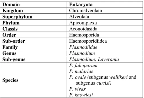
Table 1. Classification of human Plasmodium species (84,112)
Domain Eukaryota Kingdom Chromalveolata Superphylum Alveolata Phylum Apicomplexa Classis Aconoidasida Order Haemosporida Sub-order Haemosporidiidea Family Plasmodiidae Genus Plasmodium
Sub-genus Plasmodium; Laverania
Species
P. falciparum P. malariae
P. ovale (subgenus wallikeri and subgenus curtisi)
P. vivax P. knowlesi
Recently, Non-human primate (NHP) Plasmodium species have been a real concern since reports of P. knowlesi infecting humans. Plasmodium knowlesi is known as a zoonotic malaria parasite, which is normally residing in long-tailed macaques (Macaca fascicularis), pig-tailed macaques (Macaca nemestrina) and leaf monkeys (Presbytis melalophos) (100). Thirteen genes in P. falciparum and P. vivax are not found in P. knowlesi that could be the cause of a barrier to the parasite success to infect human host, however, this Plasmodium species can still be transmitted and become parasites in humans. At least 30 Plasmodium species have been identified as the cause of infection in the NHPs in which 53 host species of more than 25 genera can be infected (113,114). Five species of NHP Plasmodium have been reported as predominate in Southeast Asia. They are P. knowlesi, P. cynomologi, P. fieldi, P. inui, and P. coatneyi (84,115). Host-switching from NHP malaria parasites into humans has also been reported from Brazil with P. simium, Venezuela and Costa Rica with P. brasilianum / P. malariae, Malaysia with P. cynomolgi, and the Central African Republic with P. vivax-like strain from the great apes (116–122). P. brasilianum genome is 99.7% identical to human P. malariae and considered as an anthropozoonosis. Futhermore, P. simium is considered genetically similar and indistinguishable from P. vivax (123,124).
33 Table 2. Plasmodium of humans, primates and other mammals (103)
Genus : Plasmodium Subgenus : Plasmodium Group vivax Group malariae Group ovale Group uncertain Sub-genus : Laverania Sub-genus : Vinckeia Species
P. vivax, P. cynomolgi, P. eylesi, P. gonderi, P. hylobati, P. jefferyi, P. pitheci, P. schwetzi, P. simium, P. sylvaticum, P. youngi
P. malariae, P. brazilianum, P. inui
P. ovale, P. fieldi, P. simiovale
P. knowlesi (quotidin periodicity), P. coatneyi, P. fragile (both with tertian periodicity)
P. falciparum, P. reichenowi
Large number of species infecting rodents, bats, lemur, and other animals. Some of them of uncertain taxonomy status
3.2. Origin and evolution of Plasmodium
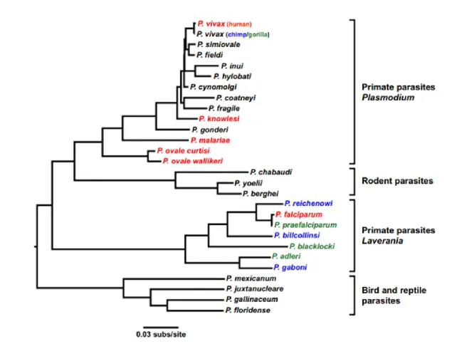
Long-standing hypotesis about the origin of Plasmodium suggested that chimpanzees and humans inherited P. falciparum-like infections from their common ancestors and co-evolved with each of their host species over millions of years. Conversely, P. vivax was believed to have appeared several hundred thousand years ago, following the cross-species of Plasmodium transmission from macaques in Southeastern Asia (125). However, the recent studies following the characterization of large numbers of additional Plasmodium parasites from African apes indicated that P. falciparum infection is relatively new for humans and arose after the acquisition of parasites from gorillas, possibly occurring in the last 10,000 years (126,127). This has put an end to the previous hypothesis. The important sign that apes have became harbouring Plasmodium infections was the evidence of three morphologically distinct forms of Plasmodium parasites in the wild-caught chimpanzees and western gorillas’s blood in Cameroon. Microscopic examination for morphological identification of parasites from the blood revealed that Plasmodium from apes suggests the existence of different Plasmodium spp., which were classified as P. reichenowi, P. rhodaini, and P.
34 schwetzi. Interestingly, these three Plasmodium resemble to P. falciparum, P. malariae, and either P. ovale or (the similar) P. vivax respectively in humans (128). Furthermore, P. falciparum and P. reichenowi were found to differ substantially in both life cycle and gametocyte morphology from other Plasmodium species. This has led to the placement of these two species separately from the other subgenus, called Laverania. Based on the sequence of the rRNA small subunit gene, a study conducted by Escalante and Ayala showed that genetic relationship amongst P. falciparum and P. reichenowi were very close relatives of each other. In contrast, both of these Plasmodium species were only distantly related to other Plasmodium spp. (Fig. 2.). If it is estimated that the rRNA gene sequence in Plasmodium spp. evolved at the same rate as expected for some bacteria, it was concluded that P. falciparum and P. reichnowi evolutionarily diverged 10 million years ago, close to time of the ancestors of human-chimpanzees. This leads to the conclusion that parasites that infect humans and chimpanzees have co-existed with their respective hosts (125,128).
Fig. 2. Evolutionary relationship of Plasmodium spp. (P.) that infect gorillas (green), chimpanzees (blue), and human (red). Phylogenetic analysis was estimated by using maximum likelihood analysis of 2.4 kb of the mitochondrial genome (128)