Supplemental material for 1
Characterization of yeast mutants lacking alkaline ceramidases YPC1 and YDC1 2
Natalia S. Voynova1,3,4, Shamroop Mallela1,3, Hector M. Vazquez1, Vanessa Cerantola1,5, Mélanie 3
Sonderegger1, Jens Knudsen2, Christer S. Ejsing2 and Andreas Conzelmann1# 4
5
Supplemental Materials and Methods 6
Materials. FM4-64 was from Molecular Probes, 5’-Fluoroorotic Acid (FOA) from Toronto Research 7
Chemicals, cycloheximide, phytosphingosine, 2,4-dinitrophenylhydrazine and Lucifer Yellow from 8
Sigma. [14C]serine was from ARC, St. Louis, MO; monomethylamine (33% in ethanol) was from 9
Fluka AG, Buchs, Switzerland. Anti-DNP antibodies were from Dako. Protease inhibitors were from 10
Roche Diagnostics GmbH, Mannheim, Germany. 3,3'-dihexyloxacarbocyanine iodide was from 11
AnaSpec. 12
Synthetic Genetic Array. SGA analysis was performed as described previously (Collins et al., 2010). 13
Briefly, the query strains (Y7092, FBY5162, FBY5173) were robotically crossed against an array of 14
4978 individual MATa knockouts of nonessential genes to generate double or triple mutant arrays. The 15
resulting double and triple mutants were then screened for genetic interactions affecting cell growth. 16
When selecting for triple mutants, the plates were further replicated in parallel on plates containing 17
also Aureobasidin A at a concentration, which did not give visible growth inhibition of WT cells (0.03 18
µg/ml). They also were replicated onto plates containing 25 µM PHS, or 100 mM Ca2+, or onto 19
inositol free medium at 37°C and at 37°C in normal medium. The measurement of growth and 20
following analysis and visualization of the high-throughput screen data were conducted with the help 21
of the ScreenMill software (Dittmar et al., 2010). Additionally, the interactions pointed by the screen 22
were verified by independent crosses, tetrad dissection or random sporulation and by serial dilution 23
plating to assess colony sizes, cloning efficiency and growth rates. 24
Metabolic labeling of cells with [14C]serine, lipid extraction, mild base treatment and thin-layer 25
chromatography. Cells were grown in synthetic minimal medium. 3.0 OD600 units of exponentially 26
growing cells (i.e. 3 ml of a culture having an OD600 of 1.0) were harvested, resuspended in 250 µl of 27
the same medium supplemented with 10 µg/ml of cycloheximide (CHX). After 10 min of 28
preincubation, 4 µCi of [14C]serine were added and cells were incubated for 40 min at 30°C. Then the 29
samples were diluted with 750 μl of fresh minimal medium supplemented with CHX and labeling was 30
continued for a further 120 min. Labeling was terminated by adding NaN3 and NaF (10 mM final 31
concentrations) and chilling cells on ice. Cells were resuspended in chloroform:methanol (2:1) and 32
broken with glass beads in the cold. The extract was kept apart and the pellet was re-extracted 33
sequentially with chloroform:methanol (1:1) and EtOH:H2O:Et2O:Pyridin: 25% NH4OH 34
(15:15:5:1:0.018), which achieves quantitative extraction of all complex sphingolipids (Hanson and 35
Lester, 1980). Extracts were combined and solvent was evaporated under vacuum in a rotary 36
evaporator. Incorporation into lipids usually amounted to 5 % of added radioactivity. Where indicated, 37
lipids were subjected to mild base hydrolysis with mono-methylamine (MMA). Lipids were 38
resuspended in 400 µl of MMA (33% in ethanol) or, as a negative control, in methanol, and incubated 39
at 53°C for 1 hour. Then, solvents were evaporated under vacuum. All lipids were resolved by 40
ascending TLC on silica gel plates after having been desalted by Folch partitioning as described (Folch 41
et al., 1957). Extracts from metabolically labeled cells were resolved with chloroform/methanol/glacial 42
AcOH (90:1:9) or CHCl3:MeOH:KCl (55:45:5) solvent systems. When the untreated and deacylated 43
lipid extract was run side by side, material from an equivalent number of cells was spotted. 44
Radioactivity was detected and quantified by one- and two-dimensional radioscanning using a 45
Berthold radioscanner and visualized by fluorography or radioimaging using the Bio-Rad Molecular 46
Imager FX. 47
Isolation of detergent resistant membranes and Triton X-100 solubilization assay. For the 48
isolation of detergent resistant membranes, published protocols were used (Bagnat et al., 2000; 49
Malinska et al., 2004). Crude membranes corresponding to 200 µg protein were incubated in 300 µl 50
cold TNE buffer (50 mM Tris-HCl, pH 7.4, 150 mM NaCl, 5 mM EDTA) containing protease 51
inhibitors (Roche Diagnostics GmbH, Mannheim, Germany) and 1% Triton X-100 for 30 min on ice. 52
Subsequently, the samples were overlaid with an Optiprep (Nycomed) step gradient and centrifuged 53
for 3 h at 208,000 × g in a Beckman SW60 rotor at 4°C. After centrifugation, six equal fractions were 54
collected, and the proteins were immunodetected on Western blots. 55
FM4-64 staining to monitor endocytosis. FM4-64 was used to stain vacuoles and endosomes as 56
described previously (Baggett et al., 2003). Yeast cells were cultured at 24˚C. Five separate aliquots of 57
1 ml each were centrifuged and cooled on ice. Each pellet was resuspended in 50 µl of ice cold FM4-58
64 (20 µg/ml). Tubes were incubated for 20 min in an ice-water bath to allow the dye to label the 59
plasma membrane. For the time point zero, 1 ml ice-cold rich medium without any carbon source was 60
added, cells were centrifuged 3 min at 300 × g, 4°C and washing was repeated. The pellet was 61
resuspended in 50 µl of rich medium with no carbon source and kept on ice from this point onward. 62
Remaining tubes were washed two times with cold rich medium containing a carbon source and finally 63
resuspended in 1 ml of the same. These tubes were placed into a water bath at 26˚C with shaking. 64
Tubes were removed after 5, 10, 20, and 45 min. When tubes were removed, the cells were washed 65
twice with ice-cold rich medium without a carbon source and resuspended in 50 µl of rich medium 66
with no carbon source and kept on ice. For fluorescent visualization, cells were mounted onto 67
Concanavalin A (ConA)–coated cover slips and observed with a rhodamine/TRITC filter. 68
Lucifer yellow (LY) accumulation in the vacuole. Fluid-phase endocytosis was assayed using the 69
dye LY as described previously (Baggett et al., 2003). Yeast cells were cultured in YPD medium 70
overnight to an OD600 of ∼ 0.1 at 30˚C. One ml aliquots of cell suspension were sedimented by 71
centrifuging 2 min at 800 × g at room temperature. The cell pellet was resuspended in 90 μl of YPD 72
and then 10 μl of 40 mg/ml of LY was added. Holes were pierced through the top of the tubes to allow 73
aeration of the cells during the LY uptake step. Tubes were incubated at 24°C for 1.5 hrs in the dark. 74
Next, 1 ml of ice-cold phosphate buffer with 10 mM of NaN3 and NaF was added and the tubes were 75
centrifuged. Washing was repeated three times, resuspending the pellet between washes. Cells were 76
mounted on ConA–coated cover slips and viewed by fluorescence microscopy using a FITC filter. 77
CPY secretion assay. Cells were grown overnight to OD600 1-2. 10 OD600 units were collected, 78
washed and resuspended in 1 ml of water. Tenfold dilutions of the various strains were deposited onto 79
YPD plates, incubated for 3 days at 30°C and the next day overlaid with nitrocellulose. After 12 h of 80
incubation at 30˚C, the nitrocellulose filter was washed with water and processed for Western blotting 81
using anti-CPY antibodies. 82
Protein carbonylation assay. The level of protein carbonylation was assessed as described before 83
(Dirmeier et al., 2002). 84
85
Supplemental Figure legends 86
Fig. S1. The localization of mtGFP, Vph1p, Sec63p, Sec7p and Sed5p is normal in yy∆∆ cells. 87
WT and yy∆∆ cells expressing either mtGFP, VPH1-GFP, SEC63-GFP, SEC7-DsRed or GFP-SED5 88
from single copy vectors were grown to exponential phase at 30°C, using galactose as a carbon source 89
for the mtGFP and Vph1p expression. mtGFP contains GFP fused to the first 69 amino acids of the 90
subunit 9 of the F0 ATPase from Neurospora crassa, under control of the GAL1 promoter 91
(Westermann and Neupert, 2000). 92
Fig. S2. yy∆∆ cells show normal kinetics of endocytosis. a, WT and yy∆∆ cells were grown to early 93
log phase on YPD medium at 24°C. Cells were incubated with FM4-64 (20 μg/ml final concentration) 94
in an ice bath for 20 min, washed and then further incubated at 26°C. After 0, 5, 10, 20 and 45 min 95
cells were visualized under the fluorescence microscope. b, exponentially growing cells were 96
incubated for 1.5 h at 24°C in rich YPD medium containing lucifer yellow (LY, 4 mg/ml), washed and 97
viewed under the fluorescent microscope. 98
Fig. S3. CPY and Gas1p are targeted normally in yy∆∆. a, tenfold dilutions of the various strains 99
were deposited onto YPD plates, incubated for 3 days at 30°C and overlaid with nitrocellulose. The 100
nitrocellulose filter was processed for Western blotting using anti-CPY antibodies. vps4∆ cells are 101
deficient in vacuolar targeting and serve as positive control. b, cell membranes of WT and yy∆∆ cells 102
containing either CAN1-GFP, GAS1-GFP, or SEC63-GFP were incubated with 1% of Triton X-100 on 103
ice for 30 min and then loaded at the bottom of a step-density Optiprep gradient (Bagnat et al., 2000). 104
After centrifugation, six fractions were collected and analyzed in a Western blot for the presence of the 105
GFP-marked proteins, the SEC63-GFP serving as a detergent sensitive control. Fractions 1-2 contain 106
the detergent resistant membranes floating on top of the gradient, fractions 4 – 6 the soluble proteins 107
not associated with detergent resistant membrane domains. 108
Fig. S4. Serine incorporation into lipids in yy∆∆ cells is qualitatively normal. a, WT and yy∆∆ 109
cells were cultured with or without 3 μg/ml of AbA for 1 h. Then the cells were labeled with 110
[14C]serine for 160 min at 30°C in the same medium as used for preincubation. The extracted lipids 111
were deacylated or not with MMA, therewith leaving sphingolipids intact but hydrolyzing labeled 112
glycerophospholipids. Lipids were resolved by TLC in chloroform:methanol:glacial AcOH (90:1:9). b, 113
the same as in A but the lipids were resolved in CHCl3:MeOH:0.25% KCl in H2O (55:45:5), with IPC-114
C, IPC-D and MIPC highlighted with a red asterisk. Cers = ceramides. 115
Fig. S5. a, hydroxylation or desaturation of fatty acids decreases their affinity for Ypc1p. 116
Reverse ceramidase activity of microsomal detergent extracts of 1∆.YPC1 cells were assayed in 117
presence of various concentrations of unlabeled fatty acids (0 – 300 nmol) as described in Fig. 5C. b, 118
long chain base specificity of Ypc1p-dependent microsomal reverse ceramidase activity. The 119
Ypc1p-dependent ceramide synthase activity was assayed under standard conditions but replacing 120
PHS by LCBs that are not normally present in yeast cells. The amounts of ceramide-[3H]C16 are 121
indicated as a percentage of the amounts obtained in the standard assay (5 nmol PHS). Result of a test 122
done in duplicate is indicated. 123
Fig. S6. Growth of ypc1∆, ydc1∆ and yy∆∆ cells on non-fermentable carbon sources. Ypc1∆, 124
ydc1∆, yy∆∆ cells and their isogenic WT were grown to exponential phase, collected and resuspended 125
at OD600 of 1.0 in media with different carbon sources, such as dextrose (2%, YPD), ethanol (3%, 126
YPEthanol), glycerol (2%, YPGlycerol) and lactate (2%, YPLactate). Cell density was measured at 127
indicated times by measuring OD600. 128
Fig. S7. Protein carbonylation in the presence of H2O2. yy∆∆ and WT strains were grown to
129
exponential phase in YPD medium, cultures were supplemented with 1 mM H2O2 and further grown 130
for 24 hours at 30˚C. After culturing, cells had reached densities of OD600 ≈ 10 and were harvested, 131
spheroplasts were prepared and lysed in hypotonic medium. ER-derived microsomes and 132
mitochondrial membranes were isolated by sedimentation at 12,100 × g, remaining cellular 133
membranes by subsequent centrifugation at 100’000 × g yielding also the cytosolic supernatant 134
fraction. 5 µg of the proteins from each fraction were derivatized with 2,4-dinitrophenylhydrazine. The 135
derivatized proteins were separated by SDS-PAGE and probed with anti-DNP antibodies on a Western 136
blot. 137
Fig. S8. Chronological life span of ypc1∆ and ydc1∆ cells. Ypc1∆::kanMX and ydc1∆::kanMX 138
deletions in the BY background (EUROSCARF collection) were grown to stationary phase (OD600 ≈ 139
15) in YPD and then transferred to sterile water (day 0). Cells were kept at 25°C without shaking and 140
CFUs were determined by plating cells at the indicated days onto YPD onto 4 plates at different 141
dilutions. Viability is given as percent of colonies counted at day 0 (=100%), which was > 400 CFUs 142
for all strains. (Due to caloric restriction and to the possibility to feed on dying cells, the CFUs drop 143
relatively slowly.) 144
Fig. S9. Localization of Ypc1p-GFP in exponentially growing and stationary cells. 145
FY1679.YPC1-GFP cells were viewed after having been grown at 30°C in complete synthetic medium 146
to late log phase viewed (OD600 = 0.9) and stationary phase (OD600 of 10). Cells were analyzed using a 147
Delta vision Deconvolution microscope (Applied Precision, Issaquah, WA) with 100x oil objective 148
and individual Z stacks are shown. (This microscope is different from the one used for Fig. 6B). 149
Diffuse cytosolic fluorescence comes from other Z stacks and is due to amplification. Comparison 150
with Nomarski pictures shows that vacuoles are spared. The white bar represents 6.4 µm. Two 151
consecutive Z stacks of the same cells are shown in the red box. 152
153 154
Supplemental Tables 155
156
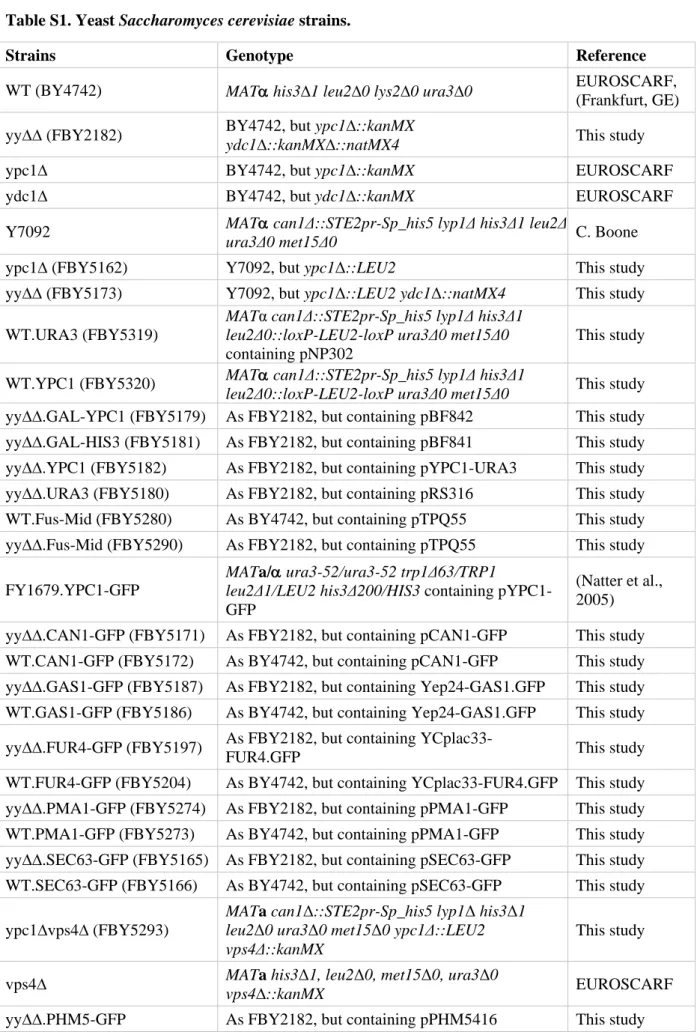
Table S1. Yeast Saccharomyces cerevisiae strains. 157
Strains Genotype Reference
WT (BY4742) MATα his3∆1 leu2∆0 lys2∆0 ura3∆0 EUROSCARF, (Frankfurt, GE) yy∆∆ (FBY2182) BY4742, but ypc1∆::kanMX
ydc1∆::kanMX∆::natMX4 This study
ypc1∆ BY4742, but ypc1∆::kanMX EUROSCARF
ydc1∆ BY4742, but ydc1∆::kanMX EUROSCARF
Y7092 MATura3Δ0 met15Δ0 α can1Δ::STE2pr-Sp_his5 lyp1Δ his3Δ1 leu2Δ C. Boone
ypc1∆ (FBY5162) Y7092, but ypc1Δ::LEU2 This study
yy∆∆ (FBY5173) Y7092, but ypc1Δ::LEU2 ydc1Δ::natMX4 This study WT.URA3 (FBY5319)
MATα can1Δ::STE2pr-Sp_his5 lyp1Δ his3Δ1 leu2Δ0::loxP-LEU2-loxP ura3Δ0 met15Δ0 containing pNP302
This study
WT.YPC1 (FBY5320) MATleu2Δ0::loxP-LEU2-loxP ura3Δ0 met15Δ0 α can1Δ::STE2pr-Sp_his5 lyp1Δ his3Δ1 This study yy∆∆.GAL-YPC1 (FBY5179) As FBY2182, but containing pBF842 This study yy∆∆.GAL-HIS3 (FBY5181) As FBY2182, but containing pBF841 This study yy∆∆.YPC1 (FBY5182) As FBY2182, but containing pYPC1-URA3 This study yy∆∆.URA3 (FBY5180) As FBY2182, but containing pRS316 This study WT.Fus-Mid (FBY5280) As BY4742, but containing pTPQ55 This study yy∆∆.Fus-Mid (FBY5290) As FBY2182, but containing pTPQ55 This study FY1679.YPC1-GFP
MATa/α ura3-52/ura3-52 trp1Δ63/TRP1
leu2Δ1/LEU2 his3Δ200/HIS3 containing pYPC1-GFP
(Natter et al., 2005) yy∆∆.CAN1-GFP (FBY5171) As FBY2182, but containing pCAN1-GFP This study WT.CAN1-GFP (FBY5172) As BY4742, but containing pCAN1-GFP This study yy∆∆.GAS1-GFP (FBY5187) As FBY2182, but containing Yep24-GAS1.GFP This study WT.GAS1-GFP (FBY5186) As BY4742, but containing Yep24-GAS1.GFP This study yy∆∆.FUR4-GFP (FBY5197) As FBY2182, but containing
YCplac33-FUR4.GFP This study
WT.FUR4-GFP (FBY5204) As BY4742, but containing YCplac33-FUR4.GFP This study yy∆∆.PMA1-GFP (FBY5274) As FBY2182, but containing pPMA1-GFP This study WT.PMA1-GFP (FBY5273) As BY4742, but containing pPMA1-GFP This study yy∆∆.SEC63-GFP (FBY5165) As FBY2182, but containing pSEC63-GFP This study WT.SEC63-GFP (FBY5166) As BY4742, but containing pSEC63-GFP This study ypc1∆vps4∆ (FBY5293)
MATa can1Δ::STE2pr-Sp_his5 lyp1Δ his3Δ1 leu2Δ0 ura3Δ0 met15Δ0 ypc1Δ::LEU2
vps4Δ::kanMX This study
vps4∆ MATa his3vps4∆::kanMX Δ1, leu2Δ0, met15Δ0, ura3Δ0 EUROSCARF yy∆∆.PHM5-GFP As FBY2182, but containing pPHM5416 This study
WT.PHM5-GFP As BY4742, but containing pPHM5416 This study yy∆∆.SNC1-GFP As FBY2182, but containing pGS416-SNC1 This study WT.SNC1-GFP As BY4742, but containing pGS416-SNC1-GFP This study yy∆∆.SSO1-GFP As FBY2182, but containing pSSO1416-GFP This study WT.SSO1-GFP As BY4742, but containing pSSO1416-GFP This study yy∆∆.STE2-GFP As FBY2182, but containing pSTE2416 This study WT.STE2-GFP As BY4742, but containing pSTE2416 This study yy∆∆.mtGFP (FBY5184) As FBY2182, but containing pYES-mtGFP This study WT.mtGFP (FBY5183) As BY4742, but containing pYES-mtGFP This study yy∆∆.VPH1-GFP (FBY5169) As FBY2182, but containing pVPH1-GFP This study WT.VPH1-GFP (FBY5170) As BY4742, but containing pVPH1-GFP This study yy∆∆.SEC7-RFP (FBY5193) As FBY2182, but containing pTQ128 This study WT.SEC7-RFP (FBY5195) As BY4742, but containing pTQ128 This study yy∆∆.SED5-GFP (FBY5201) As FBY2182, but containing pSED5-GFP This study WT.SED5-GFP (FBY5202) As BY4742, but containing pSED5-GFP This study YPK9 MATa ade2-101ochre his3-∆200 leu2-∆1
lys2-801amber trp1-∆63 ura3-52
(Jiang et al., 1998) 1∆.YPC1 YPK9 lag1∆::TRP1 containing pPK183 This study 2∆.YPC1 YPK9 lag1∆::TRP1 lac1∆::URA3 containing
pPK183
(Jiang et al., 2004) FBY7478 MAT a/α his3Δ1/his3Δ1, leu2Δ0/leu2Δ0,
lys2Δ0/LYS2, ura3Δ0/ura3Δ0, met15Δ0/met15Δ0, LYP1/lyp1Δ, CAN1/can1Δ::STE2pr-Sp_his5 FBY7479 MAT a/αhis3Δ1/his3Δ1, leu2Δ0/leu2Δ0,
lys2Δ0/LYS2, ura3Δ0/ura3Δ0, met15Δ0/met15Δ0, LYP1/lyp1Δ, CAN1/can1Δ::STE2pr-Sp_his5 ypc1::kanMX4/YPC1
ydc1::kanMX4::natMX/YDC1
This study
FBY7480 MAT a/α his3Δ1/his3Δ1, leu2Δ0/LEU2,
lys2Δ0/LYS2, ura3Δ0/ura3Δ0, met15Δ0/met15Δ0, LYP1/lyp1Δ, CAN1/can1Δ::STE2pr-Sp_his5, ypc1::kanMX4/ypc1::kanMX::ura3::LEU2, ydc1::kanMX4::natMX/ydc1::natMX This study 158 159
160
Table S2. Plasmids. 161
pNP302 CEN ARS URA3, ADH1 promoter C. De Virgilio
pYPC1-URA3 YPC1 in pNP302 This study
pBF841 2µ HIS3, GAL promoter N. Ramachandra
pBF842 YPC1 in pBF841 N. Ramachandra
pSTE2416 STE2-GFP in pRS416 CEN URA3, TPI1 promoter F. Reggiori
pPHM5416 GFP-PHM5 in pRS416 CEN URA3, TPI1 promoter (Reggiori and Pelham 2001)
pSSO1416 GFP-SSO1 in pRS416 CEN URA3, TPI1 promoter F. Reggiori
pGS416-SNC1 GFP-SNC1 in pRS416 CEN URA3, TPI1 promoter (Lewis et al., 2000) H. Pelham
pSED5-GFP GFP-SED5CENURA3
http://www2.brc.riken.jp/cache/dna/8658 A. Nakano pTQ128 SEC7-DsRed in CEN LEU2, ADH1 promoter K. Simons pYES-mtGFP mtGFP in 2µ URA3, GAL promoter (Westermann and
Neupert, 2000)
pVPH1-GFP VPH1-GFP CEN URA3 R. Schneiter
pSEC63-GFP SEC63-GFP in 2µ URA3 R. Schneiter
pCAN1-GFP CAN1-GFP in 2µ URA3, ADH1 promoter W. Tanner pFUR4-GFP YCplac33-FUR4-GFP CEN URA3, endogenous promoter W. Tanner pPMA1-GFP PMA1-GFP CEN URA3, endogenous promoter R.Schneiter YEp24-GAS1.GFP GAS1-GFP in 2µ URA3, endogenous promoter L. Popolo pTPQ55 Fus-Mid-GFP in CEN URA3, GAL promoter K. Simons
pYPC1-GFP YPC1-GFP in pRS416 URA3 TEF1 promoter (Natter et al., 2005) pPK183 YPC1 in 2µ with endogenous promoter LEU2 (Jiang et al., 2004) 162 163 Supplemental references 164 165 166
Baggett, JJ, Shaw, JD, Sciambi, CJ, Watson, HA, Wendland, B (2003) Fluorescent labeling of
167
yeast. Curr Protoc Cell Biol, Chapter 4: Unit
4.13.1-4.13.28
168
Bagnat, M, Keranen, S, Shevchenko, A, Shevchenko, A, Simons, K (2000) Lipid rafts function
169
in biosynthetic delivery of proteins to the cell surface in yeast. Proc Natl Acad Sci U S A, 97:
170
3254-3259
171
Collins, SR, Roguev, A, Krogan, NJ (2010) Quantitative genetic interaction mapping using the
172
E-MAP approach. Methods Enzymol, 470: 205-231
173
Dirmeier, R, O'Brien, KM, Engle, M, Dodd, A, Spears, E, Poyton, RO (2002) Exposure of yeast
174
cells to anoxia induces transient oxidative stress. Implications for the induction of hypoxic
175
genes. J Biol Chem, 277: 34773-34784
176
Dittmar, JC, Reid, RJ, Rothstein, R (2010) ScreenMill: a freely available software suite for
177
growth measurement, analysis and visualization of high-throughput screen data. BMC
178
Bioinformatics, 11: 353
179Folch, J, Lees, M, GH, SS (1957) A simple method for the isolation and purification of total
lipides from animal tissues. J Biol Chem, 226: 497-509
181
Hanson, BA, Lester, RL (1980) The extraction of inositol-containing phospholipids and
182
phosphatidylcholine from Saccharomyces cerevisiae and Neurospora crassa. J Lipid Res, 21:
183
309-315
184
Jiang, JC, Kirchman, PA, Allen, M, Jazwinski, SM (2004) Suppressor analysis points to the
185
subtle role of the LAG1 ceramide synthase gene in determining yeast longevity. Exp
186
Gerontol, 39: 999-1009
187Jiang, JC, Kirchman, PA, Zagulski, M, Hunt, J, Jazwinski, SM (1998) Homologs of the yeast
188
longevity gene LAG1 in Caenorhabditis elegans and human. Genome Res, 8: 1259-1272
189
Lewis, MJ, Nichols, BJ, Prescianotto-Baschong, C, Riezman, H, Pelham, HR (2000) Specific
190
retrieval of the exocytic SNARE Snc1p from early yeast endosomes. Mol Biol Cell, 11: 23-38
191
Malinska, K, Malinsky, J, Opekarova, M, Tanner, W (2004) Distribution of Can1p into stable
192
domains reflects lateral protein segregation within the plasma membrane of living S.
193
cerevisiae cells. J Cell Sci, 117: 6031-6041
194
Natter, K, Leitner, P, Faschinger, A, Wolinski, H, McCraith, S, Fields, S, Kohlwein, SD (2005)
195
The spatial organization of lipid synthesis in the yeast Saccharomyces cerevisiae derived from
196
large scale green fluorescent protein tagging and high resolution microscopy. Mol Cell
197
Proteomics, 4: 662-672
198Reggiori, F, Pelham, HR (2001) Sorting of proteins into multivesicular bodies:
ubiquitin-199
dependent and -independent targeting. EMBO J, 20: 5176-5186
200
Westermann, B, Neupert, W (2000) Mitochondria-targeted green fluorescent proteins:
201
convenient tools for the study of organelle biogenesis in Saccharomyces cerevisiae. Yeast, 16:
202
1421-1427
203 204 205