ORIGINAL ARTICLE
Skeletogenesis and sequence heterochrony in rodent
evolution, with particular emphasis on the African striped
mouse,
Rhabdomys pumilio (Mammalia)
Laura A. B. Wilson&Carsten Schradin& Christian Mitgutsch&Fernando C. Galliari& Andrea Mess&Marcelo R. Sánchez-Villagra
Received: 19 September 2009 / Accepted: 20 January 2010 / Published online: 9 March 2010 # Gesellschaft für Biologische Systematik 2010
Abstract Data documenting skeletal development in rodents, the most species-rich ‘order’ of mammals, are at present restricted to a few model species, a shortcoming that hinders exploration of the morphological and ecolog-ical diversification of the group. In this study we provide the most comprehensive sampling of rodent ossification sequences to date, with the aim of exploring whether
heterochrony is ubiquitous in rodent evolution at the onset of skeletal formation. The onset of ossification in 17 cranial elements and 24 postcranial elements was examined for eight muroid and caviomorph rodent species. New data are provided for two non-model species. For one of these, the African striped mouse, Rhabdomys pumilio, sampling was extended by studying 53 autopodial elements and examin-ing intraspecific variation. The Parsimov method of studying sequence heterochrony was used to explore the role that changes in developmental timing play in early skeletal formation. Few heterochronies were found to diagnose the muroid and caviomorph clades, suggesting conserved patterning in skeletal development. Mechanisms leading to the generation of the wide range of morpholog-ical diversity encapsulated within Rodentia may be restrict-ed to later periods in development than those studirestrict-ed in this work. Documentation of skeletogenesis in Rhabdomys indicates that intraspecifc variation in ossification sequence pattern is present, though not extensive. Our study suggests that sequence heterochrony is neither pivotal nor prevalent during early skeletal formation in rodents.
Keywords Rodent . Heterochrony . Rhabdomys . Skeletogenesis . Development . Intraspecific variation
Introduction
The clade Rodentia at present contains 2,277 members representing almost half of all living mammalian species (Wilson and Reeder2005). There is a relative dearth of data on skeletal development across mammalian clades, and the available information is limited to a few model species (e.g. the house mouse, Mus musculus; Kaufman 2008). Since
Electronic supplementary material The online version of this article (doi:10.1007/s13127-010-0020-4) contains supplementary material, which is available to authorised users.
L. A. B. Wilson (*)
:
C. Mitgutsch:
M. R. Sánchez-Villagra Paläontologisches Institut und Museum,Karl-Schmid-Strasse 4, 8006 Zürich, Switzerland e-mail: laura.wilson@pim.uzh.ch C. Schradin
Zoological Institute, Department of Animal Behaviour, University of Zurich,
Winterthurerstrasse 190, 8057 Zürich, Switzerland C. Schradin
School of Animal, Plant and Environmental Sciences, University of the Witwatersrand,
Private Bag 3, Wits 2050, Johannesburg, South Africa F. C. Galliari
Departamento Científico de Paleontología de Vertebrados, Facultad de Ciencias Naturales y Museo,
Universidad Nacional de la Plata, La Plata, Argentina
A. Mess
Museum für Naturkunde, Leibniz-Institut für Evolutions-und Biodiversitätsforschung an der Humboldt-Universität zu Berlin, 10115 Berlin, Germany
Org Divers Evol (2010) 10:243–258 DOI 10.1007/s13127-010-0020-4
Rodentia is species-rich compared to other mammalian ‘orders’, the documentation of rodent skeletal development is most markedly understudied. This shortcoming restricts studies exploring the developmental basis for the mor-phological and ecological diversification of this group, which has proven a rich avenue of research examining other aspects of prenatal development (Kavanagh et al.
2007).
Extant rodents encapsulate an array of differing traits in terms of ecology, life history, body size and locomotory habits. With the exception of Antarctica, rodents inhabit all continents and play fundamental roles in a multitude of ecosystems. Rodentia presents an ideal mammalian group in which to examine the developmental basis associated with organismal diversity (Michaux et al.2008; Monteiro et al. 2005; Roth 1996). Indeed, within the rodent sample studied here, there are representatives from two clades characterized by contrasting attributes. On the one hand, caviomorph rodents comprise relatively few species (fewer than 13% of rodents; Wilson and Reeder2005) and yet this South American radiation includes species that differ in body size by several orders of magnitude (Nowak 1999), and possess numerous different adaptations to locomotory style (Weisbecker and Schmid 2007). On the other hand, the species-rich muroid clade (1,517 species; Carleton and Musser2005) contains members that show a comparatively diminished level of anatomical diversity (Steppan et al.
2004).
One process that may be involved in the evolution of morphological traits is heterochrony (Zelditch 2001). Heterochrony, derived from classical approaches to the study of ontogeny and phylogeny (Gould 1977) and subsequently formalized (Alberch et al. 1979), refers to a change in the timing and rate of development. The approach of sequence heterochrony (sensu Smith 2001) provides a methodology to study changes in the timing of developmental events that are not explicitly characterized by size and shape, two parameters that previously governed study for the majority of classical models of heterochrony (Klingenberg1998). By considering events as discrete and sequentially occurring, the problems inherent to comparing a diverse range of species using size and time are obviated. Studies of heterochronies in ossification sequences at the marsupial/placental dichotomy (e.g. Sánchez-Villagra2002; Sánchez-Villagra et al. 2008; Sears 2009; Smith 1997; Weisbecker et al.2008) have yielded critical support for the conclusions from previous comparative studies of morpho-logical diversity in these two clades, that are characterized by extreme differences in life history, especially gestation length (e.g. Lillegraven1975; Sears2004). Within Roden-tia, postnatal sequence heterochrony examined for cranial suture closure patterning, a late time window in skeletal development, has been reported to play a role in shaping
the diversity present among members of the hystricognath clade, which includes South American caviomorphs and African phiomorphs (Wilson and Sánchez-Villagra 2009). Additionally, several studies have concentrated upon the role of growth heterochrony in rodent evolution, using morphometric approaches (e.g. Creighton and Strauss1986; Hautier et al.2008,2009; Monteiro et al.2005; Wilson and Sánchez-Villagra2010; Zelditch et al.2006).
In this study we provide the most comprehensive sampling of rodent ossification sequences to date, with the aim of exploring whether heterochrony is ubiquitous in rodent evolution at the onset of skeletal formation. In doing so, we also help address the problem of the scarcity of data for skeletogenesis in non-model rodents. We present a detailed study of the African striped mouse, Rhabdomys pumilio, a small (∼50 g) murid rodent with a widespread distribution in southern Africa (Nowak 1999). Found in many habitats, including grassland, desert and forests (Schradin 2005), R. pumilio attains approximately twice the body mass of the house mouse, Mus musculus. Moreover, unlike the latter model organism, R. pumilio is diurnal, thus lends itself to easy direct study in its natural habitat (Schradin 2006). Paternal care and communal breeding characterize the desert-living striped mice (Schradin and Pillay 2003, 2004). In addition, we provide the first ossification-sequence data for the degu, Octodon degus, a small to medium-sized, diurnal, semi-fossorial and herbivo-rous caviomorph rodent common to the central region of Chile (Mess2007; Rojas et al.1982). Studies of communal breeding in O. degus have shown that this rodent lives in groups with polygamous social structure (e.g. Ebensperger et al. 2004). Usually found in open areas, frequently close to human habitation, the degu forms extended family groups and creates complex burrow systems (Fischer1940; Woods and Boraker1975).
Material and methods Data collection
To the previously largest data set of cranial and postcranial ossification sequences (Sánchez-Villagra et al. 2008; Weisbecker et al. 2008), we add data on postcranial ossification for the degu (Octodon degus), on cranial ossification for the guinea pig (Cavia porcellus), and on cranial, postcranial and autopodial ossification for the African striped mouse (Rhabdomys pumilio). We examined both prenatal and postnatal specimens, and coded the onset of ossification for 17 cranial and 24 postcranial elements, and also for 53 autopodial elements in R. pumilio (26 manus and 27 pes elements). The cranial data set comprised seven rodent species, including five muroids and two
caviomorphs. For two species the ossification sequences were examined from specimens collected for this study; data for the remaining five species were taken from the literature. The analysis also included data from the literature for three outgroup species belonging to the Euarchonto-glires clade (Tables1,2and 3).
Evolutionary relationships among rodent species exam-ined here (Fig.1) were based upon the molecular studies of Steppan et al. (2004) and Blanga-Kanfi et al. (2009). Outgroup species relations were reconstructed from Bininda-Emonds et al. (2007).
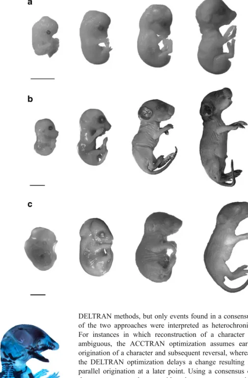
We also improve the resolution of ossification sequences for several species previously documented by Sánchez-Villagra et al. (2008) and Weisbecker et al. (2008), using a combination of additional literature sources and specimens held in the collections at the Paläontologisches Institut und Museum, Universität Zürich, Switzerland. The specimens of Cavia porcellus (Fig. 2a) and Rhabdomys pumilio (Fig.2b) were obtained from breeding colonies maintained for research at the Universität Bielefeld (Germany) and the Universität Zürich, respectively (Schradin 2006; Trillmich et al. 2003). The founder individuals of R. pumilio originated from the Geogap Nature Reserve in South Africa (29°41.56’S, 18°1.60’E). The specimens of Octodon degus (Fig. 2c) were part of the personal collection of Andrea Mess and are now deposited at the Universität Zürich.
All specimens obtained for this study were prepared using a modified version of standard enzymatic clearing and double staining (Prochel 2006) (Fig. 3). Onset of ossification for each element was recorded based upon the earliest uptake of alizarin red. Although data for several sequences of ossification were recorded from published literature, which detailed differing preparation procedures, all embryos for a given species were prepared using the
same protocol; hence the resulting ossification sequences are considered to be accurate.
Intraspecific variation in Rhabdomys pumilio
The sample of R. pumilio contained multiple specimens from several litters. We used this opportunity to examine intraspecific variation in ossification-event sequences, studies of which are rare, and so far restricted to a limited number of taxa (e.g. Colbert and Rowe 2008; Garn and Rohmann 1960; Mabee et al.2000; Moore and Townsend
2003). Out of the 61 specimens studied, 55 could be reliably assigned to ten litters as collected at time of death. Each litter contained between three and seven animals. Variation was assessed among siblings within each litter, for each subdivision of the skeleton: cranial, postcranial and autopodial. The latter approach contrasts with that for the ontogenetic sequence of ossification for R. pumilio, which was compiled by comparison of individuals across all litters. For each element within a subdivision, we compared the ossification state (ossified or not ossified) across all pups within a litter. We noted the number of litters in which we found a discrepancy in the ossification state of an element. To assess how intraspecific variability (in ossifi-cation state) affected the determination of the ossifiossifi-cation sequence, we used the quantification methods of Mabee et al. (2000), whereby we calculated the magnitude of rank difference, i.e. the degree to which a bone varied in position in the complete ossification sequence. Rank differences were calculated for all elements in each of the cranial, postcranial and autopodial sequences. The magnitude of rank difference is defined as the number of steps between the maximum and minimum position of an individual bone. We compared variation in position of a bone in relation to a
Table 1 Sources of data for ossification sequences used in this study
Taxon Common name N References Rhabdomys pumilio (Sparrman) African striped mouse 61 / 12 present study Rattus norvegicus (Berkenhout) Norway rat n.a. / 14 Strong (1925) Meriones unguiculatus (Milne-Edwards) Mongolian gerbil 187 / 8 Yukawa et al. (1999)
Peromyscus melanophrys (Coues) Plateau mouse 13 / 5 Sánchez-Villagra et al. (2008), Weisbecker et al. (2008) Mesocricetus auratus (Waterhouse) Golden hamster 168 / 8 Beyerlein et al. (1951)
Mus musculus Linnaeus House mouse n.a. / 7 Theiler (1972), Kaufman (2008) Cavia porcellus (Linnaeus) Guinea pig n.a. / 12 Petri (1935), present study Octodon degus (Molina) Degu 8 / 5 present study
Tupaia javanica Horsfield* Horsfield’s treeshrew 24 / 6 Nunn and Smith (1998), Zeller (1987), Goswami (2007) Tarsius spectrum Pallas* Spectral tarsier 21 / 6 Nunn and Smith (1998)
Homo sapiens Linnaeus* Human 60 / 15 Mall (1906), Davies and Parsons (1927), Weisbecker et al. (2008)
Asterisks (*) denote outgroup species N = number of specimens / stages examined
Table 2 Cranial events ranked according to relative timing of onset of ossification, based on observations from specimens and summaries compiled from the literature
Tarsius Homo Tupaia Rattus Mus Peromyscus Meriones Mesocricetus Rhabdomys Cavia
Premaxilla 1 2 1 2 1 1 1 2 2 1 Maxilla 1 1 1 1 1 1 2 1 2 1 Palatine 2 4 4 2 1 2 3 2 1 1 Dentary ? 1 1 1 1 1 1 1 1 1 Frontal 2 3 2 1 1 2 1 2 1 1 Parietal 3 3 2 2 1 2 4 2 1 1 Squamosal 2 3 3 2 3 4 4 2 6 1 Basioccipital 6 6 4 2 1 2 2 3 3 2 Nasal ? 4 4 3 3 4 3 3 5 2 Pterygoid ? 4 5 2 1 2 2 ? 1 3 Exoccipital 4 3 3 2 1 3 3 2 4 3 Basisphenoid 6 8 6 4 3 3 4 2 7 4 Jugal 2 3 2 3 2 4 4 5 8 1 Lacrimal 3 7 4 3 4 4 4 6 9 3 Alisphenoid 5 5 4 5 1 4 4 4 10 1 Orbitosphenoid ? 7 5 6 5 5 5 7 11 5 Periotic 7 4 7 ? 6 5 ? ? 12 5
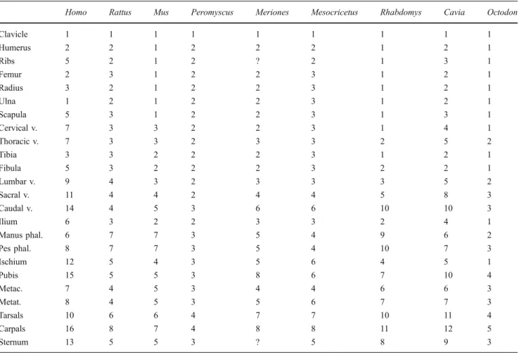
Table 3 Postcranial events ranked according to relative timing of onset of ossification, based on observations from specimens and summaries compiled from the literature
Homo Rattus Mus Peromyscus Meriones Mesocricetus Rhabdomys Cavia Octodon
Clavicle 1 1 1 1 1 1 1 1 1 Humerus 2 2 1 2 2 2 1 2 1 Ribs 5 2 1 2 ? 2 1 3 1 Femur 2 3 1 2 2 3 1 2 1 Radius 3 2 1 2 2 3 1 2 1 Ulna 1 2 1 2 2 3 1 2 1 Scapula 5 3 1 2 2 3 1 3 1 Cervical v. 7 3 3 2 2 3 1 4 1 Thoracic v. 7 3 3 2 3 3 2 5 2 Tibia 3 3 2 2 2 3 1 2 1 Fibula 5 3 2 2 2 3 2 2 1 Lumbar v. 9 4 3 2 3 3 3 5 2 Sacral v. 11 4 4 2 4 4 5 8 3 Caudal v. 14 4 5 3 6 6 10 10 3 Ilium 6 3 2 2 3 3 2 4 1 Manus phal. 6 7 7 3 5 4 9 6 2 Pes phal. 8 7 7 3 5 4 10 7 3 Ischium 12 5 4 3 5 6 4 5 1 Pubis 15 5 5 3 8 6 7 10 4 Metac. 7 4 5 3 4 4 6 6 3 Metat. 8 4 5 3 5 6 7 7 3 Tarsals 10 6 6 4 7 7 10 11 4 Carpals 16 8 7 4 8 8 11 12 5 Sternum 13 5 5 3 ? 5 8 9 3
common sequence, which was derived by determining the state (ossified or not ossified) for each element to be that shared by the majority of animals in the litter. For example, if a litter contained four animals, and if three of these displayed an ossified jugal whereas one did not, we determined that for the stage represented by that litter, the jugal was ossified. To generate a common sequence, we then compiled the information across litters.
When deriving ossification sequences for the cranial and postcranial partitions used for Parsimov analyses (Jeffery et al. 2005) and for the autopodial sequence, we took the common, or majority, pattern to be representative of the stage a given litter documented (Mabee et al.2000). Analysis of variation in ossification sequence
To examine levels of variation in sequence of a particular ossification event we standardized each rank within a sequence by expressing it as a fraction of the total number of ranks (rmax) for a given species, such that the rank
sequence for each species falls within the range between 1/rmax and 1. By scaling ranks in this manner we removed
differences in maximum rank between species consequent from differing levels of resolution between species; none-theless, the small differences in contribution to variance associated with different rmaxvalues remain inherent to this
methodology. To express variability of a particular element in the ossification sequence, we computed for each cranial and each postcranial bone, from the scaled rank values, a range in rank variation across species examined.
The frequency distribution of ranks, for each of the cranial and postcranial data sets, was calculated, in order to examine
the distribution of ossification events within the rank sequence. Maximum documented rank for each species was regressed against number of specimens studied, to test the validity of the expectation that total number of specimens studied does not instigate differences in resolution of ranks. Event pairing and Parsimov analyses
Two separate data matrices were constructed: one each for the postcranial and cranial data sets. We separated cranial and postcranial data sets, as did previous authors (e.g. Sánchez-Villagra et al. 2008; Weisbecker et al. 2008), because (A) those are two recognized modules, and (B) the resolution and availability of data for these two sets are different (Tables 2, 3). Because we have separated the skeletal regions, we cannot rule out heterochrony occurring between the cranial and postcranial modules. To identify heterochronies within the sequence of ossification, the timing of each ossification event was assessed by compar-ing the relative timcompar-ing of pairs of elements. For each data set an event-pair matrix was produced in which non-redundant pairs of events were compared: each event was coded as having occurred either earlier than (score 0), simultaneous with (1), or later than (2) each other event. The postcranial data matrix contained 276 event pairs, the cranial matrix 136 event pairs. Following the reconstruction of apomorphic character-state changes using PAUP 4.0b10 (Swofford 2002), we analyzed event-pair data using the computer program Parsimov, which reconstructs the least number of event movements that may explain all possible event-pair changes along a given branch (Jeffery et al.2005). Optimizations were performed using both ACCTRAN and
Fig. 1 Phylogenetic relation-ships among the species includ-ed in this study, reconstructinclud-ed from Steppan et al. (2004), Bininda-Emonds et al. (2007), and Blanga-Kanfi et al. (2009)
DELTRAN methods, but only events found in a consensus of the two approaches were interpreted as heterochronic. For instances in which reconstruction of a character is ambiguous, the ACCTRAN optimization assumes early origination of a character and subsequent reversal, whereas the DELTRAN optimization delays a change resulting in parallel origination at a later point. Using a consensus of these two approaches provides the most conservative estimate of heterochronic shifts present in the non-ambiguous events common to both.
Results
Event pairing and Parsimov analyses
For the cranial data set (Table2), a total of 136 event pairs yielded 79 (58.1%) parsimony-informative pairs and 57 (41.8%) variable pairs that did not provide any informative
Fig. 3 Cleared and double-stained rodents prepared at the Paläonto-logisches Institut, Zürich. Left: Octodon degus. Right: Rhabdomys pumilio. Scale: 2 mm
Fig. 2 A sample of ontogenetic series prepared for this study at the Paläontologisches Institut und Museum, Zürich. a Cavia porcellus. b Rhabdomys pumi-lio. c Octodon degus. Scales: 2 mm
signal, including 14 pairs (10.2% of total) that were autapomorphic. These autapomorphies include the parietal ossifying before the premaxilla and maxilla in R. pumilio, and in C. porcellus the pterygoid ossifying after the squamosal and basioccipital, and the jugal before the basioccipital.
Of the 276 event pairs in the postcranial data matrix (Table 3), 135 (48.9%) were phylogenetically informative, 84 (30.4%) were variable, and 57 pairs (20.7%) possessed the same character state across all species examined. In caviomorphs the metatarsals ossified after the ischium, whereas the manual phalanges ossified before the sacral vertebrae. Among the phylogenetically uninformative event pairs, an autapomorphy concerning the forelimb was distinguishable for Rattus norvegicus: both the radius and ulna ossified before the femur. In C. porcellus the metacarpals ossified before the sacral vertebrae, and the ribs after the humerus.
The consensus of ACCTRAN and DELTRAN trans-formations indicates that two early shifts occur within the Muroidea clade, one each in the cranial and postcranial skeleton (Table 4). Few heterochronies distinguished different species of muroids: those for R. pumilio involve late movements of the sternum and maxilla (Table4). The clade consisting of M. musculus and R. pumilio is characterized by an early heterochronic shift of the fibula in relation to the femur and scapula, and twinned movement of the basisphenoid and orbitosphenoid (Table4). Octodon degus is characterized by several autapomorphic character shifts: six out of seven (85.7%) relate to the postcranium, and specifically three of these six (50%; 42.8% of total) involve the vertebral column (Table4).
Skeletogenesis in Rhabdomys pumilio
Rhabdomys pumilio displays a sequence of ossification in cranial elements that is similar to that of other muroid
species studied (Table2), namely ossification of the cranio-facial bones and bones in the palatal region followed by the cranial base and then elements associated with the otic region, and lastly the periotic bone. The largest number of tied ossification events occurs in the earliest stage, with five out of 17 elements (29%) ossifying first: the palatine, dentary, frontal, parietal and pterygoid. The pattern of earliest events having the least resolution is shared with Cavia porcellus and Mus musculus; the remaining four species (57%) exhibit three or fewer ossification events occurring at the earliest moment (Table2).
The postcranial ossification sequence indicates that R. pumilio is characterized by a late ossification of the caudal vertebrae; this region is the penultimate postcranial element to ossify. Similar to Rattus norvegicus, ossification of the pubis in R. pumilio occurs earlier within the sequence than in the other rodents studied here, with this element ossifying at position seven out of eleven in R. pumilio, and at position five out of eight in R. norvegicus (Table3). In R. pumilio the first elements to ossify in the postcranium account for nine out of 24 events (38%). Octodon degus and Mus musculus share a similar concentration of relative simultaneity for the earliest events, whereas in the remain-ing five rodent species (62.5%) the clavicle ossifies first, and a greater number of events ossify in subsequent stages (Table 3).
Both the manus and pes follow similar ossification patterns concerning the spatial autopodial region (Table5), except that the proximal phalanges begin ossification before the distal phalanges in the hand, though in both the hand and foot, distal and proximal phalanges begin to ossify before their intermediate counterparts (Figs. 4, 5). In all autopods the middle metapodials are the first elements to start ossification. The capitatum (distal carpal III) starts to ossify before the trapezoid (distal carpal II) and the trapezium (distal carpal I), and the pisiform starts to ossify
Clade Event Movement … in relation to … Muroidea Nasal early Squamosal, Basioccipital
Ribs early Femur, Radius Rhabdomys Sternum late Pubis, Metatarsals
Maxilla late Palatine, Dentary, Frontal, Parietal Rhabdomys+Mus Basisphenoid twins Orbitosphenoid
Fibula late Femur, Scapula Octodon Cervical v. early Humerus, Ribs, Fibula
Sacral v. early Pes phal., Metatarsals
Caudal v. late Sacral v., Pes phal., Metatarsals, Sternum Ilium early Humerus, Ribs
Manus phal. early Thoracic v., Lumbar v. Metacarpals late Pes phal., Metatarsals Pterygoid early Premaxilla, Dentary, Jugal Table 4 Detailed
heterochro-nies for non-model species (R. pumilio and O. degus) analyzed in this study, and major clades
before the capitatum. Among the carpals, the triquetrum and the hamatum (distal carpals IV and V) consistently are the first two carpals to begin ossification (simultaneously). Metatarsal I is the last metatarsal to ossify, and it does so before any of the distal tarsals. In the hand, ossification of metacarpal I partially follows the same regime, also starting ossification later than all other metacarpals, but not before distal carpal ossification begins (Table5). Indeed, the distal carpals are the last elements to begin ossification in the hand, whereas in the foot the navicular is the last to commence ossification (Figs.4,5and 6).
Several heterochronic events characterize R. pumilio: late movement of the sternum in relation to the pubis and metatarsals, and late movement of the maxilla in relation to the palatine, dentary, frontal and parietal (Table4). Rhabd-omys pumilio and Mus musculus share twinned movement of the basisphenoid and orbitosphenoid, and late movement of the fibula in relation to the femur and scapula (Table4).
Intraspecific variation in Rhabdomys pumilio
Intraspecific variation in element ossification is present in all partitions of the skeleton (cranial, postcranial, autopo-dial). In all ten litters examined, the cranial and postcranial skeletal areas exhibit the most variation. Within the postcranial skeleton, animals in eight out of ten litters displayed variation. Hence, within each of these eight litters at least one of the 24 postcranial elements displayed an ossification state (ossified or not ossified) opposite to that displayed by the other animals in the litter. Intraspecific variation is found in six out of ten litters for the 17 cranial elements examined. Because the autopodial region contains more than twice the number of elements (53 in total) than either the postcranial or cranial skeletal areas, by sheer chance it would be more probable for intraspecific variation to be detected in this region; therefore, the reported variability among specimens in seven out of ten litters is reduced compared to the postcranial and cranial regions.
The number of different elements that vary intraspecifi-cally is low, with not more than three elements exhibiting variation among members of a litter. Among the cranial elements variation is restricted to the premaxilla, nasal, exoccipital, squamosal and basioccipital, with an average of one element out of 17 (5.8%) exhibiting variation among specimens belonging to a given litter. On average two out of 24 (8.3%) postcranial elements vary between pups from the same litter. Autopodial elements in the manus that exhibit variation in ossification are the metacarpals I, III and IV and the distal phalanx V; in the pes these are the medial phalanx V and metatarsals II, III and IV. When coupled together, variation in the manus and pes is found on average for two out of 53 elements per litter (3.7%).
The effect of intraspecific variation upon ossification sequence was determined using the rank magnitude metric of Mabee et al. (2000); see Appendices 1 and 2. For the cranial ossification sequence (Appendix 1), intraspecific variation resulted in a change in sequence position for nine out of 17 elements (52.9%), though in each case this was restricted to a magnitude rank change of 1. The postcranial results indicate that twelve out of 24 elements (50.0%) altered their position in the ossification sequence, with ten of these exhibiting a rank magnitude difference of 1, and the remaining two (pubis, lumbar vertebrae) a rank difference of 2. For the autopodial region, ten out of 26 elements (38.5%) in the hand, and twelve out of 27 (44.4%) in the foot, exhibited a variation in position (Appendix 2). Similar to the cranial and postcranial data sets, the majority of elements that moved position in the autopodial sequen-ces moved by only one place (Appendix 2). The exceptions concern the pisiform in the manus and metacarpal V, and the astragalus in the pes, each of which exhibited a magnitude or rank variation of 2.
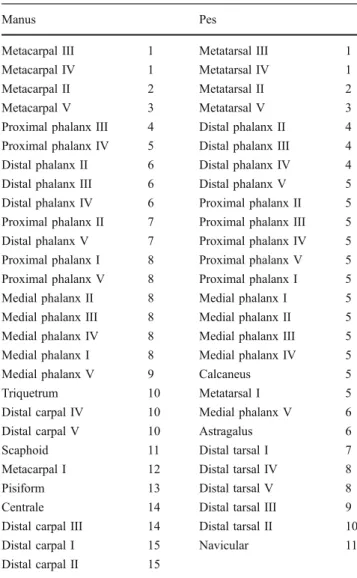
Table 5 Ranked sequences of ossification in the autopodial region of the African striped mouse, Rhabdomys pumilio
Manus Pes
Metacarpal III 1 Metatarsal III 1 Metacarpal IV 1 Metatarsal IV 1 Metacarpal II 2 Metatarsal II 2 Metacarpal V 3 Metatarsal V 3 Proximal phalanx III 4 Distal phalanx II 4 Proximal phalanx IV 5 Distal phalanx III 4 Distal phalanx II 6 Distal phalanx IV 4 Distal phalanx III 6 Distal phalanx V 5 Distal phalanx IV 6 Proximal phalanx II 5 Proximal phalanx II 7 Proximal phalanx III 5 Distal phalanx V 7 Proximal phalanx IV 5 Proximal phalanx I 8 Proximal phalanx V 5 Proximal phalanx V 8 Proximal phalanx I 5 Medial phalanx II 8 Medial phalanx I 5 Medial phalanx III 8 Medial phalanx II 5 Medial phalanx IV 8 Medial phalanx III 5 Medial phalanx I 8 Medial phalanx IV 5 Medial phalanx V 9 Calcaneus 5 Triquetrum 10 Metatarsal I 5 Distal carpal IV 10 Medial phalanx V 6 Distal carpal V 10 Astragalus 6 Scaphoid 11 Distal tarsal I 7 Metacarpal I 12 Distal tarsal IV 8 Pisiform 13 Distal tarsal V 8 Centrale 14 Distal tarsal III 9 Distal carpal III 14 Distal tarsal II 10 Distal carpal I 15 Navicular 11 Distal carpal II 15
Rank variability among rodents
For all rodents studied here, the elements in the cranial ossification sequences that exhibit the highest amount of variation in rank position are the parietal and alisphenoid, followed by the squamosal and jugal. The least variable elements within an ossification sequence are the dentary and the periotic, the latter being the last bone to ossify in all rodents examined (Table 2, Fig. 7a). In the postcranium, both the ischium and the manual phalanges were found to be most variable in position, whereas the carpals, sternum and clavicle differed least in rank position (Table 2, Fig. 7b). Across all species studied, the number of ranks extends to a maximum of twelve, and event distributions for
cranial and postcranial elements indicate that ossification-event occurrences are predominant in the earliest stages of a sequence (Fig. 8). In cranial elements, the frequency of ossification events is highest at the first stage (35; 30.4%) and progressively decreases throughout later stages. By comparison, in postcranial elements the second position in the sequence is characterized by the highest number of ossification-event occurrences (42; 23.4%), and the later stages of the sequence show increased variation in frequency of events (stages 7–12; Fig.8) compared to elements within a cranial sequence. The number of ranks in a sequence, for a given species taken from maximum value, was not signifi-cantly correlated with the number of specimens available for study; hence, resolution in a sequence is neither directly
Fig. 4 Ossification of elements of the left hand (respective up-per row) and foot (lower row), in dorsal views, of Rhabdomys pumilio; black = ossified, grey = calcified. Number of specimens examined (N)=1, except at stages a (N=16), f (6), g (2), n (9), y (4), and z (2)
improved nor reduced as a consequence of sample size (r= 0.248, p=0.553).
Discussion
Sequence heterochrony
Our results indicate that heterochrony does not play a pivotal role in early skeletal formation in rodents, based on comparisons between six muroid and two caviomorph
species. The lack of heterochrony recorded here for prenatal and early postnatal stages may indicate that heterochrony is more prominent in rodent evolution at later postnatal stages of growth (Wilson and Sánchez-Villagra2009).
We detected few shared heterochronic shifts in the cranial ossification sequence (Table4). Only the early ossification of the nasal in relation to the squamosal and basiocciptal characterizes Muroidea. Similar to the results presented herein, Sánchez-Villagra et al. (2008), in their study of mammalian cranial ossification patterns, also reported few shared shifts while, as in the study by Bininda-Emonds et al. (2003) on mammalian development, several autapomorphic shifts were documented. The lack of shared heterochronic shifts in the species examined in the present study further supports previous findings of conservatism in relative timing of vertebrate cranial developmental events (Schoch2006).
Similarly, postcranial sequence heterochronies reported here are restricted to a single event for the Muroidea clade (Table 4). Weisbecker et al. (2008) found Muroidea to be characterized by early ossification of the scapula and cervical vertebrae in relation to the forelimb and hindlimb elements, based upon the same species of rodent studied here, except O. degus, but using a lizard and alligator outgroup. Our results do not mirror the findings of Weisbecker et al. (2008) but instead yield a shared shift for Muroidea involving an early movement of the ribs in relation to the femur and radius (Table4). A similar earlier movement of the ribs was recorded by Sánchez-Villagra (2002) for R. norvegicus and M. auratus, although the feature was not shared as a synapomorphy between these
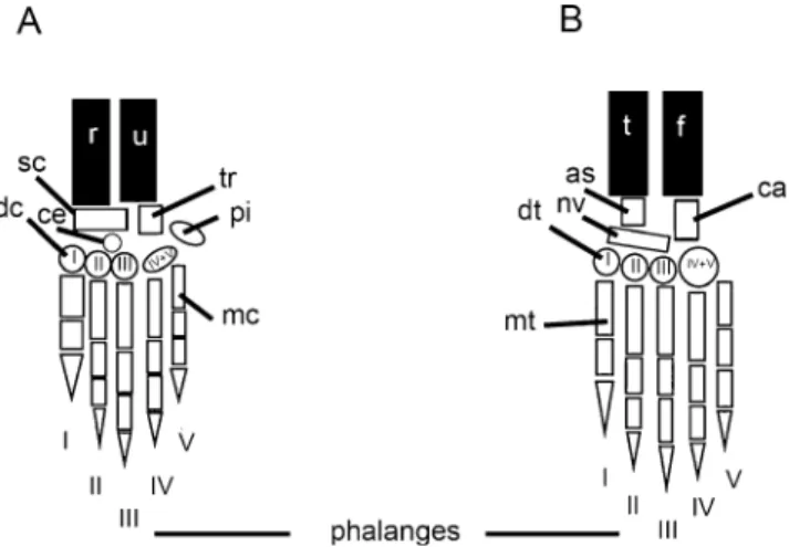
Fig. 5 Autopodial elements of the left hand a and foot b, in dorsal views, of Rhabdomys pumilio. Elements: as = astragalus, ca = calcaneus, ce= centrale, dc = distal carpal, dt = distal tarsal, f = fibula, mc = metacarpal, mt = metatarsal, nv = navicular, pi = pisiform, r = radius, sc = scaphoid, t = tibia, tr = triquetrum, u = ulna
Fig. 6 Camera lucida drawings of the right hand (shown at left) and foot, in dorsal views, of Rhabdomys pumilio. Autopodial elements: as = astragalus, ca = calcaneus, ce = centrale, dc = distal carpal, dt = distal tarsal, f = fibula, mc = metacar-pal, mt = metatarsal, nv = navicular, pi = pisiform, r = radius, sc = scaphoid, u = ulna, t = tibia, tr = triquetrum. Scales: 2 mm
two taxa. The discrepancies between our results and those of Weisbecker et al. (2008) and Sánchez-Villagra (2002) may be explained by the our relatively expanded rodent sampling, particularly its inclusion of another non-muroid species (O. degus), and by the use of more closely related non-rodent outgroups, both of which act to improve the resolution of rodent-specific shifts. Two heterochronic shifts in postcranial ossification sequence were found to characterize caviomorphs: the late ossification of metatar-sals in relation to the ischium, and early ossification of the manual phalanges in relation to the sacral vertebrae. Several autapomorphic events are also reported, including those for
Octodon that relate to early ossification of elements in the posterior skeleton, including the cervical and sacral vertebrae, and the ilium (Table 4). A lack of distinguishable hetero-chronies between the caviomorphs and the muroids may corroborate a recent finding that changes in postnatal growth pattern were equally common during the evolutionary history of both clades (Wilson and Sánchez-Villagra2010).
The considerable number of autapomorphic shifts found for the species studied here, and the comparative lack of sequence heterochronies towards the root of the phylogeny, are features in common with previous results produced with the Parsimov algorithm (Harrison and Larsson 2008), and
Fig. 7 Adjusted rank range plots of cranial a and postcranial b elements for rodents examined in this study
have been discussed before, along with issues of character non-independence (Schulmeister and Wheeler 2004). Ad-ditionally, our approach of inferring heterochronies com-mon to the consensus of ACCTRAN and DELTRAN is also highly conservative, and may further restrict the identi-fication of heterochronic shifts in our data, an issue previously acknowledged to characterize this method (e.g. Sánchez-Villagra et al. 2008; Werneburg and Sánchez-Villagra2009). Hence, in the light of methodolog-ical limitations and also considering the limited taxon sampling resulting from the difficulty of obtaining develop-mental sequences for any mammal, the role of sequence heterochrony in rodent evolution cannot be dismissed conclusively. Our study, however, uses a similar conservative approach to quantification of heterochrony comparable to that carried out in recent studies of mammals on a similar time window of development (e.g. Sánchez-Villagra et al.2008; Weisbecker et al.2008) and on embryology of the external morphology (Werneburg and Sánchez-Villagra 2010). The pattern of limited heterochrony we found in rodents does not deviate from that reported in those previous studies. Autopodial ossification
As is the case in almost all placentals, and in contrast to marsupials examined to date (Prochel and Sánchez-Villagra
2003; Prochel et al. 2004), the capitatum (distal carpal III) of R. pumilio begins ossification before the trapezoid (distal carpal II) and the trapezium (distal carpal I) (Figs. 4, 5).
Rhabdomys pumilio also displays early ossification of the pisiform in relation to the capitatum: this event has been reported by Prochel et al. (2004) to characterize Rodentia. Among the carpals, the triquetrum and the hamatum (distal carpals IV and V) consistently are the first two carpals to start ossification (simultaneously). Within the rodents examined, these two elements develop to be the largest carpals in adulthood, adding support to the notion, proposed by Huxley (1932), that the timing of initiation of an organ in the embryo is related to its size attained at adulthood. The general sequence of manual element ossification for R. pumilio follows that reported for M. musculus (Prochel et al.
2004), in that ossification begins with the humerus, radia and ulna, after which metacarpal III ossifies, followed sequentially by proximal phalanx III, distal phalanx II and medial phalanx III (Table 5). This sequence is a slight departure from that reported for C. porcellus (by Petri1935), R. norvegicus (Strong1925) and M. auratus (Beyerlein et al.
1951), where the distal phalanx begins ossification before the proximal phalanx in the aforementioned pattern. Variability in rank
In their study of mammalian cranial ossification patterns, Sánchez-Villagra et al. (2008) found the most variable cranial elements in an ossification sequence to be the basioccipital, basisphenoid, jugal, parietal, pterygoid and squamosal. In addition to the rodents examined here, the data set of Sánchez-Villagra et al. (2008) included repre-sentatives from a number of other clades, including primates, Chiroptera and Soricidae. In comparison, we find the basioccipital to display reduced difference in ossification rank across the rodent species examined, but the squamosal, jugal and basisphenoid all exhibit relatively high levels of rank variability, consistent with the previous findings.
Because the non-rodent species examined by Sánchez-Villagra et al. (2008) represented more than 85% of the sampled species, and since their analysis also encapsu-lated a greater phylogenetic breadth than that presented here, particularly with more distantly related outgroup taxa, such as turtles, it is highly likely that these factors contributed to mask the aforementioned rodent-specific patterns of rank variability identified herein. The ali-sphenoid displays the second-highest level of variation in this study, whereas Sánchez-Villagra et al. (2008) do not report elevated variation for this element. The amplified variation in alisphenoid ossification found here is the consequence of the early ossification reported for C. porcellus and M. musculus (at position one in the sequence) compared to the remaining rodents in the study sample (position four, or later) (Table2). Although neither of these two species has fewer ranks in the ossification sequence compared to the other species sampled, both
Fig. 8 Frequency variation plot of cranial and postcranial ossifying events for rodents examined in this study
species do show an increased frequency of events ossifying in the first stage and, hence, reduced resolution at the earliest point of development. This suggests that the pattern may be an artefact attributable to an issue of inadequate resolution that could be remedied by obtaining specimens covering a finer sampling of ontogenetic stages. Elements displaying the least variability in the present study are not confined exclusively to previously reported modules of the mammalian cranium (Goswami
2006). The premaxilla, orbitosphenoid and periotic are found in spatially different areas of the cranium, and other elements contained within the corresponding modules exhibit increased lability. For example, although the periotic does not vary in sequence position in the rodents studied, the other elements defined by Goswami (2006) to belong to the cranial base module (basioccipital, basisphenoid and exoccipital) exhibit variability comparable to that in elements associated with other modules (Fig.7a). While the aim here is not to explicitly test modularity within sequence heterochrony (Goswami et al.2009), the labile nature of the cranial elements we present for species within one clade of mammals suggests that developmental timing can be less integrated than previously assumed (Schoch2006).
Variability in the postcranium is highest for the ischium and both manual and pedal phalanges, and lowest for the clavicle, carpals and sternum (Fig.7b). In agreement with Prochel (2006) and Weisbecker et al. (2008) we find an early ossification of anterior postcranial elements (Table3), and elements of high and low variability are shared with those reported in the former study. The variation we find in the manual and pedal phalanges can be attributed to the late ossification of these elements in R. pumilio, R. norvegicus and M. musculus. Compared to the other species studied, these murids show earliest ossification of phalanges beginning at the second-to-last position in the ossification sequence, whereas among the remaining species sampled ossification of phalanges begins as early as the second stage (O. degus) and not later than within the first two-thirds of all events (Table3).
Intraspecific variation in ossification sequence
The incidence and extent of intraspecific variation in the ossification sequence of R. pumilio is low and mainly restricted to a few elements per region of the skeleton examined. Similar to work completed on bony fishes, we find that elements involved in intraspecific variation are not exclusively concentrated towards the end of the ossification sequence, a feature that has been cited to suggest an extension or truncation to a linear ancestral ontogeny (Mabee et al.2000). Indeed, for the autopods variation is mostly found in the first elements to begin ossification: the metacarpals and metatarsals (Figs. 5, 6; Table 5). In
contrast, Garn and Rohmann (1960) and Garn et al.
(1961a,1961,1966), in their studies of the appendicular
and axial skeleton of humans, reported variability in the ossification sequence in the hands and feet to be concentrated to the elements ossifying last; a similar situation was reported by Alberch and Blanco (1996) for sequence variation in the European fire salamander, Salamandra salamandra. Nevertheless, consistent with the present findings for R. pumilio, the above studies also documented a comparatively low overall level of intraspe-cific variation.
One might expect intraspecific variation to parallel interspecific variation to the exclusion of non-variable elements that, thus, may exhibit functionally related units reflecting modular components within the skeleton, or tightly integrated subsets (Goswami et al. 2009; Wagner
1988). We find, however, that elements involved in cases of intraspecific variation did not consistently exhibit the highest levels of interspecific variability. Variation in ossification sequence, like variation in any feature, can be explained by genetic and/or environmental factors (Wagner
1988). Indeed, Garn et al. (1966) proposed the former to provide a strong basis for variations in the human appendicular skeleton. Nevertheless, the particular influence of the genotype, and also that of environmental variables in relation to ossification sequence variations, remains unclear, which impedes development of a direct causal explanation for the variation in R. pumilio reported here.
The results of the rank-magnitude calculations
(Appendices 1 and 2) indicate that variation in sequence
position was generally low; none of the variable elements changed position by more than two ranks. The low intraspe-cific variation in R. pumilio is not surprising, especially since the sample size (55 individuals) for our study was approximately one quarter that of earlier studies, which also reported low levels of intraspecific variability (e.g. Alberch and Blanco 1996; Moore1991). Nonetheless, recent works have demonstrated and underlined that it is imperative to consider patterns of embryonic variability when examining developmental time (Colbert and Rowe2008; de Jong et al.
2009). Consideration of this notion opens a suite of questions in relation to the evolution of developmental sequences. For instance, comparisons between natural and experimentally controlled populations may help identify the respective functions of heritable variation and of environmentally induced developmental variation in the generation of intraspecific variation.
Conclusions
The present examination of ossification sequences, based upon the largest data set so far compiled for rodents,
suggests that sequence heterochrony is neither pivotal nor prevalent during early skeletal formation in this clade. Documentation of skeletogenesis in R. pumilio indicates that intraspecifc variation in ossification-sequence pattern is present, though not extensive. Further extension of the ossification data on rodents, especially the collection of information on non-model organisms, will assist our understanding of how important sequence changes are as a raw material for evolution in this species-rich clade of mammals.
Acknowledgements We thank Fritz Trillmich for kindly providing specimens of Cavia porcellus for use in this study. This work received support from the Swiss National Fond (3100A0-116013) to M.R.S.-V., a Forschungskredit (nr. 3771) from the Universität Zürich to L.A.B. W., and from the Consejo de Investigaciones Científicas y Técnicas in Argentina to F.C.G. For insightful comments that helped to improve an earlier version of this paper, we thank Olaf R. P. Bininda-Emonds and two anonymous reviewers.
Appendix 1
Intraspecific variation in ossification of cranial and post-cranial elements in Rhabdomys pumilio
Cranial element Magnitude of rank variation Postcranial element Magnitude of rank variation Premaxilla 1 Clavicle 0 Maxilla 1 Humerus 0 Palatine 0 Ribs 0 Dentary 0 Femur 0 Frontal 0 Radius 0 Parietal 0 Ulna 0 Squamosal 0 Scapula 0 Basioccipital 0 Cervical v. 0 Nasal 1 Thoracic v. 1 Pterygoid 0 Tibia 0 Exoccipital 1 Fibula 1 Basisphenoid 1 Lumbar v. 2 Jugal 1 Sacral v. 1 Lacrimal 1 Caudal v. 1 Alisphenoid 1 Ilium 1 Orbitosphenoid 1 Manus phal. 1 Periotic 0 Pes phal. 1 Ischium 1 Pubis 2 Metac. 0 Metat. 1 Tarsals 1 Carpals 0 Sternum 0 Appendix 2
Intraspecific variation in ossification of autopodial elements in Rhabdomys pumilio Manus Magnitude of rank variation Pes Magnitude of rank variation Metacarpal III 0 Metatarsal III 1 Metacarpal IV 0 Metatarsal IV 1 Metacarpal II 1 Metatarsal II 1 Metacarpal V 1 Metatarsal V 2 Proximal phal. III 0 Distal phal. II 1 Proximal phal. IV 0 Distal phal. III 1 Distal phal. II 1 Distal phal. IV 1 Distal phal. III 1 Distal phal. V 0 Distal phal. IV 1 Proximal phal. II 0 Proximal phal. II 1 Proximal phal. III 0 Distal phal. V 1 Proximal phal. IV 0 Proximal phal. I 0 Proximal phal. V 0 Proximal phal. V 0 Proximal phal. I 0 Medial phal. II 1 Medial phal. I 0 Medial phal. III 0 Medial phal. II 0 Medial phal. IV 0 Medial phal. III 0 Medial phal. I 0 Medial phal. IV 1 Medial phal. V 0 Calcaneus 1 Triquetrum 1 Metatarsal I 1 Distal carpal IV 0 Medial phal. V 0 Distal carpal V 0 Astragalus 2 Scaphoid 0 Distal tarsal I 0 Metacarpal I 0 Distal tarsal IV 0 Pisiform 2 Distal tarsal V 0 Centrale 0 Distal tarsal III 0 Distal carpal III 0 Distal tarsal II 0 Distal carpal I 0 Navicular 2 Distal carpal II 0
References
Alberch, P., & Blanco, M. J. (1996). Evolutionary patterns in ontogenetic transformation: from laws to regularities. Interna-tional Journal of Developmental Biology, 40, 845–858. Alberch, P., Gould, S. J., Oster, G. F., & Wake, D. B. (1979). Size and
shape in ontogeny and phylogeny. Palaeobiology, 5, 296–317. Beyerlein, L., Hillemann, H. H., & Van Arsdel, W. C., III. (1951).
Ossification and calcification from postnatal day eight to the adult condition in the golden hamster (Cricetus auratus). Anatomical Record, 111, 49–65.
Bininda-Emonds, O. R. P., Cardillo, M., Jones, K. E., MacPhee, R. D. E., Beck, R. M. D., Grenyer, R., et al. (2007). The delayed rise of present-day mammals. Nature, 446, 507–512.
Bininda-Emonds, O. R. P., Jeffery, J. E., & Richardson, M. K. (2003). Is sequence heterochrony an important evolutionary mechanism in mammals? Journal of Mammalian Evolution, 10, 335–361. Blanga-Kanfi, S., Miranda, H., Penn, O., Pupko, T., DeBry, R. W., &
Huchon, D. (2009). Rodent phylogeny revised: analysis of six nuclear genes from all major rodent clades. BMC Evolutionary Biology, 9, 71. doi:10.1186/1471-2148-9-71.
Carleton, M., & Musser, G. (2005). Order Rodentia. In D. W. Wilson & D. M. Reeder (Eds.), Mammal species of the world (pp. 745– 752). Washington, D.C.: Smithsonian Institution Press. Colbert, M. W., & Rowe, T. (2008). Ontogenetic sequence analysis:
using parsimony to characterize developmental sequences and sequence polymorphism. Journal of Experimental Zoology, 310B, 398–416.
Creighton, G. K., & Strauss, R. E. (1986). Comparative patterns of growth and development in cricetine rodents and the evolution of ontogeny. Evolution, 40, 94–106.
Davies, D. A., & Parsons, F. G. (1927). The age order of the appearance and union of the normal epiphyses as seen by X-rays. Journal of Anatomy, 62, 58–71.
Ebensperger, L. A., Hurtado, M. J., Soto-Gamboa, M., Lacey, E. A., & Chang, A. T. (2004). Communal nesting and kinship in degus (Octodon degus). Naturwissenschaften, 91, 391–395.
Fischer, G. M. (1940). Contribución a la anatomia de los Octodonti-dos. Boletín del Museo Nacional de Historia Natural (Santiago de Chile), 18, 103–124.
Garn, S. M., & Rohmann, C. G. (1960). Variability in the order of ossification of the bony centers of the hand and wrist. American Journal of Physical Anthropology, 18, 219–230.
Garn, S. M., Rohmann, C. G., & Apfelbaum, B. (1961a). Complete epiphyseal union of the hand. American Journal of Physical Anthropology, 19, 365–372.
Garn, S. M., Rohmann, C. G., & Wallace, D. K. (1961). Association between alternate sequences of hand-wrist ossification. American Journal of Physical Anthropology, 19, 361–364.
Garn, S. M., Rohmann, C. G., & Blumenthal, T. (1966). Ossification sequence polymorphism and sexual dimorphism in skeletal devel-opment. American Journal of Physical Anthropology, 24, 101–115. Goswami, A. (2006). Cranial modularity shifts during mammalian
evolution. American Naturalist, 168, 270–280.
Goswami, A. (2007). Modularity and sequence heterochrony in the mammalian skull. Evolution & Development, 9, 291–299. Goswami, A., Weisbecker, V., & Sánchez-Villagra, M. R. (2009).
Developmental modularity and the marsupial-placental dichoto-my. Journal of Experimental Zoology, 312B, 186–195. Gould, S. J. (1977). Ontogeny and phylogeny. Cambridge, MA: Belknap. Harrison, L. B., & Larsson, H. C. E. (2008). Estimating evolution of temporal sequence changes: a practical approach to inferring ancestral developmental sequences and sequence heterochrony. Systematic Biology, 57, 378–387.
Hautier, L., Fabre, P. H., & Michaux, J. (2009). Mandible shape and dwarfism in squirrels (Mammalia, Rodentia): interaction of allometry and adaptation. Naturwissenschaften, 96, 725–730. Hautier, L., Michaux, J., Marivaux, L., & Vianey-Liaud, M. (2008).
Evolution of the zygomasseteric construction in Rodentia, as revealed by a geometric morphometric analysis of the mandible of Graphiurus (Rodentia, Gliridae). Zoological Journal of the Linnean Society, 154, 807–821.
Huxley, J. S. (1932). Problems of relative growth. London: Methuen. Jeffery, J. E., Bininda-Emonds, O. R. P., Coates, M. I., & Richardson, M. K. (2005). A new technique for identifying sequence heterochrony. Systematic Biology, 54, 230–240.
de Jong, I. M. L., Colbert, M. W., Witte, F., & Richardson, M. K. (2009). Polymorphism in developmental timing: intraspecific heterochrony in a Lake Victoria cichlid. Evolution & Develop-ment, 11, 625–635.
Kaufman, M. H. (2008). The atlas of mouse development. London: Elsevier Academic Press.
Kavanagh, K. D., Evans, A. R., & Jernvall, J. (2007). Predicting evolutionary patterns of mammalian teeth from development. Nature, 449, 427–433.
Klingenberg, C. P. (1998). Heterochrony and allometry: the analysis of evolutionary change in ontogeny. Biological Reviews, 73, 79–123. Lillegraven, J. A. (1975). Biological considerations of the
marsupial-placental dichotomy. Evolution, 29, 707–722.
Mabee, P. M., Olmstead, K. L., & Cubbage, C. C. (2000). An experimental study of intraspecific variation, developmental timing, and heterochrony in fishes. Evolution, 54, 2091–2106. Mall, F. P. (1906). On ossification centres in human embryos less than
one hundred days old. American Journal of Anatomy: Develop-mental Dynamics, 5, 433–458.
Mess, A. (2007). Development of the chorioallantoic placenta in Octodon degus—A model for growth processes in caviomorph rodents? Journal of Experimental Zoology, 308B, 371–383. Michaux, J., Hautier, L., Simonin, T., & Vianey-Liaud, M. (2008).
Phylogeny, adaptation and mandible shape in Sciuridae (Roden-tia, Mammalia). Mammalia, 72, 286–296.
Monteiro, L. R., Bonato, V., & dos Reis, S. F. (2005). Evolutionary integration and morphological diversification in complex mor-phological structures: mandible shape divergence in spiny rats (Rodentia, Echimyidae). Evolution & Development, 7, 429–439. Moore, M. K. (1991). Comparative ontogeny of cranial ossification in the spotted salamander, Ambystoma maculatum, and the tailed frog, Ascaphus truei. M. Sc. thesis. Baton Rouge, LA: Louisiana State University.
Moore, M. K., & Townsend, V. R., Jr. (2003). Intraspecific variation in cranial ossification in the tailed frog, Ascaphus truei. Journal of Herpetology, 37, 714–717.
Nowak, R. M. (1999). Walker’s mammals of the world (6th ed.). Baltimore, MD: Johns Hopkins University Press.
Nunn, C. L., & Smith, K. K. (1998). Statistical analyses of developmental sequences: the craniofacial region in marsupial and placental mammals. American Naturalist, 152, 82–101. Petri, C. (1935). Die Skelettentwicklung beim Meerschwein, zugleich
ein Beitrag zur vergleichenden Anatomie der Skelettentwicklung der Säuger. Vierteljahrsschrift der Naturforschenden Gesellschaft in Zürich, 80, 157–240.
Prochel, J. (2006). Early skeletal development in Talpa europaea, the common European mole. Zoological Science, 23, 427–434. Prochel, J., & Sánchez-Villagra, M. R. (2003). Carpal ontogeny in
Monodelphis domestica and Caluromys philander (Marsupialia). Zoology, 106, 73–84.
Prochel, J., Vogel, P., & Sánchez-Villagra, M. R. (2004). Hand development and sequence of ossification in the forelimb of the European shrew Crocidura russula (Soricidae) and comparisons across therian mammals. Journal of Anatomy, 205, 99–111. Rojas, M. A., Montenegro, M. A., & Morales, B. (1982). Embryonic
development of the degu, Octodon degus. Journal of Reproduc-tion and Fertility, 66, 31–38.
Roth, V. L. (1996). Cranial integration in the Sciuridae. American Zoologist, 36, 14–23.
Sánchez-Villagra, M. R. (2002). Comparative patterns of postcranial ontogeny in therian mammals: an analysis of relative timing of ossification events. Journal of Experimental Zoology: Molecular Development Evolution, 294, 264–273.
Sánchez-Villagra, M. R., Goswami, A., Weisbecker, V., Mock, O., & Kuratani, S. (2008). Conserved relative timing of cranial ossification patterns in early mammalian evolution. Evolution & Development, 10, 519–530.
Schoch, R. R. (2006). Skull ontogeny: developmental patterns of fish conserved across major tetrapod clades. Evolution & Develop-ment, 8, 524–536.
Schradin, C. (2005). When to live alone and when to live in groups: ecological determinants of sociality in the African striped mouse (Rhabdomys pumilio, Sparrman, 1784). Belgian Journal of Zoology, 135(Suppl. 1), 77–82.
Schradin, C. (2006). Whole-day follows of striped mice (Rhabdomys pumilio), a diurnal murid rodent. Journal of Ethology, 24, 37–43. Schradin, C., & Pillay, N. (2003). Paternal care in the social and diurnal striped mouse (Rhabdomys pumilio): laboratory and field evidence. Journal of Comparative Psychology, 117, 317–324. Schradin, C., & Pillay, N. (2004). The striped mouse (Rhabdomys
pumilio) from the succulent karoo of South Africa: A territorial group living solitary forager with communal breeding and helpers at the nest. Journal of Comparative Psychology, 118, 37–47. Schulmeister, S., & Wheeler, W. C. (2004). Comparative and
phylogenetic analysis of developmental sequences. Evolution & Development, 6, 50–57.
Sears, K. E. (2004). Constraints on the morphological evolution of marsupial shoulder girdles. Evolution, 58, 2353–2370.
Sears, K. E. (2009). Differences in the timing of prechondrogenic limb development in mammals: the marsupial-placental dichotomy resolved. Evolution, 63, 2193–2200.
Smith, K. K. (1997). Comparative patterns of craniofacial develop-ment in eutherian and metatherian mammals. Evolution, 51, 1663–1678.
Smith, K. K. (2001). Heterochrony revisited: the evolution of developmental sequences. Biological Journal of the Linnean Society, 73, 169–186.
Steppan, S. J., Adkins, R. M., & Anderson, J. (2004). Phylogeny and divergence date estimates of rapid radiations in muroid rodents based on multiple nuclear genes. Systematic Biology, 53, 533– 553.
Strong, R. M. (1925). The order, time, and rate of ossification of the albino rat (Mus norvegicus albinus) skeleton. Amererican Journal of Anatomy: Developmental Dynamics, 36, 313–355. Swofford, D. L. (2002). PAUP, Phylogenetic Analysis Using
Parsi-mony (and other methods), version 4.0b10. Computer program. Sunderland, MA: Sinauer Associates.
Theiler, K. (1972). The House mouse: development and normal stages from fertilization to 4 weeks of age. Berlin: Springer.
Trillmich, F., Bieneck, M., Geissler, E., & Bischof, H.-J. (2003). Ontogeny of running performance in the Wild guinea pig (Cavia aperea). Mammalian Biology, 68, 214–223.
Wagner, G. P. (1988). The influence of variation and of developmental constraints on the rate of multivariate phenotypic evolution. Journal of Evolutionary Biology, 1, 45–66.
Weisbecker, V., Goswami, A., Wroe, S., & Sánchez-Villagra, M. R. (2008). Ossification heterochrony in the therian postcranial skeleton and the marsupial-placental dichotomy. Evolution, 62, 2027–2041.
Weisbecker, V., & Schmid, S. (2007). Autopodial skeletal diversity in hystricognath rodents: functional and phylogenetic aspects. Mammalian Biology, 72, 27–44.
Werneburg, I., & Sánchez-Villagra, M. R. (2009). Timing of organogenesis support basal position of turtles in the amniote tree of life. BMC Evolutionary Biology, 9, 82. doi: 10.1186/1471-2148-9-82.
Werneburg, I., & Sánchez-Villagra, M R. (2010). The early develop-ment of the Echidna, Tachyglossus aculeatus (Mammalia: Monotremata) and the grundmuster of mammalian development. Acta Zoologica. doi:10.1111/j.1463-6395.2009.00447.x. Wilson, D. E., & Reeder, D. M. (2005). Mammal species of the world:
a taxonomic and geographic reference. Washington, D.C.: Smithsonian Institution Press.
Wilson, L. A. B., & Sánchez-Villagra, M. R. (2009). Heterochrony and patterns of cranial suture closure in hystricognath rodents. Journal of Anatomy, 214, 339–354.
Wilson, L. A. B., & Sánchez-Villagra, M. R. (2010). Diversity trends and their ontogenetic basis: an exploration of allometric disparity in rodents. Proceedings of the Royal Society of London, B Biological Sciences, doi:10.1098/rspb.2009.1958
Woods, C. A., & Boraker, D. K. (1975). Octodon degus. Mammalian Species, 67, 1–5.
Yukawa, M., Hayashi, N., Takagi, K., & Mochizuki, K. (1999). The normal development of Mongolian gerbil foetuses and, in particular, the timing and sequence of ossification centres. Anatomia Histologia Embryologia, 28, 319–324.
Zelditch, M. L. (2001). Beyond heterochrony: the evolution of development. New York: Wiley-Liss Press.
Zelditch, M. L., Mezey, J., Sheets, H. D., Lundrigan, B. L., & Garland, T., Jr. (2006). Developmental regulation of skull morphology II: ontogenetic dynamics of covariance. Evolution & Development, 8, 46–60.
Zeller, U. (1987). Morphogenesis of the mammalian skull with special reference to Tupaia. Mammalia depicta, 13, 17–50.