Dynamic myosin phosphorylation regulates contractile
pulses and tissue integrity during epithelial morphogenesis
The MIT Faculty has made this article openly available.
Please share
how this access benefits you. Your story matters.
Citation
Vasquez, C. G., M. Tworoger, and A. C. Martin. “Dynamic Myosin
Phosphorylation Regulates Contractile Pulses and Tissue Integrity
During Epithelial Morphogenesis.” The Journal of Cell Biology 206,
no. 3 (August 4, 2014): 435–450.
As Published
http://dx.doi.org/10.1083/jcb.201402004
Publisher
Rockefeller University Press
Version
Final published version
Citable link
http://hdl.handle.net/1721.1/96347
Terms of Use
Creative Commons Attribution
The Rockefeller University Press $30.00
Correspondence to Adam C. Martin: acmartin@mit.edu
Abbreviations used in this paper: MBS, myosin-binding subunit; Myo-II, non-muscle myosin II; PP1c, protein phosphatase 1c; RLC, regulatory light chain; Rok, Rho-associated kinase.
Introduction
Epithelial morphogenesis is critical for organs and embryos to
change shape during development (Leptin, 2005; Heisenberg
and Bellaïche, 2013). A cell shape change that often
accom-panies epithelial remodeling is apical constriction, which
pro-motes epithelial sheet bending and cell invagination (Sawyer
et al., 2010; Martin and Goldstein, 2014). One example of
api-cal constriction–induced tissue remodeling occurs in Drosophila
melanogaster
gastrulation, when a group of 1,000 cells along
the ventral midline of the embryo undergoes apical constriction
to form a ventral furrow. These constricted cells invaginate to
become the mesoderm (Fig. 1 A; Leptin and Grunewald, 1990;
Sweeton et al., 1991). The molecular motor nonmuscle myosin
II (Myo-II), which localizes apically in Drosophila ventral
fur-row cells and other cell types that undergo apical constriction, is
thought to generate the contractile force that facilitates
invagina-tion (Young et al., 1991; Nance et al., 2003; Dawes-Hoang et al.,
2005; Hildebrand, 2005; Lee et al., 2006; Lee and Harland, 2007;
Nishimura and Takeichi, 2008; Martin et al., 2009). Although
Myo-II is clearly involved in apical constriction, the mechanism
of apical actomyosin contraction remains poorly understood.
Myo-II activity is regulated by phosphorylation of its
reg-ulatory light chain (RLC; sqh in Drosophila), which promotes
Myo-II oligomerization into minifilaments and activation of
the Myo-II motor ATPase activity (Karess et al., 1991; Sellers,
1991; Jordan and Karess, 1997). An effector of the RhoA
GTP-ase (Rho1 in Drosophila), Rho-associated coiled-coil kinGTP-ase (or
Rho-associated kinase [Rok] in Drosophila), can phosphorylate
and activate Myo-II via direct RLC phosphorylation or by
in-hibiting the phosphatase that dephosphorylates the RLC (myosin
phosphatase; Amano et al., 1996; Kimura et al., 1996; Kawano
et al., 1999; Winter et al., 2001). In the Drosophila mesoderm,
the transcription factors Twist and Snail are thought to promote
apical constriction and apical Myo-II accumulation by activating
Rho1 and its effectors, including Rok (Barrett et al., 1997; Häcker
and Perrimon, 1998; Dawes-Hoang et al., 2005;
Fig. S1 A
).
Mutants or chemical inhibition of Rok results in loss of apical
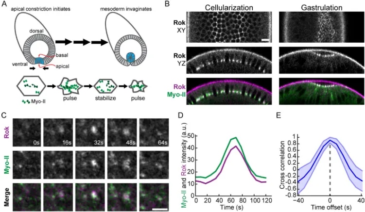
A
pical constriction is a cell shape change that
promotes epithelial bending. Activation of non
muscle myosin II (MyoII) by kinases such as Rho
associated kinase (Rok) is important to generate contrac
tile force during apical constriction. Cycles of MyoII
assembly and disassembly, or pulses, are associated
with apical constriction during Drosophila melanogaster
gastrulation. It is not understood whether MyoII phospho
regulation organizes contractile pulses or whether pulses
are important for tissue morphogenesis. Here, we show
that MyoII pulses are associated with pulses of apical Rok.
Mutants that mimic MyoII light chain phosphorylation or
depletion of myosin phosphatase inhibit MyoII contrac
tile pulses, disrupting both actomyosin coalescence into
apical foci and cycles of MyoII assembly/disassembly.
Thus, coupling dynamic MyoII phosphorylation to up
stream signals organizes contractile MyoII pulses in both
space and time. Mutants that mimic MyoII phosphoryla
tion undergo continuous, rather than incremental, apical
constriction. These mutants fail to maintain intercellular
actomyosin network connections during tissue invagina
tion, suggesting that MyoII pulses are required for tissue
integrity during morphogenesis.
Dynamic myosin phosphorylation regulates
contractile pulses and tissue integrity during
epithelial morphogenesis
Claudia G. Vasquez, Mike Tworoger, and Adam C. Martin
Department of Biology, Massachusetts Institute of Technology, Cambridge, MA 02142
© 2014 Vasquez et al. This article is distributed under the terms of an Attribution– Noncommercial–Share Alike–No Mirror Sites license for the first six months after the pub-lication date (see http://www.rupress.org/terms). After six months it is available under a Creative Commons License (Attribution–Noncommercial–Share Alike 3.0 Unported license, as described at http://creativecommons.org/licenses/by-nc-sa/3.0/).
THE
JOURNAL
OF
CELL
BIOLOGY
on August 13, 2014
jcb.rupress.org
Downloaded from
http://jcb.rupress.org/content/suppl/2014/08/04/jcb.201402004.DC2.html http://jcb.rupress.org/content/suppl/2014/07/31/jcb.201402004.DC1.html Supplemental Material can be found at:after a pulse is not stabilized, resulting in cell shape fluctuations
and inefficient constriction (Martin et al., 2009, 2010). Thus,
one model for ventral furrow formation is that cells undergo
ratch-eted apical constriction, whereby pulses drive constrictions that
are stabilized via a Twist-dependent mechanism for efficient
apical constriction. We proposed that the polarized localization
of Rok to medioapical foci results in the persistence of
medio-apical actomyosin fibers that stabilize cell shape between pulses;
we have named this cellular organization radial cell polarity
(Mason et al., 2013). The persistence of medioapical Myo-II
fibers is important for the formation of a supracellular Myo-II
meshwork that transmits tension across the ventral furrow
tis-sue (Martin et al., 2010). However, the roles of Rok and Myo-II
phosphorylation during pulsatile constriction are still unclear.
Furthermore, it is not known why cells undergo pulsatile rather
than continuous contraction to promote tissue morphogenesis.
Here, we combined live imaging and quantitative analysis
of Myo-II regulators and GFP-tagged Myo-II RLC
phosphomu-tants to define the role of coupling Myo-II activation to
up-stream signals during contractile pulses. We show that temporal
and spatial control of Myo-II phosphorylation organizes
con-tractile pulses. We suggest that the pulsatile nature of apical
constriction is required for the stable transmission of
inter-cellular forces during tissue morphogenesis.
Myo-II and lack of apical constriction, demonstrating that Rok
is necessary for cortical Myo-II localization in the ventral
fur-row (Dawes-Hoang et al., 2005; Mason et al., 2013). In
addi-tion, mutants that mimic Myo-II RLC phosphorylation progress
through development and suppress rok mutants, suggesting that
Rok increases levels of active Myo-II to promote actomyosin
contractility and morphogenesis (Jordan and Karess, 1997;
Winter et al., 2001; Royou et al., 2002). It is not clear, however,
whether coupling of Rok activity to Myo-II activation plays a
role in organizing apical actin network contraction.
We have developed a system to visualize the dynamics of
actomyosin contraction and apical constriction during
Drosoph-ila
gastrulation. In ventral furrow cells, Myo-II undergoes
cy-cles of assembly in the center of the apical surface (medioapical
cortex) followed by disassembly or remodeling, which we refer
to as pulses (Martin et al., 2009; Fig. 1 A). Phases of rapid
api-cal constriction are associated with pulses of Myo-II assembly
and the coalescence of Myo-II and F-actin structures into
medio-apical foci, which possibly represent contractions of the actin
cortex (Martin et al., 2009; Roh-Johnson et al., 2012). After
Myo-II coalescence, Myo-II structures remodel by either
disas-sembling or changing in morphology. Despite Myo-II
remodel-ing, cell shape is often stabilized between pulses, resulting in
incremental apical constriction. Although Myo-II still
under-goes pulsing in Twist knockdowns, the contracted cell shape
Figure 1. Rok colocalizes with Myo-II pulses. (A) Schematic of apical constriction of prospective mesodermal cells (shaded in blue) during Drosophila ven-tral furrow formation. (bottom) An individual cell undergoes pulsatile apical constriction. Myo-II undergoes cycles of assembly and coalescence (indicated by arrows) followed by remodeling. (B) Rok localizes with Myo-II at furrow canals during cellularization and apically (apical is up in YZ cross sections)
during ventral furrow formation. Bar, 10 µm. (C) Time-lapse images of Rok and Myo-II intensity during a pulse from rok2; Venus::Rok (WT); sqh::mCherry
embryo. Bar, 5 µm. (D) Myo-II and Rok appear simultaneously during pulse. Graph represents signal intensity of Rok and Myo-II within a single pulse event. (E) Rok signal is correlated with Myo-II signal with no temporal lag. Mean cross-correlation for different time offsets between Rok and Myo-II signals from
pulses (n = 30 pulses; shaded area is ±SD).
on August 13, 2014
jcb.rupress.org
Downloaded from
allele enabled us to examine the function of sqh
phosphomu-tants during gastrulation.
We first determined whether the different sqh
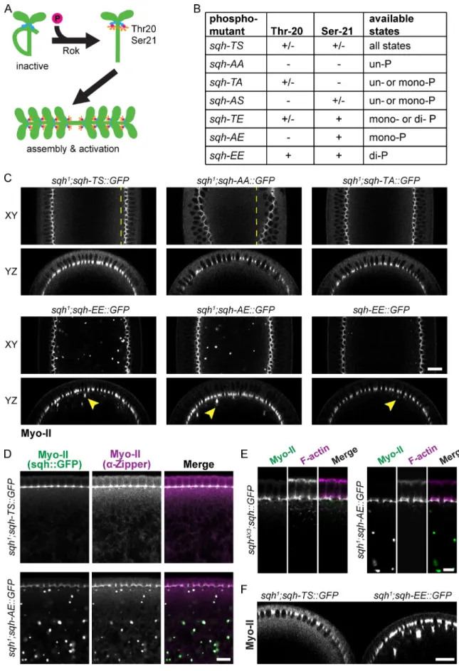
phosphomi-metic mutants activate Myo-II minifilament assembly in vivo.
During cellularization, Myo-II localizes to basal furrow canals
and cytoplasmic aggregates (Royou et al., 2004). Although we
did not observe ectopic cortical Myo-II assembly,
phosphomi-metic sqh mutants, particularly sqh-AE and sqh-EE,
accumu-lated Myo-II in aggregates that are immediately basal to the furrow
canals (Fig. 2 C and Fig. S1 E). Myo-II aggregates were reduced
in size when wild-type levels of endogenous Sqh were present
and Myo-II aggregates also contained the myosin heavy chain
(Drosophila zipper), suggesting that these aggregates are not
denatured protein (Fig. 2, C and D). Importantly, Sqh
aggre-gates do not contain F-actin, suggesting that the Sqh aggreaggre-gates
do not form by actomyosin contraction, but could result from
constitutive Myo-II minifilament assembly (Fig. 2 E). The
phos-phomimetic mutants resulted in a decrease in cytoplasmic Myo-II
levels relative to wild-type embryos, suggesting that RLC
mu-tants that mimic mono- or diphosphorylation shift the Myo-II
complex toward the active conformation in vivo, promoting
constitutive oligomerization (Fig. 2 F). Thus, our GFP-tagged
phosphomimetic RLC mutants appear to uncouple activation of
Myo-II oligomerization from phosphorylation by Rok.
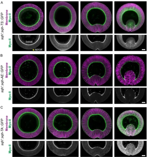
RLC phosphorylation dynamics do not trigger changes in Myo-II apical–basal localization
Apical activation of Rok has been proposed to localize Myo-II
assembly during apical constriction (Dawes-Hoang et al., 2005).
Thus, mutants that uncouple Myo-II activity from
phosphory-lation by Rok might disrupt the apical–basal localization of
Myo-II. Therefore, we tested whether the apical–basal polarity
of Myo-II localization is controlled by RLC phosphorylation and
oligomerization. Mutants that mimic mono- or
diphosphoryla-tion localized specifically to the apical surface of ventral furrow
cells, despite the presence of Myo-II aggregates throughout the
entire embryo during cellularization (Fig. 3, A and B; Fig. S2 B;
and
Video 2
). Furthermore, Myo-II was reduced in basal furrow
canals during ventral furrow invagination in phosphomimetic
mutants, similar to wild-type Myo-II, suggesting that
dephos-phorylation of the RLC is not the mechanism of basal Myo-II
loss (Fig. 3, A and B). Inactivation of the RLC phosphorylation
sites similarly did not disrupt apical Myo-II localization (Fig. 3 C,
Fig. S2 B, and Video 2). These results demonstrate that despite the
tissue-specific apical recruitment of Rok in ventral furrow cells,
regulation of oligomerization does not instruct apical Myo-II
localization at the onset of gastrulation.
Phosphomimetic RLC mutants disrupt polarized actomyosin condensation
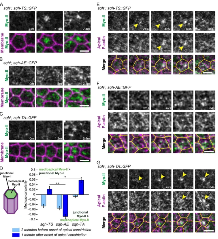
Medioapical Myo-II pulses involve the coalescence or
conden-sation of both Myo-II and F-actin into medioapical foci (Martin
et al., 2009; Mason et al., 2013). Because Rho1 and Rok are
also enriched in medioapical foci, we have proposed that the
apical domain of ventral furrow cells exhibits radial cell
polar-ity, with protein localization differing between the apical center
Results
Myo-II pulses correlate with fluctuations in apical Rok localization
Contractile pulses during ventral furrow cell apical constriction
are associated with the coalescence and increase in apical Myo-II
intensity, after which apical Myo-II structures are remodeled
(Fig. 1 A; Martin et al., 2009). Because Rok is required for
cortical Myo-II localization in the Drosophila embryo (Royou
et al., 2002; Dawes-Hoang et al., 2005), we examined whether
dynamic Myo-II localization was correlated with changes in Rok
localization. Before gastrulation, Rok localizes to basal furrow
canals and is relocalized to foci in the center of the apical
do-main during apical constriction (Mason et al., 2013; Fig. 1 B).
At the onset of apical constriction, we found that Rok intensity
exhibited clear fluctuations, with medioapical Rok foci
appear-ing and disappearappear-ing in association with Myo-II coalescence
(Fig. 1, C and D; and
Video 1
). Analysis of the time-resolved
cross-correlation between Myo-II and Rok intensity during
indi-vidual pulses demonstrated a significant correlation that peaked
at 0 s offset (Fig. 1 E). Thus, Myo-II foci appear at the same time
as Rok foci. Additionally, apical Rok intensity increased
dur-ing apical constriction, often in bursts that corresponded with
phases of rapid constriction (Fig. S1 B). The pulsatile behavior
of Rok during ventral furrow formation suggests that dynamic
changes in Myo-II phosphorylation by Rok direct contraction
pulses that result in incremental apical constriction.
RLC mutants that mimic mono- or diphosphorylation result in constitutive Myo-II oligomerization independent of Rok
To test whether dynamic Myo-II phosphorylation, and thus
changes in Myo-II minifilament assembly and motor activity, is
required for Myo-II pulses, we generated GFP-tagged sqh mutants
affecting threonine-20 and serine-21 (Fig. 2 A; Jordan and Karess,
1997). Alanine substitutions in these residues decrease Myo-II
motor ATPase activity in vitro (Kamisoyama et al., 1994), and
the sqh-AA mutant resembles a sqh null allele in vivo (Jordan
and Karess, 1997). Conversely, glutamate substitutions activate
Myo-II minifilament assembly and ATPase activity in the
ab-sence of phosphorylation in vitro, although the ATPase activity
of phosphomimetic myosin mutants is lower than that of
phos-phorylated Myo-II (Kamisoyama et al., 1994). We
differenti-ated between possible roles of mono- and diphosphorylation by
generating mutants that trap the Myo-II motor in all possible
combinations of RLC phosphorylation states that are likely
pres-ent in vivo (Fig. 2 B). The sqh phosphomutants were expressed
at similar levels as endogenous sqh and did not exhibit
gastrula-tion phenotypes in the presence of the endogenous Sqh (Figs. S1
C and
S2 A
). We reduced endogenous Sqh protein levels 90%
by making germline clones with the hypomorphic sqh
1allele
(Fig. S1, C and D). The wild-type sqh transgene (sqh-TS::GFP),
but not the inactive sqh-AA::GFP allele, rescued nuclear
migra-tion and the uneven cellularizamigra-tion phenotypes of sqh
1mutants
(Fig. 2 C; Wheatley et al., 1995; Royou et al., 2002, 2004). In
addition, we obtained gastrulating embryos from all mutants,
including the sqh-AA allele. Thus, the partial activity of the sqh
1on August 13, 2014
jcb.rupress.org
Figure 2. Characterization of sqh phosphomutant oligomerization. (A) Phosphorylation of the Drosophila Myo-II RLC sqh on threonine-20 and serine-21 is predicted to activate motor activity and promote bipolar minifilament assembly. (B) Chart summarizing phosphorylation states available to different Sqh phosphomutants. (C) Sqh mutants that mimic phosphorylation result in cytoplasmic Myo-II aggregates. Representative images of XY semisagittal sections and YZ cross sections for live cellularizing embryos. Dashed lines highlight evenness (sqh-TS) or unevenness (sqh-AA) of cellularization front. Arrowheads in-dicate examples of cytoplasmic Myo-II aggregates. Bar, 20 µm. (D) Basal Sqh aggregates colocalize with Zipper aggregates. Images are fixed embryos
on August 13, 2014
jcb.rupress.org
Downloaded from
localization by calculating the difference between medioapical
and junctional/peripheral Myo-II intensity before and after the
onset of apical constriction. Because wild-type Myo-II coalesces
toward the center of the medioapical cortex, the amount of
me-dioapical Myo-II relative to peripheral Myo-II increases when
cells begin constricting (Medioapical polarity >0; Fig. 4 D). In
contrast, Myo-II in the sqh-AE mutant did not exhibit this
me-dioapical enrichment, indicating a defect in meme-dioapical Myo-II
coalescence (Fig. 4 D). The defect in the medioapical
polariza-tion of Myo-II was specific to phosphomimetic mutants because
Myo-II in both the sqh-AA and sqh-TA mutants accumulated as
concentrated foci and the sqh-TA mutant exhibited medioapical
F-actin condensation (Fig. 4, C, D, and G; and Videos 2 and 3).
Thus, the defects in actomyosin coalescence observed in the
sqh-AE
mutants suggest that coupling of Myo-II activation to
Rok phosphorylation is critical to polarize actomyosin
contrac-tile activity within the apical domain of ventral cells.
Because Myo-II localization in the phosphomimetic
mu-tants could be influenced by the low (10%) levels of endogenous
Sqh, we sought to fully uncouple Myo-II activation from upstream
signals by activating Myo-II in a rok mutant background. Either
to the junctions (Mason et al., 2013). To test whether dynamic
regulation of RLC phosphorylation polarizes actomyosin within
the apical cortex, we combined sqh::GFP transgenes with a
membrane marker fused to mCherry (Memb::Cherry) to
visual-ize and quantify the apical organization of Myo-II. Because we
have been unable to obtain viable embryos expressing Memb::
Cherry and sqh-EE::GFP, which exhibits additional oogenesis
defects associated with constitutive Myo-II activation, we focused
our quantitative analysis on the sqh-AE mutant. We observed
similar Myo-II localization and mutant phenotypes between
sqh-AE
and sqh-EE, consistent with these mutants similarly
ac-tivating Myo-II oligomerization. The sqh-TS::GFP transgene
rescued Myo-II coalescence in sqh
1germline clone mutants,
forming medioapical foci during pulses and thus exhibiting
radial cell polarity in Myo-II accumulation (Fig. 4 A). In contrast,
Myo-II in sqh-AE mutants accumulated across the entire apical
domain, at both the junctional and the medioapical cortex, but
failed to coalesce (Fig. 4 B). Consistent with the defect in Myo-II
coalescence, the sqh-AE mutant also failed to condense apical
F-actin during Myo-II accumulation (Fig. 4, E and F; and
Video 3
).
We quantified this difference in the radial cell polarity of Myo-II
stained for Zipper (myosin heavy chain) and endogenous GFP. Bar, 10 µm. (E) Basal Sqh aggregates do not colocalize with F-actin. Images are fixed embryos stained for F-actin (phalloidin) and endogenous GFP. Bar, 5 µm. (F) Phosphomimetic Sqh mutants deplete levels of cytoplasmic Myo-II. Image is a
YZ cross section of a live sqh1; sqh-TS::GFP embryo immediately adjacent to a sqh1; sqh-EE::GFP embryo. Bar, 20 µm.
Figure 3. Dynamic RLC phosphorylation is not required to trigger basal to apical relocal-ization of Myo-II. (A) Wild-type Myo-II localizes to the apical surface and is reduced basally during ventral furrow formation. Images are fixed embryo cross sections during sequential stages of apical constriction stained for Zipper (myosin heavy chain) and neurotactin (mem-brane). (B and C) Sqh phosphomutants do not alter the basal–apical Myo-II redistribution. Embryo preparation was the same as in A, except germline clones expressing sqh-AE (B) and sqh-TA (C) were fixed. Bars, 20 µm.
on August 13, 2014
jcb.rupress.org
Figure 4. Polarized actomyosin condensation requires dynamic Myo-II phosphorylation. (A–C) Uncoupling Myo-II activation from Rok phosphorylation
disrupts medioapical Myo-II coalescence. Time-lapse images are representative cells from sqh1 germline clone embryos expressing the indicated
phos-phomutants and Memb::Cherry (membrane). Bars, 5 µm. (D) Quantification of Myo-II medioapical polarity, which is the difference between medioapical Myo-II (light green in schematic) and junctional Myo-II (green in schematic) intensities normalized by the total Myo-II intensity in a cell. Error bars are SEM (n = 120 sqh-TS cells, two embryos; n = 100 sqh-AE cells, two embryos; n = 118 sqh-TA cells, two embryos). (E–G) The sqh-AE mutant fails to
condense apical F-actin. Time-lapse images are of representative cells from sqh1 germline clone embryos expressing the indicated phosphomutants and
Utr::mCherry (F-actin). Arrowheads indicate Myo-II and F-actin condensation. Cell outlines in yellow were made by manually segmenting the subapical Utr::mCherry signal 2 µm below the apical meshwork. Bars, 5 µm. *, P < 0.05; **, P < 0.01.
on August 13, 2014
jcb.rupress.org
Downloaded from
from Rok phosphorylating theronine-20. However, the sqh-TA
and sqh-AA mutants did not exhibit Myo-II disassembly after
accumulation, demonstrating that dynamic phosphorylation
of threonine-20 is insufficient for Myo-II remodeling (Fig. 5,
C–G; and Figs. S2 C and S4 A). These data are consistent
with in vitro experiments that show phosphorylation of either
threonine-20 or serine-21 can promote minifilament formation,
but that serine-21 is most important for Myo-II ATPase
activ-ity (Kamisoyama et al., 1994; Bresnick et al., 1995),
suggest-ing that high levels of Myo-II motor activity are required to
remodel the actomyosin network after a pulse. Thus, cycling
between high and low Myo-II activity states is required for
con-tractile oscillations.
Myosin phosphatase localizes to the Myo-II contractile network and is required for contractile pulses
Continuous Myo-II accumulation in the phosphomimetic
mu-tants suggested that Myo-II dephosphorylation plays a critical
role in remodeling Myo-II pulses. Myosin phosphatase is a
multisubunit enzyme that includes a catalytic protein
phospha-tase 1c (PP1c; also referred to as PP1c in Drosophila) and a
myosin-binding subunit (MBS) that targets PP1c to Myo-II
(Hartshorne et al., 1998). Previous studies showed that the
Dro-sophila
homologues of MBS and PP1c regulate the
phosphor-ylation state of Myo-II during multiple developmental stages
(Tan et al., 2003; Ong et al., 2010; Sun et al., 2011; Majumder
et al., 2012). Therefore, we determined whether MBS could
catalyze the dynamic remodeling of Myo-II that occurs during
pulsing. During cellularization, MBS localizes to Rok/Myo-II–
containing furrow canals (Figs. 1 B and S1 F). In addition, MBS
localizes to basal Myo-II particles in sqh-TS mutants and to
basal aggregates in sqh-AE mutants, providing further evidence
that phosphomimetic mutants convert Myo-II into an active
conformation that undergoes oligomerization (Fig. S1 F).
Dur-ing ventral furrow formation, MBS is present in the apical
cyto-plasm of all cells and is enriched in the apical cortex of ventral
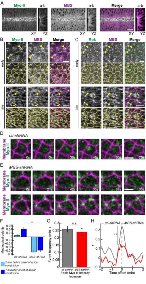
furrow cells (Fig. 6 A). MBS colocalized with both apical Myo-II
and Rok foci throughout the process of ventral furrow
forma-tion (Fig. 6, B and C). To determine whether MBS is required
for Myo-II pulses, we knocked down MBS using maternal
ex-pression of a shRNA that targets MBS (Ni et al., 2011; Fig. S1 G).
MBS knockdown resulted in apical Myo-II assembly that
ex-hibited a defect in medioapical coalescence, reminiscent of
Myo-II organization in phosphomimetic mutants (Fig. 6, D–F;
and
Video 6
). Although periods of rapid Myo-II accumulation
did occur in MBS knockdowns (Fig. 6 G), changes in Myo-II
signal were not well correlated with constriction (Fig. 6 H).
These data suggest that Myo-II dynamics in MBS knockdown
cells is uncoupled from area constriction, indicating that there is
a defect in the pulsing behavior compared with wild-type cells.
Consistent with this atypical Myo-II behavior, cells in MBS
knockdown embryos exhibited more persistent Myo-II
accumu-lation and constriction (Fig. 6 E, Fig. S4 B, and Video 6). Thus,
MBS is recruited to apical Myo-II where it is required to
polar-ize Myo-II accumulation and to generate cycles of assembly
and disassembly.
rok
1or rok
2mutant germline clones result in a loss of Myo-II
from the cortex, consistent with Rok and Myo-II
phosphoryla-tion being required for Myo-II activaphosphoryla-tion (
Fig. S3 and Video 4
;
Royou et al., 2002; Dawes-Hoang et al., 2005; Mason et al.,
2013). Previous studies suggested that sqh-EE can suppress loss
of Rok function (Winter et al., 2001; Bertet et al., 2004),
suggest-ing that Rok activates Myo-II minifilament assembly, whereras
minifilament localization is dependent on other cortical cues.
Consistent with these results, we found that sqh-EE rescued
cortical Myo-II localization in rok
1and rok
2mutant germline
clones (Fig. S3). However, despite rescuing cortical
localiza-tion in rok germline clones, sqh-EE Myo-II failed to undergo
medioapical coalescence and generate cell contractions (Video 4).
Instead, Myo-II accumulated uniformly across the apical
do-main (Fig. S3). Furthermore, the rok sqh-EE double mutants
failed to undergo mesoderm invagination (Video 4). Although
we cannot rule out the possibility that other Rok substrates are
required for Myo-II coalescence, our data suggest that simply
activating apical Myo-II is not sufficient for medioapical
acto-myosin network condensation and radial cell polarity. We favor
a model whereby spatial regulation of Myo-II by Rok within the
apical domain is required for actomyosin network contraction
during a pulse.
Myo-II phosphomutants exhibit defects in Myo-II assembly/disassembly cycles
We next determined whether dynamic Myo-II phosphorylation
is critical for temporal organization of Myo-II pulses. Similar to
Myo-II in wild-type embryos, sqh-TS::GFP displayed Myo-II
contraction pulses in sqh
1germline clones, with the appearance
of Myo-II structures lasting 20 to 30 s before being
disassem-bled or remodeled (Fig. 5 A). In contrast, phosphomimetic
mu-tants caused a gradual increase in Myo-II accumulation (Figs. 5 B
and S2 C). Myo-II persisted long after it initially appeared,
fail-ing to undergo disassembly over the course of constriction
(Fig. 5 B and
Video 5
). Based on the frequency of events
involv-ing rapid Myo-II intensity increase, we found that the sqh-AE
mutant had significantly fewer Myo-II pulses than wild-type
embryos (Fig. 5 D). Correspondingly, apical constriction in the
sqh-AE
mutant appeared more continuous, with fewer rapid
phases of constriction that were lower in magnitude than those
observed in wild-type (Fig. 5, B, E, and F; and
Fig. S4 A
).
Dur-ing pulsatile constriction, phases of rapid Myo-II accumulation
correlate with increased constriction rate (Martin et al., 2009).
However, the sqh-AE mutant displayed a significantly weaker
cross-correlation between Myo-II accumulation and the
con-striction rate, confirming our observation that mimicking Sqh
phosphorylation disrupts contractile pulses and results in more
continuous apical constriction (Fig. 5 G).
Mutants that decrease Myo-II phosphorylation resulted in
Myo-II dynamics that were distinct from the phosphomimetic
alleles. The sqh-AS mutant displayed contractile oscillations and
Myo-II disassembly similar to wild-type embryos (Fig. S2 C),
suggesting that dynamic phosphorylation of serine-21 is
suf-ficient for Myo-II remodeling. The sqh-TA mutant exhibited
phases of rapid Myo-II accumulation that were temporally
cor-related with constriction (Fig. 5, C and G), which possibly results
on August 13, 2014
jcb.rupress.org
Fig. S5 B). However, phosphomimetic mutations in other Myo-II
RLCs have been shown to not fully recapitulate the ATPase
ac-tivity of phosphorylated Myo-II in vitro (Kamisoyama et al.,
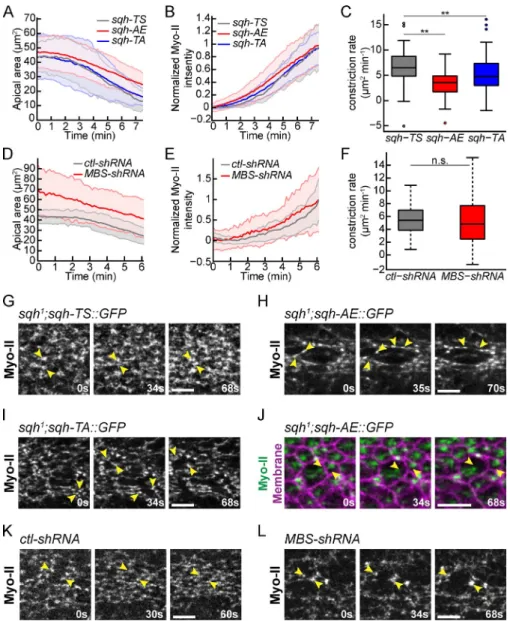
1994). Importantly, MBS depletion, which also appears to trap
Myo-II in the phosphorylated state, resulted in apical
constric-tion with similar rates to control cells, despite greater
heteroge-neity in cell size (Fig. 7, D–F). Thus, although contractile pulses
may enhance constriction rate, they do not appear to be required
for individual cell apical constriction.
Although apical constriction and tissue invagination occur
in Sqh phosphomutants, the coordination of tissue invagination
was perturbed. In wild-type ventral furrows, Myo-II foci in
neighboring cells come together steadily to form a dense
supra-cellular Myo-II meshwork across the entire tissue (Fig. 7 G and
Video 7
). Furrows in Myo-II phosphomimetic mutants underwent
Myo-II pulses are important for maintaining tissue integrity during morphogenesis
It is unknown why cells undergo pulsatile, rather than
continu-ous, contraction during tissue morphogenesis. Because we were
able to disrupt contractile pulses and promote more persistent
apical Myo-II accumulation using both Myo-II phosphomutants
and MBS depletion, we next investigated the consequences of
continuous apical constriction on tissue invagination.
Surpris-ingly, many of the phosphomutants that disrupted contractile
pulses (with the exception of the sqh-AA mutant) underwent
apical constriction and tissue invagination (Fig. 7, A–C;
Fig. S5,
A–C
; and Video 2). On average, the sqh-AE mutant cells
constricted more slowly than wild-type sqh-TS control cells,
suggesting that unregulated Myo-II may decrease the rate of
apical constriction and tissue invagination (Fig. 7, A and C; and
Figure 5. Cycles of Myo-II assembly and disassembly require dynamic RLC phosphorylation. (A–C) The sqh-AE and sqh-TA mutants inhibit Myo-II
remodel-ing associated with pulses. Time-lapse images are representative cells from sqh1 germline clone embryos expressing the indicated phosphomutants and
Memb::Cherry (membrane). Plots represent apical area and Myo-II intensity as a function of time for the same cells; the time highlighted in gray corresponds to the time time-lapse shown. Bars, 5 µm. (D) Quantification of frequency of instances where there is a rapid increase in Myo-II intensity (n = 94 sqh-TS cells, two embryos; n = 131 sqh-AE cells, three embryos; n = 92 sqh-TA cells, two embryos). (E) Quantification of frequency of instances of rapid apical area reduction (n = 94 sqh-TS cells, two embryos; n = 131 sqh-AE cells, three embryos; n = 92 sqh-TA cells, two embryos). (F) Quantification of maximal instantaneous constriction rate achieved in control or mutant cells (n = 138 TS cells, two embryos; n = 187 AE cells, three embryos; n = 172
sqh-TA cells, two embryos). (D–F) Error bars are SEM. (G) Mean cross-correlation between constriction rate and change in Myo-II intensity (n = 123 sqh-TS
cells, two embryos; n = 143 sqh-AE cells, three embryos; n = 88 sqh-TA cells, two embryos). P-values were calculated at 0 time offset. **, P < 0.01; n.s., not significant.
on August 13, 2014
jcb.rupress.org
Downloaded from
Figure 6. Myo-II contractile pulses require MBS. (A) MBS is apically enriched in ventral furrow cells. Images of XY semisagittal and YZ cross sections for fixed sqh::GFP embryos stained for MBS. Bar, 10 µm. (B) MBS structures colocalize with Myo-II foci and the supracellu-lar Myo-II meshwork (arrowheads). Images are from fixed sqh::GFP embryos stained for MBS. Bars, 5 µm. Segmented cell outlines are from subapical sqh::GFP signal. (C) MBS colocal-izes with Rok foci (arrowheads). Images are from fixed GFP::Rok embryos stained for MBS. Bars, 5 µm. Segmented cell outlines are from subapical Rok signal. (D and E) MBS knock-down inhibits Myo-II pulses. Time-lapse images are cells from embryos expressing
control-shRNA (ctl-control-shRNA) (D) or MBS-control-shRNA (E), sqh:: GFP (Myo-II), and Memb::Cherry (membrane).
(F) Quantification of Medioapical polarity in Myo-II organization (n = 115 ctl-shRNA cells, two embryos; n = 123 MBS-shRNA cells, two embryos). Error bars are SEM. (G) Frequency of rapid increases of Myo-II intensity (n = 82
ctl-shRNA cells, two embryos; n = 70 MBS-shRNA cells, three embryos). Error bars are
SEM. (H) Mean cross-correlation between constriction rate and change in Myo-II intensity for cells as a function of time offset (n = 118
ctl-shRNA cells, two embryos; n = 84 MBS-shRNA cells, three embryos). P-values were
calculated at 0 time offset. **, P < 0.01; n.s, not significant.
on August 13, 2014
jcb.rupress.org
apical constriction and the contractile force balance between
cells to maintain intercellular cytoskeletal connections in the
supracellular actomyosin meshwork that transmits tension
across the ventral furrow tissue.
Discussion
Recent studies demonstrated that pulsatile Myo-II contractions
drive diverse morphogenetic processes, including
Caenorhab-ditis elegans
embryo polarization (Munro et al., 2004),
Drosoph-ila
gastrulation (Martin et al., 2009; Roh-Johnson et al., 2012;
He et al., 2014), dorsal closure (Blanchard et al., 2010; David
et al., 2010; Azevedo et al., 2011), germband extension (Rauzi
et al., 2010; Fernandez-Gonzalez and Zallen, 2011; Sawyer et al.,
2011), oocyte elongation (He et al., 2010), and Xenopus laevis
convergent extension (Skoglund et al., 2008; Kim and Davidson,
2011; Shindo and Wallingford, 2014). Although Rok, and likely
Myo-II activation via Rok phosphorylation, is required for
con-traction (Dawes-Hoang et al., 2005; He et al., 2010; Kim and
Davidson, 2011; Mason et al., 2013), it was not clear whether
Myo-II activation simply regulates cortical Myo-II levels or
global contraction, but exhibited abnormal stretching of Myo-II
networks and separation of Myo-II foci at late stages of
fur-row invagination (Fig. 7 H and Video 7; separations observed in
16/21 sqh-AE and 14/14 sqh-EE embryos). These separations
involved recoil between Myo-II structures in adjacent cells,
resulting in gaps in the supracellular Myo-II meshwork (Fig. 7,
H and J). Furthermore, the phenotype of MBS knockdown
resembled phosphomimetic Myo-II mutants, also displaying
Myo-II separations (Fig. 7, K and L; and Video 7; separations
observed in 9/9 MBS-shRNA embryos). In the sqh-TA mutant
we also observed dynamic separation between Myo-II foci in
neighboring cells, suggesting that high levels of
phosphoryla-tion and/or Myo-II remodeling are important to maintain strong
intercellular coupling between actomyosin networks as the
tissue invaginates (Fig. 7 I and Video 7; separations observed
in 7/15 sqh-TA embryos). Although the separation between
Myo-II networks in adjacent cells is similar to cytoskeletal
separations observed in mutants that reduce cell–cell adhesion
(Martin et al., 2010), the sqh-TA and sqh-AE mutants exhibited
normal apical E-cadherin localization (Fig. S5, D–F). Thus,
dy-namic Myo-II pulses appear to be important for coordinating
Figure 7. Defects in Myo-II pulsing disrupt the integrity of the supracellular actomyosin meshwork. (A and B) Mean apical area (A) and normalized mean cellular Myo-II
inten-sity (B) for representative sqh1 germline clone
embryos expressing the indicated phospho-mutants (n = 50 sqh-TS cells; n = 49 sqh-TA cells, and n = 50 sqh-AE cells). Shaded area is ±SD. (C) Box-and-whisker plot of cell
con-striction rates for multiple sqh1 germline clone
embryos expressing the indicated phosphomu-tants (n = 102 sqh-TS cells, two embryos; n = 169 sqh-AE cells, three embryos; and n = 189
sqh-TA cells, three embryos). (D and E) Mean
apical area (D) and normalized mean cellular Myo-II intensity (E) for representative embryos expressing the indicated shRNA (n = 78
con-trol-shRNA cells; n = 31 MBS-shRNA cells).
Apical area of cells in MBS-shRNA knockdown embryos were more heterogenous and larger than that of ctl-shRNA embryos; however, they undergo apical constriction. Shaded area is ±SD. (F) Box-and-whisker plot of constriction rates for multiple embryos expressing the in-dicated shRNA (n = 104 control-shRNA cells, two embryos; n = 78 MBS-shRNA cells, three embryos). (G–L) Myo-II phosphomutants and MBS knockdown cause separation of Myo-II networks between cells. Time-lapse images
are of representative cells from sqh1 germline
clone embryos with the indicated phosphomu-tants or from embryos expressing the indicated shRNA. Arrowheads indicate contraction of an intact supracellular Myo-II meshwork in control embryos (G and K) or instances of intercel-lular Myo-II network separation in mutants or knockdowns (H–J and L). Bars, 10 µm. Box-and-whisker plots display the median (central line), 25th and 75th percentiles (box edges), the most extreme data points not considered outliers (whiskers), and outliers (plotted indi-vidually). **, P < 0.01; n.s., not significant.
on August 13, 2014
jcb.rupress.org
Downloaded from
the phenotypes of alleles that constitutively reduce
phosphoryla-tion further suggest that cycling between high and low
phos-phorylation states is required for proper Myo-II pulses.
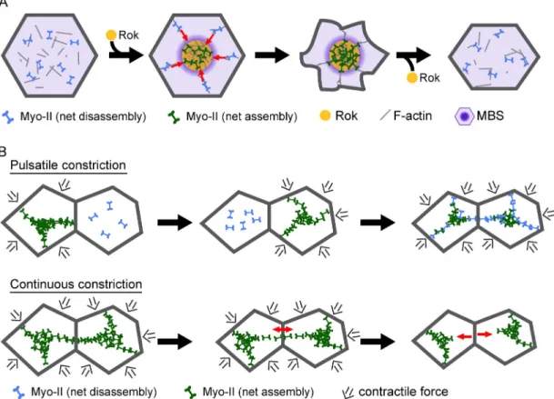
We propose a model for contractile pulses in the ventral
furrow where, in combination with unknown cortical cues that
apically localize Myo-II, local pulses of apical Rok activity
within the medioapical cortex polarize Myo-II assembly and
coalescence (Fig. 8 A). Rok foci could polarize actomyosin
condensation by generating an intracellular gradient of
minifila-ment assembly and tension that results in inward centripetal
ac-tomyosin network flow (Fig. 8 A, red arrows; Bray and White,
1988; Munro et al., 2004; Mayer et al., 2010). In addition, local
Myo-II activation by Rok foci combined with broader myosin
phosphatase activity throughout the apical cytoplasm could
gen-erate a gradient of Myo-II turnover that will concentrate Myo-II
into medioapical foci (Fig. 8 A). MBS is required to restrict
phosphorylated Myo-II to specific cell–cell interfaces during
dorsal closure, demonstrating that the balance between Myo-II
kinases and phosphatase can generate spatial patterns of Myo-II
activation in epithelial cells (Mizuno et al., 2002). Myo-II
re-modeling after coalescence could result from local decreases in
Rok activity and enrichment of apical myosin phosphatase with
Myo-II structures (Fig. 8 A). Thus, coupling Myo-II activation
to dynamic signals that regulate Myo-II phosphorylation
orga-nizes contractile pulses in space and time to drive incremental
apical constriction.
Polarized actomyosin contraction, pulses, and flows
gen-erate force and organize the actin cortex in a variety of cellular
and developmental contexts (Lecuit et al., 2011; Salbreux et al.,
2012). In contrast to the ratchet-like constriction of ventral
fur-row cells, some cell types undergo extended periods of
actomy-osin pulsing and area fluctuations without net reduction in area
(Solon et al., 2009; Blanchard et al., 2010; He et al., 2010).
Fur-thermore, directional rearrangement of cell contacts, such as
during convergent extension in the Drosophila germband, can
be achieved through planar polarized accumulation of
junc-tional Rok and Myo-II (Bertet et al., 2004; Blankenship et al.,
2006; Simões et al., 2010) in conjunction with planar polarized
medioapical actomyosin flows (Rauzi et al., 2010; Levayer and
Lecuit, 2013). Modulating the spatial and temporal regulation
of Myo-II phosphorylation and dephosphorylation provides a
possible mechanism to tune contractile dynamics and
organiza-tion to generate diverse cell shape changes (Mason and Martin,
2011). Consistent with this organizational role,
phosphomi-metic RLC mutants also disrupt the planar polarized
localiza-tion of junclocaliza-tional Myo-II in the Drosophila germband (Kasza
et al., 2014). Thus, it will be important to define the principles
that control Myo-II activity and dynamics and how tuning Myo-II
dynamics impacts force generation and tissue movement.
Role of pulsatile Myo-II contractions during tissue morphogenesis
Myo-II phosphomutants resulted in a more continuous apical
Myo-II assembly and apical constriction, enabling us to
investi-gate the role of pulsation during tissue morphogenesis.
Contin-uous Myo-II assembly and contraction in the sqh-AE mutant
resulted in a slower mean rate of apical constriction and thus
whether coupling between Myo-II activity and its regulators
or-ganizes contractile pulses in space and time. Furthermore, why
cells undergo pulsatile, rather than continuous, contraction to
drive tissue morphogenesis was unknown. We were able to
answer these questions by visualizing the consequences of
un-coupling Myo-II activation from upstream signaling pathways
on cell and tissue dynamics.
Dynamic Myo-II phosphoregulation organizes contractile pulses
We identified dynamic Myo-II phosphorylation as a key
mecha-nism that regulates contractile pulses. We found that Myo-II
pulses are associated with dynamic medioapical Rok foci and
myosin phosphatase. In addition, the phosphomimetic sqh-AE
and sqh-EE mutants, which exhibited constitutive cytoplasmic
Myo-II assembly in vivo, exhibited defects in two properties of
contractile pulses. First, phosphomimetic mutants did not
ini-tially condense apical Myo-II or F-actin into medioapical foci,
resulting in Myo-II accumulation across the apical domain and
thus a defect in Myo-II radial cell polarity. Second,
phosphomi-metic mutants continuously accumulated Myo-II in the apical
cortex, lacking clear cycles of Myo-II remodeling that are
ob-served in wild-type embryos. Although the phosphomimetic
al-leles are predicted to partially activate the Myo-II motor’s ATPase
activity compared with normal phosphorylation (Kamisoyama
et al., 1994), the similarity of the MBS knockdown phenotype
suggests that the changes in Myo-II organization and dynamics
in phosphomimetic mutants reflect defects in the control over
Myo-II dynamics rather than a reduction in motor activity. The
consequence of persistent Myo-II assembly across the apical
sur-face in phosphomimetic mutants and MBS knockdown is a more
continuous, rather than incremental, apical constriction,
demon-strating that pulsatile cell shape change results from temporal
and spatial regulation of Myo-II activity via a balance between
kinase (Rok) and phosphatase (myosin phosphatase) activity.
Mutants that decrease Myo-II phosphorylation affected
contractile pulses in a manner that was distinct from the
phos-phomimetic alleles. Both the sqh-AA and the sqh-TA mutants
exhibited Myo-II assembly into apical foci, potentially mediated
by phosphorylation of low levels of endogenous Sqh or
phos-phorylation of threonine-20, respectively. For the sqh-TA mutant,
Myo-II assembly was correlated with constriction, suggesting
that Myo-II motor activity is not rate limiting to initiate a
con-tractile pulse. However, Myo-II foci in sqh-TA and sqh-AA mutants
were not efficiently remodeled after assembly and coalescence.
The persistence of cortical Myo-II foci in sqh-AA and sqh-TA
mutants was surprising given that rok mutants and injection of
Rok inhibitor reduce cortical localization of Myo-II (Royou
et al., 2002, 2004; Dawes-Hoang et al., 2005; Mason et al., 2013).
One explanation is that high levels of Myo-II activity induce
ac-tomyosin turnover and thus could be required to remodel the
actomyosin network after contraction (Murthy and Wadsworth,
2005; Wilson et al., 2010). Alternatively, apical recruitment of
myosin phosphatase or proteins that negatively regulate Rok
could depend on Myo-II phosphorylation or actomyosin
con-traction (David et al., 2013). Although future work is needed to
address the role of Myo-II motor activity in contractile pulses,
on August 13, 2014
jcb.rupress.org
dynamics during apical constriction appear to sensitize the tissue
to loss of intercellular cytoskeletal integrity during
morphogene-sis (Fig. 8 B). Although loss of cytoskeletal continuity in
phos-phomimetic mutants does not block tissue invagination, we
speculate that dynamic Myo-II pulses are important to make
tis-sue invagination robust to changes in tensile stress. One possible
function of Myo-II pulses is to attenuate tissue tension or stiffness
during morphogenetic movements (Fischer et al., 2014). Because
pulsed Myo-II contractions are staggered between neighboring
cells, pulsation could serve as a mechanism to coordinate
con-tractile force generation across the tissue such that intercellular
connections are buffered from high levels of tension (Fig. 8 B).
Indeed, reducing adherens junction proteins sensitizes the
inter-cellular connections between cytoskeletal networks to tensile
forces generated in ventral furrow cells (Martin et al., 2010;
Sawyer et al., 2011; Spahn et al., 2012). Alternatively, remodeling
of actomyosin networks that occurs during pulses could be required
to adapt the cytoskeletal organization such that forces transmitted
between cells accommodate the changing pattern of tissue-scale
forces during the course of morphogenesis. In either case, our
data suggest that Myo-II pulsing and remodeling are important
for collective cell behavior by ensuring proper force transmission
between cells in a tissue undergoing morphogenesis.
delayed tissue invagination. This delay suggested that pulsing
might be important for the efficiency of apical constriction.
How-ever, phosphomimetic mutants might not fully recapitulate the
ATPase activity of phosphorylated Myo-II. The sqh-TA mutant,
which also perturbs Myo-II remodeling, constricted ventral
fur-row cells at a rate that is only slightly slower than wild type. In
addition, MBS knockdown, which disrupted Myo-II pulses,
ex-hibited a more variable constriction rate, but with a mean rate
comparable to control embryos. Our finding is distinct from
studies in other cell types where loss of MBS results in
exces-sive phosphorylated Myo-II accumulation and cell invagination
(Lee and Treisman, 2004; Corrigall et al., 2007). Thus, MBS
can regulate Myo-II organization and dynamics without causing
a significant increase in apical Myo-II levels. We conclude that
Myo-II pulses are not absolutely required for individual cell
apical constriction.
Although phosphomimetic mutant cells constrict and
un-dergo tissue invagination, the coordination of invagination and
the stability of the supracellular actomyosin meshwork were
per-turbed. Continuous apical constriction was associated with
ab-normal separation events between Myo-II structures in adjacent
cells, resulting in gaps or holes in the supracellular Myo-II
mesh-work. Thus, continuous Myo-II assembly and a lack of Myo-II
Figure 8. Model for mechanism and function of contractile pulses. (A) Myo-II dynamics during a contractile pulse. The contractile pulse is initiated by a local increase in Rok activity that elevates Myo-II phosphorylation and activity. Myo-II coalescence into medioapical foci results from cortical flow (red arrows) resulting from a gradient in cortical tension and net Myo-II minifilament assembly (dark green) at Rok foci with net minifilament disassembly (light blue) in regions of low Rok by myosin phosphatase (purple). Decreased Rok activity after Myo-II coalescence results in Myo-II dephosphorylation followed by remodeling of the contracted cortex. (B) Contractile pulses are required to maintain tissue integrity. Pulsatile Myo-II contraction occurs asynchronously in adjacent cells, which reduces stress at adherens junctions. In addition, Myo-II remodeling allows cells to adjust contacts to maintain stable intercellular cytoskeletal connections. Continuous Myo-II assembly and apical constriction decreases ability of actomyosin networks to dynamically adjust to changes in tissue mechanics, resulting in stretching of Myo-II structures and loss of intercellular connections (red arrows).
on August 13, 2014
jcb.rupress.org
Downloaded from
NJ) and -neurotactin were methanol/heat fixed (Fig. 3); all other immuno-stainings used PFA-fixed embryos. For methanol/heat fixations, embryos were placed in boiling Triton salt solution (0.03% Triton X-100 and 0.4% NaCl in water), cooled on ice, and then devitellinized in a 1:1 heptane/methanol solu-tion. PFA-fixed embryos were fixed in a 1:1 solution of 8% PFA in 0.1 M phos-phate buffer, pH 7.4, and heptane for 30 min, transferred to a Petri dish, and manually devitellinized using a syringe needle. After immunostaining, heat-fixed embryos were placed on a slide in mounting medium (4% N-propyl-galate in 80% glycerol) and sliced using a syringe blade to create cross sections. PFA-fixed embryos were mounted in AquaPolymount (Polysciences, Inc.). Images were acquired on a confocal microscope (LSM 710; Carl Zeiss) with a 40×/1.2 Apochromat water objective (Carl Zeiss), using argon ion, 561-nm diode, 594-561-nm HeNe, and 633-561-nm HeNe lasers. In Figs. 1 and 6, endog-enous GFP was used to visualize Myo-II and Rok, respectively.
Time-lapse imaging
Embryos were dechorionated in 50% bleach and mounted ventral side up on a slide coated with “embryo glue” (double-sided tape soaked in hep-tane). No. 1.5 coverslips were used as spacers and to create a chamber for the mounted embryo. A No. 1 coverslip completes the top of the chamber, and the chamber was filled with Halocarbon 27 oil. All imaging occurred at room temperature (23°C) on a confocal microscope with a 40×/1.2 Apochromat water objective, argon ion and 561-nm diode lasers, and a pinhole setting between 1 and 2 Airy units. Simultaneous excitation was used for live two-channel imaging of GFP/mCherry or Venus/mCherry. The band-pass filters for Venus/mCherry were set at 519–578 nm and 599– 696 nm, respectively. For Venus/mCherry two-channel imaging, we con-firmed that there was minimal spectral bleed-through between channels. The band-pass selected for GFP was 488–558 nm and for mCherry 573– 696 nm. All images were acquired using Zen software (Carl Zeiss). Image processing and analysis
Images were processed using Fiji (http://fiji.sc/wiki/index.php/Fiji) and MATLAB (MathWorks).
A Gaussian filter ( = 0.5–0.7) was applied to images. Apical images are maximum intensity projections of 2–5 µm. Subapical image and mem-brane images are single sections 1–2 µm below the apical projection.
To quantify Rok and Myo-II intensity over time, we generated kymo-graphs of pulses and acquired intensity values along a linear trace through the kymographs. To determine the phase relationship between Rok and Myo-II signals, a Pearson correlation was calculated for various time offsets where the Rok and Myo-II signals were shifted relative to each other.
We used custom MATLAB software, Embryo Development Geometry Explorer (EDGE; Gelbart et al., 2012), to segment images for quantifica-tion of apical area and Myo-II intensities. EDGE automatically segmented cell membranes; however, we manually corrected cells with errors in seg-mentation. Segmented cell membranes were subapical to the Myo-II signal. Embryos were aligned in time using the mean apical area signals of each embryo by choosing the time where the tissue begins to constrict. For quan-tification of junctional and medioapical Myo-II intensity in Fig. 4, we used a maximum intensity projection of the raw apical Myo-II signal and applied EDGE to segment images. The medioapical domain of a cell is defined by shrinking the cell’s segmented contour by 2 pixels wide; the pixel intensity in this area was used to define medioapical Myo-II, and junctional Myo-II was defined as the difference between the Myo-II intensity in the entire cell and the intensity in the medioapical area (see equations). We then calcu-lated medioapical polarity using the following equations:
total Myo-II =
∑
=I P k k P T T 1medial Myo-II =
∑
=I Pk k P m m 1junctional Myo-II = − − = =
∑
∑
I I P P k kP k k P T m m T 1 1p medial Myo-II junctional Myo-II total Myo-II
= −
Materials and methods
Fly stocks and genetics
Stocks used in this investigation are listed in Table S1. Germline clones
were generated using the FLP-DFS technique by heat shocking mutant/ovoD
larvae for 2 h at 37°C for 3–4 d (Chou and Perrimon, 1992). For two-channel imaging of Venus::Rok (UAS-driven; Simões et al., 2010; gift from J. Zallen, Sloan Kettering Institute, New York, NY) and sqh::mCherry (sqh
promoter), rok2 FRT/FM7;;mat15 sqh::mCherry/TM3 females were crossed
to ovoD FRT/Y; hsFlp UAS-Venus::Rok/CyO males, and embryos from heat
shocked rok2 FRT/ovoD FRT; hsFlp UAS-Venus::Rok/+; mat15 sqh::
mCherry/+ females were collected for live imaging. For sqh::GFP rescue of
sqh1 germline clones, sqh1 FRT/FM7; sqh-XX::GFP/CyO females were
crossed to ovoD FRT/Y; hsFlp males, the resulting larvae were heat shocked,
and sqh1 FRT/ovoD FRT; sqh-XX::GFP/hsFlp females were crossed to OreR
to collect embryos that resulted from germline clones. All sqh-XX::GFP trans-genes were expressed via the endogenous sqh promoter. Sqh phosphomu-tants were recombined with Gap43::mCherry (Memb::Cherry, plasma membrane marker, driven by sqh promoter; Martin et al., 2010) or with
Utr::mCherry (F-actin marker, driven by sqh promoter; Rauzi et al., 2010;
gift from T. Lecuit, Institut de Biologie du Développment de Marseille, Marseille, France). For sqh-EE::GFP suppression of rok mutant germline clones,
rok1 FRT/FM7; sqh-EE::GFP/CyO females were crossed to ovoD FRT/Y;
hsFlp males and heat shocked. rok1 FRT/ovoD FRT; sqh-EE::GFP/hsFlp
females were crossed to OreR males to collect embryos. In all cases, con-trol crosses lacking heat shock were performed to verify the presence of the
ovoD allele. To generate MBS knockdown embryos, females containing a
UAS-driven shRNA against MBS (MBS-shRNA) or the white gene (control) were crossed to males containing a double-maternal driver line with both
sqh::GFP and Gap43::mCherry. Both UAS stocks were gifts from N. Perrimon,
L. Perkins, and the Transgenic RNAi Project (Harvard Medical School, Bos-ton, MA). The resulting larvae were raised at 25°C and females that mater-nally expressed MBS-shRNA or ctl-shRNA and both fluorescent markers were crossed to OreR males, and the resulting embryos were imaged. Construction of GFP-tagged sqh phosphomutants
Substitution of threonine-20 and serine-21 with alanine or glutamate pre-vented or mimicked phosphorylation, respectively (Jordan and Karess, 1997; Winter et al., 2001). The sqh gene, including 5 promoter sequences and 3 termination sequences, was tagged at the carboxy terminus with eGFP. The sqh::GFP sequence is identical to the previously used sqh::mCherry construct with the exception of the fluorescent protein sequence (Martin et al., 2009). Site-directed mutagenesis of threonine-20 and/or serine-21 resi-dues was performed on sqh::GFP in the pBluescript vector using QuikChange II XL site-directed mutagenesis kit (Agilent Technologies). The 3.5-kb KpnI– XbaI sqh::GFP fragments were cloned into the pTiger transformation vector containing an attB site (pTiger courtesy of S. Ferguson, State University of New York at Fredonia, Fredonia, NY). These constructs were sent to Best-Gene Inc. for integration into either the attP1 or attP40 landing sites (see Table S1) using the C31 integrase system (Groth et al., 2004). Transgenes
integrated at these chromosome II landing sites were crossed to the sqh1
mutant. The sqh-TS::GFP transgene could rescue sqh1 mutant flies to
adult-hood; however, homozygous sqh1 adult flies were not observed with any of
the sqh::GFP phosphomutants (including sqh-AE and sqh-EE), demonstrating that dynamic myosin phosphorylation is required for development. Generation of full-length Sqh antibody and Western blotting
Full-length Sqh cDNA was cloned and purified from E. coli by GenScript (GenScript USA Inc.). Rabbit sera were obtained using the full-length Sqh as antigen (Panigen, Inc.). Polyclonal anti-Sqh antibody was affinity puri-fied using standard biochemical procedures. In brief, puripuri-fied Sqh was coupled to CNBr-activated Sepharose 4B (GE Healthcare), the Sqh-coupled resin was incubated with sera, the resin was washed, and the antibody was eluted with glycine, pH 2.5.
Western blotting was performed by grinding embryos directly in sam-ple buffer and running samsam-ples on 12% SDS-PAGE gels. Protein was trans-ferred to 0.45-µm nitrocellulose membrane (Bio-Rad Laboratories) and the indicated primary antibodies were detected using horseradish peroxidase– labeled secondary antibodies (Jackson ImmunoResearch Laboratories, Inc.). Immunohistochemistry
Antibodies and corresponding concentrations used in this investigation are
listed in Table S2. For fixed imaging, all embryos were first dechorionated in
50% bleach and then either methanol/heat fixed or PFA fixed. Embryos stained with -zipper (gift from E. Wieschaus, Princenton University, Princeton,