Journal of Antimicrobial Chemotherapy (1987) 20, 903-911
Treatment failures of cefotaxime and latamoxef in meningitis caused by
Enterobacter and Serratia spp.
Robert H. K. Engf, Charles E. Cherubin*, Jean-Claude Pecfaere' and Thomas R. Beam, Jr.'
'Infectious Disease Section, Medical Service. Veterans Administration Medical Center, East Orange, NJ 07019, and New Jersey Medical School/UMDNJ, Newark, New Jersey, U.S. A; b Department of Medicine, Bay ley Seton Hospital, Staten Island, New
York 10304, U.S.A; 'Dipartement de Microbiologie, Centre Medical Universitaire. University de Geneve, Geneve 4 CH-1211, Switzerland; 'Infectious Disease Section, Veterans Administration Medical Center, Buffalo, NY and Department of Medicine,
SUNYat Buffalo, NY 14215, U.S.A.
Despite the apparent success of several new cephalosporins in the treatment of Gram-negative bacterial meningitis, four treatment failures with cefotaxime or latamoxef were encountered (two caused by Enterobacter and two by Serratia spp.). In-vitro parameters of susceptibility of these clinical isolates were compared with those of a meningeal Ent. cloacae isolate from a successfully treated patient. The MIC and MBC values, degrees of inoculum effect, and amounts of /?-lactamase produced correlated poorly with the observed clinical outcome. However, the extent to which an isolate was killed by the cephalosporin used for treatment, in a 6-h in-vitro incubation, showed good correlation. We suggest that such a test should be used to predict clinical outcome of therapy because the other parameters such as the MIC and MBC values are not sufficiently discriminatory.
Introduction
The treatment of Gram-negative bacterial meningitis with several of the newer cephalosporins (such as cefotaxime, ceftriaxone and latamoxef) has been highly successful and documented in an extensive literature (Cherubin et al., 1982; Corrado, Gombert & Cherubin, 1982; Cherubin & Eng, 1986). However, individual reports have begun to appear describing treatment failures (Bradsher, 1982; Eng et al., 1984a). Organisms responsible for failures have included susceptible strains of Escherichia coli, Klebsiella pneumoniae and Salmonella spp. An earlier study showed that the failures were possibly attributable to a lack of rapid bactericidal activity of the new cephalosporins against the individual isolates of the patients (Eng et al., 1984a). Whether such observations can be expanded to include other members of the Enterobacteriaceae is not known.
In four treatment failures (two Enterobacter and two Serratia species) the pathogens were available and were studied in vitro for killing activity of the cephalosporins employed during treatment. The results were compared with those from a successful Correspondence: Dr Robert Eng, Medical Service (111), VA Medical Center, East Orange, NJ 07019, U.S.A.
903
treatment of enterobacter meningitis. In addition, /J-lactamase production in these and other control isolates was examined, to determine the importance of this in treatment failure.
Materials and methods Patients, bacterial isolates and antibiotics
The cases of Gram-negative bacterial meningitis were cases seen by the authors or referred to them by other Infectious Disease physicians during the period 1978 to 1980. Enterobacter cloacae EL 3513 was also included in the study. This organism was obtained from the blood of a patient and, in a rabbit model of meningitis, had been shown to be unresponsive to therapy by cefotaxime and latamoxef (Beam, 1983). A collection of nonmeningeal isolates of Ent. cloacae was also included for pMactamase studies.
Susceptibility testing
The bacterial isolates were tested for susceptibility to antibiotics in three ways. First, the isolates were tested by macro-broth dilution method with an inoculum of 5 x 10³ cfu/ml in 1 ml. Secondly, the method described by Eng, Smith & Cherubin (19846) was used to ascertain the effect of a large inoculum (5 x 10⁷ cfu/ml) on the susceptibility results. Thirdly, the method of washing the bacteria free of antibiotic before subculture was used to eliminate the antibiotic carry-over effect (Eng et al.,
1984a).
Killing kinetics
Killing curves were generated by exposing 5 x 10⁵ or 5 x 10⁷ cfu/ml bacteria in tubes containing 5 ml Mueller-Hinton broth and multiples of the MICs of the test antibiotics, as described by Eng et al. (1984a).
P-Lactamase studies
Bacteria were grown at 37°C in tryptic soy broth (Difco, Detroit, MI), with shaking at 300 rpm. Organisms were collected by centrifugation in late logarithmic phase, after 2-3 h growth, (l-2±0-4x 10⁹ cfu/ml), and washed twice in 01 M saline. Bacteria (1 ml pellet, or approximately 1 g wet weight) were resuspended in 5 ml saline and disrupted by sonic treatment (Artek Sonic 300 at 60 W) by two 3-5 min bursts. The preparation was maintained on ice during sonication. Cell debris was removed by centrifugation at 48,000 g for 20 min at 4°C and supematants were decanted. /?-Lactamase activity in the crude extract was quantified using a spectrophotometric assay at 483 nm in 0-1 M phosphate buffer, pH 70 (Ultraspec 4050, LKB produkter AG, Broma, Sweden, coupled with an Apple lie microcomputer) with nitrocefin (100 /iM final concentration) as substrate. /?-Lactamase activities were standardized per mg of protein (BCA protein assay Kontron, Zurich, Switzerland). The amount of /Mactamase produced by a strain was given as the arithmetic mean of at least three completely independent determinations.
Treatment failure in meningitis 905 /?-Lactamase induction was assessed using an adaptation of the method of Gootz & Sanders (1983), with cefoxitin, 10 mg/1 final concentration, as inducer. Controls, without cefoxitin, were run simultaneously. Kinetic constants, K,,, and V ^ , , were determined spectrophotometrically at 260 nm and 37°C after partial purification of the crude extract on Sephacryl S 200 in 0 1 M Tris-HCl buffer (pH 7-5) at 4°C. Cephaloridine was used as substrate, at concentrations varying from 001 to 0-2 mmol. K^ and V ^ values were estimated from a least-squares fit to a Lineweaver-Burk plot. Frequency of resistant clones
The frequency of resistant clones existing within the bacterial population before exposure to /Mactam agents was estimated by enumerating the cfu on Mueller Hinton agar containing cefotaxime at 32 times the MIC. The starting inoculum size was 1 0 to 2 0 x 108 cfu and the antibiotic agar plates were incubated for 24 h at 37°C.
Case studies Case 1
This case has been previously reported by Corrado et al. (1982) and only the details of therapy are summarized here. A 31 year-old man developed meningitis several days after head trauma. Gram stain of his CSF revealed numerous Gram-negative bacteria. He received 12 g cefotaxime per day. The organism was identified as Ent. aerogenes and the MIC of cefotaxime was 0 0 6 mg/1 by microdilution and 0-3 mg/1 by macrobroth dilution. The CFS cefotaxime levels ranged from 4 1 to 11-5 mg/1. The organism persisted in the CSF during therapy and appeared on Gram stain as filamentous forms. On days 2 and 3 of therapy, the same organism was isolated, with much higher MICs and MBCs of both cefotaxime and latamoxef (0-3-2-5 mg/1). This necessitated the addition of gentamicin 5 mg/kg/day intravenously and 8 mg/day intrathecally, plus carbenicillin 500 mg/kg/day intravenously. The patient did not respond and died. Case 2
A 57 year-old man developed Ent. cloacae meningitis 11 days after craniotomy for a left cerebral haematoma. Latamoxef was administered as initial therapy (12 g day). The initial MIC and MBC of the isolate were 0-3 and 4-8 mg/1, respectively, of latamoxef and 0-8 and 1-5 mg/1, respectively, of cefotaxime. Repeat lumbar puncture 48 h later showed continued presence of the organism on Gram stain and cultures yielded a more resistant strain. Cefotaxime was substituted for latamoxef at the same dose. However, subsequent CSF bactericidal assays revealed no apparent killing activity for the latter isolates. The resistant strain had MIC and MBC of 100 mg/1 for both antibiotics. Latamoxef was readministered along with 5 mg/day gentamicin intrathecally. The patient recovered after 14 days of combined therapy.
Case 3
A 32 year-old man was admitted following a motorcycle accident. CT scan showed haemorrhage into the left maxillary sinus, and an intraventricular pressure monitoring
device was inserted. On the fifth day, he developed deep coma and a temperature of KMT. Lumbar puncture yielded CSF with 20 polymorphonuclear cells and 332 RBC/mm3. Protein was 48 mg/dl and glucose 72 mg/dl. A moderate number of Gram-negative bacteria were seen per high power field, subsequently identified as Ent. cloacae. The MIC of ceftriaxone was 0-2 mg/1. The patient was started on ceftriaxone at 2 g 8 hourly.
The patient showed no clinical improvement for three days. Although the CSF was sterile at this time, the CSF leucocyte cell count had increased to 11,400/mm3, On day 8 of therapy, a repeat lumbar puncture showed fluid with 103 WBC. The CSF ceftriaxone level was 7 mg/1 and bactericidal activity was 1:1024. Progressive clinical improvement was noted between days 8 and 10 and the patient recovered without residua.
Case 4
A 57 year-old man had two surgical attempts at evacuation of a parietal hacmatoma from arteriovenous malformation. Five days after the second operation, he developed Ser. marcescens meningitis. He was treated initially with chloramphenicol, then with intrathecal amikacin for two days, then with amikacin and concurrent iv latamoxef for four days. His CSF remained culture-positive and he died. The initial isolate had MIC and MBC of 0-3 and 1-25 mg/1 latamoxef, respectively.
Case 5
A 42 year-old developed Ser. marcescens meningitis after an evacuation of cerebral haematoma sustained in a car accident. He was treated initially with penicillin and chloramphenicol until the organism was identified. Intrathecal gentamicin and 12 g/day latamoxef were administered. CSF cultures remained positive after 14 days of therapy. A variety of other antibiotics, including new cephalosporins, were added, but the patient died.
Laboratory results Susceptibility results
The MIC and MBC data are shown in Table I. The initial isolates from all the cases showed similar MICs and MBCs for all the /Mactam antibiotics tested. The isolate from the successfully treated case of Ent. cloacae meningitis (case 3) had MIC and MBC values indistinguishable from those of initial isolates from the failure cases caused by Enterobacter spp. (cases 1 and 2). The MBC values for the antibiotics used in therapy were 1-5 mg/1 ceftriaxone for case 3 as against 2-5 and 1-2 mg/1 of cefotaxime and latamoxef, respectively, for cases 1 and 2. Subsequent isolates from the failure cases caused by Enterobacter spp. were far more resistant.
The MBC values were noted to depend on the methods used. The values obtained by the standard method are shown in Table I. These values of isolates from failure cases did not differ from the value of a successfully treated case. A comparison of washing versus not washing df the organisms on Millipore filters prior to subculture showed major differences in several antibiotic-bacteria combinations. A prime example of this
Treatment failure in meningitis 907 Table I. Summary of antibiotic susceptibility of meningeal isolates'
Isolate Case 1 Ent. aerogenes initial subsequent Case2 Ent. cloacae initial subsequent Case3 Ent. cloacae Case 4 Ser. marcescens Case5 Ser. marcescens EL 3513 Ent. cloacae CTX 0-3/2-5 (8) 400/ > 400 ( - ) 0-6/1-2 (64) 100/> 100 ( - ) 0-3/1-2 (2) 0-3/> 20 (64) 0-6/1-2 (64) 0-3/5-0 (2) MIC/MBC LMX 0-3/2-5 (4) 200/200 ( - ) 0-3/5-0 (2) 10-0/> 100 ( - ) 1-2/2-5 (16) 0-3/1-2 (2) 0-3/>5 (>32) 0-3/0-3 (8) CTR 0-7/0-7 (2) 0-7/6-0 (32) 10-0/> 100 ( - ) 0-2/1-2 (8) 0-3/0-3 (2) 0-6/0-6 (4) 1-5/3-0 (8) IMP 0-6/1-2 (4) 0-3/0-3 (1) 1-2/2-5 (4) 1-2/2-5 (4) 0-6/2-5 (8) 2-5/5-0 (4) 1-2/2-5 (2) 1-5/1-5 (2)
•CTX, Cefotaxime; LMX, latamoxef; CTR, ceftriaxone; IMP, imipenem.
Inoculum effect in parenthesis is expressed as tbe ratio of the MIC for 5 x 10⁷ cfu/ml to the MIC for
5xlO'cfu/ml.
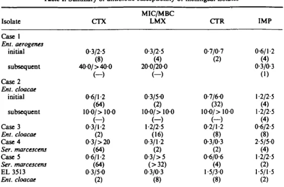
was the isolate from case 1. After a 6 h exposure to cefotaxime at 32 x MIC, there was less than a 1-log cfu reduction, but when the organisms were not washed free of the antibiotic a reading of a greater than 3-log reduction was recorded (Figure 1).
The MICs of cefotaxime, latamoxef and ceftriaxone for the isolates when the inoculum size was raised from 5 x 10³ to 5 x 10⁷ exhibited a two-fold to greater than
5 4 o At t3 2 3 E " U 1 -1 -1 -1 -1 - \ \ \ \ i i 4 8 Multiple! of MIC 16 32
Figure 1. Difference in 6 h bactericidal results when the organisms of the isolate from Case 1 were washed (O) and unwashed ( A ) before subculture.
- I I
i
~
2 i 3 - 3 . ? - 4 - 5 I CTX 2 LMX 3 CTR A LMXCoset ond cepholosporini
5 LMX EL LMX
Figure 2. Number of bacteria killed in 6 b (open bars) and in 18 h (hatched bars) by 4 x MIC of the
cephalosporin used in treatment of the meningeal isolates or of EL 3513 (the meningitis animal model strain). Antibiotic abbreviations: see Table I.
32-fold increase. Imipenem did show a slight inoculum effect, but an inoculum effect of greater than eight-fold was not noted with this agent.
Susceptibility of isolates to fi-lactam killing
The log decreases in cfu/ml of each isolate when exposed to 4 x MIC of the antibiotic used in treatment are shown in Figure 2. The major distinguishing feature of the isolate from the successfully treated case (case 3) from the antibiotic failure cases was the amount of killing attained at 6 h of incubation. The isolate from case 3 attained a 31 log reduction in cfu/ml in 6 h whereas other isolates showed a considerably smaller amount of killing.
4 8 Multiple! of MIC
16 32
Figure 3. Concentration-dependent killing of meningeal isolates at 6 h by the cephalosporin used in
treatment (case 1, cefotaxirne, O; case 2, latamoxef. A; case 3, ceftriaxone, D; case 4, latamoxef, A; and case S, latanioxef, # ) . The experiments were performed with washing of the bacteria free of antibiotics prior to quantitative subculture.
Treatment faflnre in meningitis 909
When the antibiotic concentrations were increased in the killing kinetic studies, while antibiotic carry-over was eliminated by washing, isolates from cases 4 and 5 were totally resistant to killing by cefotaxime and latamoxef, as were the Enterobacter isolates recovered during therapy from cases 1 and 2. In contrast, the isolate from the successfully treated case showed concentration-dependent killing (Figure 3).
P-Lactamase production by the isolates
/J-Lactamase production before and after cefoxitin induction in these isolates is shown in Table II. For the Enterobacter isolates initially recovered from cases 1 and 2, the amount of /Mactamase produced was 002 and 0-1 units as compared to 002 unit from case 3 (success case). After induction by cefoxitin, the units of enzymes from the isolates of cases 1 and 2 were 0-3 and 0-2, respectively as compared with 0-13 for case 3. Hence the amount of /Mactamase produced by the organism with and without cefoxitin induction did not distinguish among the isolates with regard to success or failure during treatment with stable /Mactam antibiotics. The kinetic parameters, Km and V ^ , were all characterized by low K,,,, indicating a very high affinity of the
/?-Table II. Summary of /Mactamase studies
Isolate Enterobacter Case 1 first second Case2 first second Case3 EL 3513 1228 688 1483 1394 1201 692 362 1529 4207 66 288 Serratia Case4 Case5 /7-Lactamase Without induction" (S.D.) 002 (001) 0-3 (01) 0 1 (001) 01 (0-01) 002 (001) 002 (001) 001 (0005) 0-03 (0-01) 1-51 (0-2) 401 (0-3) 4-79 (0-3) 002 (0005) 001 (0005) 002 (001) 001 (0005) 001 (0005) 001 (0005) 015(001) 012 (001) activity Induction ratio* 15 4-7 2 7 6-5 9-5 123 109 4-6 4 116 10 5 4 96 15 28 8 5-3 5-8 Kinetic K» 0-4 0-2 0-3 0-3 0-9 0-2 0-36 2-9 012 ND 019 0-73 0-37 ND 0-2 ND 3-0 0-9 0-3 values' 1 14 0-7 19 0-8 11 10 60 10 N D 11 0-7 008 ND 0-5 ND 3 0 7-3 2 0 Frequency of clones resistant to CTX at 32 x MIC 2 x l O "6 1 x l O '6 <1 x l O "8 3 x l O "7 < l x l 0 - » "Activity expressed in //mol/min/mg of protein.
'Ratio of activity following induction with 10 mg/1 cefoxitin to the uninduced level.
CKO is expressed in ptmol and V ^ in ponol/min/mg protein.
lactamase for the substrate. However, these parameters were similar for all the isolates studies, including the nonmeningeal isolates.
The organism isolated from patient 1 during treatment produced 15 times more (i-lactamase than the pretreatment isolate. Also, with cefoxitin stimulation, the treatment isolate produced four times more /Mactamase than the pretreatment isolate. For case 2, the second isolate showed a three fold increase in /Mactamase production in the presence of cefoxitin as compared to the pretreatment isolate. ^-Lactamase induction by cefoxitin appeared to be more pronounced in those organisms isolated during therapy.
Frequency of resistant clones
The frequencies of clones resistant to 32 times the MIC for cefotaxime are shown in the last column of Table II. These frequencies ranged between less than 10"8 to 2 x 10~6, with the lowest frequencies found in the organisms from cases 3 and 5.
Discussion
Bacterial meningitis is a rapidly fatal disease and host defences in the CSF are limited. To a large extent, the patient is dependent upon the bactericidal activity of antibiotics to eradicate the organisms. Gram-negative organisms have presented a therapeutic problem because many older antibiotics were ineffective in vitro and in vivo. The use of the new cephalosporins appears to be associated with increased survival rates. However, cases of Gram-negative meningitis have been reported in which treatment with the new cephalosporins was associated with failure (Bradsher, 1982; Eng et al., 1984a).
The success of the new cephalosporins was initially attributed to potent activity as reflected in low MIC values. Closer examination of the susceptibility information with respect to clinical responses has shown that low MIC values are necessary, but by no means can be used as a universal predictors of treatment success of Esch. coli and K. pneumoniae meningitis. In these two species, it is unlikely that /Mactamases play a significant role in causing treatment failure. With Serratia and Enterobacter spp., however, treatment failures of nonmeningeal sites have been ascribed to the large amount of /Mactamases produced or induced. We have examined the in-vitro parameters of susceptibility to find the parameter that best predicts success of therapy. The amount of /Mactamases produced, and induced in Cases 1 and 2 did not differ greatly from that of the other Enterobacter isolates tested. Isolates recovered during therapy and which had become resistant showed markedly greater production of /?-lactamase than the initial isolates from the same patient and, in general, the production rate is also greater than those isolates recovered from nonmeningeal sites. The amount of /Mactamase produced in the presence or absence of cefoxitin did not greatly differ between any of the groups of organisms. The frequency of resistant clones within the bacterial population of the pretreatment isolates was clearly different between the enterobacter failure and success cases. However, very low frequencies were also found among the isolates from the two serratia failure cases. The MIC values are clearly not sufficient in themselves to predict the success or failure of a treatment regimen, and the MBC results may be falsely low, unless the organisms are washed free of the test antibiotics before subculture. Yet, even with the addition of this washing step in the MBC methodology, the MBC values obtained from 18 to 24 h incubation
Treatment faflnre in meningitis 911
in themselves were not sufficiently discriminatory to predict the success of therapy. Inoculum effect of the treatment antibiotic-isolate combinations also failed to discriminate between combinations that produced clinical success or failure.
On the other hand, the bactericidal effect at 6 h correlated well with clinical response. The isolate that showed a 3-log or more decrease following 6-h incubation with antibiotic concentrations up to 16 x MIC was associated with success of therapy. Conversely, those bacterium-antibiotic combinations which did not produce a rapid decrease in cfu were associated with a poor clinical response. The 6 h bactericidal test is not difficult to perform and can be completed within one day in the microbiology laboratory; results can be available the next day. In fact, this test would require no more time for completion than the standard MBC test.
Our data are limited by the small number of cases we were able to collect. Our observation should be extended by infectious disease clinicians who treat such patients. If our conclusions can be upheld, bactericidal kinetics during the first 6 h of incubation could become a better test in the evaluation of antibiotics for treatment of Gram-negative bacterial meningitis.
Acknowledgments
We thank Dr David Droller, Dr Thomas R. Beam and Dr Myles Gombert for permission to report cases 2, 3 and 4, respectively. We also thank Frank Buccini for the technical assistance, and the Medical Media Production Service of East Orange VA Medical Center for the illustrations. This work was supported in part by the Veterans Administration and Fond National Suisse de la Recherche Scientifique (3.221.0.85).
References
Beam, T. R. (1983). Antibiotic failure in experimental meningitis. In Proceedings of 13th
International Congress of Chemotherapy (Spitzy, K. H. & Karrer, K., Eds), pp. 12-4. Verlag
H. Egenmann, Vienna.
Bush, K. & Sykes, R. B. (1986) Methodology for study of /}-lactamases. Antimicrobial Agents
and Chemotherapy 30, 6-10.
Bradsher, R. W. (1982) Relapse of gram-negative bacillary meningitis after cefotaxime therapy.
Journal of the American Medical Association 248, 1214-8.
Cherubin, C. E., Corrado, M. L., Nair, S. R., Gombert, M. E., Landesman, S. & Humbert, G. (1982). Treatment of gram-negative bacillary meningitis: role of the new cephalosporin antibiotics. Reviews of Infectious Diseases 4, Suppl., S453-64.
Cherubin, C. E. & Eng, R. H. K. (1986). Experience with the use of cefotaxime in the treatment of bacterial meningitis. American Journal of Medicine 80, 398-404.
Corrado, M. L., Gombert, M. E. & Cherubin, C. E. (1982). Designing appropriate therapy in the treatment of gram-negative bacillary meningitis. Journal of the American Medical
Association 248, 71-4.
Eng, R. H. K., Cherubin, C , Smith, S. M. & Buccini, F. (1984a). Examination of gram-negative bacilli from meningitis patients who failed or relapsed on moxalactam therapy.
Antimicrobial Agents and Chemotherapy 26, 850-6.
Eng, R. H. K. Smith, S. M. & Cherubin, C. (19846). Inoculum effect of new /Mactam antibiotics on Pseudomonas aeruginosa. Antimicrobial Agents and Chemotherapy 26, 42-47. Eng, R. H. K., Cherubin, C , Smith, S. M. & Buccini, F. (1985). Inoculum effect of /J-lactam
antibiotics on Enterobacteriaceae. Antimicrobial Agents and Chemotherapy 28, 601-6. Gootz, T. D. & Sanders, C. C. (1983). Characterization of /Mactamase induction in Enterobacter
Cloacae. Antimicrobial Agents and Chemotherapy 23, 91-7. (Manuscript accepted 13 July 1987)