157
Differential Expression of
Proinflammatory
Cytokines and Their Inhibitors
during the Course of Meningococcal Infections
Marcel van Deuren, Johanna van der Ven-Jongekrijg,
Pierre N. M. Demacker, Anton K. M. Bartelink,
Roelof van Dalen, Robert W. Sauerwein, Harald Gallati,
James L. Vannice, and Jos W. M. van der Meer
Departments oj Internal Medicine. Intensive Care. and Medical Microbiology. University Hospital Nijmegen. and Department ofInternal Medicine. Eemland Hospital.Aniersfoort,Netherlands;F.Hoffmann-La Roche Ltd.. Pharmaceutical Research. Basel. Switzerland; Synergen lnc.. Boulder. Colorado Circulating concentrations of tumor necrosis factor-a (TNF), interleukin (IL)-lP, IL-6, IL-l
receptor antagonist (lL-lra), and soluble TNF receptors p55 (sTNFr-55) and p75 (sTNFr-75) and ex vivo production ofTNF, IL-l, IL-6, and IL-lra using a whole blood culture system were measured during the acute and convalescent stages of meningococcal infection. Circulating TNF and IL-l were below detection level, whereas IL-6 and IL-lra, sTNFr-55, and sTNFr-75 were increased at admission. The ex vivo production of proinflammatory cytokines TNF, IL-l, and IL-6 was suppressed at admission and restored gradually during recovery. On the contrary, the production of the antiinflammatory IL-lra was increased at admission. The elevated concentra-tions of both IL-lra and sTNFr early in the course of infection suggest a regulatory role for these antiinflammatory compounds. The observed down-regulation of the ex vivo production ofTNF, IL-l, and IL-6 and up-regulation of the production of IL-l ra in the acute stage may indicate a protective regulation mechanism.
On contact with gram-negative bacteria or endotoxin (lipo-polysaccharide, LPS), the immune-apparatus responds with the production of a broad spectrum of cytokines. After injec-tion ofLPS in human volunteers, the plasma concentrainjec-tions of the proinflammatory cytokines tumor necrosis factor-a (TNF), interleukin (IL)-l {J, and IL-6 and the antiinflamma-tory IL-I receptor antagonist (IL-I ra) rapidly increase [1-3]. Furthermore, the plasma concentrations of soluble TNF re-ceptors p55 (sTNFr-55) and p75 (sTNFr-75) also rise [4,5]. Proinflammatory cytokines are thought to be essential for an adequate host defense. However, excessive production of TNF and IL-I causes shock, and high plasma concentrations of TNF, IL-I, and IL-6 are associated with high mortality rates [6-10]. This deleterious action of the proinflammatory cytokines is balanced by sTNFr and lL-1 ra. Soluble TNF receptors prevent the inflammatory effects of TNF by bind-ing to TNF. High sTNFr/TNF plasma ratios were found to be associated with a better prognosis [5, II]. IL-I ra blocks the proinflammatory action of IL-I by competitive binding to the IL-I receptor [12]; infusion of IL-I ra in experimental shock prevents the shock and improves survival [13-15]. A high production of IL-I ra during the acute stage of an infec-tion may therefore be beneficial.
Received 26 May 1993; revised I September 1993. Financial support: Merck Sharp& Dohme, Netherlands.
Reprints or correspondence: Dr. Marcel van Deuren, Dept. of Internal Medicine. University Hospital Nijmegen. P.O. Box 910I.6)00 HB Nijrne-gen. Netherlands.
The Journal of Infectious Diseases 1994;169:157-61 © 1994 by The University of Chicago. All rights reserved. 0022-1899/94/6901-0022$01.00
The principle sources of these cytokines are blood mono-cytes and tissue macrophages. On incubation with LPS, iso-lated monocytes or whole blood taken from healthy donors produce ex vivo cytokines in a spectrum similar to that ob-served after LPS injection in volunteers. Without LPS stimu-lation, there is no or minimal ex vivo cytokine production [16, 17]. Several investigators have reported that during the acute stage of serious infections, the ex vivo production of TNF and IL-l is depressed [18-21]. The cause of this im-paired production is unknown, but it has been considered as a down-regulated state of the cytokine-producing cells, possi-bly reflecting a protective mechanism by averting high con-centrations of these cytokines [19, 21]. This hypothesis would be supported if the production of the antiinflamma-tory cytokine IL-l ra were inversely regulated and increased during the acute stage.
We describe the pattern of circulating TNF, IL-I, IL-6, IL-I ra, sTNFr-55, and sTNFr-75 and the ex vivo production ofTNF, IL-l, IL-6, and IL-I ra during meningococcal infec-tion.
Patients and Methods
Five patients with bacteriologically proven acute meningococ-cal infections, admitted to our intensive care unit, were studied. Four patients had meningitis without severe hemodynamic complications, and I (patient 2) had mild sepsis without menin-gitis. Some clinical parameters indicating the severity of disease and prognosis are summarized in table 1. All patients received antibiotics. Dexamethasone was given in different doses over 1- 7 days (table I). Patient 2 was also treated with two exchange transfusions [22]. All patients recovered completely, except pa-tient 3 who developed sensorineural deafness.
158 van Deuren et al. JID1994; 169(January)
Table1. Characteristics of the patients with meningococcal infections at admission and their dexamethasone therapy.
Disease period Plasma Dexamethasone therapy
before Blood ArterialHeO;- Leukocytes in Leukocytes in endotoxin
Patient no., hospitalization pressure concentration cerebrospinal peripheral blood concen tration Duration Dose sex/age (years) (h) (mmHg) (rnrnol/L) fluid (X 106/L) (XI09/L) (pg/ml.) (days) (rug/kg/day)
L F/3 18 100/55 20.0 24,320 30.9 <12.5 3.5 1.000
2. F/5 16 80/50 16.9 II 7.3 162 4 0.600
3. M/6 36 110/85 17.0 13,800 19.2 <12.5 7 0.800
4. M/15 18 110/80 16.0 10.000 20.0 21 1.5 0.400
5. F/20 24 120/78 15.3 13,500 29.8 283 I 0.130
Serial plasma and serum samples were collected shortly after admission and daily for 6 days. For endotoxin measurements, 2 mL of blood was drawn into 5-mL pyrogen-free plastic vials (Falcon; Becton Dickinson Labware, Lincoln Park, NJ) contain-ing 50 IV of pyrogen-free heparin and centrifuged at 200 g for 10 min. Plasma for cytokine assays was drawn into 4-mL tubes (Vacutainer System; Becton Dickinson, Rutherford, NJ) con-taining 48 JLL of 15% EDTA(K3 ) and 250 JLL of aprotinin (10,000 kallikrein-inactivating units/rnl.; Bayer, Leverkusen, Germany). The tubes were centrifuged immediately at 2250 g for 10 min and then at 15,000 g for 5 min to remove the plate-lets. For IL-6 measurements serum was used. Aliquots were stored at - 200
e
until assay.The ex vivo production of cytokines was measured in whole blood using similar 4-mL tubes [23]. One tube was incubated without LPS; in the other, 50 JLL ofLPS (final concentration, 10 JLg/mL; Escherichia coli 055:B5; Sigma, St Louis) was added
under sterile conditions. After 24 h of incubation at 37°C, both tubes were centrifuged and handled as described above.
Endotoxin was measured in platelet-rich plasma by a chromo-genic limulus amoebocyte lysate assay (Kabi Vitrum, Stock-holm). TNF was determined by RIA as described by Van der Meer et al. [24 ] (detection level, 100 pg/rnl.). IL-I was mea-sured by RIA according to Lisi et al. [25] without chloroform extraction (detection level, 80 pg/mL). IL-6 was measured by ELISA as delineated by Barrera et al. [26] (detection level, 20 pg/rnl.). IL-I ra was determined by RIA according to Poutsiaka et al. [ 17] (detection level, 300 pg/mL). sTNFr were measured by an enzyme-linked immunobinding assay (Hoffmann-La Roche; detection level, 80 pg/ml, for sTNFr-55 and 300 pg/mL for sTNFr-75); normal values measured in 19 healthy volun-teers were 1470± 190 pg/mL (median±SD) for sTNFr-55 and 2520 ± 660 pg/mL for sTNFr-75. To minimize analytical errors, all samples from the same patient were analyzed in the same run in duplicate.
Results
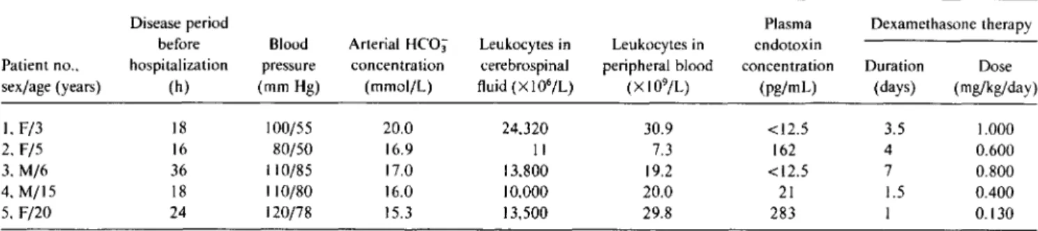
Circulating cytokines and sTNFr. The plasma concentra-tions ofTNF and IL-I were below detection level in all sam-ples. At admission, IL-6 (range, 365-2550pg/rnl.; median, 860), IL-I ra (range, 2840-4680 pg/rnl.; median, 3740), sTNFr-55 (range, 2782-5215 pg/rnl.; median, 3873), and sTNFr-75 (range, 5700-17282 pg/ml.; median, 12910)
were increased (figure I). Within 2 days, these concentra-tions fell to normal levels.
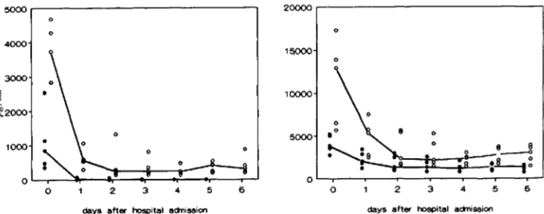
Ex vivo cytokine production. The ex vivo production of TNF without LPS stimulation was below detection level in all samples. With LPS stimulation, TNF production was be-low detection level during the acute stage of the disease. After 3 days, during convalescence, this production gradu-ally recovered to 8 10-3430 pg/mL (median, 2260) at day 6 (figure 2).
The course of IL-I production showed a similar pattern: no measurable production at admission and restored LPS-stimulated production during recovery at day 6 (range, 945-4300pg/ml.;median, 2325).
IL-6 was detectable at admission in unstimulated and in LPS-stimulated tubes. However, after correction for the cir-culating serum concentrations, the ex vivo production ofIL-6 at admission was negligible. Similar to the pattern of ex vivo production of TNF and IL-l, the LPS-stimulated pro-duction ofIL-6 was restored during recovery, reaching 165-9000 pg/mL (median, 2850) at day 6.
The ex vivo production of IL-I ra followed a different pat-tern. At admission, the production ofIL-1 ra in both unstimu-lated and LPS-stimuunstimu-lated cultures appeared to be increased. The concentration at day 0 in the unstimulated tubes ranged from 4450 to 6510 pg/rnl, (median, 5570); in the LPS-stimu-lated tubes, this was 7080-12,990 pg/rnl, (median, 11,140). The LPS-stimulated production decreased during the next 2 days toward a stable median of 6710 pg/rnl. from day 2 to day 6. The unstimulated production declined gradually to-ward a median of 1140 pg/ml, at days 4-6.
The ex vivo production of sTNFr was not measured be-cause previous studies demonstrated that TNF receptors were only minimally released in the whole blood culture sys-tem on LPS stimulation (data not shown).
A similar pattern of ex vivo production was observed in all patients, regardless of their leukocyte number or corticoste-roid dose. Thus, the observed pattern in ex vivo cytokine production was not influenced by these parameters. Discussion
We followed the concentrations of circulating cytokines and their ex vivo production during the course of
meningo-110 1994;169 (January) Cytokines in Meningococcal Infections 159 6 5 4 3 2
days after hospital actnission
o 5000 15000 10000 20000~---, 6 5 4 3 2
days after hospital actnission
o 1000 3000 4000
5000 . . - - - , Figure 1. Circulating cytokine
concentrations and their inhibitors during course of disease. Tumor necrosis factor-a (TNF) and inter-leukin (IL )-1{j were below detec-tion levels (respectively, 100 and 80 pg/mL) in all samples and there-fore are not shown. Left: patterns oflL-6(e)and IL-I receptor antag-onist (0). Right: courses of soluble TNF receptors p55 (e) and p75 (0). Median values have been in-terpolated.
coccal infections. The onset of acute meningococcal infec-tions is abrupt, with a rather short disease period before hospi-talization. As such, meningococcal infections resemble experimental models using endotoxin challenge.
In our patients, circulating TNF and IL-I concentrations were all below detection level. Several factors may be respon-sible for this negative finding. It may be possible that we missed the initial cytokinemia. The disease period before ad-mission ranged from 16 to 36 h, and it is known that after LPS injection in human volunteers, TNF and IL-l levels peak within 90-180 min [I, 2]. From clinical studies, we know that high TNF concentrations occur early during se-vere meningococcal infections [8-10]. The relatively low en-dotoxin concentrations at admission, the good prognostic score, and the absence of shock in 4 of the 5 patients may also explain the low TNF and IL-l concentrations [8-10, 27, 28]. Finally, a slight increase in TNF and IL-I could have been missed because of the relatively high detection level of our assays. Recently, we have been able to detect rapidly declining TNF and IL-l concentrations early in the course of very severe and lethal meningococcal disease (unpublished data). In the presen t series, actual activation of the cytokine
network was documented by elevated concentrations of IL-6. Furthermore, the increased sTNFr-55 and sTNFr-75 lev-els are compatible with TNF activity on target cells in tissues, because binding of TNF to its receptors on these cells leads to shedding of the extramembranous part of the receptor [5, 29]. IL-I ra concentrations were also elevated during the acute stage. The high concentrations of both sTNFr and IL-l ra earIL-ly in the course of the infection suggest a reguIL-lating role for these antiinflammatory compounds.
The LPS-stimulated ex vivo production of the proinflam-matory cytokines TNF, IL-I, and IL-6 was suppressed during the acute stage. Gradually the production capacity was re-stored during reconvalescence. In contrast, the capacity to produce ex vivo IL-I ra was regulated inversely (figure 2). The LPS-stimulated IL-I ra production was maximal during the acute stage and reached stability after 2 days. Moreover, in the unstimulated cultures at admission, a spontaneous pro-duction of IL-I ra was observed, which gradually decreased during convalescence.
We used a whole blood culture system to measure the ex vivo cytokine production. In vitro studies with isolated pe-ripheral blood mononuclear cells (PBMC) or monocytes
Figure 2. Ex vivo cytokine pro-duction of tumor necrosis factor-a (TNF). interleukin (IL)-I{j, IL-6 and IL-I receptor antagonist (IL-Ira) during course of disease. Left: median concentrations(n = 5) in whole blood cultures after 24 h ex vivo incubation without lipo-polysaccharide (LPS) stimulation. Right: median concentrations after incubation with 10p,g/mL LPS. pg/mL 15000 10000 5000 o 2 3 4 5 6
days after admission
pg/mL
15000
10000
5000
o 3 4 5 6
160 van Deuren et at. JID 1994; 169 (January)
have yielded a substantial part of our knowledge of the cyto-kine response to infectious stimuli. However, isolation of PBMC requires large blood volumes and is laborious and difficult to organize. Whole blood culture systems are a suit-able alternative in these circumstances [23, 30-32]. In addi-tion, the whole blood system represents the cytokine re-sponse of all types of cells present in blood at a certain time point and may therefore be a more realistic assessment than measurements using a fixed number of isolated and cultured cells.
Impaired ex vivo production of the proinflammatory cyto-kines TNF and IL-l during acute infection has been reported by others [18-21]. Our study confirms these observations but also shows that the impaired capacity to produce proin-flammatory cytokines does not indicate that the cells present in blood are completely refractory in terms of cytokine pro-duction. Rather, they seem to have switched from a mainly proinflammatory action (as in healthy individuals) to an an-tiinflammatory action. During recovery, the cells gradually switch again to the balanced proinflammatory status.
Further research should be done on the mechanism be-hind this programmed switch. Our findings in meningococ-cal infection may represent a process similar to endotoxin tolerance, the phenomenon that survival in experimental an-imals is markedly increased if a lethal dose of endotoxin is preceded by a smaller dose. Inhibition of the production of TNF, IL-l, and IL-6 is regarded as crucial [33]. Similarly, in human volunteers receiving endotoxin intravenously, the ex vivo production by CD 14+ cells of TNF, IL-l, and IL-6 is significantly decreased [34]. IL-4 and IL-l 0 may playa role in this process. IL-l 0 and IL-4 both down-regulate the LPS-stimulated production by human PBMC and monocytes of TNF, IL-l, and IL-6, whereas preincubation with IL-4 in-creases the synthesis of IL-l ra [35-37]. Better insight into these regulatory mechanisms may enable us to understand why in some patients the invasion in the bloodstream of
Neisseria meningitidisinduces high plasma concentrations of TNF, IL-l, and IL-6 with severe shock, whereas in others a localized meningitis develops with low systemic concentra-tions ofthese cytokines [8-10]. Further study on these regula-tory mechanisms may provide new therapeutic interven-tions.
In conclusion, antiinflammatory compounds such as sTNFr and IL-l ra are present in the circulation during early meningococcal infection. The down-regulation of the pro-duction of TNF, IL-l, and IL-6 and up-regulation of the production of IL-l ra during acute infection could serve as a mechanism of protection.
Acknowledgments
We thank G. R. Adolph (Boehringer, Vienna) for theTNFa,
P. Graber (Glaxo, Geneva) for the IL-l fJ, D. Boraschi (Sclavo,
Sienna, Italy) for the anti-IL-IfJ antibodies, andC.A.Dinarello for the anti-IL-l ra antibodies.
References
I. Hesse DK. Tracey KJ, Fong Y, et at. Cytokine appearance in human endotoxemia and primate bacteremia. Surg Gynecol Obstet 1988; 166: 147-53.
2. Cannon JG. Tompkins RG, Gelfland JA, et al. Circulating interleukin-I and tumor necrosis factor in septic shock and experimental fever. J Infect Dis 1990; 161:79-84.
3. Granowitz EV, Santos AA. Poutsiaka DD, et at. Production
ofinterleu-kin-lreceptor antagonist during experimental endotoxaemia. Lancet 1991;2: 1423-4.
4. Spinas GA, Keller U, Brockhaus M. Release of soluble receptors for tumor necrosis factor (TNF) in relation to circulating TNF during experimental endotoxinemia. J C1in Invest 1992;90:533-6. 5. Van Zee KJ. Kohno T. Fischer E. Rock CS, Moldawer LL. Lowry SF.
Tumor necrosis factor soluble receptors circulate during experimen-tal and clinical inflammation and can protect against excessive tumor necrosis factor a in vitro and in vivo. Proc Natl Acad Sci USA
1992;89:4845-9.
6. Michie HR, Spriggs DR. Manogue KR. et at. Tumor necrosis factor and endotoxin induce similar metabolic responses in human beings. Sur-gery 1988; 104:280-5.
7. Okusawa S. Gelfland JA. Ikejima T, Connolly RJ. Dinarello CA. Inter-leukin I induces a shock-like state in rabbits. Synergism with tumor necrosis factor and the effect of cyclooxygenase inhibition. 1 Clin Invest 1988;81: 1162-72.
8. Waage A. Halstensen A. EspevikT.Association between tumor necro-sis factor in serum and fatal outcome in patients with meningococcal disease. Lancet 1987;1:355-7.
9. Girardin E. Grau GE, Dayer 1M, Roux-Lombard P. 15 Study Group, Lambert PH. Tumor necrosis factor andinterleukin-Iin the serum of children with severe infectious purpura. N Engl J Med 1988;319: 397-400.
10. Waage A. Brandtzaeg P, Halstensen A. KierulfP, EspevikT.The com-plex pattern ofcytokines in serum from patients with meningococcal septic shock. Association between interleukin 6, interleukin I.and fatal outcome. 1 Exp Med 1989; 169:333-8.
II. Girardin E, Roux-Lombard P, Grau GE, et al. Imbalance between tu-mor necrosis factor-a and soluble TNF receptor concentrations in severe meningococcaemia. Immunology 1992;76:20-23.
12. Dinarello CA. Thompson RC Blocking IL-I: interleukin-I receptor antagonist in vivo and in vitro. Immunol Today 1991;12:404-10. 13. Alexander HR. Doherty GM, Buresh CM. Venzon DJ, Norton JA. A
recombinant human receptor antagonist to interleukin I improves survival after lethal endotoxemia in mice. 1 Exp Med 1991;173: 1029-32.
14. Wakabayashi G. Gelfland JA. Burke JF. Thompson RC Dinarello CA. A specific receptor antagonist for interleukin I prevents Escherichia
coliinduced shock in rabbits. FASEB J 1991;5:338-43.
15. Fischer E, Marano MA, Van Zee KJ, et at. Interleukin-I receptor block-ade improves survival and hemodynamic performance in Escherichia
coliseptic shock. but fails to alter host responses to sublethal
endo-toxemia. J Clin Invest 1992;89:1551-7.
16. Schindler R, Mancilla J. Endres S.Ghorbani,Clark SC Dinarello CA. Correlations and interactions in the production ofinterleukin-6 (IL-6). IL-I and tumor necrosis factor (TNF) in human blood mononu-clear cells: IL-6 suppresses IL-I and TNF. Blood 1990;75:40-7. 17. Poutsiaka DD, Clark BD, Vannier E. Dinarello CA. Production of
in-terleukin-I receptor antagonist and interleukin-ItI by peripheral blood mononuclear cells is differentially regulated. Blood 1991;78:
JID 1994; 169 (January) Cytokines in Meningococcal Infections 161
18. Luger A. Graf H. Schwarz HP. Stummvoll HK. Luger T A. Decreased serum interleukin I activity and monocyte interleukin I production in patients with fatal sepsis. Crit Care Med 1986; 14:458-61. 19. Helminen M. VesikariT. Interleukin-I production in bacterial
meningi-tis. Scand J Infect Dis 1990;22: 105-8.
20. Simpson SQ, Modi H. Balk RA, Bone RC'. Casey LC'. Reduced alveolar macrophage production of tumor necrosis factor during sepsis in mice and man. Crit Care Med 1991;19: 1060-6.
21. Helminen M. Interleukin-I production from peripheral blood mono-cytes in septic infections in children. Scand J Infect Dis 1991; 23:607-11.
22. Van Deuren M. Santman FW. Van Dalen R. Sauerwein RW. Span LFR. Van der Meer JWM. Plasma exchange and whole blood ex-change in meningococcal sepsis. Clin Infect Dis 1992; 15:424-30. 23. Nerad JL. Griffiths K, Van der Meer JWM. et at. lnterleukin-l d
(IL-113). IL-I receptor antagonist. andTNFaproduction in whole blood. J Leukoc BioI 1992;52:687-92.
24. Van der Meer JWM. Endres S. Lonnemann G. et al. Concentrations of immunoreactive human tumor necrosis factor alpha produced by human mononuclear cells in vitro. J Leukoc BioI 1988;43: 16-23. 25. Lisi PJ. Chu CW. Koch GA, Endres S, Lonnemann G. Dinarello CA.
Development and use of radio immunoassay for human interleukin-113. Lymphokine Res 1987;6:229-44.
26. Barrera P, Boerbooms AMT, Janssen EM, et at. Circulating soluble tumor necrosis factor receptors. interleukin-Z receptors. tumor ne-crosis factora,and interleukin-6 levels in rheumatoid arthritis. Lon-gitudinal evaluation during methotrexate and azathioprine therapy. Arthritis Rheum 1993;36: 1070-9.
27. Brandtzaeg P. Kierulf'P, Gaustad P. et at. Plasma endotoxin as a predic-tor of multiple organ failure and death in systemic meningococcal disease. J Infect Dis 1989; 159: 195-204.
28. Gedde-Dahl TW, Bjark P. Helby A, Hest JH, Bruun IN. Severity of meningococcal disease: assessment by factors and scores and impli-cations for patient management. Rev Infect Dis 1990;12:973-92. 29. Lantz M. Malik S. Slevin ML. Olsson I. Infusion of tumor necrosis
factor (TNF) causes an increase in circulating TNF-binding protein in humans. Cytokine 1992;2:402-6.
30. Van der Ven-Jongekrijg J. Demacker PMN. Van der Meer JWM. A simple method for measuring cytokine production in vitro [abstract]. Cytokine 1991 ;3:495.
31. Finch-Arietta MB. Cochran FR. Cytokine production in whole blood ex vivo. Agents Actions 1991;34:49-52.
32. Wilson BMG. Severn A. Rapson NT. Chana J. Hopkins P. A conve-nient human whole blood culture system for studying the regulation of tumor necrosis factor release by bacterial lipopolysaccharide. J Immunol Methods 1991;139:233-40.
33. Mengozzi M. Ghezzi P. Cytokine down-regulation in endotoxin toler-ance. Eur Cytokine Netw 1993;4:89-98.
34. Granowitz EV, Porat R. Mier JW. et al. Intravenous endotoxin sup-presses the cytokine response of peripheral blood mononuclear cells of healthy humans. J Immunol 1993; 151: 1637-45.
35. De Waal Malefijt R. Abrams J. Bennet B. Figdor CG, De Vries JE. Interleukin 10 (IL-I 0) inhibits cytokine synthesis by human mono-cytes: an autoregulatory role oflL- 10 produced by monocytes. J Exp Med 1991;174:1209-20.
36. Te Velde AA. Huijbens RJ, Heije K. De Vries JE. Figdor CG. Interleu-kin-d (IL-4) inhibits secretion of IL-I beta. tumor necrosis factor alpha, and IL-6 by human monocytes. Blood 1990;76: 1392-7. 37. Vannier E. Miller LC, Dinarello CA. Coordinated antiinflammatory
effects ofinterleukin 4: interleukin 4 suppresses interleukin I produc-tion but up-regulates gene expression and synthesis ofinterleukin I receptor antagonist. Proc Natl Acad Sci USA 1992;89:4076-80.