suggestive of a vector mite bite, and a positive serodiagnostic for O. tsutsugamushi without other rickettsial serologies. The reversal of an evolution apparently headed to a fatal outcome under a treatment active on O. tsutsugamushi provides a strong additional presumption. Antibody titers may seem low, but since serodiagnostics were performed tardily, these titers may have already been decreasing. Another explanation would be an impaired immune response that could explain the disease severity and the persistent efficacy of antibiotic therapy at a late stage.
Pneumonia is a well-documented complication of rickettsial diseases, but ARDS is rare (6 cases) [3, –7]. Our observation seems to confirm that western practitioners should suspect scrub typhus in travelers returning from Southeast Asia with respiratory symptoms (if travel conditions make vector bites possible), because of the potentially severe course.
The use of tetracyclines in populations at risk of scrub typhus has dramatically reduced the associated mortality rate and probably explains the decrease in severe complications [2, 8]. Other active antibiotics include chloramphenicol [2] and cip-rofloxacin [9]. Data on the clinical efficacy of fluoroquinolones are scarce. Ofloxacin was prescribed to our patient to broaden the spectrum of antibiotic therapy. When the diagnosis of scrub typhus was suspected, retrospective analysis of the clinical course strongly suggested a link between improvement and of-loxacin administration. The decision not to add a tetracycline to the therapeutic regimen was thus taken, and the patient’s condition continued to improve. Therefore, ofloxacin activity seems likely, all the more so because a spontaneously favorable outcome, impossible to rule out, is made unlikely but the se-verity of the MOF. The description of doxycycline and chlo-ramphenicol-resistant strains of O. tsutsugamushi in northern Thailand [10] would make the efficacy of fluoroquinolones in scrub typhus an interesting therapeutic alternative. Such
effi-cacy is suggested by the case that we describe here, although it is clear that evidence beyond a single case is needed.
Christophe Cracco,1 Christian Delafosse,1 Laurence Baril,2 Yannick Lefort,1 Capucine Morelot,1 Jean-Philippe Derenne,1Franc¸ois Bricaire,2 and Thomas Similowski1
Service de Pneumologie et Re´animation,1De´partement des Maladies Infectieuses et Tropicales, Parasitologie et Sante´ Publique,2Groupe Hospitalier Pitie´-Salpeˆtrie`re, Assistance Publique-Hoˆpitaux de Paris, Paris, France
References
1. Tamura A, Ohashi N, Urakami H, Miyamura S. Classification of Rickettsia
tsutsugamushi in a new genus, Orientia gen. nov., as Orientia tsutsugamushi
comb. nov. Int J Syst Bacteriol 1995; 45:589–91.
2. Sheehy T, Hazlett D, Turk RE. Scrub Typhus: a comparison of chloram-phenicol and tetracycline in its treatment. Arch Intern Med 1973; 132: 77–80.
3. Gotloib L, Barzilay E, Shustak A, Waiss Z, Lev A. Hemofiltration in severe high microvascular permeability pulmonary edema secondary to rickett-sial spotted fever. Resuscitation 1985; 13:25–9.
4. Sacks HS, Lyons RW, Lahiri B. Adult respiratory distress syndrome in Rocky mountain spotted fever. Am Rev Respir Dis 1981; 123:547–9. 5. Lopez Rodriguez A, Jerez V, Garcia Lombardo A, Rebollo J, Julia JA. ARDS
associated with boutonneuse fever. Chest 1989; 95:924–5.
6. Lee WS, Wang FD, Wang LS, et al. Scrub typhus complicating acute res-piratory distress syndrome: a report of two cases. Chung-Hua I Hsueh Tsa Chih (Taipei) 1995; 56:205–10.
7. Watt G, Strickman D. Life-threatening scrub typhus in a traveler returning from Thailand. Clin Infect Dis 1994; 18:624–6.
8. Kawamura A Jr, Tanaka H. Rickettsiosis in Japan. Jpn J Exp Med 1988; 58:169–84.
9. Eaton M, Cohen MT, Shlim DR. Ciprofloxacin treatment of typhus. JAMA 1989; 262:772–3.
10. Watt G, Chouriyagune C, Ruangweerayud R, et al. Scrub typhus infections poorly responsive to antibiotics in northern Thailand. Lancet 1996; 348: 86–9.
Acute Community-Acquired Diarrhea Requiring Hospital Admission in Swiss Children
In order to ascertain the prevalence of agents that cause childhood diarrheal illness, stool specimens of 312 con-secutive children with community-acquired diarrhea re-quiring admission were evaluated. Pathogens were de-tected in 166 (53%) of the 312 children (>2 pathogens in
28 children): Rotavirus (n p 75), Salmonella spp. (n p
), Campylobacter spp. ( ), Shigella spp. ( ),
37 n p 24 n p 5
Giardia spp. (n p 4), Yersinia spp. (n p 2), Aeromonas spp. (n p 15), Cryptosporidium (n p 15), enteropathogenic
Es-cherichia coli (n p 13), enterotoxigenic E. coli (n p 7), and
enterohemorrhagic E. coli (n p 5). In conclusion, acute
childhood diarrheal illness pathogens, such as Aeromonas,
Cryptosporidium, and diarrheagenic E. coli, account for a
large proportion of patients with a microbiologically pos-itive stool specimen.
Acute childhood diarrheal illness represents a major cause of morbidity even in temperate, industrialized areas. Infectious agents long been known to cause acute diarrheal illness in such climates areas include Salmonella spp., Shigella spp., Campy-lobacter spp., Yersinia spp., Rotavirus and Giardia lamblia [1]. Recent advances in our understanding of enteric pathogens and improved diagnostic techniques have identified new agents of diarrhea, such as Cryptosporidium parvum, diarrheagenic Es-cherichia coli, or Aeromonas spp. [1].
Because the relative frequency of the different etiologic agents
Reprints or correspondence: Dr. M. G. Bianchetti, University Children’s Hospital, Inselspital, CH-3010 Bern, Switzerland (mario.bianchetti@insel .ch).
Clinical Infectious Diseases 2000; 30:192–6
q 2000 by the Infectious Diseases Society of America. All rights reserved. 1058-4838/2000/3006-0043$03.00
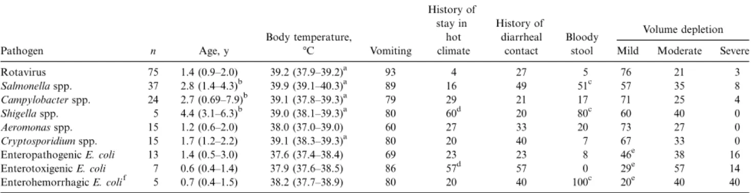
Table 1. Age, symptoms, and signs in 166 children with diarrhea, according to pathogens identified in stool specimens. Pathogen n Age, y Body temperature, 7C Vomiting History of stay in hot climate History of diarrheal contact Bloody stool Volume depletion Mild Moderate Severe
Rotavirus 75 1.4 (0.9–2.0) 39.2 (37.9–39.2)a 93 4 27 5 76 21 3 Salmonella spp. 37 2.8 (1.4–4.3)b 39.9 (39.1–40.3)a 89 16 49 51c 57 35 8 Campylobacter spp. 24 2.7 (0.69–7.9)b 39.1 (37.8–39.3)a 79 29 21 17 71 25 4 Shigella spp. 5 4.4 (3.1–6.3)b 39.0 (38.1–39.3)a 80 60d 20 80c 60 40 0 Aeromonas spp. 15 1.2 (0.6–2.0) 38.0 (37.0–39.0) 60 27 33 20 73 27 0 Cryptosporidium spp. 15 1.7 (1.2–2.2) 39.1 (38.3–39.3)a 80 20 40 7 67 33 0 Enteropathogenic E. coli 13 1.4 (0.5–3.0) 37.6 (37.4–38.4) 69 23 23 8 46e 38 16 Enterotoxigenic E. coli 7 0.6 (0.4–1.4) 37.9 (37.6–38.5) 86 57d 57 0 29e 57 14 Enterohemorrhagic E. colif 5 0.7 (0.4–1.5) 38.2 (37.7–38.9) 80 20 40 100c 20e 40 40 NOTE. Data are either median (interquartile range) or relative frequency. Data are percentages, unless otherwise indicated. Only pathogens isolated in>5 children are considered. Two or more pathogens were found in 28 of the 166 patients (this fact accounts for apparent mathematical discrepancies). E. coli, Escherichia
coli.
a
vs. Aeromonas spp. or diarrheagenic E. coli.
P!.01
b
vs. Rotavirus, Aeromonas spp., Cryptosporidium spp., and diarrheagenic E. coli.
P!.01
c
vs. Rotavirus, Campylobacter spp., Aeromonas spp., Cryptosporidium spp., and enteropathogenic or enterotoxigenic E. coli.
P!.02 d
vs. Rotavirus, Salmonella, Campylobacter, Aeromonas, Cryptosporidium, and enteropathogenic or enterohemorrhagic E. coli.
P!.05
e
vs. Rotavirus, Salmonella spp., Campylobacter spp., Shigella spp., Aeromonas spp., and Cryptosporidium spp.
P!.05
f
None of the isolates belonged to the serogroup 0157.
linked with acute infectious diarrhea greatly varies according to the geographic and socioeconomic settings [1], we undertook a study of the etiology and clinical features of community ac-quired diarrheal illness in children admitted at the Department of Pediatrics, University of Bern, Switzerland.
The Department of Pediatrics, University of Bern, Switzerland, is a 60-bed pediatric teaching hospital that serves as tertiary referral center. The catchment area has a population of approx-imately 400,000 people of various social and ethnic backgrounds. All patients aged between 5 weeks and 15 years who required hospital admission because of an acute, community-acquired diarrheal illness were eligible. Acute diarrhea was defined as an abnormal increase in stool liquidity and frequency of<9 days of duration. Children treated with antibiotics or drugs known to cause diarrhea in the preceding 2 weeks and immunocom-promised patients were excluded. Three hundred twelve con-secutive patients fulfilled these criteria and were prospectively evaluated from January 1990 through December 1994. They were 175 boys and 137 girls, ranging in age from 1.5 months to 13 years (median, 1.5 years).
The history before admission and the initial physical ex-amination were used to ascertain a recent (!10 days) stay in a
hot climate, poorly industrialized country, a recent diarrheal contact (at home or at a nursery), the quality of stools (watery or grossly bloody, by examination), the rectal body temperature (on admission), and the degree [2] of extracellular volume de-pletion (assumed to be moderate in patients with 2 and severe in those with>3 of the following signs: altered skin elasticity, sunken eyes, dry mucous membranes, absent tears, or delayed capillary refill). A complete WBC count in peripheral blood was performed in all patients. Leukocytosis was defined as a WBC count higher than the age dependent upper reference value [3]: children aged<24 months,17.53 109cells/L; children
aged 25 months to 5 years,15.53 109cells/L; and children aged >6 years,13.53 109cells/L. The leukocytes were differentiated by microscopy, and a segmented neutrophil granulocyte was defined by at least 1 indentation of the nucleus to less than one-third of the maximal nuclear diameter. A shift to the left was defined as nonsegmented polymorphonuclear cells110%
of the total WBC count [3].
In the 312 patients, a stool specimen was collected within 24 h of admission and processed for Rotavirus, Salmonella spp., Campylobacter spp., Shigella spp., Yersinia spp., Giardia lam-blia, and C. parvum at the Institute for Clinical Microbiology, University of Bern, Switzerland. Standard laboratory tech-niques were used for Salmonella spp., Campylobacter spp., Shi-gella spp., and Yersinia spp. [4]. G. lamblia and C. parvum were detected in a fresh stool specimen fixed by use of the sodium acetate-acetic acid formalin medium. The specimen was con-centrated, and a wet mount was examined for the presence of cysts and trophozoites of G. lamblia or oocysts of C. parvum, identified by staining the specimen by use of auramine-carbol-fuchsin and visualized by fluorescent microscopy [5].
Aeromonas spp., enteropathogenic E. coli, enterotoxigenic E. coli, and enterohemorrhagic E. coli were isolated at the Ref-erence Laboratory for Foodborne Diseases, University of Bern. Briefly, stool specimens were streaked onto cefsulodin-irgasan-novobiocin agar for isolation of Aeromonas spp. and Mac-Conkey agar for isolation of E. coli [6]. Suspect colonies on cefsulodin-irgasan-novobiocin agar were subcultured onto Sim-mons citrate medium and blood agar, and oxidase- and citrate-negative colonies were identified by use of standard procedures [7]. For identification of diarrheagenic E. coli, 6 representative colonies were picked from the MacConkey agar, identified by conventional procedures, and used in a colony-blot hybridi-zation assay. Probes and conditions used for the detection of
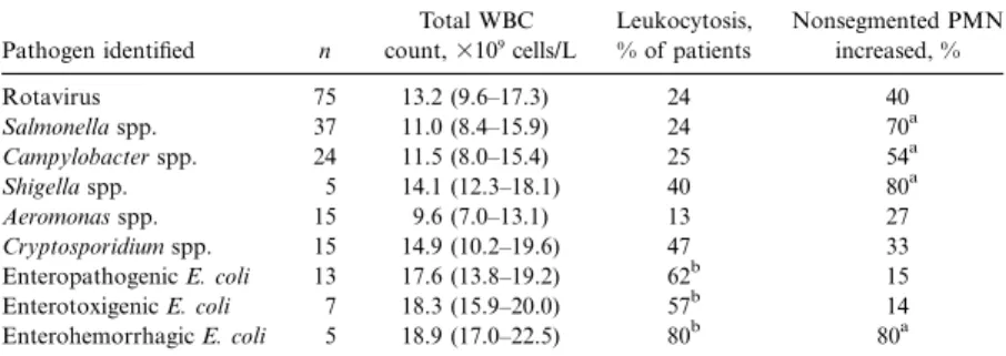
Table 2. Peripheral blood total WBC count in 166 children with diarrhea, according to pathogens identified in stool specimens.
Pathogen identified n Total WBC count,3109 cells/L Leukocytosis, % of patients Nonsegmented PMN increased, % Rotavirus 75 13.2 (9.6–17.3) 24 40 Salmonella spp. 37 11.0 (8.4–15.9) 24 70a Campylobacter spp. 24 11.5 (8.0–15.4) 25 54a Shigella spp. 5 14.1 (12.3–18.1) 40 80a Aeromonas spp. 15 9.6 (7.0–13.1) 13 27 Cryptosporidium spp. 15 14.9 (10.2–19.6) 47 33 Enteropathogenic E. coli 13 17.6 (13.8–19.2) 62b 15 Enterotoxigenic E. coli 7 18.3 (15.9–20.0) 57b 14 Enterohemorrhagic E. coli 5 18.9 (17.0–22.5) 80b 80a
NOTE. Data are either median (interquartile range) or relative frequency. Only pathogens isolated in>5 children are considered. Two or more pathogens were found in 28 of the 166 patients (this fact accounts for apparent mathematical discrepancies). Leukocytosis was defined according to the lit-erature [3] and “shift to the left” nonsegmented PMN110% of the total WBC count [3]. E. coli,
Escherichia coli; PMN, polymorphonuclear cells.
a
vs. Rotavirus, Aeromonas species, Cryptosporidium species, enteropathogenic E. coli, and
P!.01
enterotoxigenic E. coli. b
vs. Rotavirus, Salmonella species, Campylobacter species, Shigella species, Aeromonas
P!.01
species, and Cryptosporidium species.
Figure 1. Monthly distribution of admission for acute diarrheal illness in 312 consecutive patients from January 1990 through Decem-ber 1994. Patients with (m) and those without (M) isolated pathogens are distinguished.
enterotoxigenic E. coli (heat stable and heat labile enterotoxins), enteropathogenic E. coli (probes for the virulence plasmid as-sociated EAF and chromosomal eaeA genes), and enterohe-morrhagic E. coli (probes for Shigatoxin and the virulence gene eaeA) have been described elsewhere [8–10]. Enteroinvasive E. coli were detected by use of a digoxigenin-labeled ipaH probe [11] produced by PCR using the primer pair 50
-CTGGCTGAT-GCCGTGACAGC-30(forward), 50
-CGGTCAGCCACCCTC-TGAGA-30 (reverse), and genomic deoxyribonucleic acid of Shigella flexneri NZ 194-95 as a template.
Isolates of enterohemorrhagic E. coli were agglutinated in antisera against O157 and H7 antigens of E. coli in commer-cially available sera including passage of the strains in semisolid motility medium to enhance expression of flagellar antigens. The presence of O157 antigens was also assessed by aggluti-nation in a latex reagent, rapid sorbitol fermentation was as-sessed on Sorbitol-MacConkey plates (Oxoid, Basingstoke, UK), and production of glucuronidase was measured with a fluorogenic substrate (Bactident; Merck, Germany).
The results are given as relative frequency or as median and interquartile range. The x2test (with the Yates correction) and
the 2-tailed Kruskal-Wallis test (with the Bonferroni adjust-ment) were used for analysis [12]. Differences that had a prob-ability1.05 by the appropriate null hypothesis were considered insignificant.
Pathogens were detected in stool samples from 166 of the 312 patients with acute diarrheal illness included in the study (>2 pathogens were found concomitantly in 28 children): Ro-tavirus, Salmonella spp., Shigella spp., Aeromonas spp., Cryp-tosporidium spp., diarrheagenic E. coli, and Campylobacter spp. were isolated each in at least 5 children (table 1). G. lamblia (n p 4; 1.3%) and Yersinia spp. (n p 2; 0.6%) were detected in a small minority of patients. No enteroinvasive E. coli were detected.
The salient characteristics of patient history and clinical pre-sentation are summarized in tables 1 and 2. Children infected with Salmonella spp., Campylobacter spp., or Shigella spp. were significantly older than were children infected with Rotavirus, Cryptosporidium spp., Aeromonas spp., or diarrheagenic E. coli. A history of travel abroad was given by roughly two-thirds of patients infected with Shigella and enterotoxigenic E. coli, whereas on average 85% of the other pathogens were domesti-cally acquired. Although the body temperature was significantly higher in patients with acute diarrheal illness caused by Rota-virus, Salmonella spp., Campylobacter spp., or C. parvum, there was a considerable overlap with findings in patients infected with other pathogens, and clinical symptoms did not permit a clear distinction of diarrheal syndromes.
This was also true for the results of the total WBC count. Peripheral blood leukocytosis was noted in the majority of the patients with acute diarrheal illness caused by diarrheagenic E.
coli. A shift to the left was noted in the majority of the children with diarrhea caused by Salmonella spp., Campylobacter spp., Shigella spp., or enterohemorrhagic E. coli. However, grossly bloody diarrhea was present in all patients infected with en-terohemorrhagic E. coli (80% of those with shigellosis and 51% of those with salmonellosis). The extracellular volume depletion was slightly more pronounced (moderate or severe) in patients with acute diarrheal illness caused by diarrheagenic E. coli. An 8-month-old girl with bloody diarrhea caused by enterohe-morrhagic E. coli developed hemolytic uremic syndrome. Chil-dren with acute diarrheal illness were admitted throughout the year (figure 1). However, admissions tended to be more frequent during the warmer months. No etiology-specific seasonality was noted.
The results of the present study demonstrate that in this region pathogens, such as C. parvum, enteropathogenic E. coli, enterotoxigenic E. coli, enterohemorrhagic E. coli, or Aero-monas spp., play an important role in the etiology of inpatients with acute diarrheal illness, as indicated by the fact that these pathogens were isolated in stool specimens from 18% of the pediatric patients with acute community acquired diarrheal ill-ness that required admission.
The well-established pathogens Rotavirus, Salmonella spp., Campylobacter spp., Shigella spp., and Yersinia spp. [1] still represent a major cause of acute diarrheal illness in Swiss chil-dren. Compared with a survey performed at this institution from 1981 through 1984 [4], the relative frequency of acute diarrheal illness caused by Rotavirus tended to decrease (from 42% to 24%). In contrast, there was an increase in diarrhea caused by Salmonella spp. (from 9% to 12%) and Campylo-bacter spp. (from 3% to 8%).
The most frequent so-called emerging pathogen in this study was C. parvum. This protozoan was long known to cause di-arrheal diseases in the immunocompromised but is now a rec-ognized agent of acute diarrhea in all groups of patients [12]. Cryptosporidium also causes an important number of sporadic cases of diarrhea worldwide, with widely differing prevalence rates among regions [12]. The identification of Cryptosporidium is important for prognostic, epidemiological, and therapeutic reasons, since the symptoms tend to persist or recur for pro-longed periods [13]. The organism is easily transmitted from person to person, and no uniformly efficacious therapy is avail-able [12].
In this study, Aeromonas spp. was detected in 4.8% of the stool specimens. This frequency is similar to that reported from other industrialized countries (2.2%–10%). Although the epi-demiological association between diarrheal disease and the presence of Aeromonas spp. in stools has been firmly established in many studies worldwide, there is still some controversy about its role as a pathogen [7]. A common clinical picture caused by isolates of Aeromonas spp. emerges from the literature: acute-onset watery diarrhea in infants and toddlers with slightly el-evated body temperature. Vomiting is less common than in
diarrhea caused by other pathogens, and the total WBC count rarely shows a leukocytosis or a shift to the left [14].
The most significant advances in our understanding of enteric pathogens have been made in the study of strains of E. coli that cause diarrhea [15]. Enteropathogenic, enterotoxigenic, and enterohemorrhagic [15] E. coli accounted together for 8.0% of the positive stool specimens in the present study. Thus, as a group, these pathogens were as prevalent as Campylobacter spp., being the third most frequent pathogens isolated. Among the diarrheagenic E. coli, enteropathogenic E. coli was the most common type detected. Although often considered a pathogen in developing countries, 77% of the cases observed were do-mestically acquired, which concurs with recent observations in the United States [16]. The second most prevalent category of diarrheagenic E. coli were enterotoxigenic E. coli. They are the main agent of travelers’ diarrhea, and, consistent with this, all but one of the patients shedding this organism had recently traveled to areas where this pathogen is prevalent [15].
The most important new pathogen in terms of the severity of associated illness, as well as its public health impact, is en-terohemorrhagic E. coli [15]. All 5 case patients showed the typical picture of hemorrhagic colitis, including severe abdom-inal pain and grossly bloody diarrhea. Some individuals de-velop hemolytic uremic syndrome after the initial diarrheal phase of illness [17], as was observed in 1 of the patients with enterohemorrhagic E. coli infection. None of the isolates be-longed to the E. coli serogroup O157, consistent with the rarity of this strain in most European countries [9, 15]. The entero-hemorrhagic E. coli prevalence in the present study is similar to data obtained several years ago [10].
Despite attempts to identify many pathogens, no pathogen was isolated in ∼50% of cases. Several viral pathogens like Astrovirus or other enteric viruses [18, 19] as well as parasites, were not looked for.
Bettina Essers,1 Andre´ P. Burnens,2 Francesco M. Lanfranchini,1 Stefano G. E. Somaruga,1 Rodo O. von Vigier,1 Urs B. Schaad,1 , 4 Christoph Aebi,1 , 3and Mario G. Bianchetti1 1Department of Pediatrics,2Reference Laboratory for Foodborne Diseases, and3Institute for Clinical Microbiology, University of Bern, Bern;4Department of Pediatrics, University of Basel, Basel, Switzerland
References
1. Sherman PM, Petric M, Cohen MB. Infectious gastroenterocolitides in chil-dren: an update on emerging pathogens. Pediatr Clin North Am 1996; 43:391–407.
2. Gugler E. Die akute Dehydratation bei Kindern. Ther Umsch 1994; 51: 616–21.
3. Walters MC, Abelson HT. Interpretation of the complete blood count. Pediatr Clin North Am 1996; 43:599–622.
4. Nielsen J, Schilt U, Heinzer I, Schaad UB. Les diarrhe´es infectieuses de l’enfant. Schweiz Med Wochenschr 1987; 117:518–26.
5. Nguyen XM. Intestinale Cryptosporidiose: eine seltene Durchfallserkrankung beim Menschen. Schweiz Med Wochenschr 1985; 115:1205–8.
6. Altorfer R, Altwegg M, Zollinger Iten J, von Graevenitz A. Growth of
Aero-monas spp. on cefsulodin-Irgasan-novobiocin agar selective for Yersinia enterocolitica. J Clin Microbiol 1985; 22:478–80.
7. Abbott SL, Cheung WK, Kroske Bystrom S, Malekzadeh T, Janda JM. Identification of Aeromonas strains to the genospecies level in the clinical laboratory. J Clin Microbiol 1992; 30:1262–6.
8. Boss, P, Monckton RP, Nicolet J, Burnens AP. Nachweis von toxingenen verschiedener E. coli pathotypen beim schwein mit nichtradioaktiv mar-kierten sonden. Schweiz Arch Tierheilkd 1992; 134:31–7.
9. Burnens AP, Frey A, Lior AH, Nicolet J. Prevalence and clinical significance of vero-cytotoxin–producing Escherichia coli (VTEC) isolated from cattle in herds with and without calf diarrhoea. Zentralbl Veterinarmed B 1995; 42:311–8.
10. Burnens AP, Boss P, Orskov F, et al. Occurrence and phenotypic properties of verotoxin producing Escherichia coli in sporadic cases of gastroenteritis. Eur J Clin Microbiol Infect Dis 1992; 11:631–4.
11. Venkatesan MM, Buysse JM, Kopecko DJ. Use of Shigella flexneri ipaC and
ipaH gene sequences for the general identification of Shigella spp. and
enteroinvasive Escherichia coli. J Clin Microbiol 1989; 27:2687–91.
12. Guerrant RL. Cryptosporidiosis: an emerging, highly infectious threat. Emerg Infect Dis 1997; 3:51–7.
13. MacKenzie WR, Schell WL, Blair KA, et al. Massive outbreak of waterborne cryptosporidium infection in Milwaukee, Wisconsin: recurrence of illness and risk of secondary transmission. Clin Infect Dis 1995; 21:57–62. 14. San Joaquin VH. Aeromonas, Yersinia, and miscellaneous bacterial
entero-pathogens. Pediatr Ann 1994; 23:544–8.
15. Nataro JP, Kaper JB. Diarrheagenic Escherichia coli. Clin Microbiol Rev 1998; 11:142–201.
16. Bokete TN, Whittam TS, Wilson RA, et al. Genetic and phenotypic analysis of Escherichia coli with enteropathogenic characteristics isolated from Se-attle children. J Infect Dis 1997; 175:1382–9.
17. Imoberdorf G, Bianchetti MG, Rossi E, Gugler E, Oetliker OH. Ha¨moly-tisches-ura¨misches Syndrom bei Kindern: Retrospektive u¨ber 19 Jahre. Schweiz Med Wochenschr 1993; 123:1439–44.
18. Lew JF, Moe CL, Monroe SS, et al. Astrovirus and adenovirus associated with diarrhea in children in day care setting. J Infect Dis 1991; 164:673–8. 19. Caprioli A, Pezzella C, Morelli R, et al. Enteropathogens associated with
childhood diarrhea in Italy. Pediatr Infect Dis J 1996; 15:876–83.
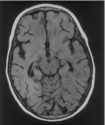
Figure 1. MRI from the brain of a 25-month-old patient with sub-acute sclerosing panencephalitis, showing increased signal intensity of the complete right hemisphere and the left frontal lobe.
Rapid Progressive Subacute Sclerosing Panencephalitis in a 2-Year-Old Child with Congenital Athyreosis
We present the unique case of a 2-year-old girl with congenital athyreosis who acquired primary measles virus infection at the age of 18 months, coincidentally with an Epstein-Barr virus infection. First neurologic symptoms of subacute sclerosing panencephalitis appeared 5 months later, and the girl died within 6 months after a rapid pro-gressive illness. Factors possibly predisposing to this extraordinary disease course—primary measles virus in-fection at an early age and lack of evidence for immuno-deficiency—are discussed.
Subacute sclerosing panencephalitis (SSPE) is a rare subacute infection of the CNS caused by measles virus (MV) [1]. The invariably fatal disease occurs several years after primary MV infection [2] and is characterized by uncontrolled replication of mutated and defective MV in neuronal and glial cells [3]. MV infection that occurs before 2 years of age is associated with a risk for SSPE that is 16 times as high as the risk associated with infection after 5 years of age [4]. SSPE that occurs before 2 years of age is extremely rare; we only found 2 cases in the literature [5, 6]. Although the characteristic course of the disease is slowly progressive, rare fulminating cases also have been reported [6, 7]. Most of these children had primary MV infection at an early age or coincidentally with a second viral infection. We report on
Reprints or correspondence: Gerd M. Lackmann, Zentrum fu¨r Kinder-heilkunde, Heinrich-Heine-Universita¨t, Moorenstr. 5, D-40225 Du¨sseldorf, Germany.
Clinical Infectious Diseases 2000; 31:196–9
q 2000 by the Infectious Diseases Society of America. All rights reserved. 1058-4838/2000/3101-0044$03.00
the case of a 2-year-old girl with congenital athyreosis who suf-fered primary MV infection coincidentally with an Epstein-Barr virus (EBV) infection at 18 months of age and who developed fulminating SSPE 5 months later.