1273
α
1,3Fucosyltransferase VI is expressed in HepG2 cells and codistributed with
β
1,4galactosyltransferase I in the Golgi apparatus and monensin-induced swollen vesicles
Lubor Borsig1, Timo Imbach, Matthias Höchli3 and
Eric G.Berger2
Institute of Physiology, University of Zürich, Winterthurerstrasse 190, CH-8057 Zürich, Switzerland and 3Elektronenmikroskopisches
Zentrallaboratorium der Universität Zürich, Gloriastrasse 30, CH-8028 Zürich, Switzerland
Received on February 15, 1999; revised on April 13, 1999; accepted on April 14, 1999
The major α1,3fucosyltransferase activity in plasma, liver, and kidney is related to fucosyltransferase VI which is encoded by the FUT6 gene. Here we demonstrate the pres-ence of α1,3fucosyltransferase VI (α3-FucT VI) in the human HepG2 hepatoma cell line by specific activity assays, detection of transcripts, and the use of specific anti-bodies. First, FucT activity in HepG2 cell lysates was shown to prefer sialyl-N-acetyllactosamine as acceptor substrate indicating expression of α3-FucT VI. RT-PCR analysis fur-ther confirmed the exclusive presence of the α3-FucT VI transcripts among the five human α3-FucTs cloned to date.
α3-FucT VI was colocalized with β1,4galactosyltransferase I (β4-GalT I) to the Golgi apparatus by dual confocal immunostaining. Pulse/chase analysis of metabolically labeled α3-FucT VI showed maturation of α3-FucT VI from the early 43 kDa form to the mature, endoglycosidase H-resistant form of 47 kDa which was detected after 2 h of chase. α3-FucT VI was released to the medium and accounted for 50% of overall cell-associated and released enzyme activity. Release occurred by proteolytical cleavage which produced a soluble form of 43 kDa. Monensin treat-ment segregated α3-FucT VI from the Golgi apparatus to swollen peripheral vesicles where it was colocalized with
β4-GalT I while α2,6(N)sialyltransferase remained associ-ated with the Golgi apparatus. Both constitutive secretion of α3-FucT VI and its monensin-induced relocation to ves-icles analogous to β4-GalT I suggest a similar post-Golgi pathway of both α3-FucT VI and β4-GalT I.
Key words: fucosyltransferase/human liver/monensin/Golgi
apparatus
Introduction
The α1,3fucosyltransferase (α3-FucT) enzyme activity has been detected in various types of tissues (Mollicone et al.,
1990, 1992; for review, see Macher et al., 1991). FucTs con-stitute a family of homologous glycosyltransferases with a high degree of identity (Lowe, 1991). This family comprises five different fucosyltransferases, named α3-FucT III to VII (Goelz et al., 1990; Kukowska-Latallo et al., 1990; Koszdin and Bowen, 1992; Weston et al., 1992a,b; Sasaki et al., 1993; Natsuka et al., 1994). These enzymes differ in their capacity to transfer fucose to distinct oligosaccharide acceptors, cation requirements and tissue specific expression (Goelz et al., 1990; Kukowska-Latallo et al., 1990; Mollicone et al., 1990, 1992; Lowe, 1991; Macher et al., 1991; Weston et al., 1992a,b; Koszdin and Bowen, 1992, Sasaki et al., 1993; Nat-suka et al., 1994). The five FucTs can be divided in two main subgroups, one comprising α3-FucTIV and VII, the other α 3-FucTIII, V, and VI which are encoded by syntenically arranged genes. These are identical to 85% rendering their spe-cific detection by molecular probes or immunological reagents difficult.
While the respective functions of α3-FucTIV and VII are being increasingly understood on the basis of their specific deletion in mice rendering them unable to synthesize selectin ligands (Lowe, 1997), the function of the three remaining FucTs needs to be further investigated. The study of develop-mental changes of tissue specific expression and the conse-quences of genetic polymorphisms all constitute approaches towards this goal (Mollicone et al., 1994). For instance the
FUT5 and FUT6 genes, respectively, encode two different
enzymes with activities exhibiting the specificity of the plasma
α1,3fucosyltransferases (Koszdin and Bowen, 1992; Weston
et al., 1992a). In several Java families the plasma α1,3fucosyltransferase activity has been found to be deficient. This deficiency was a result of a mutation in the FUT6 gene (Mollicone et al., 1994). The linkage relationship between a
α3-FucT VI mutation and deficiency of plasma fucosyltrans-ferase activity was further confirmed by work of van Dijk and associates (Brinkman-Van der Linden et al., 1996): The mis-sense mutation in the α3-FucT VI gene led to a complete absence of α3-fucosylation of serum glycoproteins. In sera of different individuals with inactivated FUT6 gene but with a functional FUT5 gene α1,3fucosyltransferase activity has not been detected (Brinkman-Van der Linden et al., 1996). This finding excluded the involvement of FUT5 in contributing to plasma FucT activity. While it seems clear that the FUT6 gene encodes the plasma α1,3fucosyltransferase activity, its tissue origin has not formally been identified. Kidney is one candi-date source of α3-FucT VI as a plasma fucosyltransferase activity. In the case of an α3-fucosylated individual with a con-genital kidney anomaly, only 10% of FucT plasma activity has been detected which could be ascribed to the myeloid type (Caillard et al., l988). Lack of expression of the Lex antigen in 1Present address: Glycobiology Program, UCSD Cancer Center, University
of California, San Diego, La Jolla, California 92093
the kidney of this individual suggested that this organ may normally contribute to plasma FucT activity (Caillard et al., l988; Mollicone et al., 1990). Another candidate for the origin of a plasma FucT activity is the liver, where transcripts of α 3-FucT VI and enzyme activity corresponding to α3-FucT VI have been detected (Mollicone et al., 1990, 1992; Johnson et
al., 1995). This organ is the source of many plasma proteins. In
addition, another glycosyltransferase, e.g. β 1,4galacto-syltransferase found in human serum originates at least par-tially from liver (Kim et al., 1972a,b).
While α3-FucT V and α3-FucT VI were investigated as recombinant enzymes expressed in CHO cells (Borsig et al., 1996, 1998) little is known on localization, intracellular trans-port, and release of endogenously expressed fucosyltrans-ferases. Since the major α1,3fucosyltransferase activity in human plasma is encoded by α3-FucT VI and putatively released from the liver, we examined whether the HepG2 hepatoma cell line expresses α3-FucT VI and investigated localization, biosynthesis, intracellular transport, and release of this enzyme in these cells. In addition, β4-GalT I and α 3-FucT VI were found to react similarly to monensin treatment which may indicate an analogous post-Golgi pathway (for review, see Dinter and Berger, 1998).
Results
Fucosyltransferase activity in HepG2 cells
Lack of fucosylation of liver-derived serum glycoproteins in patients affected by a deficiency of α3-FucT VI
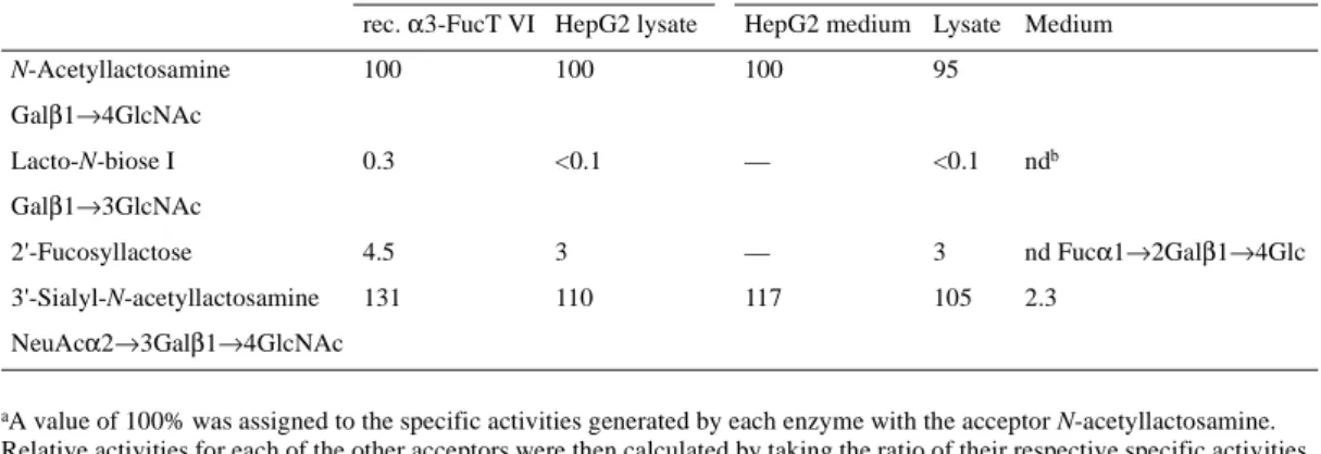
(Brinkman-Van der Linden et al., 1996) prompted us to investigate α 3-FucT activity in lysates of HepG2 cells, an established liver carcinoma cell line. To determine the nature of the α 1,3fuco-syltransferase activity detected in HepG2 cells we used the acceptor substrates listed on Table II. Acceptor substrate pref-erence of the overall fucosyltransferase activity was directed toward type 2 acceptors, N-acetyllactosamine (LacNAc), and its sialylated derivative 3’-sialyllactosamine (sLacNAc). In addition, type 1 acceptor lacto-N-biose as well as type 6 accep-tor 2’-fucosyllactose were poorly utilized (The definition of acceptor types are as follows: type 1: Galβ1→3GlcNAc; type 2: Galβ1→GlcNAc; type 6: Galβ1→4Glc). The very low ratio of utilization of type 1 to type 2 acceptors allowed to exclude expression of FucTIII to a significant amount. Preference for type 2 acceptor (neutral or sialylated) were in good agreement with the previously reported results of FucT activity in human liver cells (Jezequel-Cuer et al., 1993) and in human serum (Sarnesto et al., 1992). The majority of the α3-FucT activity in plasma is due to α3-FucT VI which is encoded by the FUT6 gene (Brinkman-Van der Linden et al., 1996). Indeed, a very similar acceptor specificity profile has been obtained with cloned α3-FucT VI expressed in CHO (Borsig et al., 1998), COS cells (Koszdin and Bowen, 1992), or insect cells (De Vries et al., 1997). To assign HepG2 cell-associated α3-FucT activity to α3-FucT VI or α3-FucT V, the enzyme activity of
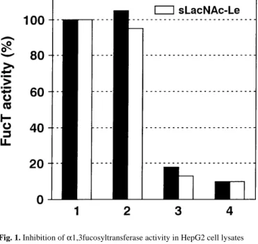
α1,3fucosyltransferase from HepG2 cell lysates was subjected to a neutralization experiment with a specific antiserum (desig-nated OLI) raised against recombinant α3-FucT VI (Borsig et
al., 1998) (Figure 1). The OLI antiserum was able to inhibit the
enzymatic activity with LacNAc and sLacNAc as respective acceptors by at least 90%. The inhibition with antibodies was similar with both acceptors suggesting the expression of either
α3-FucT VI or α3-FucT V.
α3-FucT VI is the only α1,3fucosyltransferase expressed in HepG2 cells
Since the OLI antiserum raised against α3-FucT VI also cross-reacts with α3-FucT V and α3-FucT III (Borsig et al., 1998), further determination of the α3-FucT expressed in HepG2 cells Fig. 1. Inhibition of α1,3fucosyltransferase activity in HepG2 cell lysates
using the OLI antiserum. α3-FucT VI activity was assayed in lysates of HepG2 cells as described in Materials and methods using LacNAc and sialyl-N-acetyllactosamine as acceptors, respectively. 1, Control assay mixture of 50 µl supplemented with 20 µl of H2O. 2, As in 1, with 20 µl of preimmune serum
(PIS). 3, As in 1, with 10 µl of PIS and 10 µl of OLI antiserum. 4, As in 1, with 20 µl of OLI antiserum.
Table I. PCR primers for amplifying fucosyltransferases
aU, Upper strand primer; L, lower strand primer.
bNumbers in parentheses indicates fragment size in base pairs.
mRNA species detected
Primersa
3-FucT III(585)b U: 5’ACCACTGGGATATCATGTCCAACCCTAAGT3’
L: 5’GGGCCAGGTCCTTGGGGCTCTGGAAGTCG3’
α3-FucT IV(589) U: 5’GGGGCATCCAGGCGCACACTGC3’ L: 5’CGCTCGTAGTTGGCACGGTCTG3’
α3-FucT V(811) U: 5’CCAGGGCTTATGGCAGTGGAACCTGTCAC3’ L: 5’GGGCCAGGTCCTTGGGGCTCTGGAAGTCG3’
α3-FucT VI(737) U: 5’ATCCCACTGTGTACCCTAATGG3’ L: 5’CGGCAGGAACCTCTCGTAGTTG3’
α3-FucT VII(448) U: 5’CCTGGGTGGTCAGCAACTTC3’ L: 5’CGGTCACAGATGGCACAGAAAC3’
was done by RT-PCR analysis. Based on activity measure-ments, α3-FucT VI and α3-FucT V could be expected. To be able to distinguish between α3-FucT V and α3-FucT VI expression, we used specific primers for α3-FucT V (Borsig et
al., 1998) and α3-FucT VI (Table I). Absence of a signal in controls and specific amplification from cDNA and genomic DNA showed expression solely of α3-FucT VI in HepG2 cells (Figure 2, lane 4). PCR amplification with α3-FucT V primers yielded no product (Figure 2, lane 1). RT-PCR amplification with specific primers for α3-FucT III were also negative (Fig-ure 2, lane 17). Although crossreactivity of OLI antibodies with α3-FucT IV and α3-FucT VII was not observed (data not shown), RT-PCR analysis was carried out, also with negative results (Figure 2, lanes 9, 13). Taken together, we conclude that among the five cloned α3-FucTs α3-FucT VI only is expressed in HepG2 cells.
α1,3Fucosyltransferase VI is localized to the Golgi apparatus
To determine the steady-state distribution of α 1,3fuco-syltransferases VI, HepG2 cells were subjected to indirect con-focal immunofluorescence microscopy (Figure 3). For staining of α3-FucT VI, previously characterized polyclonal affinity
purified OLI antibodies were used (Borsig et al., 1998) (Figure 3C). A specific Golgi staining was found using OLI antibodies to α3-FucT VI (Figure 3C), while preimmune serum (Figure 3A) or staining with antibodies preabsorbed with rα3-FucT VI antigen (Figure 3B) produced background staining only. Golgi location of FucT was further confirmed by double confocal immunofluorescence staining with a mono-clonal antibody to β1,4-galactosyltransferase I (Berger et al., 1986) indicating colocalization of both antigens (Figure 3C,D).
Maturation of α3-FucT VI in HepG2 cells and its release into the medium
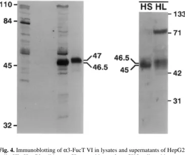
To determine the molecular weight of α3-FucT VI expressed in and released from HepG2 cells, immunoblotting of cell lysates and supernatants with OLI antibodies was carried out (Figure 4). In HepG2 cells, α3-FucT VI appeared as a 46.5 kDa protein, thus slightly smaller than the recombinant enzyme in CHO cells which was detected as a 47 kDa band (Borsig et al., 1998). The small difference might be due to cell-type-specific glyco-sylation. The enzyme released from HepG2 cells migrated as a 45 kDa protein indicating an analogous processing step for the endogenously expressed enzyme as for the recombinant enzyme described previously (Borsig et al., 1998; Grabenhorst et al., 1998). Fucosyltransferase activity was also measured in the medium (Table II). The specificity of the released enzymatic activity was similar to the intracellular enzyme. The cumula-tive amount of enzyme released was half of total activity recovered in the cell lysate and in the medium. Maturation of
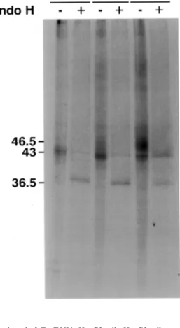
α3-FucT VI in HepG2 cells was analyzed by metabolic labe-ling followed by immunoprecipitation. HepG2 cells were sub-jected to pulse-chase analysis (Figure 5). The 43 kDa form (no chase), corresponding to the core glycosylated enzyme, par-tially shifted to 46.5 kDa after 60 min, which became prepon-derant after 2 h. The 43 kDa form was sensitive to endo-H treatment (no chase) or PNGase treatment (not shown) and was reduced to a 36.5 kDa form indicating that all four N-glyco-sylation sites are occupied. After 2 h chase, endoglycosidase-H treatment reduced the mature form to ~45 kDa (chase 120 min) while converting the nonprocessed forms to 36.5 kDa. The Table II. Measurement of Fuc-T activity in lysates of HepG2 cells and in the medium; comparison with recombinant α3-FucT
VI activity from CHO cells
aA value of 100% was assigned to the specific activities generated by each enzyme with the acceptor N-acetyllactosamine.
Relative activities for each of the other acceptors were then calculated by taking the ratio of their respective specific activities to the specific activity observed with N-acetyllactosamine;
bNot determined.
Acceptor activity of α1,3fucosyltransferase
Relative activitya (%) Activity (pmol/min/mg prot.)
rec. α3-FucT VI HepG2 lysate HepG2 medium Lysate Medium
N-Acetyllactosamine 100 100 100 95
Galβ1→4GlcNAc
Lacto-N-biose I 0.3 <0.1 — <0.1 ndb
Galβ1→3GlcNAc
2'-Fucosyllactose 4.5 3 — 3 nd Fucα1→2Galβ1→4Glc
3'-Sialyl-N-acetyllactosamine 131 110 117 105 2.3
NeuAcα2→3Galβ1→4GlcNAc
Fig. 2. RT-PCR of α1,3fucosyltransferases in HepG2 cells. After PCR analysis an aliquot was loaded on a 1.5% agarose gel containing ethidium bromide. α 3-FucT V: lanes 1–3, 7; α3-FucT VI: lanes 4–6, 8; α3-FucT IV: lanes 9–12; α 3-FucT VII: lane 13–16; α3-FucT III: 17–20; cDNA from HepG2 cells: lanes 1, 4, 9, 13, 17; RNA controls: lanes 2, 5, 10, 14, 18; H2O controls: lanes 3, 6, 11,
partial sensitivity to endoglycosidase most likely indicates that not all of the four N-glycans are converted to complex type. In summary, maturation of α3-FucT VI expressed in HepG2 cells was almost identical to the one previously observed for rα 3-FucT VI expressed in CHO cells (Borsig et al., 1998).
Colocalization of α3-FucT VI with β4-GalT I in monensin-induced swollen vesicles
Previous data have shown that monensin, an established Golgi-disturbing agent (for reviews, see Mollenhauer et al., 1990; Dinter and Berger, 1998), segregates β4-GalT I from sialyl-T by relocating β4-GalT I to peripheral swollen vesicles (Berger
et al., 1993). While the nature of these vesicles has not been
unequivocally determined, they most probably belong to a post-Golgi compartment. In support of this view, β4-GalT I
has been found to colocalize with TGN46 in monensin-induced swollen vesicles (Figure 6G/H). Since TGN46 is a well characterized marker of the TGN which recycles to the cell surface within the post-Golgi compartments (for review, see Banting and Ponnambalam, 1997) and which does not colocalize with β4-GalT I under steady-state conditions (Pres-cott et al., 1997) the structures in which both TGN46 and β 4-GalT I colocalize (Figure 6 G/H) are compatible with Golgi-derived vesicles. It was therefore of interest to investigate whether α3-FucT VI would conform to the same segregative behavior than β4-GalT I: HepG2 cells were treated with mon-ensin for 30 min and analyzed by confocal immunofluores-cence: As shown on Figure 6, α3-FucT VI (panel D) colocalizes in swollen vesicles with β4-GalT I (panel C) in monensin-treated cells, whereas ST6Gal I (panel B) and gian-tin (panel A), a putative structural protein with predominant cytoplasmic orientation (Linstedt and Hauri, 1993), remain colocalized in a nondisturbed Golgi pattern. The difference between the monensin effect on β4-GalT I and giantin is shown on Figure 6, E and F, respectively. The monensin-induced dissociation of β4-GalT I, α3-FucT VI, and TGN46 was completely reversible within 1 h after washing-out mon-ensin (not shown). Thus, β4-GalT I and α3-FucT VI, both con-stitutively secreted enzymes, reacted similarly to monensin treatment but distinctly from ST6Gal I and giantin.
Discussion
In this work we present the first localization and trafficking study of an endogenously expressed fucosyltransferase. More specifically, this work deals with the presence and expression of α3-FucT VI, the product of the FUT6 gene, in hepatocyte-derived cells. Based on α1,3fucosyltransferase activity Fig. 3. Localization of α3-FucT VI in HepG2 cells by confocal
immunofluorescence microscopy. Cells were grown and subjected to immunofluorescence labeling using the OLI antiserum as described in Materials and methods. (A) HepG2 cells stained with OLI preimmune serum; (A1) corresponding interference contrast picture (Nomarski); (B) HepG2 cells stained with OLI antibodies preabsorbed with srα3-FucT VI; (B1)
corresponding interference contrast picture (Nomarski); (C) and (D) HepG2 cells double labeling for α3-FucT VI using affinity purified OLI antibodies (C) and β1,4-galactosyltransferase I using the mAB GT2/36/118 (D). Scale bar, 10 µm.
Fig. 4. Immunoblotting of α3-FucT VI in lysates and supernatants of HepG2 cells. HL, HepG2 cell lysate; CL control lysate from CHO cells stably transfected with α3-FucT VI (19); HS, HepG2 cell supernatant, see Materials and methods. PIS, OLI stained with OLI preimmune serum; IM, OLI stained with OLI immune serum.
measurements in human liver, α3-FucT VI was already assumed to be expressed in liver cells (Mollicone et al., 1990; Johnson et al., 1995). Previous work carried out by Johnson et
al. already surmised expression of α3-FucT VI in human liver on the basis of activity measurements, immunochemical evi-dence using an antibody crossreactive with α3-FucT III and Northern analysis of human liver tissue and HepG2 cells. In this work we confirm and extend these findings by RT-PCR analysis of Hep-G2 cell mRNA showing the exclusive expres-sion of α3-FucT VI among the five human α 1,3fuco-syltransferases cloned to date. Indeed, enzyme activity in HepG2 cells measured with different acceptors showed a very similar pattern of acceptor substrate preference as already observed by the transient expression of α3-FucT VI in COS cells (Koszdin and Bowen, 1992) and its stable expression in CHO cells (Borsig et al., 1998). However, enzyme activity measurements could not unequivocally delineate the number and nature of the possible α3-FucTs which are expressed in HepG2 cells. In normal human liver, transcripts of α3-FucT V as well as α3-FucT VI have been detected (Johnson et al., 1995). In the work of Mollicone and colleagues expression of at least one α1,3fucosyltransferase enzyme in liver cells was suggested (Mollicone et al., 1990, 1992). To exclude
expres-sion in HepG2 cells of other members of the α3-FucT family, RT-PCR analysis was carried out. After careful adjustment of amplification conditions for each one of the cloned α3-FucTs only the expression of α3-FucT VI could be documented (Fig-ure 2). Using antibodies specifically recognizing α3-FucT VI though crossreacting with α3-FucT III and V (Borsig et al., 1998) we identified a band by immunoblotting which likely represents α3-FucT VI since the expression of both crossreac-tive α3-FucT III and V has been excluded on the basis of RT-PCR analysis. Moreover, α3-FucT V exceeds the size of α 3-FucT VI by 15 amino acids. Thus, crossreactive α3-FucT V would migrate on SDS–PAGE differently from α3-FucT VI.
α3-FucT III could be excluded on the basis of acceptor specif-icity: lacto-N-biose clearly was not a substrate (Table II) as would be expected in the case of α3 FucT III expression Fig. 5. Maturation of α3-FucT VI in HepG2 cells. HepG2 cells were pulsed for
20 min and chased as indicated, immunoprecipitated, and treated with endoglycosidase H as indicated. Details are described in Materials and methods.
Fig. 6. Monensin selectively disturbs β4-GalT I/α3-FucT VI structural elements of the Golgi apparatus. HepG2 cells (A–F) and fibroblasts (G, H) were treated with 2 µM monensin for 30 min and processed for dual stain immunofluorescence confocal microscopy: (A/B) giantin/ST6Gal I; (C/D) β 4-GalT I/α3-FucT VI; (E/F) β4-GalT I/giantin; (G/H) β4-GalT I/TGN46. Elements where colocalization is easily apparent are marked with an arrow. Scale bar, 10 µm.
(Johnson et al., 1995). Here we also assign α3-FucT VI to the list of late-acting Golgi-associated glycosyltransferases. Dou-ble labeling with β4-GalT I, a trans Golgi enzyme (Slot and Geuze, 1983), by using confocal microscopy, suggests colocal-ization of both enzymes which would implicate a trans locali-zation also for α3-FucT VI. Despite several efforts, ultrastructural localization of this enzyme has not been possi-ble. Circumstantial evidence suggested the presence of recom-binant α3-FucT VI in distal Golgi compartments for its ability to compete with an α2,3sialyltransferase (Grabenhorst et al., 1998).
The plasma α1,3fucosyltransferase activity is encoded by the FUT6 gene (Mollicone et al., 1994; Brinkman-Van der Linden et al., 1996). The origin of this activity remains unknown. The liver has been suggested to be one of the poten-tial candidate sources for the plasma activity (Mollicone et al., 1990, 1992; Johnson et al., 1995). The observed release of the
α3-FucT VI enzyme activity from HepG2 cells provides the first indication that the liver could be, at least in part, the source of plasma α3-FucT activity. The 50% of FucT total activity present in medium of HepG2 cells indicates efficient release from the cells. By contrast, pulse-chase analysis showed a rather slow maturation of α3-FucT VI, which reached a partial endo-H resistance only after 2 h (Figure 5). Partial endo-H resistance is a common feature for the α3-FucT VI enzyme which was already observed in stably transfected CHO cells, where even the secreted form did not reach full resistance (Borsig et al., 1998). Release of α3-FucT VI occurs upon pro-teolytical cleavage accompanied by a reduction in molecular mass of the enzyme. Observations with other glycosyltrans-ferases indicated that release of soluble forms of enzymes occurs by the action of serine-like (Strous and Berger, 1982; Masri et al., 1988; Homa et al., 1993) and cathepsin-like pro-teases (Weinstein et al., 1987). The site of action of proteolytic processing of released glycosyltransferase remains to be deter-mined. In this regard, it is interesting to observe that the post-Golgi fate of α3-FucT VI resembles in several aspects the fate of β4-GalT I. This enzyme is also easily detectable as a soluble glycosyltransferase in serum (Kim et al., 1972a,b) and other body fluids (Gerber et al., 1979), is located to the trans side of the Golgi apparatus (Roth and Berger, 1982; Slot and Geuze, 1983) and appears to share a common post-Golgi pathway with
α3-FucT VI as inferred by their dissociation from the Golgi apparatus to Golgi-derived vesicles when cells are treated with monensin (Dinter and Berger, 1998; Berger et al., unpublished observations).
The nature of the swollen vesicles induced by monensin treatment has not yet been unequivocally determined. Their appearance and location as well as codistribution of TGN46 are compatible with the view that they are TGN-derived. A number of other genuine Golgi proteins, such as ST6Gal I, giantin (as shown on Figure 6), mannosidase II, N-acetylgalactosaminyltransferase II and ST3Gal III (unpub-lished observations) also remain associated with the Golgi apparatus, indicating a specific post-Golgi behavior of β 4-GalT I and α3-FucT VI.
In summary, we show that HepG2 cells harbor and secrete
α3-FucT VI which is colocalized with β4-GalT I and which shows a trafficking behavior analogous to β4-GalT I.
Materials and methods Cell culture and RNA isolation
HepG2 cells were obtained from American Type Culture Col-lection. They were grown in Dulbecco’s modified Eagle medium (Gibco BRL) containing 10% fetal calf serum (com-plete medium). Total RNA from 1 × 10⁸ HepG2 cells was
iso-lated with guanidinium isothiocyanate followed by centrifugation on cesium chloride cushions (Sambrook et al., 1989). The mRNA was isolated from the total RNA using polyT-linked Dynalbeads (Dynal, Norway) according to the manufacturer's protocol.
RT-PCR analysis of fucosyltransferases
First strand cDNA was prepared using 2 µg of poly(A)+ RNA.
Synthesis of cDNA was carried out with 200 U of M-MLV reverse transcriptase (Gibco BRL) and 50 pmol of oligo dT primer. For PCR of fucosyltransferases specific primers were used as depicted in Table I. For α3-FucT III 30 cycles were used as follows: 1 min 95°C, 1 min 63°C, 1 min 72°C; for FucT IV 35 cycles were used as follows: 50 s at 95°C; 40 s at 60°C; 50 s at 72°C and final extension of 5 min. For α3-FucT V and α3-FucT VI 35 cycles were used as described previously (Cameron et al., 1995). For α3-FucT VII 35 cycles were used as follows: 50 s at 95°C; 40 s at 58°C; 48 s at 72°C and final extension of 5 min. PCR amplifications with 10 ng of genomic DNA for each FucT to prove the specificity of amplification were carried out. To control for genomic contaminations, each sample was amplified without reverse transcriptase or without DNA. To prove the specificity of PCR fragments, the PCR product was digested by appropriate restriction enzymes (data not shown).
Immunoblotting
HepG2 cells and CHO 61/11 cells stably transfected with recombinant α3-FucT VI were lysed in 1% (w/v) Triton X-100 in PBS. Supernatants were recovered from overnight cultures in serum-free media and concentrated 10-fold prior to analysis. Electrophoresis on 10% SDS/PAGE gel and subsequent immu-noblotting was carried out as described previously (Borsig et
al., 1998). Nitrocellulose membranes were incubated first with
affinity purified OLI antibodies (1:200) followed by goat anti-rabbit horse radish peroxidase (1:5000) and stained using the ECL developing kit according to the manufacturer's instruc-tions (Amersham, UK).
Fucosyltransferase assay
Cell extracts containing 1% Triton X-100 were prepared as described previously (Borsig et al., 1996). Protein concentra-tions of cell extracts were determined with a BCA protein assay reagent (Pierce Chemical Co., Rockford, IL). A typical 50 µl reaction mixture contained 40 mM sodium cacodylate (pH 6.2), 10 mM MnCl2, 10 mM L-fucose, 5 mM ATP, 101
µM GDP-fucose (~5000 c.p.m./nmol, mixture of GDP-[U-14C]
fucose from Amersham and GDP-fucose from Oxford Glyco-sciences), 5 mM of acceptor substrate (N-acetyllactosamine, Lacto N-biose I from Sigma, 3'-sialyl-N-acetyllactosamine or 2'-fucosyllactose from Oxford Glycosciences) and 30–60 µg of protein from cell lysates or 20–30 µl of medium. Controls without added acceptor were assayed in parallel under the same conditions. After incubation at 37°C for 2 h the reaction
mix was diluted with cold water and applied to a column con-taining Dowex 1X8-400, formate form (Kukowska-Latallo et
al., 1990). The flow-through fraction, and 2 ml of a subsequent
water elution, were collected and counted with 1 volume of Instagel (Packard, IL) in a liquid scintillation counter (Rack-beta 1219, LKB). In the case of octyl-linked acceptors, assays were performed essentially as described previously (Palcic et
al., 1989). After stopping the assay with 1 ml of water, the
assay mixture was loaded on a C18 Sep-Pak cartridge (Waters), washed three times successively with 5 ml water, and eluted with 5 ml of methanol.
Metabolic labeling and immunoprecipitation
HepG2 cells were washed with prewarmed PBS before being starved in methionine-/cysteine-free MEM medium for 20 min at 37°C. The cells were continuously labeled for 1.5 h with 50
µCi [35S] methionine/cysteine (EXPRE35S35S methionine,
cysteine labeling mix, NEN/ Du Pont, Wilmington/DE) per ml of met-/cys-free medium or 10 min (pulse/chase) with 100
µCi/ml. Cells were chased for various periods of time with complete DMEM medium and washed 2 times with ice-cold PBS. Cells were scraped off the culture dishes in 10 ml ice-cold PBS containing protease inhibitors per ml: 1 µg antipain, 1 µg aprotinin, 1 µg benzamidine, 0.5 µg leupeptin, 1 µg pep-statin A, 0.2 mM PMSF), and collected by centrifugation at 1500 × g for 5 min. Cells were homogenized by passing three times through a 25G5/
8 gauge needle in 10 ml PBS containing
1% (w/v) Triton X-100, and lysed for 30 min at 4°C while rocking. Lysates were cleared by centrifugation for 10 min at 15,000 × g at 4°C and precleared for 1 h at 4°C with 100 µl sus-pended protein A-Sepharose (Pharmacia) in 10 ml buffer A (PBS, 1% (w/v) Triton X-100). Immunoprecipitation was car-ried out essentially as described before (Borsig et al., 1998). Controls included preimmune serum and absorption with anti-gen; in both cases the specific signal was quenched (not shown). For PNG-ase F and endo-H treatment, 30 µl of 0.5% SDS, 1% β-mercaptoethanol in water was added to the washed beads and boiled for 10 min. After cooling, beads were spun down. The supernatant was adjusted to a final conc. of 1% NP-40 (w/v) and incubated for 16 h at 37°C either with 500 U of PNG-ase F (NEB, Beverly/MA) or with 50 U of endo-H (NEB). For neuraminidase treatment, to the washed beads 30µl of 50 mM sodium citrate, pH 4.5, protease inhibitors (see above) and 50 U of neuraminidase (NEB) were added and incubated for 16 h at 37°C. The reaction was stopped by adding an equal volume of 2× SDS–PAGE sample buffer and boiled for 5 min. Immunoprecipitated proteins were separated by SDS–PAGE on a 10% acrylamide gel. After electrophoresis, gels were soaked in 50% methanol/10% acetic acid, dried, and exposed to FUJI x-ray films.
Confocal laser scanning double immunofluorescence microscopy
HepG2 cells were fixed and permeabilized as described previ-ously (Borsig et al., 1996). The first antibodies were affinity purified rabbit antibodies to α3-FucT VI (OLI) raised to solu-ble recombinant α3-FucT VI (Borsig et al., 1998) or mono-clonal antibody mAB2/36/118 to human β 1,4-galactosyltransferase (Berger et al., 1986), respectively. A rabbit polyclonal antiserum to TGN46, the human homologue of rat TGN38 was obtained from α3-FucT V. Ponnambalam
(Dundee) Preimmune serum in an appropriate dilution was used. In case of preabsorption of OLI antibodies, affinity puri-fied antibodies were preincubated with 10 µg of antigen for 1 h prior to the staining procedure. Fluorescein isothiocyanate (FITC) and Texas red (TR)-conjugated secondary antibodies were obtained from Dako (anti-mouse Ig) and Organon (anti rabbit Ig). For mounting of coverslips embedding medium was used as described previously (Borsig et al., 1996). Immuno-fluorescence images were taken on a Leica microscope using dual fluorescence mode for Texas red and FITC. Single fluo-rescence images or extended focus projections were generated using the Imaris software (Bitplane, Zürich Switzerland).
Incubation of cells with monensin
Monolayers were incubated for the indicated times in complete medium to which a stock solution of monensin (Calbiochem) in ethanol (1 mg/ml) was added to a final concentration of 2µM monensin (1,4 µl/ml medium); controls were supple-mented with the same volume of ethanol. Recovery from mon-ensin treatment was carried out by replacing the monmon-ensin- monensin-supplemented medium by normal medium.
Acknowledgments
This work was supported by Grant 3100–46836.96 of the Swiss National Science Foundation to EGB. We thank C.Gasser for his help in preparing the figures and Bea Berger for assistance and Dr. Ponnambalam (Dundee) for antiserum to TGN46.
References
Banting,G. and Ponnambalam,S. (1997) TGN38 and its orthologues—roles in post-TGN vesicle formation and maintenance of TGN morphology [review]. Biochim. Biophys. Acta Mol. Cell Res., 1355, 209–217. Berger,E.G., Aegerter,E., Mandel,T. and Hauri,H.-P. (1986) Monoclonal
anti-bodies to soluble, human milk galactosyltransferase (lactose synthase A protein) Carbohydr. Res., 149, 23–33.
Berger,E.G., Grimm,K., Bächi,T., Bosshart,H., Kleene,R. and Watzele,M. (1993) Double immunofluorescent staining of α2,6 sialyltransferase and
β1,4 galactosyltransferase in monensin-treated cells: evidence for different Golgi compartments?. J. Cell. Biochem., 52, 275–88.
Borsig,L., Katopodis,A.G., Bowen,B.R. and Berger,E.G. (1998) Trafficking and localization studies of recombinant α-1,3-fucosyltransferase vi stably expressed in cho cells. Glycobiology, 8, 259–268.
Borsig,L., Kleene,R., Dinter,A. and Berger,E.G. (1996) Immunodetection of
α1–3 fucosyltransferase (FucT-V). Eur. J. Cell Biol.,70, 42–53. Brinkman-Van der Linden,E.C.M., Mollicone,R., Oriol,R., Larson,G., Van
den Eijnden,D,H. and Van Dijk,W. (1996) A missense mutation in the FUT6 gene results in total absence of α 3-fucosylation of human α (1)-acid glycoprotein. J. Biol. Chem., 271, 14492–14495 (published erratum appeared in J. Biol. Chem., 271, 267–94, 1996).
Caillard,T. Le Pendu,J., Ventura,M., Mada,M., Rault,G., Mannoni,P. and Oriol,R. (l988) Failure of expression of α-3-L-fucosyltransferase in human serum is coincident with the absence of the x (or Lex) antigen in the kidney
but not on leucocytes. Immunogenetics, 5, 15–23.
Cameron,H.S., Szczepaniak,D. and Weston,B.W. (1995) Expression of human chromosome 19p α (1,3)-fucosyltransferase genes in normal tissues— alternative splicing, polyadenylation and isoforms. J. Biol. Chem., 270, 20112–20122.
De Vries,T., Palcic,M.P., Schoenmakers,P.S., Van Den Eijnden,D.H. and Joziasse,D.H. (1997) Acceptor specificity of GDP-Fuc:Gal
β1→4GlcNAc-R α3-fucosyltransferase VI (FucT VI) expressed in insect cells as soluble, secreted enzyme. Glycobiology, 7, 921–927.
Dinter,A. and Berger,E.G. (1998) Golgi-disturbing agents [review]. Histo-chem. Cell Biol., 109, 571–590.
Gerber,A.C., Kozdrowski,I., Wyss, SR. and Berger,E.G. (1979) The charge heterogeneity of soluble human galactosyltransferases isolated from milk, amniotic fluid and malignant ascites. Eur. J. Biochem., 93, 453–460. Goelz,S.E., Hession,C., Goff,D., Griffiths,B., Tizard,R., Newman,B.,
Chi-rosso,G. and Lobb,R. (1990) ELFT—a gene that directs the expression of an ELAM-1 ligand. Cell, 63, 1349–1356.
Grabenhorst,E., Nimtz M., Costa,J. and Conradt,H.S. (1998) In vivo specifi-city of human α1,3-fucosyltransferases III–VII in the biosynthesis of Lewisx and sialyl-Lewisx motifs on complex-type N-glycans. J. Biol.
Chem., 273, 30985–30994.
Homa,F.L., Hollander T., Lehman,D.J., Thomsen D.R.and Elhammer,A.P. (1993) Isolation and expression of a cDNA clone encoding a bovine UDP-GalNAc:polypeptide N-acetylgalactosaminyltransferase. J. Biol. Chem.,
268, 12609–12616.
Jezequel-Cuer,M., N'Guyen-Cong,H., Biou,D. and Durand,G. (1993) Oli-gosaccharide specificity of normal human hepatocyte α1–3 fucosyltrans-ferase. Biochim. Biophys. Acta, 1157, 252–258.
Johnson,P.H., Donald,A.S., Clarke,J.L. and Watkins,W.M. (1995) Purifica-tion, properties and possible gene assignment of an α 1,3-fucosyltrans-ferase expressed in human liver. Glycoconj. J., 12, 879–893.
Kim,Y.S., Perdomo,J., Whitehead,J.S. and Curtis,K.J. (1972a) Glycosyltrans-ferases in human blood. II. Study of serum galactosyltransferase and N-acetylgalactosaminyltransferase in patients with liver disease. J. Clin. Invest., 51, 2033–2039.
Kim,Y.S., Perdomo,J. and Whitehead,J.S. (1972b) Glycosyltransferases in human blood. I. Galactosyltransferase in human serum and erythrocyte membranes. J. Clin. Invest., 51, 2024–2032.
Koszdin,K.L. and Bowen,B.R. (1992) The cloning and expression of a human
α-1,3 fucosyltransferase capable of forming the E-selectin ligand. Bio-chem. Biophys. Res. Commun., 187, 152–157.
Kukowska-Latallo,J.F., Larsen,D.R., Nair,R.P. and Lowe,J.B. (1990) A cloned human cDNA determines expression of a mouse stage-specific embryonic antigen and the lewis blood group α (1,3/1,4) fucosyltrans-ferase. Gene Dev., 4, 1288–1304.
Linstedt,A.D. and Hauri,H.P. (1993) Giantin, a novel conserved Golgi mem-brane protein containing a cytoplasmic domain of at least 350 kDa. Mol. Biol. Cell, 4, 679–93.
Lowe,J.B. (1991) Molecular cloning, expression and uses of mammalian glyc-osyltransferases [review]. Semin. Cell Biol., 2, 289–307.
Lowe,J.B. (1997) Selectin ligands, leukocyte trafficking and fucosyltrans-ferase genes. Kidney Int., 51, 1418–1426.
Macher,B.A., Holmes, EH., Swiedler S.J., Stults,C.L. and Srnka,C.A. (1991) Human α1–3 fucosyltransferases [review]. Glycobiology, 1, 577–584. Masri,K.A., Appert,H.E. and Fukuda,M.N. (1988) Identification of the
full-length coding sequence for human galactosyltransferase (β -N-acetylglu-cosaminide: β1,4-galactosyltransferase). Biochem. Biophys. Res. Com-mun., 157, 657–663.
Mollenhauer,H.H., Morré,D.J. and Rowe,L.D. (1990) Alteration of intracellu-lar traffic by monensin; mechanism, specificity and relationship to toxicity [review]. Biochim. Biophys. Acta, 1031, 225–246.
Mollicone,R., Gibaud,A., François,A., Ratcliffe,M. and Oriol,R. (1990) Acceptor specificity and tissue distribution of three human α-3 fucosyl-transferases. Eur. J. Biochem., 191, 169–176.
Mollicone,R., Candelier,J.J., Mennesson,B., Couillin,P., Venot,A.P. and Oriol,R. (1992) Five specificity patterns of (1–3)-α-L-fucosyltransferase activity defined by use of synthetic oligosaccharide acceptors. Differential expression of the enzymes during human embryonic development and in adult tissues. Carbohydr. Res., 228, 265–276.
Mollicone,R., Reguigne,I., Fletcher,A., Aziz,A., Rustam,M., Weston,B.W., Kelly,R.J., Lowe,J.B., Oriol,R. (1994) Molecular basis for plasma α (1,3)-fucosyltransferase gene deficiency (FUT6). J. Biol. Chem., 269, 12662– 12671.
Natsuka,S., Gersten,K.M., Zenita,K., Kannagi,R. and Lowe,J.B. (1994) Molecular cloning of a cDNA encoding a novel human leukocyte α -1,3-fucosyltransferase capable of synthesizing the sialyl Lewis x determinant. J. Biol. Chem., 269, 16789–16794.
Palcic,M., Heerze,L., Srivastava,O. and Hindsgaul,O. (1989) A bisubstrate analog inhibitor for α (1–2)-fucosyltransferase. J. Biol. Chem., 264, 17174–17181.
Prescott,A.R., Lucocq,J.M., James,J., Lister,J.M. and Ponnambalam,S. (1997) Distinct compartmentalization of TGN46 and β-1,4-galactosyltransferase in Hela cells. Eur. J. Cell, Biol., 72, 238–246.
Roth,J. and Berger,E.G. (1982) Immunocytochemical localization of galactos-yltransferase in HeLa cells: co-distribution with thiamine pyrophosphatase in trans-Golgi cisternae. J. Cell Biol., 93, 223–229.
Sambrook,J., Fritsch,E.F. and Maniatis,T. (1989) Molecular Cloning: A Labo-ratory Manual. 2nd ed. Cold Spring Harbor LaboLabo-ratory, Cold Spring Har-bor, NY.
Sarnesto,A., Kohlin,T., Hindsgaul,O., Thurin,J. and Blaszczyk-Thurin,M. (1992) Purification of the secretor-type β-galactoside α 1–2-fucosyltrans-ferase from human serum. J. Biol. Chem., 267, 2737–2744.
Sasaki,K., Watanabe,E., Kawashima,K., Sekine,S., Dohi,T., Oshima,M., Hanai,N., Nishi,T. and Hasegawa,M. (1993) Expression cloning of a novel Gal β (1–3/1–4) GlcNAc α2,3-sialyltransferase using lectin resistance selection. J. Biol. Chem., 268, 22782–22787.
Slot,J.W. and Geuze,H.J. (1983) Immunoelectron microscopic exploration of the Golgi complex. J. Histochem. Cytochem., 31, 1049–1056.
Strous,G.J.A.M. and Berger,E.G. (1982) Biosynthesis, intracellular transport and release of the Golgi enzyme galactosyltransferase (lactose synthetase A protein) in HeLa cells. J. Biol. Chem., 257, 7623–7628.
Weinstein,J., Lee,E.U., McEntee,K., Lai, P.-H. and Paulson,J.C. (1987) Pri-mary structure of β-galactoside α2,6-sialyltransferase. Conversion of membrane-bound enzyme to soluble forms by cleavage of the NH2
-termi-nal sig-termi-nal anchor. J. Biol. Chem., 262, 17735–17743.
Weston,B.W., Nair,R.P., Larsen,R.D. and Lowe,J.B. (1992a) Isolation of a novel human α (1,3)fucosyltransferase gene and molecular comparison to the human Lewis blood group α (1,3/1,4)fucosyltransferase gene. Syn-tenic, homologous, nonallelic genes encoding enzymes with distinct acceptor substrate specificities. J. Biol. Chem., 267, 4152–4160. Weston,B.W., Smith,P.L., Kelly,R.J. and Lowe,J.B. (1992b) Molecular
clon-ing of a fourth member of a human α (1,3)fucosyltransferase gene family. Multiple homologous sequences that determine expression of the Lewis x, sialyl Lewis x and difucosyl sialyl Lewis x epitopes [published erratum appeared in J. Biol. Chem., 1993, 268, 18398]. J. Biol. Chem., 267, 24575– 24584.