Riding the sulfur cycle ^ metabolism of sulfonates and sulfate esters
in Gram-negative bacteria
Michael A. Kertesz *
Institute of Microbiology, Swiss Federal Institute of Technology, ETH-Zentrum, CH-8092 Zu«rich, Switzerland Received 2 May 1999; received in revised form 1 October 1999; accepted 27 October 1999
Abstract
Sulfonates and sulfate esters are widespread in nature, and make up over 95% of the sulfur content of most aerobic soils. Many microorganisms can use sulfonates and sulfate esters as a source of sulfur for growth, even when they are unable to metabolize the carbon skeleton of the compounds. In these organisms, expression of sulfatases and sulfonatases is repressed in the presence of sulfate, in a process mediated by the LysR-type regulator protein CysB, and the corresponding genes therefore constitute an extension of the cys regulon. Additional regulator proteins required for sulfonate desulfonation have been identified in Escherichia coli (the Cbl protein) and Pseudomonas putida (the AsfR protein). Desulfonation of aromatic and aliphatic sulfonates as sulfur sources by aerobic bacteria is oxygen-dependent, carried out by the K-ketoglutarate-dependent taurine dioxygenase, or by one of several FMNH2-dependent monooxygenases.
Desulfurization of condensed thiophenes is also FMNH2-dependent, both in the rhodococci and in two Gram-negative species. Bacterial
utilization of aromatic sulfate esters is catalyzed by arylsulfatases, most of which are related to human lysosomal sulfatases and contain an active-site formylglycine group that is generated post-translationally. Sulfate-regulated alkylsulfatases, by contrast, are less well characterized. Our increasing knowledge of the sulfur-regulated metabolism of organosulfur compounds suggests applications in practical fields such as biodesulfurization, bioremediation, and optimization of crop sulfur nutrition. ß 1999 Federation of European Micro-biological Societies. Published by Elsevier Science B.V. All rights reserved.
Keywords: Sulfonate; Sulfate ester; Organosulfur metabolism; Sulfur cycle; Sulfatase; Sulfonatase
Contents
1. Introduction . . . 136
2. Bacterial responses to sulfate limitation . . . 136
3. Sulfonates and sulfate esters in nature . . . 138
3.1. Naturally occurring sulfonates . . . 138
3.2. Production, role and fate of sulfonated xenobiotics . . . 139
3.3. Naturally occurring sulfate esters . . . 139
4. Sulfatases ^ from bacteria to humans . . . 140
4.1. Bacterial arylsulfatases . . . 141
4.2. Alkylsulfatases . . . 147
4.3. Carbohydrate sulfatases . . . 150
5. Novel oxygenases in the desulfonation of aliphatic sulfonates . . . 152
5.1. Taurine desulfurization; K-ketoglutarate-dependent dioxygenases . . . 154
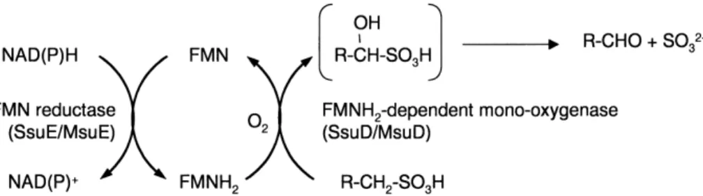
5.2. Desulfurization of methanesulfonate and other alkanesulfonates; FMNH2-dependent monooxygenases . . . 154
6. Desulfonation of aromatic sulfonates ^ a further adaptation . . . 157
7. Biodesulfurization of condensed thiophenes . . . 160
8. Regulation of bacterial organosulfur metabolism . . . 161
9. Uptake of sulfonates and sulfate esters . . . 165
10. Organosulfur utilization without oxygen . . . 165
* Tel.: +41 (1) 632 33 57; Fax: +41 (1) 632 11 48; E-mail: kertesz@micro.biol.ethz.ch
11. Conclusions and perspectives . . . 166 Acknowledgements . . . 167 References . . . 167
1. Introduction
All organisms require sulfur for growth. In bacteria sul-fur makes up 0.5^1% of the cell dry weight, and is needed primarily as a component of the amino acids cysteine and methionine. Sulfur also plays an essential role in a variety of enzyme cofactors, including biotin, coenzyme A, coen-zyme M, thiamine and lipoic acid, and is critical in many redox processes, both as a building block for iron-sulfur centers and as the redox-active component of disul¢de bonds. Sulfur is common in the environment, making up about 0.1% of the earth's crust [1], but much of this ma-terial is inaccessible to living organisms. Sulfur for biosyn-thetic processes is derived from the assimilation of inor-ganic sulfate by plants and bacteria (animals are unable to synthesize methionine and can synthesize cysteine, the key intermediate in most pathways of sulfur metabolism, only by transsulfurylation from dietary methionine [2]). Cys-teine biosynthesis by the sulfate assimilation pathway pro-ceeds by the transport of inorganic sulfate into the cell, its activation by conversion to 3P-phosphoadenosine-5P-phos-phosulfate (PAPS), subsequent reduction to sul¢te and thence to sul¢de, and transfer of this sul¢de onto an or-ganic moiety to yield the target molecule cysteine (Fig. 1). The biochemistry and genetics of this process have been extensively characterized in both plants and bacteria, and have been the subject of recent reviews [3^5]. However, perhaps because the growth media traditionally used in the laboratory contain high levels of inorganic sulfate (minimal media contain 1^15 mM sulfate [6]; LB medium contains only about 100^150 WM sulfate, but excess cys-teine), cysteine biosynthesis starting from sulfur sources other than sulfate has not been investigated in detail until quite recently. The purpose of this article is to review recent advances in the genetics and biochemistry of how Gram-negative bacteria utilize organosulfur compounds, in particular sulfonates (R-SO3
3) and sulfate esters
(R-OSO3
3), to provide themselves with cysteine and
methio-nine for growth. It will concentrate on assimilative sulfur metabolism by aerobic bacteria, though in some contexts it has been necessary to include comparisons with anaer-obic organisms and with yeasts and other eukaryotes. For a more detailed description of dissimilative organosulfur utilization by sulfate-reducing bacteria and other anaer-obes the reader is directed to recent reviews of this subject [7,8]. Sulfur metabolism in yeasts and ¢lamentous fungi has also been reviewed recently [9,10].
Our understanding of the biochemical processes in-volved in organosulfur utilization by bacteria has leapt
forward in recent years, with the characterization of sev-eral new sulfur-regulated desulfurizing enzyme systems [11^16]. Additional information has become available through the sequencing of bacterial genomes, 21 of which have been completely sequenced at the time of writing. Where possible, data from these projects have been in-cluded in this article. Homologues of many of the genes that are involved in organosulfur metabolism can be iden-ti¢ed in other contexts on the chromosomes of other or-ganisms, yielding further information about the substrate £exibility and regulatory complexity of their expression. Conclusions based on this kind of information are neces-sarily speculative, but may provide useful starting points for future research, and help to put biochemical and phys-iological £esh on the genetic skeleton supplied by the ge-nome sequences.
Studies of bacterial assimilative organosulfur metabo-lism are intimately connected with studies of cysteine bio-synthesis. Bacterial sulfur assimilation has frequently been assumed to follow the characterized pathway that has been established in enteric bacteria, regulated on a global level as part of the cys regulon [5,17]. More recent studies have shown that in non-enteric bacteria, the regulation of cysteine biosynthesis may be somewhat di¡erent (e.g. in Bacillus [18] or in Pseudomonas [19,20]). The initial pro-teomic study described below suggested that organosulfur metabolism might be controlled by a separate regulatory network to cysteine biosynthesis [21], but by now it has become clear that in most Gram-negative bacteria it is also part of the cys regulon, which is much more extensive than previously recognized (as foreseen by Kredich in his re-view in 1996 [5]).
2. Bacterial responses to sulfate limitation
In the laboratory, bacteria are usually grown either in mineral salts media that provide them with a large excess of sulfur in the form of inorganic sulfate, or in complex media containing not only sulfate but also amino acid sulfur derived from cell hydrolysates. How do they react when they are starved of inorganic sulfate or cysteine? In the absence of these two compounds, bacteria synthesize speci¢c proteins which are required for mobilization of alternative sulfur sources, and for using the last traces of available sulfate. Appropriate sulfate-limited (but not growth-limited) conditions can be provided by supplying the cell with sulfur in organically bound form, e.g. sulfo-nates, sulfate esters, methionine, sulfamates (R-NHSO3),
organosul¢des (R-SS-RP) or thioethers (R-S-RP), in the absence of inorganic sulfate. The cellular response has been examined in several proteomic studies [20^22].
Under sulfate-limited conditions a set of extra proteins was synthesized by several species of bacteria, and identi-¢ed by di¡erential screening with two-dimensional electro-phoretic techniques [20^22]. These so-called sulfate starva-tion-induced proteins (SSI proteins) are synthesized only in the absence of `preferred sulfur sources', which vary according to species, but include sulfate, cysteine and thi-ocyanate. SSI proteins have been found in Gram-positive and Gram-negative species, in soil bacteria and in human pathogens (Pseudomonas putida, Staphylococcus aureus, Escherichia coli, Pseudomonas aeruginosa). They are not synthesized after heat shock, under phosphorus or nitro-gen starvation conditions, or when the cells are grown anaerobically, and therefore appear to constitute a speci¢c response to the absence of sulfate or a related metabolite [20].
The SSI proteins of E. coli and P. aeruginosa have been characterized by N-terminal sequencing, and by mass spectrometric ¢ngerprinting [20,22^24] (Table 1). The SSI proteins fall into three categories.
(i) They are enzymes and transport systems involved in scavenging and metabolizing alternative sulfur sources from the environment. In this capacity the SSI response can be seen as functionally equivalent to the pho response, which governs the utilization of phosphonates and phos-phate esters as a response to phosphos-phate limitation in a variety of species [25^28]. In E. coli and P. aeruginosa these SSI proteins include periplasmic binding proteins for sulfate, cystine and sulfonates, which are anticipated to be involved in sulfur scavenging, and a variety of sul-fonatases (Table 1).
(ii) They can be low-sulfur copies of important cellular proteins, in which cysteine or methionine residues which are not critical for function have been replaced by other amino acid residues. Cyanobacteria, for instance, synthe-size a second set of phycobilisomal proteins with reduced cysteine/methionine content under sulfate-limited growth conditions [29]. In P. aeruginosa a low-sulfur version of the lipoprotein NlpA was found, which may have struc-tural relevance under sulfate-limited growth conditions. Synthesis of several antioxidant proteins was also upregu-lated during sulfate starvation (Table 1), including alkyl-hydroperoxide reductase and a thiol-speci¢c antioxidant protein. Synthesis of these proteins may be a response to peroxide intermediates generated as byproducts of sulfo-nate metabolism (see below), or may have a more direct link to sulfur metabolism, but this has not yet been ex-plored.
(iii) They could be enzymes involved in mobilization of intracellular sulfur storage compounds (the total sulfur content of E. coli, for instance, drops by half during sulfur limitation [30], implying that viability is maintained via a redistribution of sulfur within the cell). As yet, no such SSI proteins have been identi¢ed. It is interesting that many of the SSI proteins identi¢ed in these studies [20,22^24] had previously been undetected, since even in detailed studies such as the E. coli proteome analysis car-ried out by the Neidhardt group, the growth media con-tained 286 WM sulfate [6], and the SSI proteins were there-fore synthesized only at low levels.
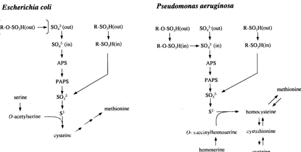
The above study in our laboratory was the ¢rst to in-vestigate the synthesis of a set of proteins as part of a global sulfate starvation response in bacteria, but a similar global response to limitation for preferred sulfur sources has been recognized in yeast and in ¢lamentous fungi Fig. 1. Cysteine biosynthetic pathways in E. coli and P. aeruginosa. In enteric bacteria, sul¢de is transferred to a serine moiety to give cysteine, whereas in P. aeruginosa the acceptor molecule is O-succinylhomoserine, yielding homocysteine. Although E. coli does not desulfurize sulfate esters, other enteric bacteria do so, using periplasmic sulfatases (square brackets, top left). APS: adenosine-5P-phosphosulfate; PAPS: 3P-phosphoadenosine-5P-phosphosul-fate.
[9,31,32]. In bacteria, quite a number of enzymes are known whose synthesis is regulated by sulfur supply, pri-marily those of the cysteine biosynthetic pathway (Fig. 1). The corresponding cys genes in enteric bacteria are acti-vated by the CysB protein, and constitute the cys regulon. During growth in the presence of excess inorganic sulfate expression of the cys genes is reduced to 40^50% of fully derepressed values [5]. This degree of repression is, how-ever, much less than for the SSI proteins detected in our initial study, many of which could not be observed at all in cells grown with sulfate [20^22].
Arylsulfatase is the best characterized bacterial enzyme that is known to be strongly regulated by sulfate starva-tion, in bacteria, algae [33,34], and ¢lamentous fungi [31,35]. Surprisingly, it was not identi¢ed by the di¡eren-tial two-dimensional analysis technique described above, because the amount of arylsulfatase protein synthesized even under fully derepressed conditions was below the detection limit of the method used [11]. Since arylsulfatase activity is strongly upregulated in the absence of sulfate in a variety of bacterial species [21,35], this suggests that sul-fate-regulated enzymes may be synthesized at lower levels than other proteins (corresponding to the cell's lower re-quirement for sulfur, compared to other macronutrients). It is likely, therefore, that further sulfate-regulated pro-teins still remain undetected.
The data summarized in Table 1 suggest that the bac-terial response to sulfate limitation is largely an adaptation that allows the cells to use sulfonates or sulfate esters as an alternative source of sulfur for growth, in the absence of preferred sulfur sources. These are usually sulfate, sul-¢te, sul¢de or cysteine, but methionine is also a preferred sulfur source in fungi [9,31], and thiocyanate represses the
SSI response in pseudomonads [21]. Since sulfonates and sulfate esters are not generally accepted to be common classes of compounds, we must next ask why such an adaptation may have evolved, and where sulfonates and sulfate esters occur in nature.
3. Sulfonates and sulfate esters in nature
Plants can only synthesize cysteine from inorganic sul-fate. However, in soil environments sulfate often consti-tutes only 1^5% of the total available sulfur, and it has now been conclusively shown that most of the sulfur present is in organically bound form, either as sulfate es-ters (over 50% of total sulfur in grassland soils [36]) or as sulfonates (e.g. in forest soils [37]) (Fig. 2). Sulfate for plant nutrition is provided in these systems by sulfur cy-cling in soil organic matter, catalyzed predominantly by microbial action. Anaerobic soils and sediments also con-tain considerable quantities of sul¢de, due to the action of sulfate-reducing bacteria, and aquatic environments are usually high in inorganic sulfate (both limnic and marine environments ^ seawater contains an average of 28.7 mM inorganic sulfate [38]). Xenobiotic and naturally occurring sulfonates and sulfate esters available to bacteria as sour-ces of sulfur or carbon are described below.
3.1. Naturally occurring sulfonates
Naturally occurring sulfonates have until quite recently been regarded as few in number, and of minimal impor-tance in the biological sulfur cycle, though they often have important biological functions. They include coenzyme M
Table 1
Sulfate-regulated proteins of E. coli and P. aeruginosaa
Protein Gene locus Function Reference
P. aeruginosa
PA1 sbp periplasmic sulfate binding protein [20]
PA2, PA11, PA13 ssuEADCBF, msuEDC alkanesulfonate desulfurization [20,24]
PA11 lsfA thiol-speci¢c antioxidant [20,24]
PA9 tauABCD taurine desulfurization [20,24]
PA7 nlpA lipoprotein [20,24]
PA4 atsK unknown [20,24]
PA19 similar to E. coli £iY putative amino acid transport protein [24]
PA17 similar to P. putida asfC putative periplasmic sulfonate binding protein [24]
PA14 similar to E. coli ahpC alkylhydroperoxide reductase subunit [24]
AtsA atsA arylsulfatase [11]
AtsRBC atsR, atsBC transport of sulfate esters [136]
E. coli
TauA, TauD tauABCD taurine desulfurization [218]
SsuE, SsuD ssuEADCB alkanesulfonate desulfurization [22]
Sbp sbp periplasmic sulfate binding protein [22]
FliY £iY periplasmic cystine binding protein [22]
AhpC ahpC alkylhydroperoxide reductase subunit [22]
aIn E. coli, expression of the genes of the cys regulon is also repressed during growth in the presence of sulfate [5]. This is presumably also the case in
in methanogenic bacteria [39], taurine in mammals [40], methanesulfonate as a degradation product of dimethyl-sul¢de produced by marine algae [41], and sulfoquinovose in the plant sulfolipid [42] (Table 2). Aeruginosin B, a phenazine derivative synthesized by P. aeruginosa, is the only known natural aromatic sulfonate [43]. The sulfur in aerobic soils is almost entirely found in organically bound form (Fig. 2), and has been characterized on the basis of chemical reactivity as hydriodic acid-reducible sulfur (sul-fate ester sulfur), Raney-nickel reducible sulfur (amino acid sulfur) and residual organically bound sulfur (sulfo-nate sulfur). Whereas this last assignment appears some-what generous, it has been con¢rmed by very recent stud-ies of sulfur composition in marine sediments, and in soils. Using X-ray absorption near edge structure spectroscopy, Vairavamurthy and co-workers have shown that up to 37% of the sulfur in several sediment and humic substance samples was indeed sulfonate sulfur [44], and that sulfo-nates made up 20^40% of the organic sulfur in marine sediments down to at least 50 cm depth [45]. Low molec-ular mass sulfonates have also been found in micromolar concentrations in the porewater of marine microbial mats [46]. Sulfonates are also present in all soil strata, including the humus layer. They may be derived from biogenic sul-fonates such as plant sulfonolipid, or they may be oxida-tion products of cysteine, as has been suggested for the sulfonates present in humic substances carried in river water [47]. It has also been suggested that sulfonates may arise in sediments by a chemical process involving the addition of sul¢de to carbon-carbon double bonds [48]. Since sul¢de is omnipresent in soils and sediments, due to the action of sulfate-reducing bacteria, this theory appears attractive. In situ labelling studies using radiola-belled sulfate in forest soils have shown not only that sul-fate is rapidly incorporated into the sulfonate fraction, but that there is a signi¢cant £ux of sulfur through this sulfo-nate pool, and that it ultimately reappears as sulfate [36,49,50]. Humus chemistry is complex, due to the enor-mous structural diversity of the substrate [51], but it seems clear that both sulfonate synthesis and desulfonation re-actions take place in humus at considerable rates, and that much of this activity is microbially mediated.
Sulfur present in humus will ultimately make its way into sulfur-containing compounds in oil and coal.
Re-search into speci¢c desulfurization of such compounds, without degrading the backbone carbon structure, makes up an important objective of many research groups [52]. In the past 20 years, success in reducing the sulfur content of oil, primarily by hydrodesulfurization, has led to a dra-matic decrease in SO2 pollution in the industrialized
world. An unexpected result of this success is the emer-gence of the phenomenon of sulfur limitation for crops, as the plants can no longer absorb the sulfur they require from the atmosphere, but are forced to rely on the lower levels of inorganic sulfate provided by the soil.
3.2. Production, role and fate of sulfonated xenobiotics Xenobiotic sulfonates are used in a wide variety of ap-plications, as diverse as dyestu¡s and brighteners, deter-gents, cement additives, and industrial chemical intermedi-ates. Many of these compounds, and intermediates from their synthesis, are released into the environment and can be detected in rivers [53^56] or in sewage sludge-amended soils [57,58]. Most applications make use of the amphi-philic nature of aromatic sulfonates, with a highly charged sulfonate group (pKa6 0) attached to a lipophilic
aro-matic ring. The most widespread aroaro-matic sulfonates are probably the linear alkylbenzenesulfonate surfactants (LAS), with an annual world production in 1995 of 2.8 mil-lion tons, predicted to reach 4 milmil-lion tons by 2005 [59]. Although LAS are not toxic to higher organisms [60], they may have toxic e¡ects on algae and invertebrates at the levels found in polluted waters [61^64]. They accumulate in some natural compartments, particularly in sediments and other anaerobic environments [65^67], but under aer-ated conditions are generally subject to ready biodegrada-tion, and are quickly mineralized.
3.3. Naturally occurring sulfate esters
Unlike aryl sulfonates, aromatic sulfate esters are not common xenobiotic compounds. The widespread occur-rence of bacterial arylsulfatases therefore provokes the question of the identity of the natural substrates of these enzymes. In part, the problem is a misleading one. Aryl-sulfatases have often been de¢ned as such because they are capable of hydrolyzing standard aromatic sulfate sub-strates such as 4-nitrophenyl sulfate or nitrocatechol sul-fate [68], although their natural substrates are aliphatic or carbohydrate sulfates. Nonetheless, aromatic sulfates are certainly found in nature. In mammals sulfation is a key step in the detoxi¢cation and excretion of aromatic xeno-biotics, since the sulfate conjugates are generally more water-soluble and less reactive than the parent com-pounds. Sulfation may occur either directly on a hydroxyl group (e.g. of dietary catechols), or after initial hydroxy-lation by cytochromes P450 [69], and the conjugates are subsequently excreted in the urine or in the bile. It should be noted that although this mechanism is geared towards Fig. 2. Sulfur speciation in lakewater [323], and in forest [37] and
grass-land [36] soils. The sulfur composition is shown as inorganic sulfate (E), sulfate ester sulfur ( ), amino acid sulfur (u), and sulfonate sulfur (F). For lakewater, amino acid and sulfonate sulfur are combined in one fraction.
detoxi¢cation, in occasional cases it may also lead to bio-activation of e.g. aromatic amines or benzylic alcohols to carcinogenic derivatives [70]. Bacteria also utilize this strategy for xenobiotic detoxi¢cation [71], though this does not appear to be widespread.
Sulfation of tyrosine residues is a common post-lational modi¢cation that occurs in the eukaryotic trans-Golgi network [72^74], and up to 1% of the tyrosines in a cell may be sulfated [75]. In mammals, sulfation and de-sulfation reactions regulate the concentrations of biologi-cally active molecules such as estrogens, which are trans-ported around the body as the sulfate esters and hydrolyzed in the target tissue to the active steroid [70]. Sulfate esters are therefore common, biologically active molecules to which bacteria are commonly exposed.
As described above, a large proportion of the sulfur in various soil and sediment environments is present as sul-fate esters. These may arise in humus itself, through chem-ical sulfation of lignin-derived phenols, or they may be biogenic in nature. In addition to the aromatic sulfates described above, carbohydrate sulfates contribute to nat-ural deposition of sulfate esters. Glycosaminoglycans (hep-arin and heparan sulfate, chondroitin sulfate, dermatan sulfate and keratan sulfate) are highly O-sulfated and N-sulfated, and are important components of connective tissue, playing roles in modulation of many extracellular processes [76]. In vivo, these polymers are subject to lyso-somal hydrolysis [77] (most of the mammalian sulfatases characterized so far are speci¢c for carbohydrate sulfates [78]). A variety of glycosaminoglycan lyases have also been characterized from bacterial sources [76]. These are extracellular enzymes that cleave the glycosaminoglycan backbone to yield di- to hexasaccharides. Most of the bacteria that catalyze this reaction have been isolated from soil, suggesting that soil environments are rich in glycosaminoglycan products, presumably of animal origin. Alkyl sulfate surfactants in detergent formulations have now been largely displaced from the market by the alkyl-benzenesulfonates described above, though alkyl sulfates are still in use in special laundry applications, and in prod-ucts such as toothpastes, antacids or car-cleaning sham-poos [79], and are therefore released to the environment. It has long been recognized that both primary and secondary alkyl sulfate surfactants are rapidly degraded by environ-mental bacteria [60], and so they are not present in high concentrations in the environment. A number of naturally occurring alkyl sulfate esters are also known, ranging from methyl sulfate to long-chain, partially chlorinated aliphatic sulfates derived from algae (reviewed in [35]). The ability of bacteria from pristine sites to degrade xenobiotic alkyl-sulfates is believed to derive from their previous exposure to these natural compounds [80], since alkylsulfatases are found even in sites which have not been exposed to sur-factant pollution [80,81].
From the above, it is clear that a variety of natural and xenobiotic sulfonates and sulfate esters are to be found in
the environment. In principle, bacteria can utilize these compounds in at least two ways. They can degrade the carbon skeleton to provide carbon and energy for growth, in which case the sulfur atom is released in excess of growth requirements for sulfur. In this case, the processes concerned are regulated, as far as they have been inves-tigated, primarily by substrate induction of the corre-sponding genes, and by catabolite repression. Alterna-tively, sulfonates and sulfate esters can be desulfurized to provide sulfur for growth, a process which is controlled by the sulfur supply to the cell (i.e. derepression in the absence of preferred sulfur sources such as sulfate). Both carbon-cycle and sulfur-cycle regulation have been ob-served for metabolism of organosulfates and organosulfo-nates in laboratory studies with pure bacterial cultures. In the following sections, the emphasis will be on metabolism of organosulfur compounds as part of the sulfur cycle in bacteria, concentrating on aerobic systems.
4. Sulfatases ^ from bacteria to humans
Sulfate esters provide a readily available source of sulfur for bacteria in soil and enteric environments, and micro-organisms have responded with the synthesis of a battery of sulfatase enzymes. Many of these appear to be related, and their activity is dependent on a formylglycine residue within the active site. Although several bacterial arylsulfa-tases are well characterized, the structure of the alkylsul-fatase enzymes and the genetic details of their expression are still almost completely unknown, and elucidation of these problems will provide interesting challenges in the future.
Sulfatases (EC 3.1.6.-) have been isolated from bacteria [35], fungi [9,31], algae [34,82,83], sea urchins [84^86], and higher eukaryotic organisms [78], but there is only scanty evidence for their occurrence in plants [87^89]. Because arylsulfatase activity is widely distributed in soils, it is often used as a test of soil quality, along with e.g. urease, amylase and alkaline phosphatase (e.g. [87,90^92]). At ¢rst sight, there appears to be a contradiction in the simulta-neous presence of high levels of sulfate esters and sulfa-tases in soils, but this can be rationalized if one considers that much of the sulfate ester sulfur may be protected from sulfatase attack by soil structure [93^95]. In humans most sulfatases are located in the acidic environment of the lysosomes, and are involved in the desulfation of gly-cosaminoglycans such as heparan sulfate, chondroitin fate and dermatan sulfate [77,78]. Other mammalian sul-fatases are located in the microsome and in the endoplasmic reticulum/Golgi apparatus [78]. As already noted, although these enzymes are known as arylsulfa-tases, their natural substrates are predominantly carbohy-drate sulfates, and not aromatic sulfate esters. Genetic de¢ciencies in most of the individual sulfatase genes are associated with speci¢c human diseases [78,96]. In
addi-tion, there is a rare lysosomal storage disease in humans called multiple sulfatase de¢ciency (MSD), in which all the sulfatases are defective. This syndrome is caused by the loss of the post-translational modi¢cation system that gen-erates the essential formylglycine in the active site of all the sulfatases [97].
Bacterial arylsulfatases and alkylsulfatases were last re-viewed in detail in 1982 [35]. Since that time there has been a considerable increase in our understanding of the genet-ics of arylsulfatases, and much progress has been made in understanding the mechanism of sulfatase action, both in eukaryotes and in microorganisms.
4.1. Bacterial arylsulfatases
Bacterial arylsulfatases have been identi¢ed and studied in a variety of species, including the enterobacteria (Kleb-siella [98^100], Salmonella [101,102], Enterobacter [103,104], Proteus [105,106], Serratia [107]), pseudomo-nads (Pseudomonas [11,108,109], Comamonas [110]), myco-bacteria, and cyanobacteria [111]. In recent years, how-ever, genetic and biochemical studies have concentrated primarily on the arylsulfatase enzymes from Pseudomonas aeruginosa and Klebsiella pneumoniae, at least in part be-cause these enzymes are closely related to the human ar-ylsulfatases. Earlier work has been comprehensively re-viewed [35], and is therefore only summarized brie£y here. Two main conclusions can be drawn from the early biochemical studies: (i) most of the strains studied con-tained multiple arylsulfatase isozymes, and (ii) the arylsul-fatase enzymes can be divided into two groups according to pH optimum, with one group showing optimal activity at pH values 6.5^7.1, and the other group at higher pH values of 8.3^9.0. Thus, Proteus rettgeri was reported to contain nine arylsulfatase isozymes, with pH optima at 6.7 and 8.3 [105,112], Serratia marcescens contains multiple isozymes with a broad pH optimum of 6.8^8.2 [107], P. aeruginosa has two isozymes with pH optima 9.0 and 8.4 [109,113], and Salmonella typhimurium has ¢ve en-zymes, all with optimum pH 6.7 [102]. It should be noted, however, that multiple arylsulfatase bands have also been reported in two species (P. aeruginosa and K. pneumoniae) where later work revealed only one functional arylsulfa-tase gene. The earlier observations of multiple isozymes may therefore be in part artefactual. Some of the earliest work on arylsulfatase was done with Enterobacter (Aero-bacter) aerogenes (ATCC 9621). This strain has since been reclassi¢ed as K. pneumoniae, and so the data obtained [103,104,114,115] must be compared with later studies with the latter species.
Arylsulfatases are also commonly found in multiple forms in the mycobacteria, but there has been no attempt to characterize the enzymes in detail. However, arylsulfa-tase activity patterns have been used as a means of iden-tifying and distinguishing di¡erent Mycobacterium species [116,117], and the presence of arylsulfatase is routinely
used as a phenotypic test for the genus Mycobacterium [118].
4.1.1. The arylsulfatase enzymes of P. aeruginosa and K. pneumoniae
The bacterial arylsulfatases that have been studied most in the last decade are the enzymes from K. pneumoniae and P. aeruginosa. These two proteins are relatively similar at the protein sequence level (37% amino acid identity, see Fig. 3), and in size (60 kDa for the Pseudomonas enzyme [11], and 62 kDa for the mature Klebsiella sulfatase [119]. However, there are also signi¢cant di¡erences, both at the protein and at the gene level. The Klebsiella sulfatase is a periplasmic enzyme, and analysis of the gene sequence reveals a typical signal peptide, whereas the Pseudomonas enzyme is an intracellular protein, since the N-terminal amino acid sequence corresponded with the 5P-end of the gene, and attempts to detect the Pseudomonas enzyme in periplasmic shock £uids were unsuccessful. The pH opti-mum for the Klebsiella enzyme is 7.5 [120], whereas the Pseudomonas sulfatase shows highest activity at a more alkaline pH value of 8.9 [11]. Two arylsulfatase isozymes were originally reported in P. aeruginosa [109,113], which di¡ered in their isoelectric points and pH optima. Un-fortunately these data were obtained with an incompletely characterized strain, and in more recent work, using the standard strain PAO1 [121], only one isozyme was de-tected by native isoelectric focussing, and in-gel activity staining [11]. In addition, deletion of the arylsulfatase gene (atsA) in this strain led to complete loss of arylsulfa-tase activity. The previously reported isozymes may there-fore have been either experimental artifacts or degradation products, as has been suggested for additional sulfatase bands that appeared during puri¢cation of the Klebsiella enzyme [35]. However, it should be noted that two further putative sulfatase genes have been detected during se-quencing of the P. aeruginosa genome [122] (see below), and it is possible that these genes are cryptic in strain PAO1 but were expressed in the strain studied previously. In addition to the enzymes described above (Fig. 3), a further sulfatase has recently been characterized from the marine bacterium Alteromonas carrageenovora [83]. The Alteromonas enzyme is considerably smaller (36 kDa) than the other bacterial sulfatases, and does not display sequence similarity to any of the sulfatases above. It shows a broad optimum in its activity at pH 8.5. The enzyme appears to be located in the periplasm, and is probably involved in desulfation of sulfated polysaccharides [83]. Although expression of the Alteromonas arylsulfatase is not repressed by sulfate [83], the protein is low in cysteine (only one residue), a typical characteristic of sulfate-regu-lated enzymes (cf. the Klebsiella arylsulfatase (zero cys-teines) and the Pseudomonas arylsulfatase (zero cyscys-teines)). 4.1.2. Post-translational modi¢cation of sulfatases
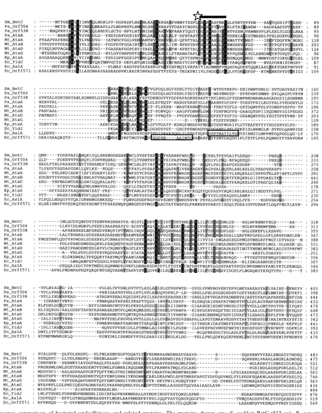
ex-cept for the Alteromonas enzyme, contain a conserved ami-no acid sequence motif in the active site, which is required for enzyme activity. This sulfatase motif (C/S-X-P-X-R-X4-TG) is conserved in both prokaryotic and eukaryotic
sulfatases, and in all cases examined it directs a post-trans-lational modi¢cation of the initial cysteine or serine in the sequence to a 2-oxoalanine (formylglycine; FGly) residue. The presence of this FGly residue has been demonstrated Table 2
in the human lysosomal arylsulfatases A [123,124] and B [125]. A lack of the ability to carry out the CysCFGly modi¢cation is the cause of the lysosomal storage disease MSD, in which the activities of all the lysosomal sulfatases
are severely reduced [97]. The FGly residue is also found in the sulfatase of the alga Volvox carteri [126], and in the two bacterial sulfatases from P. aeruginosa [13] and K. pneumoniae [98]. In P. aeruginosa it is formed by mod-Fig. 3. Sequence comparison of bacterial arylsulfatases and related proteins. The proteins shown are: S. meliloti BetC (512 aa), P. aeruginosa Orf504 (504 aa), Orf538 (538 aa) and AtsA (536 aa), M. tuberculosis AtsA (787 aa), AtsB (970 aa), AtsD (787 aa) and AtsG (465 aa), K. pneumoniae AtsA (464 aa), E. coli YidJ (497 aa), AslA (551 aa) and Orf f571 (571 aa). The alignment was done with CLUSTALW, and identical residues are indicated by shading. The cysteine/serine that is modi¢ed to formylglycine is indicated with a star, and the consensus modi¢cation sequence motif (PROSITE Sul-fatase box 1) is overlined. In the AslA and Orf f571 proteins, this consensus sequence does not align well with the other sequences, and is boxed sepa-rately.
i¢cation of a cysteine residue, as in the eukaryotic sulfa-tases, whereas in K. pneumoniae it arises by modi¢cation of a serine residue. Modi¢cation of the bacterial sulfatases was demonstrated by reduction of the aldehyde group of the puri¢ed enzymes with [3H]sodium borohydride, tryptic
digestion, and radiosequencing of the corresponding pep-tide. This revealed that cysteine-51 of the Pseudomonas arylsulfatase and serine-72 of the Klebsiella enzyme had been modi¢ed, and the nature of the modi¢cation was con¢rmed by MALDI-MS of the peptide in its native state, and after treatment with nitroaniline, which reacts with aldehyde groups [13]. A striking di¡erence between the two enzymes was that the Klebsiella sulfatase was only circa 40% modi¢ed [98,119], whereas the Pseudomonas en-zyme was 100% converted to the FGly form [13], probably re£ecting a di¡erence in the e¤ciency of the cysteine and serine modi¢cation processes.
X-ray structural analysis of the human arylsulfatases A and B [127,128] has shown that the FGly in these proteins is present in the active site of the enzyme, as its hydrate or sulfate ester hemiacetal, at the base of a positively charged pocket. This hydrate is thought to play an essential role in the putative catalytic mechanism outlined in Fig. 4. Re-placement of the cysteine in the active site of the human sulfatases by serine led to a complete loss of enzyme ac-tivity [125]. It is therefore particularly interesting that the serine residue of the Klebsiella sulfatase can be modi¢ed to FGly. The initial study showed that this was carried out with arylsulfatase isolated from K. pneumoniae [98], but the enzyme was also active when expressed in E. coli [99,119]. The Pseudomonas enzyme was also active when expressed in E. coli, but all activity was lost when the cysteine-51 residue was replaced by a serine or an alanine [13]. Identical results were obtained when the Pseudomonas sulfatase and its C51S and C51A mutant forms were trans-formed into a vatsA strain of P. aeruginosa or into K. pneumoniae [13]. These data suggest that two separate systems exist for modi¢cation of cysteine or serine residues within the conserved sulfatase motif, and that the Ser-spe-ci¢c modifying system of E. coli is incapable of recogniz-ing the serine form of the Pseudomonas sulfatase.
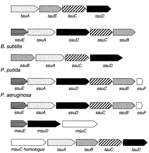
The proposed classi¢cation of sulfatase-modifying en-zymes into Cys-speci¢c and Ser-speci¢c systems has mech-anistic implications (Fig. 5). Conversion of a serine residue to FGly requires merely an oxidation, whereas the mod-i¢cation of cysteine to FGly proceeds formally via oxida-tion to the thio-aldehyde, followed by a hydrolytic step. The activation enzymes for serine and cysteine have not yet been identi¢ed in any organism, but they are therefore likely to be oxidoreductases. Schirmer and Kolter [129] have compared this reaction with the oxidative conversion of aspartate to oxaloacetate via iminoaspartate [130], but this has not yet been tested experimentally. These authors postulate an oxygen-dependent reoxidation of the cofac-tor. However, there must be other mechanisms, since the Pseudomonas arylsulfatase can be overexpressed in active
form in E. coli cells grown under anaerobic conditions [131]. As mentioned above, the Cys-type Pseudomonas fatase is an intracellular enzyme, whereas the Ser-type sul-fatase of Klebsiella is periplasmically located. This corre-lation between sulfatase type and putative cellular location has also been found in all other hypothetical sulfatases that have been identi¢ed through genome analysis (see below) This leads to the interesting possibility of a spatial separation between the two modifying systems, i.e. that the Cys-modifying system is located in the cytoplasm, whereas the Ser-modifying enzyme either is located in the periplasm or is obligatorily linked to transport through the membrane. An analogous situation to the latter is found for human sulfatases, for which the modi¢cation process occurs at a late stage of protein translocation into the endoplasmic reticulum [125].
Searching the databases for the consensus sequence for the arylsulfatase consensus motif led to the discovery of several other enzymes that contain this motif and catalyze related reactions. Thus, the alkaline phosphatase of P. aeruginosa (accession number AF047381) and the phos-phonate monoester hydrolase of Burkholderia caryophylli [133] also contain the modi¢cation sequence, though it has not yet been shown that either of these is indeed modi¢ed in the active site. In addition, if the essential core sequence C/S-X-P-X-R [119] is combined in a search with mis-matched versions of the auxiliary element AALLTGR, then further proteins are found that almost match the consensus [134]. It will be interesting to determine in fu-ture whether any of these indeed contain FGly, and if this modi¢cation therefore plays a wider role in cellular metab-olism.
4.1.3. Arylsulfatase genes in P. aeruginosa and K. pneumoniae
An obvious candidate for the Ser-modi¢cation enzyme is found immediately when the genetic structure of the arylsulfatase gene in K. pneumoniae is examined. This is the atsBKpgene product, which is encoded in a bicistronic
operon with the atsAKp gene, encoding the arylsulfatase
(Fig. 6) [99,119] (for clarity's sake, a subscript is added to the ats genes here, Kp for K. pneumoniae, and Pa for P. aeruginosa). The atsBKp gene product is required for
synthesis of active arylsulfatase, and was originally pro-posed to be a transcriptional activator for the atsBA op-eron [135]. However, the 45-kDa AtsB protein does not show any of the motifs expected for a transcriptional reg-ulator (e.g. helix-turn-helix DNA binding regions), and expression of the atsAKp gene behind the strong lac
pro-moter in E. coli in the absence of atsBKp yielded only
inactive arylsulfatase [119]. When this inactive arylsulfa-tase was puri¢ed, it was found that the serine at position 72 was completely unmodi¢ed. However, co-expression of the atsAKp and atsBKp genes on separate plasmids led to
synthesis of an active sulfatase, in which the arylsulfatase ser-72 was 50% modi¢ed to FGly [119]. The AtsB protein
contains 12 cysteine residues in three clusters whose ar-rangement (cluster 1: C-X3-C-X2-C; cluster 2: C-X5
-C-X14-C; cluster 3: C-X2-C-X5-C-X3-C-X18-C) is
reminis-cent of [Fe-S] iron sulfur reminis-centers [129]. This structure is consistent with the putative function of the modi¢cation enzyme as an oxidoreductase. The deduced protein does not carry a signal peptide, and appears to be a soluble, cytoplasmic protein. Since the Klebsiella arylsulfatase is a periplasmic enzyme, this implies that modi¢cation of the serine-72 residue occurs prior to translocation into the periplasm. An arylsulfatase-negative mutant of K. pneumo-niae could be complemented with the atsBAKpgenes,
dem-onstrating that this strain contains only one sulfatase iso-zyme that was expressed under the conditions used [99].
In P. aeruginosa the ats genetic organization is some-what di¡erent. Since the Pseudomonas arylsulfatase is a cytoplasmic enzyme, an uptake system is required to trans-port the charged sulfate esters into the cell. The genes encoding such an uptake system are indeed found up-stream of the atsAPagene (Fig. 6) ([136], accession number
Z48540). Similarity searches revealed that the atsBCPa
genes are similar to the permease and ATP binding com-ponents of ABC-type transporters, respectively. Trans-porters of this type are normally composed of two perme-ase components within the cell membrane, each of which is made up of six transmembrane helices, and two ATP binding subunits which are located peripherally to the cy-toplasmic side of the cell membrane [137,138]. In addition, a high-a¤nity substrate binding protein located in the periplasm is responsible for delivering the speci¢c sub-strate to the membrane component. The 58-kDa atsBPa
gene product is larger than most characterized permease components, and contains 12 putative transmembrane helices. It is therefore equivalent to a dimer of six-helix components, and presumably interacts with two molecules of the AtsC protein, which was identi¢ed as the ATP bind-ing subunit by the presence of the conserved Walker mo-tifs A and B [139,140]. The atsB and atsC genes overlap by four nucleotides. A fourth gene, atsRPa, lies divergent to
the atsBCA operon, and encodes a periplasmic protein. The role of these proteins was con¢rmed by insertional inactivation of either the atsRPa or atsBPa genes with a
gene cassette encoding gentamicin resistance (Gm) [136]. Both atsR: :Gm and atsB: :Gm mutant strains were unable to grow with aromatic sulfates, and whereas the atsR: :Gm mutant still produced arylsulfatase, the polar e¡ect of the atsBPa insertion led to a loss of arylsulfatase activity as
well. Complementation of the atsBPa mutant with the
atsAPa gene under lac control led to restoration of
aryl-sulfatase activity but not of growth with arylsulfates. The atsRPa: :Gm and atsBPa: :Gm mutants are also
de-fective in utilization of hexyl sulfate as a sulfur source, and the AtsRBC transport system therefore constitutes a gen-eral transporter for sulfate esters. It also plays a role in the transport of aromatic sulfonates, as discussed below. In-terestingly, the Pseudomonas arylsulfatase does not
hydro-lyze alkylsulfate esters, so an alkylsulfatase gene must also be present elsewhere on the chromosome.
P. aeruginosa contains only one gene with signi¢cant similarity to the atsBKp gene, nirJ, whose gene product
shows 25% identity to AtsBKp in the N-terminal domain.
However, since the nirJ gene is part of an extensive ANR-regulated operon encoding the dissimilatory nitrite reduc-tase [141], it seems unlikely that the NirJ protein is in-volved in sulfatase maturation. It is also interesting to note that Synechocystis contains an atsBKp homologue
(ORF slr1507 [142]), but does not contain any genes en-coding putative sulfatases. Together, these data suggest that (i) although the atsBKp gene product is essential for
Ser-type sulfatase maturation in enteric bacteria [119], oth-er species have developed altoth-ernative strategies to carry out this modi¢cation, and (ii) there may be other proteins that require the FGly modi¢cation for activity.
4.1.4. Putative sulfatase genes in bacterial genome sequences
In recent decades there has been no systematic search for bacterial species that possess arylsulfatase activity, and since earlier screens were done before it was recognized that expression of bacterial arylsulfatase genes is often repressed in the presence of sulfate [35], the value of these studies is somewhat limited. Since the start of the `genomic age' it has become possible to complement biochemical searches by in silico screening of published bacterial ge-nome sequences for open reading frames with sequence similarity to known sulfatase genes. The arylsulfatase gene sequences from P. aeruginosa, K. pneumoniae and A. carrageenovora were used as probes to search the non-redundant GenBank database, the completed bacterial ge-nomes, and the P. aeruginosa partial genome sequence for arylsulfatase homologues using the BLAST algorithm [143].
The results are summarized in Figs. 3 and 6, and were somewhat surprising. Putative sulfatases were found in E. coli, P. aeruginosa, and Mycobacterium tuberculosis, i.e. in exactly those species where arylsulfatase enzymes have already been studied. In the 19 other complete ge-nomes and 20 incomplete genome projects examined, only three species contained arylsulfatase homologues: Borde-tella pertussis [144] and Salmonella typhi [144] each con-tained two putative Cys-type sulfatases, and Yersinia pestis [144] had two Cys-type and one Ser-type sulfatases. The sulfatase sequences in these three organisms were not ex-amined further, as the genome sequences are not yet com-plete. P. aeruginosa, M. tuberculosis and E. coli were also found to contain multiple arylsulfatase genes [122,145,146]. Thus, P. aeruginosa contained three arylsul-fatase copies, of which one is presumably choline sularylsul-fatase [147], a second is an arylsulfatase [11], and the third is similar to a published phosphonate monoester hydrolase [133]. M. tuberculosis contained four di¡erent sequences with similarity to arylsulfatases [146]. The best described
set of sulfatase genes is the three putative sulfatases found in E. coli [29], and is particularly interesting because this organism does not grow with sulfate esters as sulfur sour-ces, and the putative sulfatase genes are all cryptic. All the putative sulfatase genes encoded proteins which contained the FGly modi¢cation sequence, carrying either a cysteine or a serine in the position to be modi¢ed. The putative Ser-type sulfatases also carried a putative signal sequence, for export to the periplasm, as noted by Schirmer and Kolter [129], whereas the Cys-type sulfatases appear to be cytoplasmic.
More information can be obtained from the genetic en-vironment of the putative sulfatase genes (Fig. 6). The Ser-type sulfatases found in enteric bacteria (aslA and orf f571 in E. coli and atsAKp in K. pneumoniae) are all found in
close proximity to a gene encoding the putative activator protein (aslB, orf f390 and atsBKp, respectively). An
acti-vator of this sort was not found near the genes encoding the Cys-type sulfatases (indeed, there is no evidence that either P. aeruginosa or M. tuberculosis contain an atsBKp
homologue). The Cys-type sulfatases almost all occur in gene clusters which encode transport proteins of some sort, underlining the putative location of the Cys-type sul-fatases within the cell, and perhaps providing some evi-dence about their function. Thus, the atsRBC genes of P. aeruginosa are thought to be involved in uptake of ali-phatic and aromatic sulfate esters, and the genes preceding the choline sulfatase in this species may be connected with choline sulfate transport. The E. coli yidJ gene, encoding a putative Cys-type sulfatase (Fig. 6), is distal to a gene encoding a putative glucose transporter, implying a possi-ble function in glucose-sulfate uptake.
The presence of multiple arylsulfatase genes in each ge-nome where sulfatase genes were found makes it tempting to draw conclusions about the occurrence of arylsulfatase isozymes often reported earlier [35]. For example, if one of the three sulfatases present on the genome of P. aeruginosa were cryptic in one isolate, and expressed in another, this would explain the observed di¡erences in the number of isozymes in di¡erent reports [11,109]. Several species of enteric bacteria, in particular, seem to contain several sul-fatase genes. It will be interesting to learn whether these are all pseudogenes, or whether the encoded proteins are expressed under conditions that have not yet been discov-ered and catalyze the desulfation of substrates that have not yet been tested.
4.1.5. Regulation of bacterial arylsulfatase expression With the exception of the arylsulfatase from A. carra-geenovora [83], expression of all the bacterial arylsulfatases which have been studied biochemically is subject to repres-sion during growth in the presence of inorganic sulfate, and derepression during growth with organosulfur sources such as methionine or alkanesulfonates [11,21,35]. This regulatory pattern strongly suggests that the role of these enzymes in nature is exclusively in the assimilation of
sul-fur for bacterial growth, and that the arylsulfatases are an important part of the bacterial sulfate starvation response. However, arylsulfatase expression is repressed not only by sulfate, but also during growth with cysteine or with cysteine biosynthetic intermediates such as sul¢te or sul-¢de. This pattern of regulation is reminiscent of the con-trol of cysteine biosynthesis, which has been best charac-terized in enteric bacteria [5,17]. In these species, activation of the genes of the cys regulon is mediated by the CysB protein, together with N-acetylserine, which acts as a co-inducer, and with sul¢de or thiosulfate, which act as anti-inducers. In P. aeruginosa, expression of atsB: :lacZ and atsR: :lacZ fusions was also found to re-quire an active CysB protein [136]. Although the pathways of cysteine and methionine biosynthesis in P. aeruginosa and E. coli di¡er slightly [19,20,148] (Fig. 1), this demon-strates that the arylsulfatase gene cluster constitutes an extension of the cys regulon in the former species.
The question of how sulfate, sul¢te and sul¢de cause gene repression in P. aeruginosa is more complex. In prin-ciple, sulfate may exert its repressive e¡ect either directly or after conversion to another metabolite, probably (but not certainly) in the cysteine biosynthetic pathway. In or-der to isolate the e¡ects of di¡erent intermediates of the pathway, de¢ned mutants were constructed in the cysN (sulfate activation) and cysI (sul¢te reductase) genes of P. aeruginosa, and the ability of sulfate, sul¢te or sul¢de to repress arylsulfatase synthesis in these mutants was studied (Table 3). Inorganic sulfate did not repress sulfa-tase formation in a cysN mutant, but did so in a cysI strain, suggesting that either PAPS or sul¢te was acting as a repressor. cysN and cysI mutants grown with sul¢de also did not synthesize arylsulfatase, and since cysteine does not repress arylsulfatase completely in P. aeruginosa [20], it seems likely that sul¢de is also a repressor. At least two independent compounds are hence active as repressors in vivo in this species. Alternatively, the active repressor of sulfatase synthesis is a separate, uncharacterized molecule that can be synthesized from either of these two precur-sors.
A similar study was carried out in K. pneumoniae by Adachi et al. [100], but with very di¡erent results. Here, too, a strategy was adopted that made use of mutants in cysteine biosynthesis, but the mutants were de¢ned only physiologically, and not at a genetic level. Synthesis of arylsulfatase was repressed independently by intracellular sulfate and by cysteine, but not by sul¢te, sul¢de or thio-sulfate directly. Unfortunately, the role of CysB in regu-lation of arylsulfatase expression in K. pneumoniae has not yet been examined.
Arylsulfatase synthesis in enteric bacteria is regulated not only by sulfur supply, but also by monoamine com-pounds such as tyramine, dopamine, or norepinephrine. In the presence of tyramine, sulfate-mediated repression of arylsulfatase synthesis is overridden, and arylsulfatase is expressed, together with other genes of the monoamine
(moa) regulon, including monoamine oxidase (maoA), tyr-amine oxidase (tynA) and several uncharacterized genes [135]. This e¡ect has been shown to be mediated by the positive regulator protein MoaR, a 26-kDa protein which is related to response regulators of the two-component sensor-regulator family. Expression of moaR is subject to catabolite repression [149], and the expression of arylsul-fatase as part of the moa regulon may therefore imply its involvement in central metabolism. It has indeed been proposed [135] that the monoamine regulon in enteric bac-teria is primarily concerned with utilization of dopamine sulfate, since dopamine, like many other phenols and cat-echols, is transported in human systems as its less reactive sulfate ester. A similar derepression of arylsulfatase ex-pression by monoamine compounds has been observed in several other enteric bacteria, including S. typhimurium, S. marcescens, and also E. coli, in which the cryptic atsA gene was found to be induced by tyramine, using an atsA: :lacZ fusion as a reporter system [150]. Tyramine does not derepress arylsulfatase synthesis in P. aeruginosa [11].
A mutant of K. pneumoniae has also been reported in which arylsulfatase was expressed constitutively, regardless of the sulfur source supplied [100]. The wild-type pheno-type was regained when the mutant was transformed with the atsRKpgene [151]. The encoded protein showed
signi¢-cant similarity (89% identity) to the dihydrofolate reduc-tase of E. coli. Expression of the atsR gene on a plasmid led to increased dihydrofolate reductase activity in the cells, and the E. coli folA gene was also able to comple-ment the atsR mutation in K. pneumoniae. However, the
signi¢cance of this link between arylsulfatase synthesis and C-1 metabolism remains unclear.
A further important regulatory aspect of arylsulfatase synthesis is how synthesis of the corresponding modi¢ca-tion system(s) is controlled. Very little is yet known on this subject, but it is clear already that the modi¢cation sys-tems are not regulated by sulfur supply, since in E. coli both the Pseudomonas and Klebsiella arylsulfatases are ex-pressed in active form after growth in LB medium, if the atsA genes are expressed behind the lac or T7 promoters [13,119]. It seems possible that the modi¢cation systems are important in another role that has yet to be identi¢ed, and that their expression is therefore either constitutive or regulated by other factors.
4.2. Alkylsulfatases
Alkylsulfatases di¡er from arylsulfatases not only in their substrate range, but also from a mechanistic point of view. Whereas arylsulfatases cleave the O-S bond of their substrates (a mechanism which is consistent with (but not indicative of) the presence of a FGly hydrate in the active site (Fig. 4)), the long-chain alkylsulfatases break the C-O bond and do not form a covalent en-zyme-sulfate intermediate (reviewed in [35]). It therefore seems unlikely that the long-chain alkylsulfatases share the FGly modi¢cation found in the arylsulfatases. By con-trast, the short-chain alkylsulfatase synthesized by the cor-yneform strain B1a also catalyzes cleavage of the O-S bond [152], but no further data are yet available concern-ing its mechanism.
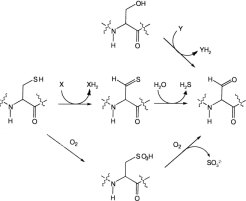
Fig. 4. Proposed mechanism of sulfate ester cleavage in arylsulfatases (adapted from [96]). The hydrate form of the active site formylglycine attacks the sulfate ester, breaking the S-O bond and forming a covalent enzyme-sulfate intermediate. This intermediate decomposes to regenerate the aldehyde form, which is then rehydrated in the active site by addition of a water molecule.
All of the alkylsulfatases described to date come from Gram-negative bacteria, though a Bacillus cereus strain is known that will grow with dodecyl sulfate [153]. Alkylsul-fatases were included in an extensive review in 1982 [35], which detailed many of the earlier results obtained. Much of this work was carried out with the strain Pseudomonas C12B, an isolate from a sewage outlet which synthesizes as
many as ¢ve separate alkylsulfatases. Two of these are speci¢c for primary sulfates, but di¡er in the pattern of their regulation, one being constitutively expressed, and the other inducible. The remaining three alkylsulfatases are speci¢c for secondary sulfate esters; two of these are constitutive enzymes that speci¢cally hydrolyze either D
-orL-isomers, while the third secondary alkylsulfatase is an
inducible enzyme. The two inducible enzymes are also re-pressed by primary alcohols (the products of the sulfatase reaction), and by tricarboxylic acid cycle intermediates, suggesting that catabolite repression plays a role in regu-lation of these enzymes.
This regulatory pattern immediately suggests that these enzymes are not part of the sulfur cycle, but are used to provide carbon and energy for bacterial growth. It should be noted, however, that most alkylsulfatase studies have been done with enzymes from bacterial strains that were isolated on the basis of their ability to mineralize sulfate esters completely (Pseudomonas C12B, for instance, was
isolated for its ability to grow with dodecylsulfate as car-bon source [154]). In their earlier review, Dodgson et al. [35] listed only two alkylsulfatases whose expression was
repressed by inorganic sulfate: the lithocholate sulfate sul-fatase of P. aeruginosa [155] and cholinesulsul-fatase of Pseu-domonas V-A [156]. In both these studies it is notable that the sulfate ester was supplied solely as sulfur source, and carbon for growth was provided by benzoate or citrate, respectively. It seems likely that more sulfate-regulated bacterial alkylsulfatases still await discovery if suitable growth conditions are chosen.
Studies with the Pseudomonas C12B alkylsulfatases
re-vealed that these were speci¢c for medium- or long-chain sulfate esters, but had little or no activity with substrates with a chain length less than C5. However, organisms have
subsequently been isolated from soil, canal water and sew-age which are able to grow with short-chain alkylsulfates as carbon source [157], and several of these have been examined in more detail. The sulfatase of the coryneform isolate B1A was active on C3^C7 primary alkylsulfates,
but not on longer or shorter-chain homologues, nor on choline sulfate [152]. Methyl and ethyl sulfates acted as inhibitors of this enzyme (Ki values of 5.3 mM and 3.9
mM respectively), though these compounds were not themselves hydrolyzed by the enzyme. In two environmen-tal isolates, Agrobacterium sp. strain M3C and Hyphomi-crobium sp. strain MS223, methyl sulfate was not metab-olized by a hydrolytic mechanism, but via an oxidative pathway [158,159]. Although initial studies had suggested that the Hyphomicrobium pathway might involve a hydro-lytic enzyme, an elegant study using 13C-labelled methyl
sulfate demonstrated that in both species methylsulfate is Fig. 5. Mechanisms for formylglycine formation in Cys-type and Ser-type arylsulfatases. Conversion of serine requires only an oxidative step, with un-known electron acceptor, whereas cysteine conversion requires an additional hydrolytic step. Alternatively, cysteine oxidation to cysteate and subsequent oxygenolytic cleavage would also lead to the desired aldehyde.
converted directly to formaldehyde and sulfate, and that methanol is not an intermediate in this process [160]. A mechanism was proposed involving initial oxygenation to methanediol monosulfate by a monooxygenase, followed by spontaneous decay of the hemiacetal to yield formal-dehyde and sulfate. Electron microscopic studies have re-vealed that whereas the long-chain-speci¢c sulfatases of Pseudomonas sp. strain C12B were localized on the outer
cell wall, the short-chain-speci¢c enzymes in a coryneform species were intracellular [161].
From a genetic point of view, very little is known about alkylsulfate degradation, either in the carbon cycle or in the sulfur cycle. Gene sequences have been determined for a choline sulfatase (betC) from Sinorhizobium meliloti [162], for a bile acid sulfatase from Comamonas testoster-oni [163,164], and for a putative dodecylsulfate sulfatase (sdsA) from the detergent-degrading strain Pseudomonas sp. ATCC 19151 [165,166]. The proteins encoded by the two latter genes appear to be related, and do not contain an FGly modi¢cation motif. The betC gene from S. meli-Fig. 6. Genetic structure of selected bacterial sulfatase gene clusters. Hypothetical function assignment of uncharacterized ORFs was done using BLAST [143]. The sequences are from: E. coli [129,145], P. aeruginosa [11,122,136], K. pneumoniae [99,119], S. meliloti [162]. Genes containing Ser-type sulfa-tases are shaded black, and those encoding Cys-type sulfasulfa-tases are shaded gray.
loti, by contrast, belongs to the same family as the eukary-otic arylsulfatases and the P. aeruginosa arylsulfatase, and is a Cys-type sulfatase that appears to be located within the cell (no signal sequence was found) (Fig. 6).
The alkylsulfatase gene of Pseudomonas sp. ATCC 19151, sdsA, was identi¢ed by complementation of several mutants derived from a previously described SDS-utilizing strain. Two genes were identi¢ed that were required for SDS utilization, sdsA and sdsB. The putative SdsB protein is a member of the LysR family of transcriptional regula-tors, and is closely related to the regulatory protein re-quired for arylsulfonate desulfonation in P. putida, de-scribed below. The regulatory role of the SdsB protein was con¢rmed by the fact that the SDS-negative pheno-type of an sdsB mutant could be complemented by an sdsA gene expressed constitutively behind the T7 pro-moter. SdsB therefore seems to be required for expression of the sdsA gene, which encodes a 59-kDa protein. This protein was expressed in E. coli, but did not show detect-able sulfatase activity. The lack of a signal peptide sug-gests that the encoded protein is intracellular, which con-trasts with the observations that sulfatase activity in Pseudomonas sp. ATCC 11951 is periplasmically located, and it was suggested that the secretion sequence might be an atypical one, and not recognized in the heterologous host. Homologues of SdsA are found in E. coli (YjcS; 73 kDa, 32% amino acid identity to SdsA), and in P. aerugi-nosa (30% identity over the full length of the protein), and are related to lactamases from several organisms.
A homologue of SdsA (27% identity) has also been identi¢ed in Comamonas (Pseudomonas) testosteroni, where it catalyzes the hydrolysis of bile acid sulfates [163]. The organism was identi¢ed in a collection of strains for its ability to utilize lithocholic acid as a source of carbon and sulfur for growth, and the sulfatase was iso-lated and found to be a homodimer of 53-kDa subunits. Sulfatase activity was Mn2-dependent. Synthesis of
the sulfatase was found to be completely repressed by 800 WM inorganic sulfate. Since the organism was grown with 2 mM lithocholic acid sulfate as carbon source, it is not clear why the excess sulfate released as a result of the di¡erential cellular requirements for carbon and sulfur did not repress synthesis of the enzyme in vivo. One can spec-ulate that the sulfatase is involved only in release of the sulfur, whereas the carbon-based degradation of the ste-roid skeleton was carried out by a separate system, leaving the sulfate ester bond intact. A second, biochemically sim-ilar sulfatase has recently been isolated from the same organism [167], but sequence data have not yet been re-ported.
A further type of alkylsulfatase has recently been re-ported in S. meliloti [162]. The BetC protein catalyzes the hydrolysis of choline sulfate to choline, and is part of a pathway that synthesizes the osmoprotectant glycine betaine. However, the organism can also utilize choline sulfate as a source of carbon, nitrogen and sulfur for
growth. Expression of the sulfatase was induced by cho-line or chocho-line sulfate, but not by sulfate limitation. This induction was slightly decreased under osmotic stress, and the sulfatase may therefore be involved in the metabolism of choline, rather than in osmoprotectant synthesis (cho-line sulfate itself showed only low osmoprotectant activ-ity). Choline sulfatase has also been characterized from P. aeruginosa [147]. Its synthesis is tightly regulated in this species, since the level of enzyme produced depended on whether the substrate was being utilized by the cell as a source of carbon (22.8 nmol min31 mg31 protein),
nitro-gen (18.5 nmol min31 mg31), or sulfur (4.1 nmol min31
mg31). The product of the putative P. aeruginosa choline
sulfatase gene (orf504, Fig. 6) [122] shows 45% identity to the S. meliloti BetC protein, and both contain the signa-ture for a Cys-type sulfatase. Inhibitor studies carried out with the P. aeruginosa choline sulfatase [147] are consis-tent with a FGly residue constituting the active site as in arylsulfatases, but the presence of the modi¢cation has not yet been demonstrated.
4.3. Carbohydrate sulfatases
Most of the eukaryotic sulfatases are in fact carbohy-drate sulfatases, acting on glycosaminoglycans as sub-strates [78]. Several bacterial sulfatases have also been characterized that belong to this group, including enzymes from Proteus vulgaris, Flavobacterium heparinum (now called Cytophaga heparina), and Bacteroides thetaiotaomi-cron. Glycosaminoglycan breakdown by these bacteria has been reviewed in detail [76], and involves the cleavage of the polymer by a lyase, usually extracellular, followed by desulfation of the resultant sulfated disaccharides and hy-drolysis to the monosaccharide level by a glucuronidase. Bacteria that can carry out this process have been isolated primarily from soil, and degrade glycosaminoglycans as a source of carbon and energy for growth. No attempts appear to have been made to isolate bacteria that can utilize glycosaminoglycans solely as sulfur source for growth.
A further family of bacterial carbohydrate sulfatases has also recently been identi¢ed, which cleave the sulfate moiety from mucin. Mucin is particularly heavily sulfated in the mouth and in the colon, where bacteria are nor-mally common, and the sulfation appears to help provide the mucin with a degree of protection from bacterial at-tack [168]. Strains of B. thetaiotaomicron, P. aeruginosa and Burkholderia cepacia were found to contain high levels of mucin sulfatase activity [169,170], a ¢nding which is of importance in studies of cystic ¢brosis, since the latter two species contribute signi¢cantly to the severity of this disease. Interestingly, an E. coli strain was also found to contain mucin sulfatase activity [170], and this may suggest a role for the uncharacterized sulfatase genes previously identi¢ed in the E. coli genome sequence [129].
![Fig. 2. Sulfur speciation in lakewater [323], and in forest [37] and grass- grass-land [36] soils](https://thumb-eu.123doks.com/thumbv2/123doknet/14895367.651233/5.894.93.406.109.210/fig-sulfur-speciation-lakewater-forest-grass-grass-soils.webp)
![Fig. 4. Proposed mechanism of sulfate ester cleavage in arylsulfatases (adapted from [96])](https://thumb-eu.123doks.com/thumbv2/123doknet/14895367.651233/13.894.226.674.709.1060/fig-proposed-mechanism-sulfate-ester-cleavage-arylsulfatases-adapted.webp)
![Fig. 6. Genetic structure of selected bacterial sulfatase gene clusters. Hypothetical function assignment of uncharacterized ORFs was done using BLAST [143]](https://thumb-eu.123doks.com/thumbv2/123doknet/14895367.651233/15.894.244.652.104.845/genetic-structure-selected-bacterial-sulfatase-hypothetical-assignment-uncharacterized.webp)
![Fig. 7. Proposed mechanism for taurine desulfonation by the K-ketoglutarate-dependent taurine dioxygenase TauD [12]](https://thumb-eu.123doks.com/thumbv2/123doknet/14895367.651233/19.894.69.823.940.1113/proposed-mechanism-taurine-desulfonation-ketoglutarate-dependent-taurine-dioxygenase.webp)
![Fig. 10. Genes required for growth with aromatic sulfonates in P. putida S-313. Three separate gene clusters (asf, ssu and ats) were identi¢ed by trans- trans-poson mutagenesis and selection for desulfonation-negative mutants [185]](https://thumb-eu.123doks.com/thumbv2/123doknet/14895367.651233/24.894.182.724.110.398/required-aromatic-sulfonates-separate-clusters-mutagenesis-selection-desulfonation.webp)
