DOI 10.1007/s00402-009-0957-y
O R T H O P A E D I C S U R G E R Y
Autologous chondrocyte implantation versus ACI using
3D-bioresorbable graft for the treatment of large full-thickness
cartilage lesions of the knee
Christoph Erggelet · Peter C. Kreuz · Eike H. Mrosek · Jan C. Schagemann · Andreas Lahm ·
Pascal P. Ducommun · Christian Ossendorf
Received: 17 February 2009 / Published online: 27 August 2009 © Springer-Verlag 2009
Abstract
Background In autologous chondrocyte implantation (ACI), the periosteum patch which is sutured over the carti-lage defect has been identiWed as a major source of compli-cations such as periosteal hypertrophy. In the present
retrospective study, we compared midterm results of
Wrst-generation ACI with a periosteal patch to second Wrst-generation ACI using a biodegradable collagen Xeece (BioSeed-C) in 82 patients suVering from chronic posttraumatic and degen-erative cartilage lesions of the knee.
Methods Clinical outcome was assessed in 42 patients of group 1 and in 40 patients of group 2 before implantation of
the autologous chondrocytes and at a minimum follow-up
of 2 years using the ICRS score, the modiWed Cincinnati
score and the Lysholm score.
Results Although patients treated with BioSeed-C had more previous surgical procedures on their respective
knees, highly signiWcant improvements (P < 0.001) were
assessed in both groups at comparable outcome levels: the ICRS score improved from grade D (poor) preoperatively to grade C (fair); the modiWed Cincinnati knee score from 3.26 to 6.4 (group 1) and 3.3 and 6.88 (group 2). Lysholm score improved from 33 to 70 points (group 1) and from 47 to 78 points (group 2), respectively. Revision surgery was due to symptomatic periosteal hypertrophy (n = 4), graft failure (n = 3), plica syndrome (n = 2) synovectomy (n = 1) (group 1); and graft failure (n = 2), debridement (n = 1), synovectomy (n = 2) (group 2).
Conclusion These results suggest that BioSeed-C is an
equally eVective treatment option for focal degenerative
chondral lesions of the knee in this challenging and com-plex patient proWle.
Keywords Knee · Cartilage · Biosurgery · Trauma · Degenerative defects
Introduction
The treatment of cartilage defects represents a common, complex and multifaceted task for orthopaedic surgeons; particularly, in young patients suVering from large carti-lage defects, there are only limited conservative and
sur-gical treatment options. Thus, several eVorts to restore
articular cartilage were undertaken [4, 11, 14, 20, 23,
38]. However, these cartilage repair techniques provide
only limited durability of the repair tissue [29] or
C. Erggelet · P. C. Kreuz
Department of Orthopaedics and Trauma Surgery, University of Freiburg, Hugstetter Strasse 49, 79095 Freiburg, Germany
E. H. Mrosek
Department of Orthopaedic Surgery, Kantonsspital St. Gallen, Rohrschacher Strasse 95, 9007 St. Gallen, Switzerland
J. C. Schagemann
Department of Orthopaedics, University of Luebeck, Ratzeburger Allee 160, 23538 Luebeck, Germany
A. Lahm
Section of Orthopaedic Research and Cell Biology, Department of Orthopaedics and Orthopaedic Surgery, University of Greifswald, F.-Sauerbruch-Strasse, 17475 Greifswald, Germany
P. P. Ducommun
Department of Surgery, Division of Plastic,
Reconstructive & Aesthetic Surgery, Balgrist University Hospital, University of Zurich, Forchstrasse 340, 8008 Zurich, Switzerland
C. Ossendorf (&)
Department of Surgery, Division of Trauma Surgery,
University of Zurich, Raemistrasse 100, 8091 Zurich, Switzerland e-mail: c.ossendorf@gmx.net
suitable for small defects only [6]. In contrast, autolo-gous chondrocyte implantation (ACI) introduced in 1994, lacks these disadvantages, but provides
hyaline-like cartilage [4, 35–37].
Autologous chondrocyte implantation has been recom-mended as the treatment of choice for symptomatic
carti-lage defects larger than 2 cm [2] and for failed cartilage
repair procedures and more than 10,000 patients worldwide
have been treated with ACI already [26]. Encouraging short
to midterm results of ACI were reported from case series
[7, 9, 24, 25, 33, 39], while randomised clinical trials could
not clearly demonstrate superiority of ACI compared with either microfracture or osteochondral cylinder
transplanta-tion [3, 12, 13, 17, 18]. First-generation ACI involves
har-vesting a periosteal Xap from the tibia that later has to be Wxed to the surrounding cartilage requiring a contained car-tilage lesion. Application in some regions of the knee may thus be delicate or even impossible. Notably, the periosteal Xap covering the defect has been identiWed as a potential source of complications resulting in reoperations in up to
25% of the patients [19, 24, 27, 28, 37]. To eliminate this
signiWcant disadvantage, to prevent chondrocytes from dediVerentiation, to improve adhesion and to facilitate handling, ACI has been reWned through the development of bioresorbable matrices (e.g., MACI, BioSeed-C). With these grafts, it is no longer necessary to have a stable carti-lage rim surrounding the defect and non-contained lesions can be addressed as well as contained lesions. Addressing defects on the medial or lateral femoral condyle that are
smaller than 2 cm [2], BioSeed-C can be implanted
arthro-scopically [8].
BioSeed-C is now frequently applied to a signiWcant patient population. Theoretically, the advantages of this technique would make it a better choice for the treatment of full-thickness cartilage lesions of the knee outclassing stan-dard periosteum ACI. However, no study has compared BioSeed-C to the standard method of periosteum ACI yet. Therefore, orthopaedic surgeons cannot be sure of whether they deprive their patients of a potentially better articular cartilage repair if keeping at periosteum ACI. To determine which of the two techniques should be chosen to ensure best treatment results, to optimise the outcome and the ben-eWt for the patients suVering from full-thickness cartilage lesions of the knee and to assess typical complications of both techniques, we performed a retrospective study to evaluate and to compare the clinical outcomes of standard ACI to ACI using a biodegradable polymer-Xeece.
Materials and methods
In this clinical non-randomised retrospective study, 82 patients (42 women, 40 men) meeting the following
criteria were included: (1) clinical and radiographic [16] focal
full-thickness cartilage lesions of the knee on the medial or lateral femoral condyle, patella or trochlea; (2) lesion
grades III–IV in the Outerbridge classiWcation [34]; (3)
symptom-like pain or dysfunction of the knee joint and (4) given patient’s oral consent following adequate infor-mation about mode of data acquisition, processing, analy-sis, interpretation and publication, according to regional guidelines.
Patients fulWlling at least one of the following criteria were not included in this study: (1) severe osteoarthritis; (2) diVuse tricompartmental degeneration; (3) multiple small
lesions <1 cm [2]; (4) bilateral chondrocyte implantation;
(5) age over 65 years; (6) presence of active infection; (7) injuries or aZictions of regions other than the knee; (8) gravity; (9) neoplasms; (10) radicular pain; (11) paralysis; (12) psychiatric diseases and (13) Wbromyalgia.
Between March 1997 and October 2004, 82 patients were treated with ACI for full-thickness cartilage lesions of the knee in one hospital. They formed two groups: group 1 was treated with standard periosteum ACI and group 2 with ACI using a biodegradable Xeece (BioSeed-C). There were 29 men and 13 women in group 1 and 22 men and 18 women in group 2. Mean defect size was 6.38 cm² (2–17.5) in group 1 and 4.6 cm² (2–15) in group 2 (P < 0.001). Aver-age Aver-age was 34 (16–53) and 36 years (17–64), respectively. In both groups, defects were mainly situated on the medial femoral condyle (group 1: n = 29, group 2: n = 27). Mean follow-up of the periosteum ACI group was 36 months (24–63), of the BioSeed-C group 24 months (24 months). All but three patients of group 1 had at least one previous surgical procedure on the knee, with a mean of 1.94 (0–4). In group 2, all patients had previous surgical procedures of the knee with a mean of 3.35 (1–11), including meniscecto-mies (group 1: n = 20, group 2: n = 20), reconstructions of the anterior cruciate ligament (group 1: n = 10, group 2:
n = 12) and cartilage repair procedures like abrasion
arthro-plasty (group 1: n = 10, group 2: n = 0), microfractures and drillings (group 1: n = 17, group 2: n = 13), respectively. In patients with the disruption of the anterior cruciate liga-ment, ACL reconstruction is required and was performed together with ACI; so was done with malalignment. More detailed information about the study cohorts is given in
Table1.
ICRS score [15], Lysholm score [21], Cincinnati
score [31, 32] and the modiWed Cincinnati knee score
[30] were used for evaluation of outcome. Data
acquisi-tion was performed by an independent investigator. Clinical evaluation and scoring were done preopera-tively and at follow-up. To calculate levels of signiW-cance, the paired and unpaired Wilcoxon-rank sum test was done with SPSS for Windows Version 11.0 (SPSS, Chicago, IL, USA).
Ta ble 1 C h ar ac te ri st ic s of t h e t w o pa ti en t g ro u p s co m p ar ed i n t h e pr es en t st udy C h ar ac ter is ti c G rou p 1 (c on ve nt io na l A C I, n = 4 2) G rou p 2 (b io de gr ad ab le X ee ce , n =4 0 ) Ge n d er 2 8 ma le , 1 4 f ema le 2 2 ma le , 1 8 f ema le A g e (y ea rs ) 34 ( ra n g e 16 –53 ) 3 6 ( ra nge 17– 63 ) H ei g ht ( cm ) 177 (r an ge 1 62– 19 6) 1 7 5 ( ra ng e 16 0– 189 ) W ei g h t ( k g ) 73 ( ra n g e 50 –92 ) 7 6. 88 (r an ge 5 4–1 02 ) BM I 23 ( 1 8 –29 ) 2 5 ( ra nge 19– 34 ) T re at ed k n ee 2 6 ri gh t, 16 l eft 21 ri gh t, 19 l ef t D ef ect s ize ( cm 2) 6 .3 8 (2 –1 7. 5) 4. 6 ( 2 – 1 5 ) C ar ti lag e gr ad e 3 8 gr ad e IV , 4 gr ad e III (1 st l esi on ); 9 gr ad e IV , 2 gr ade II I ( 2 n d l esi on ) 4 0 gr ad e I V ( 1 st l esi on ); 1 gr ad e II , 1 1 gr ad e IV ( 2 n d l es io n ) Lo cal is at io n (1 st l esi o n ) 29 m edi al fe m o ra l co ndy le , 9 l at era l f em o ra l c ond yl e, 7 t ro ch le a, 4 pa te ll a 2 7 me d ia l, 3 la te ra l, 6 p at ell a, 4 tr o ch le a N u m b er o f pr ev io us su rg ic al pr oc edu re s Me an : 1. 97 ; 3 £ 0, 10 £ 1, 1 7 £ 2, 9 £ 3, 3 £ 4 M ea n: 3. 35 ; 3 £ 1, 15 £ 2, 6 £ 3, 7 £ 4, 5 £ 5, 2 £ 6, 1 £ 7, 1 £ 11 P re v io u s s u rg ic al pr oc ed ur es 2 0 m en is ce ct o m ie s, 1 7 d ri ll ing s/ m icr of ra ct ur es , 1 0 ACL re co n stru ctio n s, 1 0 a b ra si o n a rth ro p la sti es , 4 l at er al re le as es , 1 Ali C ro g iu s 2 0 me n is ce ct o mi es , 1 3 d rill in g s/ mic ro fra ct u re s, 1 2 AC L r ec o n stru ct io n s, 2 l at era l r el ea se s, 1 co ll at er al l ig am ent re co ns tr uc ti on , A d d it ion al pr oce d u re s 6 A C L r ec ons tr u ct ions , 6 h igh-ti b ia l o st eo tom ie s, 8 OAT S , 2 mic ro fra ct ur es 10 A C L re co ns tr uc ti o ns , 1 0 h igh -t ib ia l o st eo tom ie s, 2 d rill in g / mic ro fr ac tu re s, 1 la te ra l r el ea se , 1 me d ia l ca p su la r sh if t
Surgical technique
For both procedures, periosteum ACI and BioSeed-C, the
cartilage defect was assessed arthroscopically for deWnite
indication to ACI. Approximately, 250 mg of articular car-tilage was taken as a biopsy from a lesser or non-weight bearing region of the knee as the linea terminalis or the intercondylar notch. The biopsy was placed in transport container provided by the commercial cell culturing com-pany and sent to the respective comcom-pany’s cell culturing facility (Genzyme Biosurgery, Cambridge, MA, USA; Biotissue Technologies, Freiburg, Germany). There, chon-drocytes were expanded in vitro. For conventional ACI, these chondrocytes were brought into a suspension at for later injection. For BioSeed-C, the chondrocytes were
rear-ranged three dimensionally in Wbrin and a polymer-based
scaVold of polyglycolic/polylactic acid (polyglactin, vicryl) and polydioxanone.
The implantation of the cultured autologous chondrocytes was performed at a date according to the laboratory guide-lines. Under general or local anaesthesia and antibiotic prophylaxis, a medial or lateral arthrotomy was performed preparing the cartilage defect in a tourniquet-controlled
bloodless Weld. The cartilage lesion was carefully debrided
back to healthy cartilage building a stable rim.
For periosteum ACI, a template was Wtted to defect size and periosteum was harvested from the lateral aspect of the
tibia using this template. This periosteal Xap was Wtted to
defect size and sutured into the defect cambium layer down (Vicryl 6-0) leaving a gap for the injection of the cultured
chondrocytes (Fig.1 b, arrowhead). The rim was sealed
with Wbrin glue. The chondrocyte suspension was injected
under the periosteal Xap and the Xap was Wnally secured
and sealed with Wbrin glue and a Wnal suture.
BioSeed-C is a resorbable composite material consisting
of polyglaction 910 and poly-p-dioxanone collagen Xeece
seeded with autologous chondrocytes in a
three-dimen-sional matrix. It contains 20 £ 106cells/cm3 Wxed with
approximately 5–7 mg Wbrin adhesive and will be resorbed after 2–3 months. For the BioSeed-C procedure, the defect was debrided to a rectangular shape down to the
subchon-dral bone using a sharp curette (Fig.2d). A template was
taken and the Xeece Wtted to size. Using a resorbable thread,
the graft (Fig.2a) was armed with a double-knot loop at
each corner (Vicryl 2-0). One threefold knot approximately
1 cm from the edge secures the sling (Fig.2b). An
addi-tional knot approximately 1 cm out moors the sling and serves as a pulley. Using k-wires (1.7 mm, 35 cm long with eye), four drill holes on the corners of the defect were made
to Wx the implant transosseously (Fig.2c). Firm action on
the pulleys guided the knots into the transosseous drill
holes at the corners of the defect (Fig.2d). The pulley
slings were cut close to their dermal exit (Fig.2e).
For both procedures, arthrotomy was closed under care-ful haemostasis. For postoperative management, continu-ous passive motion machines were used starting the Wrst postoperative day. Range of motion initially was limited to 40° of Xexion and was increased in stages of 5°–10° per day limited to 90° of Xexion. The operated knee was unloaded for 6 weeks with approximately 15% of the body weight. Physiotherapy was prescribed individually. Starting the seventh postoperative week, load was increased gradu-ally and speciWc isometric strengthening exercises were performed. Light exercise such as walking or cycling was
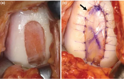
Fig. 1 Periosteum ACI: the
cartilage defect was debrided back to healthy cartilage (left). A periost patch harvested from the tibia was sutured into the defect cambium layer down. On the upper left side, one suture was skipped (arrow) which will be done later to allow injection of the autologous chondrocyte suspension (right)
allowed after the sixth postoperative month and return to contact sports was not recommended before 12 months postoperatively.
Results
Intraoperatively, no loosening, ablation or derangement of either transplant occurred. No knee joint infection and no allergic reaction occurred in either group. Postoperatively, neither an extension lag nor a Xexion deWcit could be observed. One patient of the BioSeed-C group had a mod-erate eVusion at follow-up, but not other problems and did well later on. The rate of follow-up in the periosteum group was 100%. In the BioSeed-C group, the grafts of one patient were removed in a peripheral institution due to soft regenera-tion tissue after 9 months. This patient was registered as a treatment failure. Another patient developed paraplegia dur-ing follow-up and, therefore, was excluded from the study. One patient was lost to follow-up due to unknown address.
At follow-up, mean scores signiWcantly increased in both groups compared with baseline: in the periosteal group, the ICRS score improved from grade D (poor) pre-operatively to grade C (fair) post-op (P < 0.0001) with a
range from grade A (excellent) to grade D (poor) (Fig.3a).
The modiWed Cincinnati knee score improved from 3.26 to 6.44 (patient) (P < 0.0001) and from 3.36 to 6.88
(physi-cian) (P < 0.0001) (Fig.3b, c). Mean Lysholm score
improved from 33 to 70 points (P < 0.0001) (Fig.3d).
In BioSeed-C group, like in the periosteum group, the ICRS score improved from grades D to C as well
(P < 0.0001) (Fig.3a). The modiWed Cincinnati knee score
increased from 3.3 to 6.4 (patient) (P < 0.0001) and from
4.9 to 6.5 (physician) (P < 0.0001) (Fig.3b, c). Mean
Lys-holm score enhanced from 47 to 78 points (P < 0.0001)
(Fig.3d). There was no signiWcant diVerence between
groups 1 and 2 in the ICRS score rating (P = 1) and in the Lysholm score (P = 0.065). In the overall rating of the
modiWed Cincinnati knee score (patient), the diVerence
between groups was not statistically signiWcant (P = 0.39). However, the physician rating was signiWcantly better in the periosteum group (P = 0.033).
In the periosteum group, 10 patients required revision surgery, due to symptomatic periosteal hypertrophy (n = 4) or graft failure (n = 3), plica syndrome (n = 2) and synovec-tomy (n = 1).
In the BioSeed-C group, reoperations were necessary and regarded as directly related to ACI in Wve patients. One patient underwent graft removal in a peripheral institution and was thus registered as a treatment failure. Two patients had a synovectomy and one patient had a debridement and one a total knee replacement, respectively.
Discussion
The aim of the present study was to compare the clinical outcome of standard periosteum ACI to BioSeed-C (ACI
using a biodegradable Xeece) in patients with focal
degenerative cartilage defects of the knee. Patients of both treatment groups showed highly signiWcant increases in the respective scores used for outcome measurement Fig. 2 BioSeed-C is delivered
as a 20 £ 30 £ 2 mm polymer-Xeece seeded with 2 £ 106 autologous chondrocytes (a). The graft is armed with vicryl sutures at every corner which serve as a pulley (b) and are Wxed transosseously using 1.7-mm-k-wires in an insideout technique (c); that way, the graft is smoothly Wtted into the defect (d) and securely Wxed in a press-Wt technique (e)
demonstrating major improvements in activities of daily living, ability to work and in sports. Patients treated with periosteal ACI required twice as many reoperations as patients treated with BioSeed-C. In the ICRS score, the patient rating of the modiWed Cincinnati knee score and the Lysholm score, the outcome was comparable between the periosteum and the BioSeed-C group. However, in the objective and strict ICRS score and in the patient rating of the modiWed Cincinnati knee score, patients treated with periosteum ACI scored slightly, but not signiWcantly better, whereas in the Lysholm score, it was vice versa. In contrast, in the physician’s rating, BioSeed-C patients scored slightly worse than periosteum patients, because of better baseline ratings.
The patients of the two groups compared in this study diVered in defect size, number of previous or concomitant surgical procedures and length of the postoperative follow-up period. This may inXuence the results. However, the
impact of this potential eVect is unknown. The group
treated with conventional ACI had larger defects and longer follow-up. However, a direct comparison of the two groups may be inappropriate, as there were signiWcantly more pre-vious surgical procedures in the group treated with Bio-Seed-C.
When compared with case series, randomised studies evaluating ACI versus other cartilage repair surgeries as
reported previously are favourable [3, 12, 13, 17, 18]. A
recent systematic review comparing ACI and osteochondral autograft transfer with each other with one another and with traditional abrasive techniques demonstrated no clear
supe-riority of ACI over microfracture [22] in line with the
Wnd-ings published by other authors [17, 18]. However, in
another study, ACI showed a superior cartilage regenerate
than microfracture at same clinical short-term results [39].
When comparing the two ACI methods, the present study shows that these are equally eVective. Advantages of the BioSeed-C techniques comprise less morbidity, as no periosteum has to be harvested and the option of arth-roscopical implantation. Defects of the femoral condyles can be addressed arthroscopically depending on lesion size and location which is associated with faster recov-ery after surgrecov-ery and with better cosmetical results. Fur-ther advantages of BioSeed-C, are that it is easier to apply, is more stable than the periosteum cartilage trans-plant and triggers less second look surgeries, because there can be no periosteum hypertrophy. This may partly be due to the lacking periosteum hypertrophies fre-quently causing reoperations in patients treated with ACI using periosteum. ACI involves an open technique with inherent disadvantages such as adhesions and prolonged recovery. Therefore, an arthroscopical approach to ACI as published previously is desirable and could
poten-tially reduce postoperative morbidity [8]. The authors
believe that this is the Wrst study comparing standard
ACI to a 3D Xeece technique (BioSeed-C), representing
a non-randomised retrospective, comparative study with at least 40 patients in each group. However, the outcome of this study should be interpreted with care and
statisti-cal eVects associated with small numbers of patients may
be considered.
First-generation tissue engineering grafts such as perios-teum ACI have been demonstrated to be an appropriate therapy for the regeneration of posttraumatic defects
[5,10]. However, second generation cartilage tissue
Fig. 3 Clinical outcome after
2 years as measured by the ICRS, modiWed Cincinnati knee score and the Lysholm score. All scores showed highly signiWcant (P < 0.0001) improvements at follow-up. The ICRS score improved from grade IV at baseline to grade III at follow-up (a). The modiWed Cincinnati knee score as evaluated by patient (b) and physician (c) showed highly signiWcant improvements in the evaluated follow-up period. The Lysholm score improved highly signiWcant as well (d) 0 1 2 3 4
Bas eline Follow-up Baseline Follow-up
ACI Periosteum ACI BioSeed-C
Score 0 1 2 3 4 5 6 7 8 9 10
Baseline Follow-up Baseline Follow-up
ACI Periosteum ACI BioSeed-C
Score 0 1 2 3 4 5 6 7 8 9 10
Baseline Follow-up Baseline Follow-up
ACI Periosteum ACI BioSeed-C
Score 0 10 20 30 40 50 60 70 80 90 100
Baseline Follow-up Baseline Follow-up
ACI Periosteum ACI BioSeed-C
S core *** *** *** *** *** *** *** *** (a) (b) (c) (d)
engineering grafts using various matrices were recently considered to be technically more attractive. For instance, in a series of 47 cartilage defects, similar outcomes were
obtained clinically as well as histologically [1]. In a
multi-center study evaluating Hyalograft C, which consists of autologous chondrocytes embedded in a derivative of hyal-uronic acid, more than 90% of patients showed
improve-ments of the ICRS score [23]. Clinical assessment as done
by the Meyers score, the Lysholm score and in the ICRS scores was improved in a prospective study investigating
5-year results of matrix associated ACI [2].
In summary, we regard the outcome of this study as a
decent treatment result for this diYcult and highly
demand-ing patient proWle, particularly because patients treated with BioSeed-C had the same outcome levels, e.g., in knee-related performance and quality of life as the patients treated with periosteum ACI, although they have had more surgeries on their knees before.
Concerning its advantages in comparison to periosteum ACI, we conclude that BioSeed-C is equally eVective as periosteum ACI.
ConXict of interest statement All authors certify they have not
signed any agreementwith a commercial interest related to this study which would in any way limit publicationof any and all data generated for the study or to delay publication for any reason.
References
1. Bartlett W, Skinner JA, Gooding CR et al (2005) Autologous chondrocyte implantation versus matrix-induced autologous chon-drocyte implantation for osteochondral defects of the knee: a pro-spective, randomised study. J Bone Joint Surg 87-B:640–645 2. Behrens P, Bitter T, Kurz B et al (2006) Matrix-associated
autolo-gous chondrocyte transplantation/implantation (MACT/MACI): 5-year follow-up. Knee 13:194–202
3. Bentley G, Biant LC, Carrington RW et al (2003) A prospective, randomised comparison of autologous chondrocyte implantation versus mosaicplasty for osteochondral defects in the knee. J Bone Joint Surg 85-B:223–230
4. Brittberg M, Lindahl A, Nilsson A et al (1994) Treatment of deep cartilage defects in the knee with autologous chondrocyte trans-plantation. N Engl J Med 331:889–895
5. Browne JE, Anderson AF, Arciero R et al (2995) Clinical outcome of autologous chondrocyte implantation at 5 years in US subjects. Clin Orthop Relat Res 436:237–245
6. Curl WW, Krome J, Gordon ES et al (1997) Cartilage injuries: a review of 31, 516 knee arthroscopies. Arthroscopy 13:456–460 7. Erggelet C, Browne JE, Fu F et al (2000) Autologous chondrocyte
transplantation for treatment of cartilage defects of the knee joint: clinical results. Zentralbl Chir 125:516–522
8. Erggelet C, Sittinger M, Lahm A (2003) The arthroscopic implan-tation of autologous chondrocytes for the treatment of full-thick-ness cartilage defects of the knee joint. Arthroscopy 19:108–110 9. Erggelet C, Steinwachs MR, Reichelt A (2000) The operative
treatment of full thickness cartilage defects in the knee joint with autologous chondrocyte transplantation. Saudi Med J 21:715–721 10. Fu FH, Zurakowski D, Browne JE et al (2005) Autologous chondrocyte implantation versus debridement for treatment of
full-thickness chondral defects of the knee: an observational co-hort study with 3-year follow-up. Am J Sports Med 33:1658–1666 11. Hangody L, Karpati Z (1994) New possibilities in the management of severe circumscribed cartilage damage in the knee. Magy Trau-matol Ortop Kezseb Plasztikai Seb 37:237–243
12. Horas U, Pelinkovic D, Herr G et al (2003) Autologous chondro-cyte implantation and osteochondral cylinder transplantation in cartilage repair of the knee joint: a prospective, comparative trial. J Bone Joint Surg 85-A:185–192
13. Horas U, Schnettler R, Pelinkovic D et al (2000) Osteochondral transplantation versus autogenous chondrocyte transplantation: a prospective comparative clinical study. Chirurg 71:1090–1097 14. Hubbard MJ (1996) Articular debridement versus washout for
degeneration of the medial femoral condyle: a Wve-year study. J Bone Joint Surg Br 78:217–219
15. International Cartilage Repair Society (1999) http://www.carti-lage.org
16. Kellgren JH, Lawrence JS (1957) Radiological assessment of rheumatoid arthritis. Ann Rheum Dis 16:485–493
17. Knutsen G, Drogset JO, Engebretsen L et al (2007) A randomized trial comparing autologous chondrocyte implantation with micro-fracture: Wndings at Wve years. J Bone Joint Surg 89-A:2105–2112 18. Knutsen G, Engebretsen L, Ludvigsen TC et al (2004) Autologous chondrocyte implantation compared with microfracture in the knee: a randomized trial. J Bone Joint Surg Am 86-A:455–464 19. Kreuz PC, Steinwachs M, Erggelet C et al (2007) ClassiWcation of
graft hypertrophy after autologous chondrocyte implantation of full-thickness chondral defects in the knee. Osteoarthritis Carti-lage 15:1339–1347
20. Lexer E (1908) Substitution of whole or half joints from freshly amputated extremities by free plastic operation. Surg Gynecol Ob-stet 6:601–608
21. Lysholm J, Gillquist J (1982) Evaluation of knee ligament surgery results with special emphasis on use of a scoring scale. Am J Sports Med 10:150–154
22. Marcacci M, Berruto M, Brocchetta D et al (2005) Articular carti-lage engineering with hyalograft C: 3-year clinical results. Clin Orthop Relat Res 435:96–105
23. Matsusue Y, Yamamuro T, Hama H (1993) Arthroscopic multiple osteochondral transplantation to the chondral defect in the knee associated with anterior cruciate ligament disruption. Arthroscopy 9:318–321
24. Micheli LJ, Browne JE, Erggelet C et al (2001) Autologous chon-drocyte implantation of the knee: multicenter experience and min-imum 3-year follow-up. Clin J Sport Med 11:223–228
25. Minas T (2001) Autologous chondrocyte implantation for focal chondral defects of the knee. Clin Orthop Relat Res 391:349– 361
26. Minas T (2003) Autologous chondrocyte implantation in the arthritic knee. Orthopedics 26:945–947
27. Minas T (1999) The role of cartilage repair techniques, including chondrocyte transplantation, in focal chondral knee damage. Instr Course Lect 48:629–643
28. Minas T, Peterson L (1999) Advanced techniques in autologous chondrocyte transplantation. Clin Sports Med 18:13–44, v–vi 29. Nehrer S, Spector M, Minas T (1999) Histologic analysis of tissue
after failed cartilage repair procedures. Clin Orthop Relat Res 365:149–162
30. Noyes FR, Barber-Westin SD (1997) Arthroscopic-assisted allo-graft anterior cruciate ligament reconstruction in patients with symptomatic arthrosis. Arthroscopy 13:24–32
31. Noyes FR, Barber SD, Mooar LA (1989) A rationale for assessing sports activity levels and limitations in knee disorders. Clin Orthop Relat Res 246:238–249
32. Noyes FR, McGinniss GH, Mooar LA (1984) Functional disability in the anterior cruciate insuYcient knee syndrome: review of knee
rating systems and projected risk factors in determining treatment. Sports Med 1:278–302
33. Ossendorf C, Kreuz PC, Steinwachs MR et al (2007) Autologous chondrocyte implantation for the treatment of large full-thickness cartilage lesions of the knee. Saudi Med J 28:1251–1256 34. Outerbridge RE (1961) The etiology of chondromalacia patellae.
J Bone Joint Surg 43-B:752–757
35. Peterson L (1996) Articular cartilage injuries treated with autolo-gous chondrocyte transplantation in the human knee. Acta Orthop Belg 62(Suppl 1):196–200
36. Peterson L, Brittberg M, Kiviranta I et al (2002) Autologous chon-drocyte transplantation: biomechanics and long-term durability. Am J Sports Med 30:2–12
37. Peterson L, Minas T, Brittberg M et al (2002) Two- to 9-year out-come after autologous chondrocyte transplantation of the knee. Clin Orthop Relat Res 374:212–234
38. Steadman JR, Rodkey WG, Rodrigo JJ (2001) Microfracture: sur-gical technique and rehabilitation to treat chondral defects. Clin Orthop Relat Res 391:S362–S369
39. Steinwachs M, Kreuz PC (2007) Autologous chondrocyte implan-tation in chondral defects of the knee with a type I/III collagen membrane: a prospective study with a 3-year follow-up. Arthros-copy 23:381–387