Dynamics of re-expansion of atelectasis during general anaesthesia
6
0
0
Texte intégral
(2) Rothen et al.. Table 1 Patient characteristics (mean (SD) and median (range)). Sex (M/F) Non-smoker/smoker Age (yr) Weight (kg) Height (cm) BMI (kg m-2). Mean (SD). Median (range). 7/5 9/3 51 (14) 81 (15) 174 (10) 26.4 (2.9). – – 55 (30–69) 80 (58–101) 176 (158–195) 26.5 (20.8–31.2). Patients and methods A total of 12 patients, undergoing neurosurgical procedures or surgery of the eye, at the University Hospital Uppsala, Sweden, were included in the study (Table 1). Another four patients refused to participate (refusal rate 25%). No patient had cardiac or pulmonary disease, as assessed by patient history and clinical examination. The study was approved by the Ethics Committee of the University Hospital of Uppsala, Sweden. Informed consent was obtained from each subject. In the awake patient, CT of the lungs was performed at end-expiration with the subject lying supine on the CT table and breathing air. Anaesthesia was induced, and an arterial cannula was inserted into a radial artery. After a minimum of 15 min of anaesthesia (see below), the CT scan was repeated and a sample of arterial blood was obtained in duplicate for blood-gas analysis. Thereafter, the VC manoeuvre was initiated and several CT scans were obtained (see below). Five minutes later, another sample of arterial blood was obtained. Finally, the patient was moved from the x-ray department to the operating theatre to undergo surgery. Details of anaesthesia have been described previously.16 In brief, anaesthesia was induced with fentanyl 2–3 µg kg–1 and propofol 2 mg kg–1 i.v., followed by continuous infusion of propofol 5–10 mg kg–1 h–1. The dose of these drugs was adjusted according to clinical signs of depth of anaesthesia (arterial pressure and heart rate). During induction, the lungs were ventilated manually via a face mask with 100% oxygen. To facilitate orotracheal intubation, patients received pancuronium 0.1 mg kg–1; additional doses of 1–2 mg were given when needed. The lungs were ventilated mechanically at a rate of 10 bpm with 40% oxygen in nitrogen (Servo Ventilator 900C, Siemens Elema, Lund, Sweden). The I:E ratio was 1:2. No positive end-expiratory pressure was used. Ventilation was adjusted to maintain an end-tidal carbon dioxide concentration of 4.0–4.5% (CO2 ¨ analyser Eliza, Engstrom, Bromma, Sweden). The resulting tidal volume was mean 8.7 (SD 0.8) ml kg–1. Pulse oximetry was used throughout the study (Biox 3740, Ohmeda, Lousiville, CO, USA). Heart rate and arterial pressure (Riva-Rocci method) were recorded every 5 min.. Computed tomography of the lungs Atelectasis was studied by CT (Somatom Plus 4, Siemens, Erlangen, Germany). The technique has been described in. detail previously.12 13 Subjects were in the supine position with their arms alongside the body. A frontal scout view, covering the chest, and a CT scan in the transverse plane, 1 cm cranial to the top of the right diaphragm, were obtained at end-expiration (i.e. functional residual capacity (FRC)) in the awake subject and after induction of anaesthesia. Scan time was 750 ms and slice thickness was 8 mm. With a matrix of 5123512, the resulting picture element (pixel) was approximately 1.531.5 mm. To identify atelectasis, a magnified image of the dorsal portion of the CT scan of both the right and left lung was made (Sienet Magic-View, Siemens, Erlangen, Germany). The dorsal border between the thoracic wall and the dense areas was drawn manually, whereas the ventral border between inflated lung tissue and atelectasis was identified by the region-of-interest (ROI) programme. All pixels with attenuation values between –100 and 1100 Hounsfield units (HU) were considered to represent atelectatic lung tissue.12 Furthermore, the total intra-thoracic area (including the mediastinum) was measured, including all pixels with attenuation values between –1000 HU and 1100 HU. During the VC manoeuvre (see below), 10 CT scans were performed between 1 and 26 s. In an attempt to examine the same region of the lung during the VC manoeuvre, helical scanning covering 6 cm13 was performed during the first part of the VC manoeuvre. Scanning started at the same level as the scan performed before the VC manoeuvre. Duration of helical scanning was 6 s, table feed was 10 mm and slice thickness was 8 mm. Helical scanning was followed immediately by dynamic scanning (same position as the final scan during helical scanning). One scan was obtained every 6 s for the next 18 s. Rotation time was 750 ms and slice thickness 8 mm. The estimated total x-ray exposure for each patient was 3 mSv (total average exposure in Sweden55 mSv per year).. Analysis of arterial blood All patients had an arterial cannula. Arterial blood was collected for blood-gas and oxygen saturation analysis (ABL 300 and OSM3, Radiometer, Copenhagen, Denmark) in duplicate,15 min after induction of general anaesthesia and 5 min after the re-expansion manoeuvre.. Vital capacity manoeuvre The VC manoeuvre was performed as follows. The ventilator was switched to pressure-controlled mode, using an inspiratory pressure of 20 cm H2O. After two breaths, the end-expiratory hold button was pressed, inspiratory pressure was changed immediately to 40 cm H2O and the longest possible inspiratory time was selected by changing ventilatory frequency to 6 bpm and inspiratory time to 80% of the total respiratory cycle. This resulted in an active inspiratory phase of 8 s. The x-ray exposure programme was started, the end-expiratory hold button was released and the lungs were inflated by the ventilator (5VC manoeuvre). During this inflation manoeuvre, the inflation-hold button. 552.
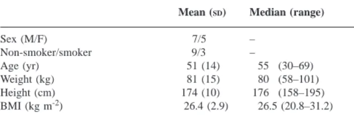
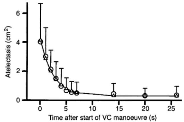
(3) Re-expansion of atelectasis. Table 2 Analysis of arterial blood before and after the VC manoeuvre Before pH 7.44 (0.03) PaCO2 (kPa) 4.8 (0.6) 17.2 (4.0) PaO2 (kPa) SaO2 (%) 98.3 (0.9) Bicarbonate (mEq litre-1) 24.6 (1.2) BE (mEq litre-1) 0.1 (1.4). After. P. 7.44 (0.04) 4.7 (0.7) 22.2 (6.0) 98.7 (0.5) 24.8 (1.1) 0.2 (1.1). 0.2 0.3 0.013 0.1 0.5 0.9. of the ventilator was pressed to prolong the inspiratory phase. Inflation was maintained until the last CT scan was taken 26 s after initiation of the VC manoeuvre. Thereafter, the ventilator was switched back to volume-controlled mode, as used before the VC manoeuvre. Inspiratory pressure (airway pressure Paw) was measured in the ventilator on the inspiratory side. The latter data were recorded with a personal computer for further analysis (LabVIEW 3.1, National Instruments, Austin, TX, USA).. Fig 1 Atelectasis before and during the VC manoeuvre. Mean values (s) and SD (error bars) are shown. Also shown is a curve with negative exponential decay, fitted to individual data (n), connected by a line. For further details, see text.. A curve with negative exponential decay was fitted to the data according to:. Statistical analysis Unless otherwise stated, values are mean (SD). To compare data between repeated measures (e.g. atelectasis or thoracic area in the awake state, after induction of anaesthesia and after the VC manoeuvre), the Friedman two-way analysis of variance was used. The Wilcoxon signed rank test was used as a follow-up procedure to make pairwise comparisons. Further, change in atelectasis over time was analysed using an exponential regression model. For all calculations, the SYSTAT 7.0 computer software package (SPSS Arlington, VA, USA) was used.. Results Awake and after induction of general anaesthesia Overall, measurements in the awake subjects were in the normal range. In one patient, a small amount of densities was found in the dependent part of the lungs. No other abnormalities were present on CT scan. Mean thoracic area close to the diaphragm was 357 (75) cm2. After induction of anaesthesia, all patients developed atelectasis. Mean amount, as measured in the scan close to the diaphragm, was 4.0 (2.7) cm2 (median 3.9 (range 0.7–8.0) cm2). Mean thoracic area decreased significantly to 322 (54) cm2 (P50.002). The partial pressure of oxygen and carbon dioxide in arterial blood are given in Table 2.. VC manoeuvre There was an immediate and fast decrease in the amount of atelectasis (Figs 1, 2). Seven seconds after the start of the VC manoeuvre, some atelectasis was still present in three subjects. During the rest of the VC manoeuvre, atelectasis remained unchanged in two of these three subjects (with 1.3 cm2 and 0.8 cm2 of atelectasis at 26 s after the start of the VC manoeuvre, respectively) and decreased further in one subject (from 2.3 cm2 at 7 s to 1.6 cm2 at 26 s).. atelectasis (cm2)5a1y03e–(time (s)/τ) where y053.9 cm2 (95% CI 3.1–4.6 cm2), τ52.6 s (95% CI 1.8–4.3 s) and a50.3 cm2 (95% CI –0.1–0.7 cm2). Note: y0 represents the amount of atelectasis before the start of the VC manoeuvre, τ5 time constant, e5base of natural logarithms and a5correction factor.18 The change in thoracic area is shown in Figure 3. Again, the most prominent change occurred during the first few seconds of the VC manoeuvre. Mean area was 427 (75) cm2 at 7 s and 419 (77) cm2 at 26 s. The change in Paw during the VC manoeuvre is shown in Figure 3. Note that Paw increased within the first second of the VC manoeuvre. There was a decrease in Paw from 41 (3) cm H2O at 1 s to 40 (2) cm H2O at 7 s, and to 37 (4) cm H2O at 26 s. Compared with the value before the VC manoeuvre, arterial oxygen tension increased by a mean of 5 kPa (P50.013) (Table 2). There was no significant change in any other variable measured in arterial blood. There were no clinically relevant changes in heart rate or arterial pressure during the study.. Discussion Our study has allowed a detailed analysis of the change in the amount of atelectasis during a vital capacity (VC) manoeuvre in anaesthetized subjects undergoing mechanical ventilation, with no history of lung disease. Furthermore, it has confirmed previous investigations, demonstrating that virtually all atelectatic tissue may be re-expanded by a VC manoeuvre, and that this results in improved oxygenation of arterial blood.14–16 In our previous studies, the lungs were inflated up to a Paw of 40 cm H2O, and this pressure was maintained for 15 s. Previously, analysis of lung tissue by CT was performed after completion of the VC manoeuvre only. However, in our study, the change in amount of collapsed. 553.
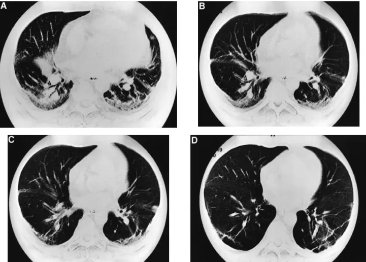
(4) Rothen et al.. Fig 2 CT scan during the VC manoeuvre. A5At start of VC manoeuvre; manoeuvre; and D53.5 s after the start of the VC manoeuvre.. Fig 3 Thoracic area and airway pressure (measured in the ventilator on the inspiratory side) before and during the VC manoeuvre (mean (SD)). Note the fast increase in Paw within the first second of the VC manoeuvre. aw5Awake.. lung tissue was followed during re-expansion. Interestingly, atelectasis was reduced mostly during the first 7–8 s of the VC manoeuvre. In three of 12 patients, some dense areas were still present 7 s after the start of the VC manoeuvre. In these subjects, there was only a minor or no change during the rest of the VC manoeuvre. In the analysis by negative exponential decay, this persistent amount of atelectasis was reflected by a correction factor of 0.3 cm2. This correction factor suggests (although the 95% confid-. B51. s after the start of the VC manoeuvre;. C51.5. s after the start of the VC. ence interval of the correction factor includes 0 cm2) that not all lung tissue may be re-expanded by the VC manoeuvre, even if an airway pressure of 40 cm H2O were maintained for a much longer period. The time constant (2.6 s) suggests that atelectasis was reduced to 50% of the initial value after 1.8 s (‘half-life’5 0.693τ). It may also be estimated that on average, atelectasis was reduced to 5% of the initial value after 7.8 s (533τ).18 In summary, the most important change in terms of re-opening of collapsed lung tissue during a VC manoeuvre occurred during the initial few seconds of the procedure. One might speculate that more time is needed to reexpand large (e.g. lobar) atelectasis. The small number of subjects in our study does not allow conclusions to be drawn in terms of time needed to re-expand such extensive atelectasis. However, as already noted, even in the subjects with the highest amount of atelectasis, re-opening occurred mostly during the initial seconds of the VC manoeuvre with virtually no change later than 7–8 s after the start of the manoeuvre. These findings suggest that time is an important determinant for re-opening of previously collapsed lung units. Obviously, airway pressure is another relevant factor.13 Applying the same airway pressure for a prolonged time does not necessarily result in re-opening of. 554.
(5) Re-expansion of atelectasis. more lung units if the critical opening pressure of the respective lung units is not exceeded. Thus extending the time of inflation further may simply increase adverse cardiovascular effects without additional beneficial effects in terms of improved oxygenation. The flow pattern used during re-expansion may also be important. In our study, a pressure-controlled mode was used for the VC manoeuvre (see also Fig. 3). Thus there was a decelerating, initially high inspiratory flow. Using other flow patterns, such as continuous or accelerating flow, may result in a different change in atelectasis over time. Indeed, manual inflation of the lungs, aiming at an inflation pressure of 40 cm H2O, was found to result in slower re-expansion of collapsed lung tissue (own unpublished observations). Apart from cardiocirculatory compromise, a further possible adverse effect of a VC manoeuvre is disruption of the alveolocapillary barrier or barotrauma. It has been shown that in small animals, periods of hyperinflation as short as 2 min may alter microvascular permeability, resulting in increased extravascular lung water.19 In that study, the lungs were inflated to a Paw of 35 mm Hg (48 cm H2O), corresponding to a tidal volume of 46 ml kg–1. In contrast, inflation of the entire lung in rabbits to 40 cm H2O for 20 min did not produce a protein-permeable epithelium or accumulation of alveolar liquid, whereas distension of small areas of the lung by the same pressure and for the same time produced protein-permeable epithelium.20 Furthermore, in dogs, a higher Paw of 70 mm Hg was required to cause stress failure of pulmonary capillaries,21 and in thoroughbred horses, an even greater Paw of approximately 100 mm Hg (i.e. 135 cm H2O) was necessary to cause such disruptions.22 In addition, there was no correlation between ‘high’ ventilatory pressures and the development of pneumothorax or other air leaks in patients with sepsis-induced acute respiratory distress syndrome.23 Thus it is difficult, if not impossible, to draw firm conclusions from these findings with respect to the risk of barotrauma caused by hyperinflation in adults with healthy lungs. The VC manoeuvre used in our study resulted in a volume not larger than vital capacity in the awake, supine subject.13 Therefore, we believe that during mechanical ventilation, such a manoeuvre may still be used in healthy adults with an adequate clinical indication, such as significant hypoxaemia caused by atelectasis. When a lung unit is re-opened, the composition of inspiratory gas plays an important role for renewed collapse.14 16 Re-expansion may have a sustained effect for at least 40 min if 40% oxygen in nitrogen is used.15 However, if 100% oxygen is used for the VC manoeuvre and for ventilation of the lungs thereafter, lung collapse reappears within a few minutes16 What are the clinical implications of our findings? No clinically adverse events were caused by the VC manoeuvre. In patients with cardiac compromise however, the VC manoeuvre may result in a clinically important reduction in cardiac output and a decrease in systemic arterial pressure.17 Indeed, we have observed in clinical practice that inflation. of the lungs, as proposed previously (Paw of 40 cm H2O, maintained for 15 s), may result in a marked change in haemodynamic variables (e.g. in patients with cardiovascular compromise or inadequate fluid load to the circulatory system). Thus if the time of maximal inflation of the lungs is halved, the margin of safety of such a manoeuvre increases. As a consequence, a shorter inflation time may be used as the first-line measure in patients where atelectasis is suspected to be the main cause of impaired oxygenation during general anaesthesia with mechanical ventilation.7 9 13 14 Such a manoeuvre re-opens most collapsed lung units and consequently improves oxygenation in the majority of patients. One should remember that in patients with chronic obstructive pulmonary disease, anaesthesia causes a small amount of atelectasis only, with impaired oxygenation being caused primarily by ventilation–perfusion mismatch.24 Under such circumstances, any further expansion of lung tissue may be of limited value or even harmful because of possible regional over-inflation. Another method of re-opening collapsed lung is positive end-expiratory pressure (PEEP). Interestingly, if used in patients with healthy lungs, PEEP reduces the amount of atelectasis but may have a varying effect on pulmonary shunt and result in increased deadspace.25 Further, atelectasis re-develops within a few minutes after cessation of PEEP.26 Thus if sustained re-opening of atelectasis and reduction of pulmonary shunt are the main goals, a VC manoeuvre may be more appropriate than PEEP,15 although we are not aware of a direct comparison of these two procedures. In summary, in adults with healthy lungs, inflation of the lungs to an airway pressure of 40 cm H2O, maintained for 7–8 s only (using a constant pressure, decelerating flow mode), may re-expand essentially all previously collapsed lung tissue, as seen on CT, and improve oxygenation. The previously proposed time for a VC manoeuvre thus may be halved in such subjects.. Acknowledgements This study was supported by grants from the Swedish Medical Research Council (Grant No. 4315), the Swedish Heart–Lung Fund and the Department of Anaesthesia and Intensive Care Medicine, University of Bern, Switzerland. We thank Marianne Almgren and Lena Haglund, technicians, for assistance, Peter Vock, MD, Department of Radiology University Hospital, Bern, for technical support during analysis of CT data, and Thomas Schnider, MD, for helpful suggestions concerning statistics.. References. 555. 1 Campbell EJM, Nunn JF, Peckett BW. A comparison of artificial ventilation and spontaneous respiration with particular reference to ventilation–bloodflow relationships. Br J Anaesth 1958; 30: 166–75 ´ ´ 2 Cote CJ, Goldstein EA, Cote MA, Hoaglin DC, Ryan JF. A singleblind study of pulse oximetry in children. Anesthesiology 1988; 68: 184–8 3 Moller JT, Johannessen NW, Berg H, Espersen K, Larsen LE. Hypoxaemia during anaesthesia—an observer study. Br J Anaesth 1991; 66: 437–44 4 Bendixen HH, Hedley-Whyte J, Laver MB. Impaired oxygenation.
(6) Rothen et al.. 5. 6 7. 8 9. 10. 11. 12. 13. 14. 15. in surgical patients during general anesthesia with controlled ventilation. N Engl J Med 1963; 269: 991–6 Prys-Roberts C, Nunn JF, Dobson RH, Robinson RH, Greenbaum R, Harris RS. Radiologically undetectable pulmonary collapse in the supine position. Lancet 1967; II: 399–401 Damgaard-Pedersen K, Qvist T. Pediatric pulmonary CT-scanning. Anaesthesia-induced changes. Pediatr Radiol 1980; 9: 145–8 Brismar B, Hedenstierna G, Lundquist H, Strandberg Å, Svensson L, Tokics L. Pulmonary densities during anesthesia with muscular relaxation—a proposal of atelectasis. Anesthesiology 1985; 62: 422–8 Warner DO, Warner MA, Ritman EL. Atelectasis and chest wall shape during halothane anesthesia. Anesthesiology 1996; 85: 49–59 Gunnarsson L, Tokics L, Gustavsson H, Hedenstierna G. Influence of age on atelectasis formation and gas exchange impairment during general anaesthesia. Br J Anaesth 1991; 66: 423–32 Kisala JM, Ayala A, Stephan RN, Chaudry IH. A model of pulmonary atelectasis in rats: activation of alveolar macrophage and cytokine release. Am J Physiol 1993; 264: R610–14 Froese AB, McCulloch PR, Sugiura M, Vaclavik S, Possmayer F, Moller F. Optimizing alveolar expansion prolongs the effectiveness of exogenous surfactant therapy in the adult rabbit. Am Rev Respir Dis 1993; 148: 569–77 Lundquist H, Hedenstierna G, Strandberg Å, Tokics L, Brismar B. CT-assessment of dependent lung densities in man during general anaesthesia. Acta Radiol 1995; 36: 626–32 Rothen HU, Sporre B, Engberg G, Wegenius G, Hedenstierna G. Re-expansion of atelectasis during general anaesthesia: a computed tomography study. Br J Anaesth 1993; 71: 788–95 Magnusson L, Zemgulis V, Tenling A, et al. Use of a vital capacity maneuver to prevent atelectasis after cardiopulmonary bypass. Anesthesiology 1998; 88: 134–42 Rothen HU, Sporre B, Engberg G, Wegenius G, Hedenstierna G. Reexpansion of atelectasis during general anaesthesia may have a prolonged effect. Acta Anaesthesiol Scand 1995; 39: 118–25. ¨ 16 Rothen HU, Sporre B, Engberg G, Wegenius G, Hogman M, Hedenstierna G. Influence of gas composition on recurrence of atelectasis after a reexpansion maneuver during general anesthesia. Anesthesiology 1995; 82: 832–42 17 Pinsky MR. The hemodynamic consequences of mechanical ventilation: an evolving story. Intensive Care Med 1997; 23: 493–503 18 Nunn JF. The wash-out or die-away exponential function. In: Nunn JF, ed. Nunn’s Applied Respiratory Physiology. Oxford: ButterworthHeinemann, 1993; 586–90 19 Dreyfuss D, Soler P, Saumon G. Spontaneous resolution of pulmonary edema caused by short periods of cyclic overinflation. J Appl Physiol 1992; 72: 2081–9 20 Egan EA. Lung inflation, lung solute permeability, and alveolar edema. J Appl Physiol 1982; 53: 121–5 21 Mathieu-Costello O, Willford DC, Fu Z, Garden RM, West JB. Pulmonary capillaries are more resistant to stress failure in dogs than in rabbits. J Appl Physiol 1995; 79: 908–17 22 Birks EK, Mathieu-Costello O, Fu Z, Tyler WS, West JB. Very high pressures are required to cause stress failure of pulmonary capillaries in thoroughbred racehorses. J Appl Physiol 1997; 82: 1584–92 23 Weg JG, Anzueto A, Balk RA, et al. The relation of pneumothorax and other air leaks to mortality in the acute respiratory distress syndrome. N Engl J Med 1998; 338: 341–6 24 Gunnarsson L, Tokics L, Lundquist H, et al. Chronic obstructive pulmonary disease and anaesthesia: formation of atelectasis and gas exchange impairment. Eur Respir J 1991; 4: 1106–16 25 Tokics L, Hedenstierna G, Strandberg Å, Brismar B, Lundquist H. Lung collapse and gas exchange during general anesthesia: effects of spontaneous breathing, muscle paralysis, and positive endexpiratory pressure. Anesthesiology 1987; 66: 157–67 26 Hedenstierna G, Tokics L, Lundquist H, Andersson T, Strandberg Å, Brismar B. Phrenic nerve stimulation during halothane anesthesia. Anesthesiology 1994; 80: 751–60. 556.
(7)
Figure
Documents relatifs
