New concepts of atelectasis during general anaesthesia
L. Magnusson* and D. R. Spahn
Department of Anaesthesiology, University Hospital, CHUV, CH-1011 Lausanne, Switzerland
*Corresponding author. E-mail: [email protected]Br J Anaesth 2003; 91: 61±72
Keywords: anaesthetic techniques; complications, atelectasis; lung, atelectasis; measurement techniques, tomography
At the beginning of the last century, Pasteur described postoperative pulmonary atelectasis,58 analysed
post-operative pulmonary complications (PCC; see below) and noted: `when the true history of postoperative lung compli-cations comes to be written, active collapse of the lung, from de®ciency of inspiratory power, will be found to occupy an important position among determining causes'.59
Indeed, atelectasis occurs regularly during general anaes-thesia induction,46 persists postoperatively44 and may
contribute to signi®cant morbidity7 8and additional
health-care costs.43
This review article will review the mechanism of perioperative atelectasis, discuss its clinical signi®cance and describe preventive measures.
Gas exchange and general anaesthesia
In 1964, Nunn56showed that during routine anaesthesia and
spontaneous ventilation, gas exchange was altered by shunt and uneven ventilation perfusion ratios. He concluded that to ensure the maintenance of a normal arterial PO2, the
alveolar PO2 had to be as high as 200 mm Hg and this
required an inspired oxygen concentration (FIO2) of 35%.
Since then it has been commonly accepted that all general anaesthesia should use at least 30±35% oxygen.
Atelectasis and general anaesthesia
Atelectasis was early suspected as a cause of impaired oxygenation during general anaesthesia. Bendixen and colleagues5postulated that spontaneous ventilation without
periodic deep breaths may lead to progressive atelectasis, with increased shunting and decreased pulmonary compli-ance, and that these changes were reversible by hyperin¯a-tion of the lungs. They showed that general anaesthesia without supplemental oxygen reduced PaO2 by 22% and
compliance by 15% and that three successive hyperin¯a-tions of the lungs restored both arterial oxygen tension and lung compliance to control values, suggesting that periodic deep breaths prevented progressive atelectasis and shunting.
In the 1980s, atelectasis was shown by computed tomo-graphy (CT) in anaesthetized patientsÐneonates as well as adults.7 14 Lung densities were seen in anaesthetized
children and were called `con¯uent high absorptive areas' but such areas were not found in the scans performed under sedation.14 In 1985, Brismar and colleagues7showed that
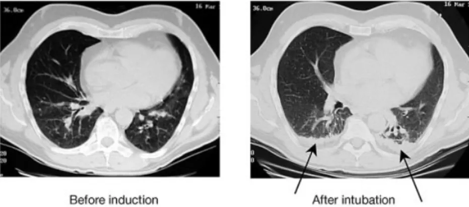
within 5 min of induction of anaesthesia, crest-shaped changes of increased density appeared in the dependent regions of both lungs (Fig. 1). In 1989, Hedenstierna and colleagues33also found densities in anaesthetized animals,
with the same location and attenuation as in anaesthetized humans.57 Microscopy showed that the densities were
atelectatic lung regions. It was concluded that these densities in dependent regions during anaesthesia were caused by atelectasis.
Since then atelectasis has been studied extensively. Atelectasis on a CT scan is de®ned as pixels with attenuation values of ±100 to +100 Houns®eld units (HU).47 These occur in the most dependent parts of the
lungs and are found in almost 90% of all patients who are anaesthetized.47 They develop with both i.v. and
inhala-tional anaesthesia and whether the patient is breathing spontaneously or is paralysed and ventilated mechanic-ally.88 The only anaesthetic so far tested that has not
produced atelectasis is ketamine,93 although when the
patient was paralysed, atelectasis also appeared in these subjects. On the contrary, epidural anaesthesia caused no or little atelectasis and no change in shunting, ventilation/ perfusion (VA/Q) matching or oxygenation.71 91
Good correlations have been found between gas exchange impairment and the amount of atelectasis (r=0.93 for atelectasis and intrapulmonary shunt; r=0.99 for atelectasis and oxygenation).35 Most atelectasis occurs near the
diaphragm in the supine patient and less towards the apex (Fig. 2).72 In most patients the atelectasis may not appear
severe, but collapsed lung comprises four times more lung tissue than aerated regions.72 Thus, in the average patient
the atelectasis may contain 15±20% of the lung tissue close to the diaphragm and about 10% of the total lung tissue. In DOI: 10.1093/bja/aeg085
extreme cases almost half the lung can be collapsed during anaesthesia, before any surgery has taken place or com-monly after cardiac surgery!90 Atelectasis can persist for
two days after major surgery45 but disappears within 24 h
after laparoscopy in non-obese subjects.19
After cardiac surgery with cardiopulmonary bypass (CPB) atelectasis is more prominent than after other forms of surgery, even thoracotomies. In an animal model, extensive atelectasis was seen 1 h after CPB, which was well correlated with intrapulmonary shunt.49 In man,
prominent atelectasis in the dorsal part of the lungs has been found on the ®rst day after cardiac surgery.90
Measurement of atelectasis
Atelectasis is not seen on conventional chest radiograph unless it becomes massive.47 Since 1980, atelectasis has
been examined by CT scanning in awake32or anaesthetized
lung-healthy patients,29 32 33 47 in the extremes of age
(paediatric populations43 86 87 and patients over 80 years
of age30), in morbidly obese patients,19 89in patients with
chronic obstructive lung disease,31 smokers,89 and in
patients with adult respiratory distress syndrome (ARDS).65
The CT scan method starts with a frontal scoutview of the chest to de®ne the borders of the lungs and to guide the settings for subsequent scans. One or more transverse CT scans are then made. To avoid excessive radiation, if successive examinations are planned, only one or two transverse slices are done. If only one CT scan is to be done then the whole lung can be studied. This shows a limitation of the method, because a single level may not re¯ect the entire lung. The level most often used is 1 cm above the right diaphragm, equivalent to the interventricular septum (Fig. 1). This level appears to be the best compromise between the most affected bases of the lungs and the less affected apex7 78and the amount of atelectasis measured at
this level correlates well with gas exchange impair-ment.6 55 71
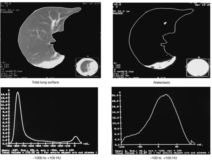
The images are analysed by computer. The entire right and left lungs can be selected as a region of interest by drawing the external boundaries of the lungs at the inside of the ribs and the internal boundaries along the mediastinal organs. The total area of the lungs is measured by including pixels with density values between ±1000 and +100 HU (Fig. 3). Densities considered to indicate atelectasis are identi®ed in dependent lung regions and outlined manually. Atelectasis is then calculated by including all pixels within these regions with HU between ±100 and +100. Manual delineation of atelectasis has only a small bias compared with computerized evaluation.47
With this technique, the partial volume effect may interfere with the measurement of atelectasis.17 Dense
tissue adjacent to the lung can in¯uence the average slice
Fig 2 Two-dimensional representation of a volume image from an anaesthetized subject. The surface of the lung is shown in shades of grey and atelectasis is shown in white. The anteroposterior view (top) shows the right and left lungs, with a visible cardiac shadow (arrow). The lateral view (bottom) shows the atelectatic regions in the most dependent lung (from Warner and colleagues98).
Fig 1 Examples of CT scans of a patient with healthy lungs, before and after induction of anaesthesia. The CT slices are 1 cm above the level of the right diaphragm. Arrows indicate lung densities, thought to represent atelectasis (from Rusca and colleagues84).
density because of the limited spatial resolution of the system. The same pixel may contain both lung tissue and some adjacent dense tissue. This is called the partial volume effect and it becomes greater when the overall area of the lung is small. Therefore, this effect will be reduced on CT scans taken 1 cm above the right diaphragm where the lung area is great. Moreover, when studies are made of repeated CT scans, this systematic error should not interfere with changes seen over time.
Dependent lung densities have been shown to be atelectasis.33 47 Nevertheless, blood is a component of
lung tissue and has nearly the same density as atelectasis. Therefore, variations in the amount of blood during a study may alter or even explain dependent lung densities.17
Variation in the amount of blood in a dependent lung region may occur with variation in intrathoracic pressure. However, basal densities are found during anaesthesia with stable intrathoracic pressure when FIO2is increased and
thus cannot be explained only by variations in blood content.81In addition, no densities appear when pressure is
changed while low oxygen concentration is used, suggesting that the amount of blood plays only a marginal role in appearance of dependent lung densities during general anaesthesia and that these densities even in human patients are atelectasis.
In most studies, dependent lung densities are called atelectasis. It would be better to be more descriptive and use the description lung densities in the title, summary and results, and only explain in the discussion that these densities are probably caused by atelectasis. Nevertheless, in this review lung densities will be considered as atelectasis.
Causes of atelectasis formation during
general anaesthesia
Pulmonary atelectasis may be caused by a variety of factors, which have been classi®ed into three basic mechanisms. Compression atelectasis occurs when the transmural pressure distending the alveolus is reduced. Absorption
Fig 3 Measurement of atelectatic surface by CT of the lung at the level of the interventricular septum and corresponding histograms. The total lung surface has densities between ±1000 and +100 HU. Atelectatic lung surface has densities between ±100 and +100 HU (from BenoõÃt and colleagues6).
atelectasis occurs when less gas enters the alveolus than is removed by uptake by the blood. Loss-of-surfactant atelectasis occurs when the surface tension of an alveolus increases because of reduced surfactant action. Any of these factors may contribute to atelectasis during anaesthesia and the postoperative period.
Compression atelectasis
The rapid formation of atelectasis on induction of anaes-thesia, being detectable as soon as CT scans can be made, and the fast reappearance after discontinuation of PEEP suggested that the atelectasis was caused by compression of lung tissue rather than by resorption of gas behind occluded airways.7The ®nding that atelectasis could be reduced by
phrenic nerve stimulation34provided further support to this
hypothesis, as did the absence of atelectasis during ketamine anaesthesia.93The latter two studies may indicate that loss
of inspiratory muscle tone is an important factor in atelectasis formation. It may thus be that the greater abdominal pressure is more easily transmitted into the thoracic cavity when the diaphragm has a reduced tone or is paralysed, as during anaesthesia. The classic study by Froese and Bryan22 showed that diaphragm motion in
spontaneously breathing normal volunteers is changed when the volunteers are paralysed with neuromuscular blocking agents. The authors concluded that in the supine position during spontaneous ventilation, the dependent part of the diaphragm had the greatest displacement. However, after neuromuscular block and positive pressure ventilation, exactly the opposite was seen: the non-dependent part had the greatest displacement. Also, Krayer and colleagues,41
using CT scans, found altered diaphragmatic motion during general anaesthesia and mechanical ventilation. In addition, Warner and colleagues98 found alterations in the
end-expiratory position of chest wall structures during general anaesthesia (Fig. 4), and Reber and colleagues74 showed
that general anaesthesia induced a cephalad displacement of the most dorsal part of the diaphragm.
Thus, compression atelectasis occurs during general anaesthesia and is caused by chest geometry and diaphragm position and motion.
Absorption atelectasis
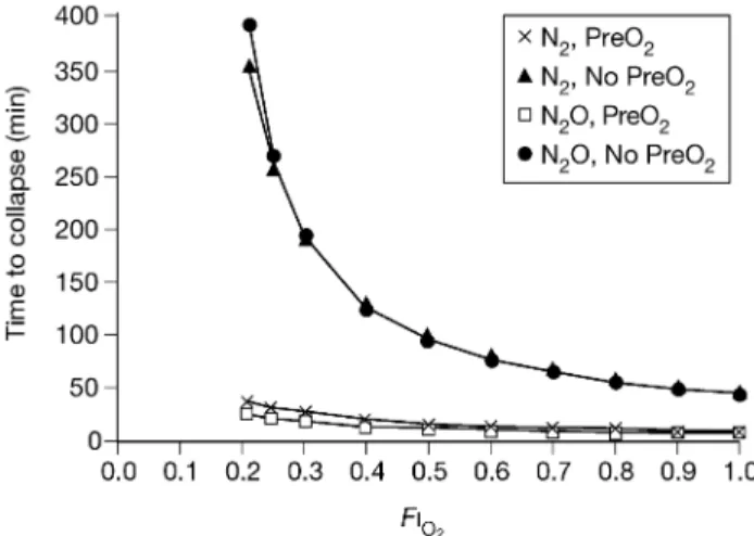
Absorption atelectasis can occur by two mechanisms. The ®rst mechanism is complete airway occlusion, which creates a pocket of trapped gas in the distal lung unit. The pressure in the pocket initially is close to atmospheric pressure. Mixed venous blood continues to perfuse the pocket, and since the sum of the gas partial pressures in the mixed venous blood is subatmospheric, gas uptake from the pocket by the blood continues and the pocket collapses.46The rate
of absorption of gas from an unventilated lung area increases with an increasing FIO2(Fig. 5).
37
The second mechanism is when the inspired VA/Q ratio is
less than a critical value. If the inspired VA/Q ratio of a lung
unit is reduced, a point is reached where the rate at which inspired gas entering the alveolus is exactly balanced by gas uptake from the alveolus into the blood. This point is known as the critical VA/Q ratio.15If the inspired VA/Q ratio is less
than this, the lung unit will collapse. This is likely when FIO2
is high and the gas uptake is large. Conversely, a reduction in the amount of atelectasis is seen when lower concentra-tions of oxygen are used at induction,73 83during
mainten-ance of general anaesthesia,80or just before extubation.6
Loss-of-surfactant atelectasis
Recurrence of atelectasis within 5 min after a vital capacity manoeuvre (VCM; see below) at FIO2=1.0
81or immediately
after removal of PEEP at FIO2=0.4
7 35suggests an instability Fig 4 Diagram of a midsagittal section of the thorax while awake (solid
lines) and while anaesthetized (dashed lines). Note the alteration in the position of the diaphragm (cephalad motion in the dependent portion; from Warner and colleagues98).
Fig 5 Time to collapse of an unventilated lung compartment. PreO2=3
min of preoxygenation. The inert gas breathed after induction was either nitrogen or nitrous oxide. Collapse occurred more quickly after preoxygenation. The greater the inspired oxygen fraction after induction, the faster the collapse. Time to collapse is largely independent of whether the inspired gas mixture after induction contains nitrogen or nitrous oxide (from Joyce and colleagues38).
in the alveoli that have been collapsed. It is possible that atelectasis, once formed, impedes surfactant function so that such a region is prone to collapse again after having been reopened. A VCM may promote surfactant production or release, and distribution of surfactant over the alveolar surface may cause a longer lasting protection against new collapse.50 Indeed, it has been shown that large gasps
increase the proportion of active forms of alveolar surfactant.58
In summary, all three mechanisms (compression, absorp-tion and loss of surfactant) may contribute to atelectasis formation during general anaesthesia. Absorption and compression are the two mechanisms most implicated in perioperative atelectasis formation. Indeed, Rothen and colleagues80 have shown that intrapulmonary shunt is
correlated to the amount of atelectasis and that poorly ventilated lung units (`low VA/Q') are correlated with
airway closure measured by the difference in closing volume and expiratory reserve volume (CV±ERV). There is no correlation between CV±ERV and atelectasis. Taken together, the amount of atelectasis and airway closure may explain 75% of the deterioration in gas exchange seen during general anaesthesia.
Factors in¯uencing atelectasis formation
Fraction of inspired oxygen
High oxygen concentration has often been associated with atelectasis formation. When an FIO2of 1.0 is used after a
VCM, atelectasis recurs within 5 min.81On the other hand,
when 40% oxygen is used, atelectasis will not recur for at least 40 min.79 81 In order to avoid atelectasis formation,
lower oxygen concentration has been used during induction of general anaesthesia. With 100% oxygen, shunt increased from 0.3% to 6.5%, with atelectasis formation correspond-ing to an area of 8.0 cm2. With 30% oxygen, shunt increased
to only 2.1%, with minimal atelectasis (0.2 cm2).83Without
any preoxygenation, no atelectasis was seen directly after induction, but when FIO2 was increased to 1.0 before
intubation, atelectasis appeared.73 82 Moreover, increasing
FIO2at the end of surgery to 1.0 before extubation will also
favour atelectasis formation, persisting in the postoperative period.6 These results suggest that the composition of
inspired gas is important in atelectasis formation during general anaesthesia. A smaller FIO2may increase the risk of
hypoxaemia if airway management is dif®cult, and therefore the use of lower FIO2 at induction of anaestheisa has not
been recommended. Moreover, the standard use of 30±40% oxygen during general anaesthesia has been challenged recently. Using 80% oxygen compared with 30% reduces the incidence of postoperative nausea and vomiting from 30% to 17%27 and ondansetron is no more effective than
supplemental oxygen in preventing postoperative nausea and vomiting.25It has also been shown that 80% oxygen as
compared with 30% oxygen during general anaesthesia
augments antimicrobial and pro-in¯ammatory responses in alveolar macrophages.40 Increased antimicrobial function
may be bene®cial for pulmonary defence. Perhaps even more importantly, use of high oxygen concentrations (FIO2=0.8) during colorectal resection halved the incidence
of surgical wound infection compared with an FIO2of 0.3.
26
The cost of supplemental oxygen is trivial, so the use of 80% oxygen may be an economical way to reduce postoperative infections.
Akca and colleagues2found that the use of 80% rather
than 30% oxygen for colon resection did not affect the incidence and severity of atelectasis or gas exchange ef®ciency. Preoxygenation with 80% oxygen is associated with only 0.8% of atelectasis directly after intubation, compared with 6.8% following preoxygenation with 100% oxygen. However, the time to reach 90% oxygen saturation during apnoea is decreased by more than 1 min compared with 100% oxygen (307 s vs 391 s, respectively).18
Such studies suggest that an FIO2 of 0.8 may offer
advantages during general anaesthesia despite the potential effect on atelectasis formation, particularly since when PEEP is used after a VCM, atelectasis does not recur despite the use of 100% oxygen.55
Obesity
In 1987, Strandberg and colleagues89 found a weak
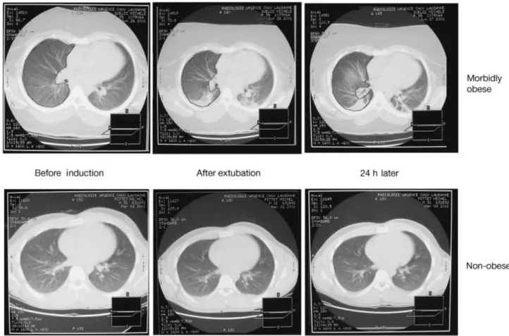
correlation (r=0.34) between obesity [calculated by Broca's index: weight (kg)/(height (cm) ±100)] and the area of lung densities seen directly after induction of anaesthesia. More recently, it has been shown that during general anaesthesia, morbidly obese patients had more atelectasis than non-obese patients. Atelectasis persisted for at least 24 h in morbidly obese patients whereas it disappeared in the non-obese (Fig. 6).19Functional residual
capacity (FRC) is lower in morbidly obese patients, the alveolar±arterial oxygenation gradient (A±aDO2) is
in-creased and intra-abdominal pressure is higher.62±64 The
different mechanics of the respiratory system and the hypoxia found in the morbidly obese patients are largely explained by a reduction in lung volume by increased intra-abdominal pressure.62When PEEP was applied, respiratory
function improved in morbidly obese patients but not in non-obese patients,66 and the reverse Trendelenburg
pos-ition improves oxygenation and lung mechanics in morbidly obese patients.67
In morbidly obese patients, avoiding atelectasis forma-tion may be particularly dif®cult but at the same time particularly important.
Chronic obstructive pulmonary disease
In contrast, patients with chronic obstructive pulmonary disease develop only a small shunt and almost no atelectasis during anaesthesia. However, they develop a more severe VA/Q mismatch. Hyperin¯ation of the lungs may make
them resist collapse or airway closure may prevent gas from leaving the alveoli (gas trapping).31
Other factors
Atelectasis during anaesthesia is found in all ages, from the newborn14 86 to patients over 80 years of age.30
Interestingly, the magnitude of atelectasis seems to be independent of age in adults, and 80-year-old patients have no more atelectasis than younger patients.30 In children,
densities appear rapidly in the dependent lung regions following induction of anaesthesia, while atelectasis was not seen in sedated children.43 87 88 This is important because
these atelectasis may obscure pulmonary metastases in 68% of children.86
In contrast to the circumstances in adults, atelectasis occurs even when preoxygenation is avoided and FIO2<0.4 is
used intraoperatively.87Atelectasis in the dependent regions
of the lung in children during anaesthesia cannot be explained by reabsorption of oxygen alone. Although the inward recoil of a child's lungs is similar to that of young adults, the outward recoil of the chest wall is less.16 This
results in a decrease in the FRC, and the less negative intrathoracic pressure increases the tendency both to airway closure and to the development of atelectasis.4 23
Less contribution of the rib cage to ventilation has been demonstrated in children during halothane anaesthesia.95In
infants, contraction of the diaphragm may cause paradoxical inward movement of the highly deformable chest wall, which could reduce ventilatory ef®ciency and increase diaphragmatic fatigue. Closing volume is greater in young children, in whom the elastic supporting structure of the lung is incompletely developed. This puts an infant at greater risk for atelectasis since airway closure can occur even during tidal breathing. The infant's lung is less compliant in relation to the chest wall: the net effect is a lower resting volume (FRC) than that seen in adults. Moreover, in children, PEEP (5 cm H2O) maintained
throughout anaesthesia is able to recruit all the available alveolar units and promote the disappearance of atelectatic areas in dependent pulmonary regions.87
Importance of atelectasis on patient outcome
Since most of atelectasis appearing during general anaes-thesia resolves within 24 h after surgery19 one may argue
that there is no need to prevent or study atelectasis since it may have no long-lasting effects. Indeed, often the lung dysfunction is transient and normal lung function resumes soon after anaesthesia and surgery. Nevertheless, patients do
Fig 6 Samples of CT scans of a morbidly obese and a non-obese patient before anaesthesia, after extubation and 24 h later. These slices were taken at the level of the interventricular septum (from Eichenberger and colleagues19).
develop perioperative respiratory complications.8 11 Since
the number of anaesthetic procedures in the Western world is considerable (60±70 000 occasions per million inhabit-ants, with more than half of these being general anaesthe-sia), even a small fraction of complications results in a large number of patients.
Some pulmonary complications occur during or imme-diately after anaesthesia, mainly hypoxaemia, and some will occur later, mainly pneumonia.
Perioperative hypoxaemia
Mild-to-moderate hypoxaemia, de®ned as an arterial satur-ation of 85±90%, occurs in approximately half of all patients undergoing elective surgery and can last from a few seconds to up to 30 min.54More alarming is the fact that about 20%
of the patients may suffer from severe hypoxaemia (oxygen saturation <81% for up to 5 min) during anaesthesia54and
13% in the post-anaesthesia care unit (PACU).53
Thirty-three percent of hypoxaemic events occur during induction of anaesthesia, one-third intraoperatively and one-third during awakening and in the PACU.11 Nowadays, more
frequent peripheral arterial saturation monitoring may reduce intraoperative hypoxaemic events compared with the 1980s.
Hypoxaemia during induction of anaesthesia
In the UK during the 1980s, three pregnant women died annually during induction of general anaesthesia because of failure to ventilate or intubate. It is estimated that dif®culty in airway management during induction of anaesthesia accounts for 600 patient deaths per year.39Dif®cult airway
management is not easily anticipated; therefore this com-plication may arise during every anaesthesia induction. During apnoea, oxygenation depends on the oxygen stores, which are small and are mainly in three compartments: the lungs, plasma and red cells. The normal store of oxygen is approximately 1500 ml, and may be increased to 3700 ml
with preoxygenation with 100% oxygen.99 Half of this
increase is from the increase in the oxygen concentration in the FRC. Therefore, prevention of atelectasis formation, which diminishes FRC, during induction of anaesthesia is important for all patients.
A greater oxygen store allows a greater margin of safety, with more time for airway management. Time to a 90% oxygen saturation is longest when anaesthesia induction is done with 100% oxygen, although this is associated with signi®cant atelectasis formation.18 However, such
atelec-tasis can be prevented by application of an end-expiratory positive pressure during anaesthesia induction despite the use of 100% oxygen84 and this will prolong the time to
desaturation by more than 2 min (unpublished observa-tions). In patients at increased risk of rapid desaturation, greater oxygen stores would be especially useful. In a mathematical model, effective preoxygenation in a `stand-ard adult' results in a time to decrease arterial saturation to 85% of 502 s.20This is reduced to 180 s in the 10-kg infant
and 171 s in the morbidly obese (Fig. 7).
Hypoxaemia during awakening and in the PACU
Transport from the operating room to the PACU is another period particularly at risk for hypoxaemic events. During this transport, patients may be without monitoring of oxygen saturation and without supplemental oxygen. At arrival in the PACU, 20% of patients may have an oxygen saturation <92% and in 10% the saturation may be <90%.51Age and obesity increase the risk. In the PACU within 3 h of surgery, 7% of patients will have at least one episode of desaturation <90%, and 3% will desaturate to <85%. This proportion is greater for thoraco-abdominal procedures, when more than half of the patients will have oxygen saturation <90% and 20% of patients will have severe hypoxaemia (<85%).102 Despite the use of 40% oxygen
given by face mask, 15% of patients will have an oxygen saturation below 92% lasting more than 30 s.85 During a Fig 7 Changes of arterial oxygen saturation (SaO2) during apnoea in children, in an obese adult and in a `typical' postoperative patient compared with
stay in the PACU, 25% of all patients will have at least one episode of desaturation.
Children, particularly young children, are also subject to hypoxaemia in the immediate postoperative period.11 101On
arrival in the PACU, 50% of children will have an arterial saturation <95% and 8% will be <90%. If the transport time was greater, the number of subjects with oxygen saturation levels <95% increased.21
In a large study with more than 24 000 patients, 0.9% had an hypoxaemic event in the PACU requiring a speci®c intervention other than only supplemental oxygen.75
Hypoxaemic events appear to prolong stay in the PACU, cause more intensive care admissions, and increase the incidence of cardiac complications. In another study, elderly cardiovascular patients (more than 80 years old) who sustained mild hypoxaemia (longer than 5 min) or severe desaturation (<80%) after surgery were more likely to experience silent myocardial ischaemia.24 Other studies
have shown that postoperative hypoxaemia is linked with ECG abnormalities76or delirium.1
There is no clear evidence that atelectasis is the cause of all these postoperative hypoxaemic events. Respiratory depression from residual anaesthetic may contribute. In a typical postoperative scenario, hypovolaemia, reduced car-diac output, anaemia, increased VA/Q mismatch, increased
shunt, hypoventilation and reduced alveolar volume can all contribute to more rapid onset of hypoxaemia (Fig. 7).20
It seems likely that preventing atelectasis formation during the whole perioperative period will increase the oxygen stores of the body. This increase in oxygen stores may reduce postoperative hypoxaemia. This may be particularly important in aged, obese and un®t patients.
Postoperative pulmonary complications
In studies on postoperative pulmonary complications (PPC), atelectasis and pneumonia are often considered together
because the changes associated with atelectasis may predispose to pneumonia. A continuum exists from non-infectious PPC (atelectasis) to non-infectious PPC (exacerbation of chronic bronchitis or pneumonia). In studies of non-cardiac surgery, the frequency of PPC and non-cardiac compli-cations (which have always received more attention) are comparable. For example, in adult men after elective abdominal surgery, PPC are more frequent than cardiac complications (estimated rates of 9.6% and 5.7%, respect-ively) and are associated with a longer hospital stay and greater healthcare costs.44 Pulmonary complications
ac-count for 24% of deaths within 6 days of surgery.8 9
Postoperative pneumonia is associated with a 30±46% mortality rate, and pneumonia causes 30±60% of the infections related to mortality.42 61 Some patients have
increased risk of PPC. Obese patients have a 25±30% risk of developing PPC.9 94 PPC are more common after some
types of surgery, for example oesophagectomy (17±50% PPC)13 28 or major head and neck surgery (15% PPC).52
Despite the lack of direct evidence of a correlation between atelectasis and pneumonia, reducing or avoiding atelectasis may diminish PPC, particularly in some patients94or after
some types of surgery28and thus improve outcome.
Prevention of atelectasis formation
A VCM can completely abolish atelectasis that develops after induction of general anaesthesia.78Lung in¯ation to an
airway pressure of 20 cm H2O did not affect atelectasis; an
airway pressure of 30 cm H2O reduced atelectasis; only with
a pressure of 40 cm H2O maintained for 15 s is atelectatic
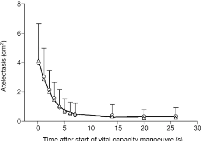
lung tissue fully re-expanded. This pressure is equivalent to in¯ation to vital capacity, and thus this manoeuvre has been called the VCM. More recently, it has been shown that this manoeuvre needs to be maintained for only 7±8 s in order to re-expand all previously collapsed lung tissue (Fig. 8).77
This manoeuvre not only has a `cosmetic' effect on CT scans but also improves oxygenation. When the inspired oxygen concentration was 40%, PaO2increased from 17.7
kPa to 22.2 kPa after the VCM.77The safety of this in¯ation
manoeuvre has been questioned but no adverse haemody-namic or pulmonary effects have been reported. In animal experiments, repeated VCM had no deleterious pulmonary effects as measured by extravascular lung water, pulmonary clearance of 99mTc-DTPA (which is a marker of the
functional integrity of the alveolocapillary barrier) and light microscopy.48
Tusman and colleagues97 studied an alternative
man-oeuvre. They increased both PEEP to 15 cm H2O and tidal
volume to either 18 ml kg±1or to a volume that caused a
peak airway pressure of 40 cm H2O, and maintained this for
10 breaths. PEEP was then decreased stepwise to 5 cm H2O
and tidal volume reduced to 9 ml kg±1. This procedure
increased PaO2, which persisted for 120 min. The same
method (Fig. 9) was also successful in augmenting arterial oxygenation during one-lung ventilation.95
Fig 8 Atelectasis before (time=0) and during the vital capacity manoeuvre. Mean values andSD(error bars) are shown. An exponential decay curve is ®tted to individual data (from Rothen and colleagues77).
The application of a PEEP of 10 cm H2O has been tested
in several studies and will consistently reopen collapsed lung tissue.7 32 92However, some atelectasis persists in most
patients. Further increases in PEEP level could re-expand this persistent atelectasis but PEEP may not be ideal. Firstly, shunt is not reduced and the arterial oxygenation is not always improved. Persistent shunt may be explained by the redistribution of blood ¯ow towards the most dependent parts of the lung when intrathoracic pressure is increased, so that residual atelectasis lung receives a larger share of the pulmonary blood ¯ow when PEEP is applied.100 The
increased intrathoracic pressure will also impede venous return and reduce cardiac output. This will decrease venous oxygen tension and augment the impact of shunted blood and perfusion of poorly ventilated regions on arterial oxygenation. Secondly, the lung may re-collapse rapidly after discontinuation of PEEP. Within 1 min after cessation of PEEP the collapse is as large as it was before the application of PEEP.34 However, PEEP applied
immedi-ately after a VCM will completely prevent recurrence of atelectasis, even when 100% oxygen is used.55
During induction of anaesthesia, application of PEEP (6 cm H2O) can prevent formation of atelectasis84 and can
increase the margin of safety before intubation. Application of PEEP (10 cm H2O) in morbidly obese patients is also
very effective for the prevention of atelectasis during induction.12
Clarke and colleagues10compared four treatments
(man-ual in¯ations, large tidal volumes, PEEP, and pressure
control inverse ratio ventilation [IRV]), using the A±aDO2
as the measure of atelectasis during anaesthesia, to deter-mine if any of these treatments affected atelectasis after anaesthesia. PEEP and IRV were most effective in reducing intraoperative A±aDO2, and no treatment affected
post-operative A±aDO2. However, since increasing FIO2even for
a few minutes before extubation can cause atelectasis, this could explain why no difference was seen after anaesthesia in Clarke's study.
Large tidal volumes (22 ml kg±1) do not improve
oxygenation in morbidly obese patients during general anaesthesia.3With these large tidal volumes, peak
inspira-tory airway pressure increased to 38 cm H2O but with an
end-inspiratory airway pressure of only 28 cm H2O. This
plateau pressure is far from the 40 cm H2O airway pressure
applied for 10 s that will relieve atelectasis in non-obese patients. The pressure generated in these obese patients was probably insuf®cient to re-expand the atelectasis and improve gas exchange.
Allowing spontaneous breathing during mechanical ven-tilation, even as little as 10±20% of the total venven-tilation, improves gas exchange. This can be done with airway pressure release ventilation (APRV) or biphasic positive airway pressure (BiPAP), as it is frequently called in Europe. Putensen and colleagues69 found better
oxygena-tion in animals with APRV than with convenoxygena-tional mech-anical ventilation. Patients with ARDS showed similar improvement.68 The long-term effects of APRV (72-h
ventilation) in patients with acute lung injury were compared with control patients receiving pressure-con-trolled, time-cycled mechanical ventilation.70 Patients
receiving APRV had greater respiratory compliance, PaO2
and cardiac output. APRV was associated with fewer days of ventilation (15 days with APRV vs 21 days with pressure-controlled ventilation). The better cardiopulmonary func-tion was attributed to recruitment of collapsed lung units. Spontaneous breathing contributed only 10±20% of total ventilation in these studies. Perhaps gas exchange would also improve with this technique during general anaesthesia in normal patients.
Use of the BiPAP system with inspiratory and expiratory positive airway pressure set at 12 and 4 cm H2O,
respect-ively (12/4) to treat obese patients for the ®rst 24 h after gastroplasty signi®cantly reduced pulmonary dysfunction, indicated by forced vital capacity, forced expiratory volume in 1 s (FEV1) and oxygen saturation, and pulmonary
function recovers more rapidly (Fig. 10).36 With lower
BiPAP pressure (8/4 cm H2O) or only supplemental oxygen
via a face mask, the pulmonary dysfunction was more severe and lasted longer. Atelectasis was not measured in this study but it is possible that the positive airway pressure applied after extubation could reduce atelectasis, explaining in part the improved lung mechanics.
By combining some of these techniques, it could be possible to prevent atelectasis formation during general anaesthesia.
Fig 9 Schematic representation of the alveolar recruitment strategy. In pressure control ventilation, the pressure amplitude of 20 cm H2O
remains constant throughout the manoeuvre. Each pressure step is maintained for 1 min. After airway pressures of 40/20 cm H2O, the
pressures are reduced to 30/10 cm H2O. The initial settings are then
resumed. (Paw, pulmonary airway pressure; Pip, peak inspiratory pressure; from Tusman and colleagues96).
Conclusion
Atelectasis that develops during general anaesthesia may lead to perioperative pulmonary complications. Prevention of atelectasis formation is therefore an important goal.
References
1 Aakerlund LP, Rosenberg J. Postoperative delirium: treatment with supplementary oxygen. Br J Anaesth 1994; 72: 286±90 2 Akca O, Podolsky A, Eisenhuber E, et al. Comparable
postoperative pulmonary atelectasis in patients given 30% or 80% oxygen during and 2 hours after colon resection. Anesthesiology 1999; 91: 991±8
3 Bardoczky GI, Yernault JC, Houben JJ, d'Hollander AA. Large tidal volume ventilation does not improve oxygenation in morbidly obese patients during anesthesia. Anesth Analg 1995; 81: 385±8
4 Beardsmore CS, Stocks J, Helms P. Elastic properties of the respiratory system in infants. Eur Respir J Suppl 1989; 4: 135±9 5 Bendixen HH, Hedley-White J, Laver MB. Impaired oxygenation
in surgical patients during general anesthesia with controlled ventilation. A concept of atelectasis. N Engl J Med 1963; 269: 991±6
6 BenoõÃt Z, Wicky S, Fischer J-F, et al. The effect of increased FIO2 before tracheal extubation on postoperative atelectasis. Anesth Analg 2002; 95: 1777±81
7 Brismar B, Hedenstierna G, Lundquist H, et al. Pulmonary densities during anesthesia with muscular relaxation ± a proposal of atelectasis. Anesthesiology 1985; 62: 422±8
8 Brooks-Brunn JA. Postoperative atelectasis and pneumonia. Heart Lung 1995; 24: 94±115
9 Brooks-Brunn JA. Predictors of postoperative pulmonary complications following abdominal surgery. Chest 1997; 111: 564±71
10 Clarke JP, Schuitemaker MN, Sleigh JW. The effect of intraoperative ventilation strategies on perioperative atelectasis. Anaesth Intens Care 1998; 26: 262±6
11 Cote CJ, Goldstein EA, Cote MA, Hoaglin DC, Ryan JF. A single-blind study of pulse oximetry in children. Anesthesiology 1988; 68: 184±8
12 Coussa M, Proietti S, Frascarolo P, Spahn D, Magnusson L. Continuous positive airways pressure prevents atelectasis formation during induction of general anaesthesia in morbidly obese patients. Swiss Med Wkly 2002; 132: 53
13 Crozier TA, Sydow M, Siewert JR, Braun U. Postoperative pulmonary complication rate and long-term changes in respiratory function following esophagectomy with esophagogastrostomy. Acta Anaesthesiol Scand 1992; 36: 10±15 14 Damgaard-Pedersen K, Qvist T. Pediatric pulmonary
CT-scanning. Anaesthesia-induced changes. Pediatr Radiol 1980; 9: 145±8
15 Dantzker DR, Wagner PD, West JB. Proceedings: Instability of poorly ventilated lung units during oxygen breathing. J Physiol 1974; 242: 72
16 Davis GM, Coates AL, Dalle D, Bureau MA. Measurement of pulmonary mechanics in the newborn lamb: a comparison of three techniques. J Appl Physiol 1988; 64: 972±81
17 Drummond GB. Computed tomography and pulmonary measurements. Br J Anaesth 1998; 80: 665±71
18 Edmark L, Enlund M, Kostova-Aherdan K, Hedenstierna G. Atelectasis formation and apnoea tolerance after pre-oxygenation with 100%, 80%, or 60% oxygen. Anesthesiology 2001; 95: A1330
19 Eichenberger A-S, Proietti S, Wicky S, et al. Morbid obesity and postoperative pulmonary atelectasis: an underestimated problem. Anesth Analg 2002; 95: 1788±92
20 Farmery AD, Roe PG. A model to describe the rate of oxyhaemoglobin desaturation during apnoea. Br J Anaesth 1996; 76: 284±91
21 Fossum SR, Knowles R. Perioperative oxygen saturation levels of pediatric patients. J Post Anesth Nurs 1995; 10: 313±19 22 Froese AB, Bryan AC. Effects of anesthesia and paralysis on
diaphragmatic mechanics in man. Anesthesiology 1974; 41: 242±55 23 Gerhardt T, Bancalari E. Chestwall compliance in full-term and
premature infants. Acta Paediatr Scand 1980; 69: 359±64 24 Gill NP, Wright B, Reilly CS. Relationship between hypoxaemic
and cardiac ischaemic events in the perioperative period. Br J Anaesth 1992; 68: 471±3
25 Goll V, Akca O, Greif R, et al. Ondansetron is no more effective than supplemental intraoperative oxygen for prevention of postoperative nausea and vomiting. Anesth Analg 2001; 92: 112±17
26 Greif R, Akca O, Horn EP, Kurz A, Sessler DI. Supplemental perioperative oxygen to reduce the incidence of surgical-wound infection. Outcomes Research Group. N Engl J Med 2000; 342: 161±7
27 Greif R, Laciny S, Rapf B, Hickle RS, Sessler DI. Supplemental oxygen reduces the incidence of postoperative nausea and vomiting. Anesthesiology 1999; 91: 1246±52
28 Grif®n SM, Shaw IH, Dresner SM. Early complications after Ivor Lewis subtotal esophagectomy with two-®eld lymphadenectomy: risk factors and management. J Am Coll Surg 2002; 194: 285±97 29 Gunnarsson L, Strandberg A, Brismar B, et al. Atelectasis and gas exchange impairment during en¯urane/nitrous oxide anaesthesia. Acta Anaesthesiol Scand 1989; 33: 629±37
30 Gunnarsson L, Tokics L, Gustavsson H, Hedenstierna G. In¯uence of age on atelectasis formation and gas exchange impairment during general anaesthesia. Br J Anaesth 1991; 66: 423±32
31 Gunnarsson L, Tokics L, Lundquist H, et al. Chronic obstructive pulmonary disease and anaesthesia: formation of atelectasis and gas exchange impairment. Eur Respir J 1991; 4: 1106±16 32 Hachenberg T, Lundquist H, Tokics L, Brismar B, Hedenstierna
G. Analysis of lung density by computed tomography before and
Fig 10 Effect of bi-level positive airway pressure (BiPAP) on FEV1.
*P<0.05 compared with the other two groups (from Joris and colleagues36).
during general anaesthesia. Acta Anaesthesiol Scand 1993; 37: 549±55
33 Hedenstierna G, Lundquist H, Lundh B, et al. Pulmonary densities during anaesthesia. An experimental study on lung morphology and gas exchange. Eur Respir J 1989; 2: 528±35
34 Hedenstierna G, Tokics L, Lundquist H, et al. Phrenic nerve stimulation during halothane anesthesia. Effects of atelectasis. Anesthesiology 1994; 80: 751±60
35 Hedenstierna G, Tokics L, Strandberg A, Lundquist H, Brismar B. Correlation of gas exchange impairment to development of atelectasis during anaesthesia and muscle paralysis. Acta Anaesthesiol Scand 1986; 30: 183±91
36 Joris JL, Sottiaux TM, Chiche JD, Desaive CJ, Lamy ML. Effect of bi-level positive airway pressure (BiPAP) nasal ventilation on the postoperative pulmonary restrictive syndrome in obese patients undergoing gastroplasty. Chest 1997; 111: 665±70
37 Joyce CJ, Baker AB, Kennedy RR. Gas uptake from an unventilated area of lung: computer model of absorption atelectasis. J Appl Physiol 1993; 74: 1107±16
38 Joyce CJ, Williams AB. Kinetics of absorption atelectasis during anesthesia: a mathematical model. J Appl Physiol 1999; 86: 1116±25
39 King TA, Adams AP. Failed tracheal intubation. Br J Anaesth 1990; 65: 400±14
40 Kotani N, Hashimoto H, Sessler DI, et al. Supplemental intraoperative oxygen augments antimicrobial and pro-in¯ammatory responses of alveolar macrophages. Anesthesiology 2000; 93: 15±25
41 Krayer S, Rehder K, Vettermann J, Didier EP, Ritman EL. Position and motion of the human diaphragm during anesthesia-paralysis. Anesthesiology 1989; 70: 891±8
42 Kroenke K, Lawrence VA, Theroux JF, Tuley MR. Operative risk in patients with severe obstructive pulmonary disease. Arch Intern Med 1992; 152: 967±71
43 Lam WW, Chen PP, So NM, Metreweli C. Sedation versus general anaesthesia in paediatric patients undergoing chest CT. Acta Radiol 1998; 39: 298±300
44 Lawrence VA, Hilsenbeck SG, Mulrow CD, et al. Incidence and hospital stay for cardiac and pulmonary complications after abdominal surgery. J Gen Intern Med 1995; 10: 671±8
45 Lindberg P, Gunnarsson L, Tokics L, et al. Atelectasis and lung function in the postoperative period. Acta Anaesthesiol Scand 1992; 36: 546±53
46 Loring SH, Butler JP. Gas exchange in body cavities. In: Farhi LE, Tenney SM, eds. Handbook of Physiology. Section 3, The Respiratory System. Volume 4, Gas Exchange. Bethesda, Maryland: Am Physiol Soc, 1987; 283±95
47 Lundquist H, Hedenstierna G, Strandberg A, Tokics L, Brismar B. CT-assessment of dependent lung densities in man during general anaesthesia. Acta Radiol 1995; 36: 626±32
48 Magnusson L, Tenling A, Lemoine R, et al. The safety of one, or repeated, vital capacity maneuvers during general anesthesia. Anesth Analg 2000; 91: 702±7
49 Magnusson L, Zemgulis V, Wicky S, et al. Atelectasis is a major cause of hypoxemia and shunt after cardiopulmonary bypass: an experimental study. Anesthesiology 1997; 87: 1153±63
50 Mason RJ. Surfactant secretion. In: Robertson B, van Golde LMG, Batenburg JJ, eds. Pulmonary Surfactant. From Molecular Biology to Clinical Practice. Amsterdam: Elsevier, 1992; 295±312
51 Mathes DD, Conaway MR, Ross WT. Ambulatory surgery: room air versus nasal cannula oxygen during transport after general anesthesia. Anesth Analg 2001; 93: 917±21
52 McCulloch TM, Jensen NF, Girod DA, Tsue TT, Weymuller EA
Jr. Risk factors for pulmonary complications in the postoperative head and neck surgery patient. Head Neck 1997; 19: 372±7 53 Moller JT. Anesthesia related hypoxemia. The effect of pulse
oximetry monitoring on perioperative events and postoperative complications. Dan Med Bull 1994; 41: 489±500
54 Moller JT, Johannessen NW, Berg H, Espersen K, Larsen LE. Hypoxaemia during anaesthesiaÐan observer study. Br J Anaesth 1991; 66: 437±44
55 Neumann P, Rothen HU, Berglund JE, et al. Positive end-expiratory pressure prevents atelectasis during general anaesthesia even in the presence of a high inspired oxygen concentration. Acta Anaesthesiol Scand 1999; 43: 295±301 56 Nunn JF. Factors in¯uencing the arterial oxygen tension during
halothane anaesthesia with spontaneous respiration. Br J Anaesth 1964; 36: 327±41
57 Nyman G, Funkquist B, Kvart C, et al. Atelectasis causes gas exchange impairment in the anaesthetised horse. Equine Vet J 1990; 22: 317±24
58 Oyarzun MJ, Iturriaga R, Donoso P, et al. Factors affecting distribution of alveolar surfactant during resting ventilation. Am J Physiol 1991; 261: 210±17
59 Pasteur W. Massive collapse of the lung. Lancet 1908; 1351±3 60 Pasteur W. Active lobar collapse of the lung after abdominal
operations. Lancet 1910; 2: 1080±3
61 Pedersen T, Viby-Mogensen J, Ringsted C. Anaesthetic practice and postoperative pulmonary complications. Acta Anaesthesiol Scand 1992; 36: 812±18
62 Pelosi P, Croci M, Ravagnan I, et al. Respiratory system mechanics in sedated, paralyzed, morbidly obese patients. J Appl Physiol 1997; 82: 811±18
63 Pelosi P, Croci M, Ravagnan I, et al. The effects of body mass on lung volumes, respiratory mechanics, and gas exchange during general anesthesia. Anesth Analg 1998; 87: 654±60
64 Pelosi P, Croci M, Ravagnan I, Vicardi P, Gattinoni L. Total respiratory system, lung, and chest wall mechanics in sedated-paralyzed postoperative morbidly obese patients. Chest 1996; 109: 144±51
65 Pelosi P, Crotti S, Brazzi L, Gattinoni L. Computed tomography in adult respiratory distress syndrome: what has it taught us? Eur Respir J 1996; 9: 1055±62
66 Pelosi P, Ravagnan I, Giurati G, et al. Positive end-expiratory pressure improves respiratory function in obese but not in normal subjects during anesthesia and paralysis. Anesthesiology 1999; 91: 1221±31
67 Perilli V, Sollazzi L, Bozza P, et al. The effects of the reverse Trendelenburg position on respiratory mechanics and blood gases in morbidly obese patients during bariatric surgery. Anesth Analg 2000; 91: 1520±5
68 Putensen C, Mutz NJ, Putensen-Himmer G, Zinserling J. Spontaneous breathing during ventilatory support improves ventilation-perfusion distributions in patients with acute respiratory distress syndrome. Am J Respir Crit Care Med 1999; 159: 1241±8
69 Putensen C, Rasanen J, Lopez FA. Ventilation-perfusion distributions during mechanical ventilation with superimposed spontaneous breathing in canine lung injury. Am J Respir Crit Care Med 1994; 150: 101±8
70 Putensen C, Zech S, Wrigge H, et al. Long-term effects of spontaneous breathing during ventilatory support in patients with acute lung injury. Am J Respir Crit Care Med 2001; 164: 43±9 71 Reber A, Bein T, Hogman M, et al. Lung aeration and pulmonary gas exchange during lumbar epidural anaesthesia and in the lithotomy position in elderly patients. Anaesthesia 1998; 53: 854±61
72 Reber A, Engberg G, Sporre B, et al. Volumetric analysis of aeration in the lungs during general anaesthesia. Br J Anaesth 1996; 76: 760±6
73 Reber A, Engberg G, Wegenius G, Hedenstierna G. Lung aeration. The effect of pre-oxygenation and hyperoxygenation during total intravenous anaesthesia. Anaesthesia 1996; 51: 733±7 74 Reber A, Nylund U, Hedenstierna G. Position and shape of the diaphragm: implications for atelectasis formation. Anaesthesia 1998; 53: 1054±61
75 Rose DK, Cohen MM, Wigglesworth DF, DeBoer DP. Critical respiratory events in the postanesthesia care unit. Patient, surgical, and anesthetic factors. Anesthesiology 1994; 81: 410±18 76 Rosenberg J, Rasmussen V, von Jessen F, Ullstad T, Kehlet H. Late postoperative episodic and constant hypoxaemia and associated ECG abnormalities. Br J Anaesth 1990; 65: 684±91 77 Rothen HU, Neumann P, Berglund JE, et al. Dynamics of
re-expansion of atelectasis during general anaesthesia. Br J Anaesth 1999; 82: 551±6
78 Rothen HU, Sporre B, Engberg G, Wegenius G, Hedenstierna G. Re-expansion of atelectasis during general anaesthesia: a computed tomography study. Br J Anaesth 1993; 71: 788±95 79 Rothen HU, Sporre B, Engberg G, Wegenius G, Hedenstierna G.
Re-expansion of atelectasis during general anaesthesia may have a prolonged effect. Acta Anaesthesiol Scand 1995; 39: 118±25 80 Rothen HU, Sporre B, Engberg G, Wegenius G, Hedenstierna G.
Airway closure, atelectasis and gas exchange during general anaesthesia. Br J Anaesth 1998; 81: 681±6
81 Rothen HU, Sporre B, Engberg G, et al. In¯uence of gas composition on recurrence of atelectasis after a reexpansion maneuver during general anesthesia. Anesthesiology 1995; 82: 832±42
82 Rothen HU, Sporre B, Engberg G, et al. Prevention of atelectasis during general anaesthesia. Lancet 1995; 345: 1387±91 83 Rothen HU, Sporre B, Engberg G, et al. Atelectasis and
pulmonary shunting during induction of general anaesthesia ± can they be avoided? Acta Anaesthesiol Scand 1996; 40: 524±9 84 Rusca M, Wicky S, Proietti S, et al. Continuous positive airways
pressure prevents atelectasis formation during induction of general anaesthesia. Anesthesiology 2001; 95: A1331
85 Russell GB, Graybeal JM. Hypoxemic episodes of patients in a postanesthesia care unit. Chest 1993; 104: 899±903
86 Sargent MA, McEachern AM, Jamieson DH, Kahwaji R. Atelectasis on pediatric chest CT: comparison of sedation techniques. PediatrRadiol 1999; 29: 509±13
87 Sera®ni G, Cornara G, Cavalloro F, et al. Pulmonary atelectasis during paediatric anaesthesia: CT scan evaluation and effect of positive endexpiratory pressure (PEEP). Paediatr Anaesth 1999; 9: 225±8
88 Strandberg A, Tokics L, Brismar B, Lundquist H, Hedenstierna G.
Atelectasis during anaesthesia and in the postoperative period. Acta Anaesthesiol Scand 1986; 30: 154±8
89 Strandberg A, Tokics L, Brismar B, Lundquist H, Hedenstierna G. Constitutional factors promoting development of atelectasis during anaesthesia. Acta Anaesthesiol Scand 1987; 31: 21±4 90 Tenling A, Hachenberg T, Tyden H, Wegenius G, Hedenstierna
G. Atelectasis and gas exchange after cardiac surgery. Anesthesiology 1998; 89: 371±8
91 Tenling A, Joachimsson PO, Tyden H, Wegenius G, Hedenstierna G. Thoracic epidural anesthesia as an adjunct to general anesthesia for cardiac surgery: effects on ventilation±perfusion relationships. J Cardiothorac Vasc Anesth 1999; 13: 258±64
92 Tokics L, Hedenstierna G, Strandberg A, Brismar B, Lundquist H. Lung collapse and gas exchange during general anesthesia: effects of spontaneous breathing, muscle paralysis, and positive end-expiratory pressure. Anesthesiology 1987; 66: 157±67
93 Tokics L, Strandberg A, Brismar B, Lundquist H, Hedenstierna G. Computerized tomography of the chest and gas exchange measurements during ketamine anaesthesia. Acta Anaesthesiol Scand 1987; 31: 684±92
94 Tseuda K, Debrand M, Bivins BA, Wright BD, Griffen WO. Pulmonary complications in the morbidly obese following jejunoileal bypass surgery under narcotic anesthesia. Int Surg 1980; 65: 123±9
95 Tusiewicz K, Bryan AC, Froese AB. Contributions of changing rib cage±diaphragm interactions to the ventilatory depression of halothane anesthesia. Anesthesiology 1977; 47: 327±37
96 Tusman G, Bohm SH, Melkun F, et al. Alveolar recruitment strategy increases arterial oxygenation during one-lung ventilation. Ann Thorac Surg 2002; 73: 1204±9
97 Tusman G, Bohm SH, Vazquez de Anda GF, do Campo JL, Lachmann B. `Alveolar recruitment strategy' improves arterial oxygenation during general anaesthesia. Br J Anaesth 1999; 82: 8±13
98 Warner DO, Warner MA, Ritman EL. Atelectasis and chest wall shape during halothane anesthesia. Anesthesiology 1996; 85: 49±59
99 West JB. Pulmonary Pathophysiology. Baltimore: Williams and Wilkins, 1987
100 West JB, Dolley CT, Naimark A. Distribution of blood ¯ow in isolated lung: relations to vascular and alveolar pressure. J Appl Physiol 1964; 19: 13±24
101 Xue FS, Huang YG, Tong SY, et al. A comparative study of early postoperative hypoxemia in infants, children, and adults undergoing elective plastic surgery. Anesth Analg 1996; 83: 709±15
102 Xue FS, Li BW, Zhang GS, et al. The in¯uence of surgical sites on early postoperative hypoxemia in adults undergoing elective surgery. Anesth Analg 1999; 88: 213±19