ORIGINAL PAPER
Randomised trial of oral versus sequential intravenous/oral
cephalosporins in children with pyelonephritis
Thomas J. Neuhaus&Christoph Berger&
Katja Buechner&Paloma Parvex&Gian Bischoff& Philippe Goetschel&Daniela Husarik&Ulrich Willi& Luciano Molinari&Christoph Rudin&Alain Gervaix& Urs Hunziker&Sergio Stocker&Eric Girardin& David Nadal
Received: 2 October 2007 / Accepted: 6 November 2007 / Published online: 12 December 2007
# Springer-Verlag 2007
Abstract The hypothesis was tested that oral antibiotic treatment in children with acute pyelonephritis and scintig-raphy-documented lesions is equally as efficacious as sequential intravenous/oral therapy with respect to the incidence of renal scarring. A randomised multi-centre trial was conducted in 365 children aged 6 months to 16 years with bacterial growth in cultures from urine collected by catheter. The children were assigned to receive either oral ceftibuten (9 mg/kg once daily) for 14 days or intravenous
ceftriaxone (50 mg/kg once daily) for 3 days followed by oral ceftibuten for 11 days. Only patients with lesions detected on acute-phase dimercaptosuccinic acid (DMSA) scintigraphy underwent follow-up scintigraphy. Efficacy was evaluated by the rate of renal scarring after 6 months on follow-up scintigraphy. Of 219 children with lesions on acute-phase scintigraphy, 152 completed the study; 80 (72 females, median age 2.2 years) were given ceftibuten and 72 (62 females, median age 1.6 years) were given ceftriaxone/
\DO00638; No of Pages
TJN, CB and KB contributed equally to this work. EG and DN share senior authorship.
Trial number: Register of the Swiss national agency for therapeutic products (Swissmedic).Reference number: IKS 2001S03204 T. J. Neuhaus
Department of Nephrology,
University Children’s Hospital Zurich, Steinwiesstrasse 75, 8032 Zurich, Switzerland T. J. Neuhaus
:
S. StockerDepartment of General Paediatrics, University Children’s Hospital Zurich, Steinwiesstrasse 75, 8032 Zurich, Switzerland C. Berger
:
K. Buechner:
D. Nadal (*) Department of Infectious Diseases, University Children’s Hospital Zurich, Steinwiesstrasse 75, 8032 Zurich, Switzerland e-mail: [email protected]U. Willi
Department of Radiology,
University Children’s Hospital Zurich, Steinwiesstrasse 75, 8032 Zurich, Switzerland L. Molinari
Child Development Center, University Children’s Hospital Zurich, Steinwiesstrasse 75, 8032 Zurich, Switzerland
P. Parvex
:
A. Gervaix:
E. Girardin Department of Nephrology,University Children’s Hospital Geneva, 1211 Geneva 14, Switzerland G. Bischoff
:
U. Hunziker Department of Paediatrics, Cantonal Hospital Winterthur, 8400 Winterthur, Switzerland P. GoetschelDepartment of Paediatrics, City Hospital Zurich-Triemli, 8063 Zurich, Switzerland D. Husarik
Department of Nuclear Medicine, University Hospital Zurich, 8091 Zurich, Switzerland C. Rudin
Department of Nephrology, University Children’s Hospital Basle, 4005 Basle, Switzerland
ceftibuten. Patients in the intravenous/oral group had significantly higher C-reactive protein (CRP) concentrations at baseline and larger lesion(s) on acute-phase scintigraphy. Follow-up scintigraphy showed renal scarring in 21/80 children treated with ceftibuten and 33/72 with ceftriaxone/ ceftibuten (p=0.01). However, after adjustment for the confounding variables (CRP and size of acute-phase lesion), no significant difference was observed for renal scarring between the two groups (p=0.2). Renal scarring correlated with the extent of the acute-phase lesion (r=0.60, p<0.0001) and the grade of vesico-ureteric reflux (r=0.31, p=0.03), and was more frequent in refluxing renal units (p=0.04). The majority of patients, i.e. 44 in the oral group and 47 in the intravenous/oral group, were managed as out-patients. Side effects were not observed. From this study, we can conclude that once-daily oral ceftibuten for 14 days yielded compa-rable results to sequential ceftriaxone/ceftibuten treatment in children aged 6 months to 16 years with DMSA-documented acute pyelonephritis and it allowed out-patient management in the majority of these children.
Keywords Ceftibuten . Child . Pyelonephritis . Scintigraphy . Out-patient
Abbreviations
CRP C-reactive protein DMSA Dimercaptosuccinic acid VUR Vesico-ureteric reflux
Introduction
In infants and young children, upper and lower urinary tract infections are common [1], and a febrile urinary tract infection is considered to be an upper urinary tract infection (i.e. pyelonephritis). Up to two thirds of children with acute pyelonephritis show a lesion (defect) on technetium-99m-labelled dimercaptosuccinic acid (DMSA) scintigraphy during the acute phase [5, 14, 21, 25]. Only these children develop post-infectious renal scarring and the incidence is 29– 60% [5,15,21,24,30,35]. Long-term follow-up of children with renal scarring showed a significant reduction of renal function [20, 32, 33]. In contrast, children with a febrile urinary tract infection without lesion on acute-phase DMSA scintigraphy did not develop renal scarring [14,15,30].
Although the recommended initial treatment for infants and young children with pyelonephritis is the intravenous administration of antibiotics [1,5,6,8], we hypothesised that oral antibiotics are equally as efficacious as sequential intravenous/oral treatment with respect to the incidence of renal scarring 6 months after acute pyelonephritis with scintigraphy-documented acute lesions. Several lines of
evidence support our hypothesis. The prevalence of bacter-aemia in pyelonephritis is 6.1–22.7% in infants younger than 2 months of age [7,25] and 9.3% in infants younger than 6 months [2]. In contrast, children older than 6 months of age either never have bacteraemia [2] or only rarely show signs of bacteraemia [25]. Thus, oral antimicrobial therapy of acute pyelonephritis appeared to be a safe option, particu-larly for children older than 6 months of age. Indeed, the results from two prior prospective trials suggested that acute pyelonephritis in children could be treated with oral anti-biotics [14,21]. Two additional aspects need consideration. First, it has to be shown that the oral route is as efficacious as the parenteral route also in patients at the highest risk for renal scarring, i.e. children with systemic inflammation (high C-reactive protein [CRP]) and initial scintigraphic lesions. Second, oral treatment might allow full out-patient manage-ment with major benefits to patients and parents. Ceftibuten, an oral cephalosporin, was deliberately chosen for three reasons: (i) good antimicrobial activity, (ii) lack of resistance against pathogens of community-acquired urinary tract infection and (iii) its long half-life, allowing once-daily oral dosing [4,19,23,34].
To test our hypothesis, we conducted a prospective randomised controlled multi-centre trial to compare the effect of oral treatment with once-daily ceftibuten for 14 days with the effect of the sequential intravenous/oral regimen (3 days of intravenous treatment with ceftriaxone, followed by 11 days with oral cephalosporin) [5]. Where it was in accordance with the local hospital guidelines, patients were planned to be treated as out-patients, since oral and out-patient management might reduce the burden on hospital staff and costs. Only children older than 6 months of age with acute pyelonephritis and acute lesions on DMSA scintigraphy [10,
11, 29]—those at highest risk for sequelae, i.e. renal scarring—were included.
Methods
Five Swiss paediatric hospitals collaborated in a prospec-tive, investigator-initiated clinical trial to compare the effects of oral and sequential intravenous/oral antibiotic regimens in children with pyelonephritis and acute lesions on DMSA scintigraphy. Patients were recruited from 1 July 2001 to 30 April 2004 and follow-up ended on 31 December 2004. The study was approved by the national agency for therapeutic products (Swissmedic) and the local ethical committees of all of the participating centres. Written informed consent was obtained from at least one parent of each patient. The study was carried out indepen-dently of the financial supporter. Financial industry support for the central collection and analysis of data was sought
only after the study had been approved by the national and local authorities.
Patient recruitment
Patients aged 6 months to 16 years with acute community-acquired pyelonephritis treated at the University Children’s Hospitals of Zurich, Geneva and Basle or in the depart-ments of paediatrics of the hospitals of Winterthur and Zurich-Triemli, were eligible. Laboratory tests on admis-sion included blood count, CRP, plasma creatinine, blood cultures, urinary dipstick and/or microscopic urinalysis (cell count in non-centrifuged urine), and urine culture. The urine samples were collected by bladder catheterisation. The diagnosis of acute pyelonephritis was considered to be probable in children with fever (rectal temperature >38°C or axillary temperature >38.5°C), an abnormal urinary dipstick test (leukocyte esterase≥1+, or nitrite positive) or microscopic urinalysis (pyuria with≥10 white blood cells/μl), and serum CRP concentration >10 mg/l (normal <4 mg/l). Additional clinical signs were not mandatory (e.g. abdominal or flank pain in children old enough to report pain accurately, irritability, vomiting, diarrhoea or feeding problems in infants). Positive urine culture was defined as growth≥104 colony-forming units/ml of a single bacterium. Patients known to have isolated vesico-ureteric reflux (VUR), megaureter or duplex kidney, independently of antibiotic prophylaxis (e.g. cotrimoxazole, trimethoprim or nitrofur-antoin) were also included. The exclusion criteria were age <6 months, antibiotic pre-treatment of acute infection, other abnormalities of the urinary tract, known impaired renal function, patients on immunosuppressive therapy and known hypersensitivity to cephalosporins. Patients were also ex-cluded at the discretion of the treating physician if the clinical condition suggested septicaemia or if other reasons precluded oral treatment.
Randomisation
Each centre was provided, as many times as necessary, with blocks of 24 sealed envelopes containing an equal number of assignments for the two antibiotic regimens. A statistician provided the computer-generated code and an independent clerk sealed and bundled the opaque envelopes, so that the person enrolling the patient into the study would not have known the patient’s assignment. Patients were locally randomised at the time of admission. For practical reasons, all patients with a presumed clinical diagnosis of probable pyelonephritis were randomised at their first consultation in the emergency unit (i.e. before the results of the urine culture and DMSA scintigraphy were available). Thus, stratification for potentially confounding variables (e.g. grade of inflam-mation [CRP], size of lesion on acute-phase DMSA
scintig-raphy and the presence/grade of VUR) was not performed. Only patients with acute pyelonephritis and lesions on acute-phase scintigraphy, as assessed by the treating physicians, underwent a follow-up scintigraphy.
Antimicrobial therapy and prophylaxis
Antibiotic therapy was started immediately after the urine and blood samples had been analysed. For the“oral-only group,” patients received 9 mg/kg body weight of ceftibuten [34] (Cedax, Essex, Lucerne, Switzerland; in Switzerland, it is licensed for children ≥6 months of age) orally twice on the first day and once daily thereafter for 14 days. In the “intravenous/oral group,” patients received 50 mg/kg cef-triaxone (Rocephin®, Roche, Basle, Switzerland) intrave-nously once daily for 3 days and then 9 mg/kg body weight of ceftibuten orally for 11 days. Ceftibuten is commercially available in most European countries and in the US.
Before starting the clinical study, the antimicrobial susceptibility of Gram-negative bacteria, isolated from positive urine cultures consecutively collected from in-patients and out-in-patients at one centre (University Child-ren’s Hospital Zurich), was tested. Of 285 Gram-negative bacterial isolates, 243 were sensitive to ceftibuten and ceftriaxone; only Pseudomonas aeruginosa (n=27) and Enterobacter cloacae (n=15), two unusual pathogens in community-acquired pyelonephritis, were resistant. Only 63% and 41% of the isolates were sensitive to amoxicillin/ clavulanic acid and amoxicillin, respectively.
Deviation from the assigned antimicrobial treatment regimen was permitted in the following events: (i) clinical deterioration as assessed by the treating physician, (ii) presumed side effects of the study drugs, (iii) resistance of bacteria grown in initial urine culture to the study anti-biotics and (iv) bacterial growth on follow-up urine culture on days 3–4.
All patients were followed at each study centre at least until day 3; patients with persistent fever at day 3 were followed until defervescence. The patients were then followed clinically by their local paediatrician, who in-formed the study centres in case of recurrent urinary tract infection. At the end of the 14-day treatment, antibiotic prophylaxis was started either with cotrimoxazole (12– 18 mg/kg body weight in one or two doses daily) or nitrofurantoin (1–1.5 mg/kg body weight in one or two doses daily) according to the Swiss guidelines [13] and was discontinued only if voiding cystourethrography showed neither VUR nor other malformations of the urinary tract. Outcome measures
Urine culture and serum CRP were repeated on days 3–4 of treatment. Renal ultrasound was done within 24 hours,
whereas renal DMSA scintigraphy was performed within 5 days, since the nuclear medicine department does not offer DMSA scanning 7 days a week. Thus, some minor lesions might become invisible between the onset of antibiotic therapy and scanning, leading to false-negative DMSA scintigraphy results. Of the children enrolled, only those with lesions on acute-phase scintigraphy were followed with a second scintigraphy (see below). Investigations were com-pleted 2–8 weeks after the acute episode by voiding cystourethrography (at least two fillings were performed) to detect VUR (grade I–V). Specimens for urine culture were obtained at the voiding cystourethrography and when the children had fever or symptoms of urinary tract infection. The study patients were followed at voiding cystourethrography and after 6 months at follow-up scintigraphy. Parents were asked whether their child had had fever or recurrence of urinary tract infection and whether the prophylaxis had been given as prescribed. In addition, the safety and convenience of treatment were evaluated in terms of the discontinuation of therapy and the incidence of side effects.
The primary end point was—only in patients with lesions on acute-phase scintigraphy—the development of renal scarring in both groups (oral and intravenous/oral) after at least 6 months, documented by follow-up DMSA scintigraphy. The 6-month interval between the acute-phase and follow-up scintigraphy was chosen because acute pyelonephritic defects can persist for up to 5 months [16]. The secondary end point was the effect of VUR on the development of renal scarring.
Renal DMSA scintigraphy
Scintigraphy was performed 3 hours after the intravenous injection of 99mTc-DMSA (Mallinckrodt Medical B.V., Petten, Netherlands). The administered dose was weight-adjusted according to the recommendations of the Pediatric Committee of the EANM (http://www.eanm.org/scientific_info/guidelines/ gl_paed_dmsa_scin.php?navId=54), with a minimal dose of 15 MBq and a maximal dose of 100 MBq. Two (one anterior and one posterior view) to six views (one anterior, one posterior, two posterior oblique and two anterior oblique views) were obtained, but tomography was not systematically performed. Each kidney (i.e. renal unit) was assessed separately. Images were acquired on dual-head gamma cameras (Zurich: Body Scan, Siemens, Erlangen, Germany; Zurich-Triemli: Prism 2000 XP with pinhole collimator, Philips (former Picker), Eindhoven, Netherlands; Winterthur: E Cam, Siemens, Erlangen, Germany; Basel: Axis, Philips, Eindhoven, Netherlands) or a triple-head gamma camera (Geneva: Toshiba GCA-9300 A/HG, Toshiba Medical Sys-tems, Tokyo, Japan).
Two paediatric nuclear medicine experts, unaware of the treatment assigned to the patients, independently interpreted all of the initial and follow-up scintigraphies. Disagree-ments were resolved by joint evaluation. The progression of renal lesions was assessed by topographic analysis of each lesion. Renal scars in the follow-up scintigraphy were defined as persistent or partially reversible lesions at the same location of the lesion in the acute-phase scintigraphy. The relative size of each lesion was estimated by relating the surface of the lesion to the surface of the kidney in the view of the scintigraphy where the size of the lesion was most pronounced. The grading of the size of the lesion(s) was as follows: grade 0=normal, I=1–9%, II=10–24%, III=25–50% and IV=>50%.
Statistical analysis
On the basis of our previous study [5], we estimated that the incidence of renal scarring was approximately 35%. This is a non-inferiority trial where a maximal difference of 20% between the percentages of renal scarring of the two groups is considered to be acceptable. For a power of 90% and a type 1 error level of 5% (two tailed), the sample size should be 98 children completing the study in each group; for a power of 80%, the sample size should be at least 71 children in each group (Pass 6.0, NCSS, Kaysville, UT, USA). The Mann-Whitney U-test was used for non-parametric comparison between the two groups. Proportions between the groups were compared by theχ2 -test.
Despite randomisation, the oral and intravenous/oral groups showed statistically significant differences in CRP concentration at baseline and size of the initial lesion on acute-phase DMSA scintigraphy. Therefore, to compare proportions between groups, logistic regression was used, and the proportions were adjusted for these two confound-ing variables. Spearman rank correlation was calculated between the size of renal scarring and the CRP concen-tration at baseline, the size of the lesion on acute-phase scintigraphy and the grade of VUR. A p-value < 0.05 was considered to be statistically significant (software: S-Plus 7.0 for Windows, Insightful, Seattle, WA).
Competing interest and the role of the funding source Two authors (TJN and DN) applied for financial support from the Essex Company and signed the contract; all of the other authors have no competing interest. The study sponsor had no role in: (i) study design, (ii) collection, analysis and interpretation of the data, (iii) writing the report or (iv) submission of the manuscript for publication.
Results Patient data
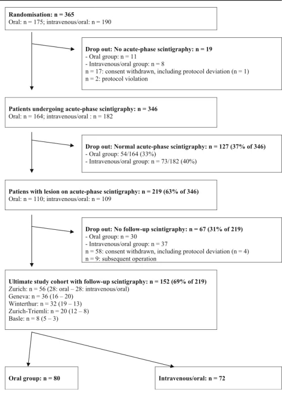
A total of 365 patients were randomised (Fig.1). No acute-phase scintigraphy was available for 19 (5%) patients and they were excluded. Consent was withdrawn in 17 patients, including one patient in the oral group with a protocol deviation. Two further patients were excluded due to protocol violation; one was given cefixime instead of ceftibuten and the other was administered medication only after day 3.
Acute-phase DMSA scintigraphy was performed in 346 patients; 219 patients (63%) showed one or more lesions and were planned to have follow-up scintigraphy. Of these, 67 patients (31%) had no secondary scintigraphy and did not complete the study, including four patients who deviated from the assigned antimicrobial regimen for medical reasons and nine patients undergoing a subsequent operation (for details, see Patients not completing the study). Under the study protocol, 127 patients with normal acute-phase scintigraphy had no follow-up.
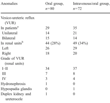
The cohort completing the study with follow-up scintigra-phy consisted of 152 patients with acute pyelonephritis and lesions on acute-phase scintigraphy. There were 80 patients in the oral group and 72 patients in the intravenous/oral group (Fig. 1). The clinical and laboratory characteristics at baseline (Table 1) and the prevalence of VUR (Table 2) did not significantly differ between the two groups, except for serum CRP concentration at baseline (p=0.04). All patients had either an abnormal urinary dipstick test or pyuria with≥10 white blood cells/μl. Plasma creatinine was normal in all participants. Escherichia coli were the predominant isolated pathogen (137/152: 90%); Proteus spp., Klebsiella pneumoniae and Enterococcus spp. were isolated in only a few cases (Table1). All isolated bacteria except Enterococcus spp. were sensitive to both ceftibuten and ceftriaxone.
The clinical course was comparable in both groups (Table 1). By day 3, approximately 90% of the patients were afebrile and CRP levels decreased to about 60% in both groups; seven and eight patients from the oral and intravenous/oral group, respectively, had persistent fever. In two of them (both from the oral group), the general condition did not improve and therapy was switched to intravenous treatment with amoxicillin/clavulanic acid and ceftriaxone. One patient in the intravenous/oral group had E. coli bacteraemia; the clinical course was uneventful without changing the therapeutic regimen. Urine culture grew cephalosporin-resistant Enterococcus spp. in three patients in the intravenous/oral group. At day 3, the urine
was sterile in all of them and two patients were afebrile. Thus, antimicrobial therapy was not changed. Urine cultures obtained from every patient on days 3–4 of treatment were all sterile. Both antimicrobial regimens were well tolerated and no side effects (e.g. skin rash, diarrhoea and gall or kidney stones) were observed.
All 92 patients (61% of the study cohort) at the University Children’s Hospitals Zurich and Geneva were treated as out-patients. The majority of patients from the other centres were hospitalised for an average of 3 days. Acute-phase DMSA scintigraphy
Eight patients from each group had acute-phase bilateral lesions, resulting in 88 and 80 affected renal units in the oral and intravenous/oral groups, respectively (Table3). In the majority of renal units, the size of the acute lesion was grade I or II. Extensive lesions (grade III and IV) were less frequent in the oral group than in the intravenous/oral group (11/160 vs. 22/144 renal units; p=0.03;χ2-test).
Renal scarring on follow-up DMSA scintigraphy
Among the 152 children, 54 developed renal scarring, including 21 patients from the oral group and 33 (46%) from the intravenous/oral group (Table 3). Among patients with bilateral acute-phase lesions, none from the oral group but three of eight patients from the intravenous/oral group developed bilateral scars. All scars were localised to the site of the acute-phase lesion(s). Renal scarring correlated with the size of the acute renal lesion (r=0.60, p<0.0001; Table 3). After statistical adjustment for the two confounding variables (i.e. CRP concentration at baseline and size of the acute-phase scintigraphic lesion), there was no significant difference between the percentages of renal scarring in the two groups, either expressed per patient or per renal unit (adjusted p-values; Table 3). All three patients from the intravenous/oral group with Enterococcus spp. whose urine had became sterile at day 3 and both patients from the oral group with protocol deviation developed renal scarring.
The prevalence of VUR did not significantly differ between the two groups. VUR was present in 93 (31%) of 304 renal units with grades I–II, III and IV in 71, 15 and 7 units, respectively (Table 2). Scarring in patients with unilateral VUR was always restricted to the refluxing unit. The correlation between VUR and renal scarring was analysed in a double manner (Table4). If VUR and scarring were expressed per patient, renal scarring was not signifi-cantly different in patients with and without VUR. However, if VUR and scarring were analysed per renal unit, scarring was more frequent in refluxing units (p=0.04; χ2-test). In
addition, scarring was correlated with the grade of VUR (r= 0.31, p=0.03). All seven renal units with grade IV VUR developed scars and renal units with grade III or IV VUR (11/22; 50%) had significantly more scarring compared to renal units with grade I–II VUR (16/71; 23%: p=0.02; χ2
-test).
The frequency of renal scarring was not different between infants <1 year of age (14/46) and older children (40/106). In addition, the frequency of renal scarring did not differ between the two centres conducting out-patient management only and the three centres carrying out
in-patient management (Table 5). In addition, there was no difference in the frequency of scarring among the five recruitment centres (data not shown).
Urinary tract infection was reported in 27 patients (18%) before entry into the study, though it was not specified as cystitis or pyelonephritis (Table 1). Three of 14 patients from the oral group showed renal scarring, compared with seven of 13 patients from the oral/intravenous group. Six patients had a recurrent febrile urinary tract infection between the acute-phase and follow-up scintigraphy. Both patients from the oral group had no scarring. In contrast, all
Randomisation: n = 365
Oral: n = 175; intravenous/oral: n = 190
Drop out: No acute-phase scintigraphy: n = 19
- Oral group: n = 11
- Intravenous/oral group: n = 8
n = 17: consent withdrawn, including protocol deviation (n = 1) n = 2: protocol violation
Patients undergoing acute-phase scintigraphy: n = 346
Oral: n = 164; intravenous/oral : n = 182
Drop out: Normal acute-phase scintigraphy: n = 127 (37% of 346)
- Oral group: 54/164 (33%)
- Intravenous/oral group: n = 73/182 (40%)
Patiens with lesion on acute-phase scintigraphy: n = 219 (63% of 346)
Oral: n = 110; intravenous/oral: n = 109
Drop out: No follow-up scintigraphy: n = 67 (31% of 219)
- Oral group: n = 30
- Intravenous/oral group: n = 37
n = 58: consent withdrawn, including protocol deviation (n = 4) n = 9: subsequent operation
Ultimate study cohort with follow-up scintigraphy: n = 152 (69% of 219)
Zurich: n = 56 (28: oral – 28: intravenous/oral) Geneva: n = 36 (16 – 20)
Winterthur: n = 32 (19 – 13) Zurich-Triemli: n = 20 (12 – 8) Basle: n = 8 (5 – 3)
Oral group: n = 80 Intravenous/oral: n = 72
Fig. 1 Randomisation and pa-tient flow chart
four patients from the intravenous/oral group had scars at the same location as the lesions on acute-phase scintigra-phy. As it is difficult, if not impossible, to distinguish between fresh lesions and old scars, additional analysis was undertaken after the exclusion of the 33 patients with previous or recurrent pyelonephritis. Renal scarring was not significantly different between the patients in the oral group (18/64) and in the intravenous/oral group (22/55; p=0.17). Patients not completing the study
Of 365 randomised patients, 86 failed to adhere to the protocol and did not complete the study; 19 patients had no acute-phase scintigraphy and 67 patients with lesions on acute-phase scintigraphy had no follow-up scintigraphy (Fig.1). Five patients deviated from the assigned treatment regimen for medical reasons. One patient was randomly placed in the oral group and switched to intravenous therapy due to recurrent vomiting; acute-phase scintigraphy was not performed but scintigraphy, performed after antireflux surgery 2 years later, was normal. Four patients with lesions on acute-phase scintigraphy showed persistent
fever beyond day 3 without apparent improvement of the general condition. One patient from the oral group was switched to intravenous meropenem. Two patients from the intravenous/oral group were maintained on ceftriaxone for 7 days. Another patient from the intravenous/oral group with Enterococcus spp. in urine culture was switched to amoxicillin/clavulanic acid. The parents withdrew consent for follow-up scintigraphy in those four patients and in 54 additional patients. Nine patients underwent surgery; ureter-ocystoneostomy in five patients with VUR, pyeloplasty in three patients with pyeloureteric junction obstruction and nephrectomy of a multi-cystic dysplastic kidney in one patient. None of these children had follow-up scintigraphy. In one centre (University Children’s Hospital Zurich), all eligible patients were recorded in a “log book” and any reasons for refusal or withdrawal were recorded. There were 292 children with a febrile urinary tract infection, of whom, 40 patients were excluded in accordance with the exclusion criteria. Of the 252 eligible study candidates, 125 (50%) could not be randomised for two reasons: 65 Swiss patients whose parents did not consent and 60 immigrants whose parents had a language barrier, rendering informed consent impossible. Thus, 127 patients were randomised; 48 patients were not enrolled because no acute-phase scintigraphy had been performed (parental withdrawal of consent: n=10; protocol violation: n=1) or the acute-phase scintigraphy was normal (with bacteriuria: n=29; with negative urine culture: n=8), 23 patients with acute-phase lesions had no follow-up scintigraphy and 56 patients completed the study.
Table 1 Baseline characteristics of 152 children with lesions on acute-phase dimercaptosuccinic acid (DMSA) scintigraphy
Characteristics Oral group, n=80 Intravenous/oral group, n=72 Sex Male 8 10 Female 72 62 Age (years) Median 2.2 1.6 Interquartile range 0.9–4.9 1.0–4.4 Previous urinary tract
infections None 66 59 ≥1 14 13 Fever (≥38.5°C) before treatment (days) Median 3 2.5 Fever on day 3 7 8 C-reactive protein (CRP, mg/l)
at baseline: median (range) 100 (8–327)a 128 (12–378)a
at day 3: median (range) 59 (10–404) 75 (12–378) Bacterial urinary pathogens
Escherichia coli 73 64 Proteus spp. 1 1 Klebsiella pneumoniae 1 0 Enterococcus spp. 0 3 Data not available 5 6 Positive blood cultures
(E. coli)
0 1
a
p=0.04, Mann-Whitney test
Table 2 Associated renal and urological anomalies Anomalies Oral group,
n=80 Intravenous/oral group, n=72 Vesico-ureteric reflux (VUR) In patientsa 29 35 Unilateral 14 21 Bilateral 15 14 In renal unitsb 44 (28%) 49 (34%) Left 26 29 Right 18 20 Grade of VUR (renal units) I–II 34 37 III 7 8 IV 3 4 Hydronephrosis 1 0 Hypospadia glandis 0 1 Duplex kidney and
ureterocele
1 0
Discussion
This prospective multi-centre randomised trial focussed on children with community-acquired acute pyelonephritis and renal lesions on acute-phase DMSA scintigraphy, i.e. patients at risk for renal scarring. In patients aged 6 months to 16 years, we showed that oral treatment with once-daily ceftibuten is safe and is equally as efficacious with respect to renal scarring after 6 months when compared with a sequential intravenous/oral regimen. The oral antimicrobial was well-tolerated and side effects, including skin rash, diarrhoea or gall bladder or kidney stones, were not observed. Thus, this study identifies a treatment modality for acute pyelonephritis in children that offers the benefits of entire out-patient management.
The study was designed to compare the efficacy of oral with sequential intravenous/oral antimicrobial treatment for children with pyelonephritis at risk of renal scarring.
Patients were randomised for the treatment regimen before scintigraphic lesions were verified for two reasons: (i) only children with lesions in acute-phase scintigraphy are likely to develop renal scars [14, 15, 30] (ii) acute-phase scintigraphy cannot be performed immediately after the diagnosis of acute pyelonephritis. However, to improve the diagnostic accuracy of acute pyelonephritis, DMSA scin-tigraphy is ideally carried out within two days. In the future, magnetic resonance imaging (MRI) might offer both the use of non-ionizing radiation and a better distinction between acute lesion and scar [18].
The presence of renal scars is a surrogate outcome. Although only a few studies have analysed the long-term outcome, renal scars can result in significant sequelae [20,
32, 33]. The follow-up of children with renal scarring for 16–26 years after childhood pyelonephritis showed a significant reduction of individual glomerular filtration rate in the unilaterally scarred kidney [32,33]. In addition, the
Table 3 Acute-phase lesions and scarring in renal units on DMSA scintigraphy
Characteristics Oral group, n=160 renal units Intravenous/oral group, n=144 renal units Adjusted p-value* Acute lesion Scarring Acute lesion Scarring
Renal units: acute lesions 88a 80a
Scarring
Patients 21 33 0.2
Renal units (% of all units) 21 (13%) 36b(25%) 0.5 Renal units: acute lesion grade I 49 29
Scarring 4 3
Renal units: acute lesion grade II 28 29
Scarring 11 13
Renal units: acute lesion grade III 9 17
Scarring 5 15
Renal units: acute lesion grade IV 2 5
Scarring 1 5
a
Eight patients in each group with acute-phase bilateral lesions
b
Three patients with bilateral scarring
*p-values adjusted for CRP concentration at baseline and size of lesion on acute-phase scintigraphy
Table 4 Renal scarring and vesico-ureteric reflux (VUR)
Characteristics Oral group, n=80 Intravenous/oral group, n=72 p-value Scarring in patients with reflux 9/29 18/35 n.s. Scarring in patients without reflux 12/51 15/37 n.s.
All 152 patients n.s.
Scarring in patients with reflux: 27/64 vs. without reflux: 27/88
Scarring in renal units with ipsilateral reflux n.s.
All units 9/44 18/49
Left unit 8/26 11/29
Right unit 1/18 7/20
All 304 renal units 0.04
Scarring in units with reflux: 27/93 vs. without reflux: 30a/211
a
long-term outcome at a mean age of 41 years (and mean follow-up of 37 years) in children with both pyelonephritis and VUR showed a lowered glomerular filtration rate (83% of patients), hypertension (50%) and proteinuria (25%) in patients with bilateral scars [20]. An increased tendency for hypertension was also found in patients with unilateral scarring [20].
The proportions of renal scarring in patients (oral group: 26%; intravenous/oral group: 46%) were comparable with previous scintigraphy investigations that documented renal scarring in 29–60% of children after acute pyelonephritis [5,15, 16,21,24, 30,31, 35]. These results indicate that mere oral ceftibuten therapy is as efficacious as parenteral therapy, i.e. a 3-day or even a 10-day course of intravenous antibiotic therapy with respect to renal scarring. However, antibiotic therapy either intravenous or oral is not able to prevent renal scarring in a substantial proportion of children with pyelonephritis. The percentage of renal scarring in our study appeared to be lower in the oral treatment group than in the intravenous/oral group. Yet, despite randomisation, the two groups differed significantly in the number of patients with previous and recurrent infections, CRP concentration at baseline and the size of the lesion in acute-phase scintigraphy. The latter variable has a signifi-cant effect on the evolution of renal scarring, as observed in our previous study [5]. After adjustment for these con-founding variables, the final analysis showed a similar frequency of renal scarring in both groups. Children aged >1 year did not develop scars more frequently than children <1 year of age. This is in contrast to previous studies, including our own [5,15,24].
There was no significant difference of renal scarring between in-patients and out-patients in both the oral and the intravenous/oral groups. Also taking into consideration that allocation to in-patient or out-patient treatment was deter-mined by local hospital guidelines and not by random-isation, this finding is valid and reliable, as the treatment of every patient was guided by a defined protocol. Thus, if out-patient treatment is chosen, clinical evaluation and urinalysis at day 3 and day 14 are recommended. If the patient is afebrile and urinalysis is normal, routine follow-up urine culture is not mandatory. Oral out-patient management might reduce the costs, though this was not formally evaluated here.
In this study, renal scarring correlated significantly with the VUR per renal unit (but not per patient) and the grade of VUR, in particular, grades III or IV VUR. Renal scarring in patients with unilateral VUR was always restricted to the refluxing unit. Published studies on the effects of VUR on renal scarring are controversial. Some studies showed a significant influence [12,24,26], while others did not [5,
22]. There were, however, methodological inconsistencies. For example, not all studies discriminated between lower and upper urinary tract infection or between VUR per patient and per renal unit.
Almost all pathogens isolated in this study of acute community-acquired pyelonephritis were E. coli susceptible to cephalosporins. Thus, antimicrobial treatment with the chosen cephalosporin is safe. This is of considerable relevance, given that the prevalence of E. coli resistant to aminopenicillins may be up to 50% [27]. The specific advantage of the oral cephalosporin ceftibuten for out-patient management is its once-daily oral dosing, based on a long T1/2 with plasma concentrations above minimal inhibitory concentration for >16 hours; it has an excellent oral bioavailability and 60–70% of the drug is excreted in the urine. The recommended dose is 9 mg/kg body weight given once daily [4, 17, 23, 34]. Side effects in this and other studies are rare [28, 34] and ceftibuten has been previously examined in children with urinary tract infection [3,31]. Infections with intrinsically cephalosporin-resistant Enterococcus spp. in this study were rare (4/365; 1%); in three of these cases, the urine was sterile on day 3, suggesting a good clinical response and, therefore, not urging the treating physicians to change therapy. Neverthe-less, all three patients developed renal scars. Thus, patients with acute pyelonephritis treated with a cephalosporin should be switched to aminopenicillin upon the growth of enterococci in urine cultures.
Our results are in agreement with two recent prospective trials which suggested that acute pyelonephritis can be treated with oral antibiotics. In the study by Montini et al. [21], amoxicillin/clavulanic acid was given for 10 days to 135 children aged 1 month to 7 years. Renal scarring developed in 26/96 patients (28%), with lesions on acute-phase scintigraphy comparable to the 26% found among children with oral treatment in our study and in earlier studies [5, 15, 16, 24, 30, 35]. However, the high
Table 5 Renal scarring in patients with out-patient and in-patient management
Management All patients Oral group Intravenous/oral group
n Scars n Scars n Scars
Out-patient 92 35 44 14 48 21
In-patient 60 19 36 6 24 13
χ2
prevalence of resistance of common urinary pathogens against amoxicillin/clavulanic acid in our unit (>30% of E. coli isolates are resistant against amoxicillin/clavulanic acid) and many other centres precludes the empiric use of this antimicrobial [27]. In the study by Hoberman et al. [14], cefixime was administered orally for 14 days to 153 children younger than 2 years. The renal scarring assessed at 6 months was lower, with 16.9% in patients having lesions on acute-phase scintigraphy. These patients, how-ever, had mild infection, as the mean initial CRP concen-tration (11.9 mg/l) was low. In contrast, median CRP was 100 mg/l in our study, i.e. a 10-fold higher range, indicating more pronounced inflammation, which, in turn, may explain the higher rate of renal scarring. Also, Montini et al. [21] reported an initial CRP concentration nine-fold above the upper normal value, with a similar range of scarring in children. Nevertheless, by bag urine collection, they may have included children with false-positive urine cultures, as urine collected by catheter was not a prerequi-site for inclusion as in our study and the study by Hoberman et al. [14]. Thus, compared with these two studies, the reported study here included children with more severe disease, covered a wider age range and renal scarring was correlated to VUR per renal unit, and, most impor-tantly, showed no difference for in- and out-patient management, respectively. Overall, given the similar fre-quencies of renal scarring after oral and sequential intravenous/oral antimicrobial treatment in all three studies, the oral-only regimen appears to be an alternative, offering the benefits of out-patient management, including the avoidance of painful and labourious intravenous approach and the reduction of burdens on both parents and hospital staff.
This study required the randomisation and enrolment of a substantially larger number of patients than the number of patients eventually qualifying for the analysis of renal scarring. In agreement with the literature [5, 14, 21, 25], one third of the enrolled patients showed no lesions in acute-phase scintigraphy and were subsequently excluded from follow-up analysis, as they are not at risk of renal scarring [14, 15,30]. In addition, almost one third of the children with lesions in the acute-phase scintigraphy did not undergo follow-up scintigraphy, primarily due to the withdrawal of parental consent. Although this resulted in a rather considerable reduction of the number of eventually eligible patients, skewing of the study results is not likely. The drop-outs were evenly distributed among the random-isation groups and the participating centres. Nevertheless, the relatively high number of drop-outs recorded in this study may mirror the limited willingness of parents and their children to participate in prospective clinical trials, in particular, if the study lasts several months beyond the
acute phase of the illness and resolution of clinical symptoms and includes procedures (e.g. injection of radioactive substances, perceived as invasive by some parents).
A further difficulty of the scintigraphy studies (including the present research) is to make the distinction between acquired renal scarring and congenital dysplasia in patients with persistent photon defect on DMSA scintigraphy [9]. Without exact patient history or previous imaging, the aetiology of scars cannot always be determined. To limit this bias, renal scarring was defined in our study as persistent or partially reversible lesions at the same location of the lesion in the acute-phase scintigraphy. In addition, renal ultrasound was not suggestive of renal dysplasia in any of the patients.
In summary, our hypothesis was validated. Oral antimi-crobial therapy with once-daily ceftibuten for 14 days, with its attendant benefits, was shown to be equally as efficacious as sequential treatment with intravenous cef-triaxone for 3 days followed by 11 days of oral ceftibuten for pyelonephritis in children.. The optimal duration of antimicrobial therapy, however, and the role of oral regimen in selected children (i.e. <6 months of age or with complex urological malformations) [6] remain to be determined.
References
1. American Academy of Pediatrics, Committee on Quality Im-provement, Subcommittee on Urinary Tract Infection (1999) Practice parameter: the diagnosis, treatment, and evaluation of the initial urinary tract infection in febrile infants and young children. Pediatrics 103:843–852
2. Bachur R, Caputo GL (1995) Bacteremia and meningitis among infants with urinary tract infections. Pediatr Emerg Care 11:280– 284
3. Banfi A, Gabriele G, Hill-Juarez JM, Kaufman A, Moens E, group amotcutiis (1993) Multinational comparative trial of ceftibuten and trimethoprim-sulfamethoxazole in the treatment of children with complicated or recurrent urinary tract infections. Pediatr Infect Dis J 12:S84–S91
4. Barr WH, Affrime M, Lin CC, Batra V (1995) Pharmacokinetics of ceftibuten in children. Pediatr Infect Dis J 14:S93–S101 5. Benador D, Neuhaus TJ, Papazyan JP, Willi UV, Engel-Bicik I,
Nadal D, Slosman D, Mermillod B, Girardin E (2001) Rando-mised controlled trial of three day versus 10 day intravenous antibiotics in acute pyelonephritis: effect on renal scarring. Arch Dis Child 84:241–246
6. Bloomfield P, Hodson EM, Craig JC (2003) Antibiotics for acute pyelonephritis in children. Cochrane Database Syst Rev: CD003772
7. Crain EF, Gershel JC (1990) Urinary tract infections in febrile infants younger than 8 weeks of age. Pediatrics 86:363–367 8. de La Vaissière B, Castello B, Quinet B, Cohen R, Grimprel E
(2006) Management of acute pyelonephritis in patients older than 3 months: survey conducted in 39 paediatric emergency departments of the Ile de France Region in 2004. Arch Pediatr 13:245–250
9. Ditchfield MR, Grimwood K, Cook DJ, Powell HR, Sloane R, Gulati S, De Campo JF (2004) Persistent renal cortical scintigram defects in children 2 years after urinary tract infection. Pediatr Radiol 34:465–471
10. Goldraich NP, Goldraich IH (1995) Update on dimercaptosuccinic acid renal scanning in children with urinary tract infection. Pediatr Nephrol 9:221–226
11. Goldraich NP, Ramos OL, Goldraich IH (1989) Urography versus DMSA scan in children with vesicoureteric reflux. Pediatr Nephrol 3:1–5
12. González E, Papazyan JP, Girardin E (2005) Impact of vesicoure-teral reflux on the size of renal lesions after an episode of acute pyelonephritis. J Urol 173:571–574; Discussion 574–575 13. Group SPN (2001) Treatment of urinary tract infections in
children. Paediatrica 12:16–21
14. Hoberman A, Wald ER, Hickey RW, Baskin M, Charron M, Majd M, Kearney DH, Reynolds EA, Ruley J, Janosky JE (1999) Oral versus initial intravenous therapy for urinary tract infections in young febrile children. Pediatrics 104:79–86
15. Jakobsson B, Berg U, Svensson L (1994) Renal scarring after acute pyelonephritis. Arch Dis Child 70:111–115
16. Jakobsson B, Svensson L (1997) Transient pyelonephritic changes on 99mTechnetium-dimercaptosuccinic acid scan for at least five months after infection. Acta Paediatr 86:803–807
17. Jones RN (1995) Ceftibuten: a review of antimicrobial activity, spectrum and other microbiologic features. Pediatr Infect Dis J 14: S77–83
18. Kavanagh EC, Ryan S, Awan A, McCourbrey S, O’Connor R, Donoghue V (2005) Can MRI replace DMSA in the detection of renal parenchymal defects in children with urinary tract infec-tions? Pediatr Radiol 35:275–281
19. Klepser ME, Marangos MN, Patel KB, Nicolau DP, Quintiliani R, Nightingale CH (1995) Clinical pharmacokinetics of newer cephalosporins. Clin Pharmacokinet 28:361–384
20. Lahdes-Vasama T, Niskanen K, Rönnholm K (2006) Outcome of kidneys in patients treated for vesicoureteral reflux (VUR) during childhood. Nephrol Dial Transplant 21:2491–2497
21. Montini G, Toffolo A, Zucchetta P, Dall’Amico R, Gobber D, Calderan A, Maschio F, Pavanello L, Molinari PP, Scorrano D, Zanchetta S, Cassar W, Brisotto P, Corsini A, Sartori S, Da Dalt L, Murer L, Zacchello G (2007) Antibiotic treatment for pyelone-phritis in children: multicentre randomised controlled non-inferi-ority trial. BMJ 335:386
22. Moorthy I, Easty M, McHugh K, Ridout D, Biassoni L, Gordon I (2005) The presence of vesicoureteric reflux does not identify a
population at risk for renal scarring following a first urinary tract infection. Arch Dis Child 90:733–736
23. Neu HC (1995) Ceftibuten: minimal inhibitory concentrations, postantibiotic effect and beta-lactamase stability—a rationale for dosing programs. Pediatr Infect Dis J 14:S88–S92
24. Orellana P, Baquedano P, Rangarajan V, Zhao JH, Eng ND, Fettich J, Chaiwatanarat T, Sonmezoglu K, Kumar D, Park YH, Samuel AM, Sixt R, Bhatnagar V, Padhy AK (2004) Relationship between acute pyelonephritis, renal scarring, and vesicoureteral reflux. Results of a coordinated research project. Pediatr Nephrol 19:1122–1126
25. Pitetti RD, Choi S (2002) Utility of blood cultures in febrile children with UTI. Am J Emerg Med 20:271–274
26. Polito C, Rambaldi PF, Signoriello G, Mansi L, La Manna A (2006) Permanent renal parenchymal defects after febrile UTI are closely associated with vesicoureteric reflux. Pediatr Nephrol 21:521–526
27. Prère MF, Licznar P, Decramer S, Fayet O (2004) E. coli from urinary tract infections and acute pyelonephritis of children: 1% of strains are resistant to a subset of third generation cephalosporins. Pathol Biol (Paris) 52:497–500
28. Reidenberg BE (1995) Worldwide safety experience with cefti-buten pediatric suspension. Pediatr Infect Dis J 14:S130–S133 29. Rushton HG, Majd M (1992) Dimercaptosuccinic acid renal
scintigraphy for the evaluation of pyelonephritis and scarring: a review of experimental and clinical studies. J Urol 148:1726–1732 30. Stokland E, Hellström M, Jacobsson B, Jodal U, Sixt R (1996) Renal damage one year after first urinary tract infection: role of dimercaptosuccinic acid scintigraphy. J Pediatr 129:815–820 31. Vilaichone A, Watana D, Chaiwatanarat T (2001) Oral ceftibuten
switch therapy for acute pyelonephritis in children. J Med Assoc Thai 84(Suppl 1):S61–S67
32. Wennerström M, Hansson S, Hedner T, Himmelmann A, Jodal U (2000) Ambulatory blood pressure 16–26 years after the first urinary tract infection in childhood. J Hypertens 18:485–491 33. Wennerström M, Hansson S, Jodal U, Sixt R, Stokland E (2000)
Renal function 16 to 26 years after the first urinary tract infection in childhood. Arch Pediatr Adolesc Med 154:339–345
34. Wiseman LR, Balfour JA (1994) Ceftibuten. A review of its antibacterial activity, pharmacokinetic properties and clinical efficacy. Drugs 47:784–808
35. Zaki M, Badawi M, Al Mutari G, Ramadan D, Adul Rahman M (2005) Acute pyelonephritis and renal scarring in Kuwaiti children: a follow-up study using 99mTc DMSA renal scintigraphy. Pediatr Nephrol 20:1116–1119