Publisher’s version / Version de l'éditeur:
Journal of Proteome Research, 9, 2, pp. 912-923, 2010-02-01
READ THESE TERMS AND CONDITIONS CAREFULLY BEFORE USING THIS WEBSITE. https://nrc-publications.canada.ca/eng/copyright
Vous avez des questions? Nous pouvons vous aider. Pour communiquer directement avec un auteur, consultez la première page de la revue dans laquelle son article a été publié afin de trouver ses coordonnées. Si vous n’arrivez pas à les repérer, communiquez avec nous à [email protected].
Questions? Contact the NRC Publications Archive team at
[email protected]. If you wish to email the authors directly, please see the first page of the publication for their contact information.
NRC Publications Archive
Archives des publications du CNRC
This publication could be one of several versions: author’s original, accepted manuscript or the publisher’s version. / La version de cette publication peut être l’une des suivantes : la version prépublication de l’auteur, la version acceptée du manuscrit ou la version de l’éditeur.
For the publisher’s version, please access the DOI link below./ Pour consulter la version de l’éditeur, utilisez le lien DOI ci-dessous.
https://doi.org/10.1021/pr900788a
Access and use of this website and the material on it are subject to the Terms and Conditions set forth at
Activity-based proteome profiling of hepatoma cells during Hepatitis C
virus replication using protease substrate probes
Blais, David R.; Brûlotte, Marc; Qian, Yiming; Bélanger, Sylvie; Yao, Shao
Q.; Pezacki, John Paul
https://publications-cnrc.canada.ca/fra/droits
L’accès à ce site Web et l’utilisation de son contenu sont assujettis aux conditions présentées dans le site LISEZ CES CONDITIONS ATTENTIVEMENT AVANT D’UTILISER CE SITE WEB.
NRC Publications Record / Notice d'Archives des publications de CNRC:
https://nrc-publications.canada.ca/eng/view/object/?id=e090e08c-f4e8-4585-a807-f25105fad5bc
https://publications-cnrc.canada.ca/fra/voir/objet/?id=e090e08c-f4e8-4585-a807-f25105fad5bc
Hepatitis C Virus Replication Using Protease Substrate Probes
David R. Blais,
†Marc Bru
ˆ lotte,
†,‡Yiming Qian,
†,§Sylvie Be´langer,
†Shao Q. Yao,
|and
John Paul Pezacki*
,†,‡,§Steacie Institute for Molecular Sciences, National Research Council Canada, 100 Sussex Drive, Ottawa, ON, K1A 0R6, Canada, Department of Biochemistry, Microbiology and Immunology, University of Ottawa, 451 Smyth Road, Ottawa, ON, K1H 8M5, Canada, Department of Chemistry, University of Ottawa, 10 Marie
Curie Private, Ottawa, ON, K1N 6N5, Canada, and Departments of Chemistry and Biological Sciences, National University of Singapore, 3 Science Drive 3, Singapore, 117543
Received September 4, 2009
Activity-based protein profiling (ABPP) offers direct insight into changes in catalytic activity of enzyme classes in complex proteomes, rather than protein or transcript abundance. Here, ABPP was performed in Huh7 hepatoma cell lines with a group of ABPP probes composed of an N-acetylated amino acid, that mimic the P1position in protease peptide substrates. Five different probes bearing distinct amino acids (Ser, Thr, Phe, Glu and His) labeled 54 differentially active proteins, including proteases, other hydrolases, oxidoreductases and isomerases. Four of the six protease families were targeted based on their P1substrate preferences. The broader specificity of the labeling observed could be explained by the substrate-based targeting nature and the electrophilic properties of the ABPP probes. When applied to Huh7 cells stably replicating hepatitis C virus (HCV) subgenomic replicon RNA, four proteins showed reduced activity, while three proteins had increased activity during HCV replication. These differentially active hits included carboxylesterase 1, cathepsin D, HSP105, protein disulfide isomerase 1 and A6, chaperonin containing TCP1 and isochorismatase domain containing 1, which demonstrated substrate preferences by being labeled by specific substrate probes. This illustrates the broader activity-based profiling capabilities of these substrate-activity-based probes to reveal novel enzyme candidates and their potential roles during HCV replication.
Keywords: activity-based protein profiling • amino acid coupled quinolimine methide probes •
carboxylesterase 1•hepatitis C•hydrolases•liver•proteases•protein disulfide isomerase
Introduction
The hepatitis C virus (HCV) is a growing threat to global health affecting over three percent of the global population.1 With no vaccines available and limited clinical treatments that are only successful in a small subset of patients, a high proportion of chronic HCV infections progress into liver steatosis, cirrhosis and eventually hepatocellular carcinoma, making HCV the leading cause of liver disease.2 HCV has a small genome of ∼9.6 kb (Figure 3a) and relies heavily on host factors and enzymes for its propagation. Host-mediated post-translational modifications, such as glycosylation of E1 and E2,3 phosphorylation of NS5A,4 as well as direct interaction with
host cell geranylgeranylated FBL2 protein5and liver microRNA miR-1226,7 are deemed essential for proper viral infectivity. Even in the early stage of the viral cycle, the ∼3000 aa translated HCV polyprotein requires the action of both viral and cellular proteases to generate the 10 or so mature viral proteins.8-11 Two virus-encoded proteases, the NS2-3 autoprotease and the NS3 serine protease, are responsible for the maturation of the nonstructural(NS)proteinsattheC-terminusofthepolyprotein.12,13 The cleavage of the structural component of viral particles, consisting of the capsid core protein and two envelope glyco-proteins E1 and E2, rely on at least a host cellular signal peptidase (SP).8,14 Another host protease, a signal peptide peptidase (SPP), is involved in completing the maturation of the HCV core by cleaving the protein from the membrane of the endoplasmic reticulum for its relocation on the surface of lipid droplets.9,11
Despite the importance of proteases in HCV pathogenesis and in numerous physiological processes, the majority of the ∼500 human proteases remain uncharacterized to date.15As the catalytic state of proteases is tightly regulated by several post-translational modifications, such as inactive zymogen processing, phosphorylation, nitrosylation, cofactors and
in-* To whom correspondence should be addressed: Steacie Institute for Molecular Sciences, National Research Council Canada, 100 Sussex Drive, Room 2107, Ottawa, ON, K1A 0R6, Canada. Tel.: 613-993-7253. Fax: 613- 941-8447. E-mail: [email protected].
†Steacie Institute for Molecular Sciences, National Research Council
Canada.
‡Department of Biochemistry, Microbiology and Immunology, University
of Ottawa.
§Department of Chemistry, University of Ottawa. |
Departments of Chemistry and Biological Sciences, National University of Singapore.
hibitors, novel experimental approaches are needed to address the potential significant divergence between protease abun-dance and activity. Activity-based protein profiling (ABPP) was developed to offer direct insight into changes in catalytic activity of enzyme classes in complex proteomes16-18and can be used to functionally annotate the enzyme function of proteins.19 ABPP employs active site-directed probes that consist of small molecule inhibitors linked to reporter tags.16 Given the importance of proteases and their broad diversity, numerous activity-based probes were developed to target their different classes,20,21such as serine,22,23cysteine,17,24aspartic25 and metalloproteases.26,27Nondirected sulfonate ester28and R-chloroacetamide29probes were also used to target a wider range of proteases through their reactivity with numerous enzyme families. However, the specificity of these nondirected probes is limited as they were shown to react with noncatalytic amino acid residues.30With the broad diversity of protease, there has been no report on activity-based probes capable of targeting the remaining classes of proteases, that is, glutamate and threonine (besides proteasome), due to the lack of known mechanism-based irreversible inhibitors that can covalently modify catalytically active site residues. Herein we report the usage of amino acid coupled quinolimine methide probes31,32 designed to mimic different protein substrates and broaden the activity-based profiling of protease families during HCV replication in human hepatocytes.
Experimental Procedures
Tissue Culture and Reagents. The human hepatoma cell line
Huh7 was grown in monolayers with Dulbecco’s Modified Eagle Medium (DMEM) supplemented with 100 nM nonessential amino acids, 50 U/mL penicillin, 50 µg/mL streptomycin and 10% fetal bovine serum (FBS) (Cansera, Rexdale, ON). The Huh7 cells stably expressing the subgenomic replicons (Figure 3a) (genotype 1B, con1) (passage number between 40 and 50) were maintained in the same culture medium supplemented with 250 µg/mL G418 Geneticin (Gibco-BRL, Burlington, ON). The pFK-I389neo/NS3-3′/5.1 subgenomic replicon (Figure 3a) was kindly provided by Ralf Bartenschlager (Institute of Hy-giene, University of Heidelberg, Germany). The activity-based substrate probes (Figure 1a) were synthesized as previously reported.31,32The probes were stored at -20 °C in methanol until added directly to the proteome extracts for activity-based protein labeling. Mass spectrometry (TOF MS ES+) was per-formed to ensure no degradation of the probe had occurred. Chemicals were provided from Sigma-Aldrich, unless indicated otherwise.
Active Proteome Extraction and Labeling. Subconfluent
cells (80-90%) were washed and pooled with ice-cold 10 mM sodium phosphate buffer, pH 7. The cells were then subjected to Dounce-homogenization at 30% power (T8 Ika-Werke homogenizer, GMBH, Staufen, Germany) and sonicated (20 pulses, 50% duty cycle, Sonifier 250, Branson Ultrasonic, Danbury, CT) in ice-cold sodium phosphate buffer supple-mented with 1% Triton X-100. The crude proteome extract was cleared by ultracentrifugation at 100 000g, 4 °C for 45 min, quantified with the BCA protein assay (Pierce, Rockford, IL) and diluted with sodium phosphate buffer to a final protein concentration of 1 mg/mL. The active proteome labeling conditions were optimized as follow (Supplemental Figure S1): the proteome extract (1 mg/mL in sodium phosphate buffer) was incubated in the dark with 5 µM (for Phe) or 10 µM (for Ser, Thr, His and Glu) probe (from a 500 or 1000 µM stock in
methanol, final methanol concentration of 1%) for 1 h at 37 °C, with occasional mixing. After 1 h, the reaction was quenched by precipitating the proteome with acetone: 5 vol of ice-cold acetone was added to the sample, frozen at -80 °C for 30 min and centrifuged at 15 000g, 4 °C for 15 min to remove salts and unreacted probe.
2D Analysis of Labeled Proteomes. Precipitated protein
pellets (200 µg) were resuspended in 125 µL of isoelectric focusing (IEF) buffer (7 M Urea, 2 M thiourea, 4% CHAPS, 1% dithiothreitol (DTT)) containing 0.2% Ampholytes pH 3-10 (biolytes, Bio-Rad, Hercules, CA). IPG strips pH 3-10 NL, 7 cm (Bio-Rad) were passively rehydrated with the resuspended proteomes for 16 h. The rehydrated protein IPG strips were then submitted to isoelectric focusing using the Protean IEF Cell (Bio-Rad) with the following conditions: 200 V rapid ramp for 30 min, 500 V rapid ramp for 30 min, 2 h linear ramp to 6500 V, and 2 h focusing (rapid ramp) at 6500 V. The accumulated voltage-hour (Vh) felt between a range of 12 000-17 500 Vh.
Prior to SDS-PAGE separation, proteins immobilized on the IPG strips were reduced (1% DTT) and alkylated (4% iodoacet-amide) in SDS equilibration buffer (6 M urea, 30% glycerol, 2% SDS, 50 mM tris-HCl, pH 8.8). The SDS-PAGE separation was performed on a 10% gel (1.5 mm thickness) at constant 150 V per gel. The gels were then scanned for fluorescence using the Typhoon 9410 (GE Healthcare, Piscataway, NJ) and were
Figure 1.Structure and protocol of substrate based probes used to target the active Huh7 proteome during HCV replication. (a) Chemical structure of substrate-based probes composed of unique amino acid side chains (R) corresponding to the P1site
residues in protease substrates. (b) Scheme of the ABPP protocol used in this study to identify protease targets. The active proteome isolated from naı¨ve Huh7 cells or Huh7 stably express-ing the HCV replicon was labeled through a nucleophile (nu) residue at the active site of the enzyme targeting the reactive group of the protease probe. After separation of the labeled proteome by two-dimensional gel electrophoresis, the fluores-cently tagged proteins were identified by LC-MS/MS.
subsequently incubated overnight in fixing solution (50% (v/ v) ethanol, 5% (v/v) acetic acid). Finally, gels were stained with silver nitrate and scanned using the Fluor-S imager (Biorad) in order to visualize the entire cellular proteome.
Identification of Labeled Protein Candidates. The
fluores-cent gel image was aligned with the silver stained gel image to locate the labeled protein spots. Protein candidates displaying heat-sensitive activity were selected as specific targets, while candidates showing heat-insensitive reactivity were considered as nonspecific targets. The selected spots were manually excised under a laminar flow hood. Gels spots were destained with potassium ferricyanide solution (15 mM potassium ferri-cyanide, 50 mM sodium thiosulfate), rinsed three times with water and then shrunk with acetonitrile. The gel pieces were reswelled with 20 µL of trypsin solution (0.01 µg/µL in 50 mM ammonium bicarbonate), and incubated overnight at 37 °C. Peptides were identified by LC-MS/MS on a LTQ XL (Ther-mo) mass spectrometer. The LTQ XL mass spectrometer (Thermo) was coupled to a MDLC chromatography system (GE Healthcare). The samples were first injected onto a 0.3 × 5 mm C18 micro precolumn cartridge (Dionex/LC Packings) to re-move salts and other soluble contaminants. Samples were then separated on a 5 cm × 75 µm BioBasic C18, 5 µm particle, PicoFrit column (New Objective) with a flow rate of ∼300 nL/ min using a 30 min gradient: 0-30% acetonitrile/0.1% formic acid over 14 min, 30-50% acetonitrile/0.1% formic acid over 14 min, 50-90% acetonitrile/0.1% formic acid over 2 min. MS and MS/MS data were collected in enhanced profile and normal centroid mode (scan rate, ∼8000 amu/s; scan range, 400-1600), respectively. MS/MS was performed on the three most abundant multiple-charged peaks for each MS scan.
Peak lists were generated using the default parameters by Extract-MSN in BioWorksBrowser 3.2 (ThermoFisher). LC-MS/ MS spectra were searched against the National Centre for Biotechnology Information nonredundant database (October 16, 2006; 2 879 860 sequence entries; 1 012 985 077 residues) using Mascot daemon version 2.0 (Matrix Science, London, U.K.). Search parameters used for queries were trypsin cleavage, e1 missed cleavages, (1.5-Da peptide tolerance, (1.2-Da MS/ MS tolerance, with no fixed modifications and the following variable modifications: carbamidomethyl (Cys), oxidation (Met). The results were then evaluated manually: protein identification required the matching of a least two peptides per protein, a MASCOT score above 100 and a corresponding pI and mass according to their position after 2D gel electrophoresis.
Mapping the Activity-Based Reactivity of PDI toward the Phe Probe. Recombinant human protein disulfide isomerase
(PDI) (Cedarlane Laboratories Ltd., Hornby, ON) at 0.8 µg/µL (0.5 µg) was preincubated for 30 min at room temperature (RT) either in presence or absence of 0.7 mM of dinitrothiocy-anobenzene (DNTB) following the treatment with 5 µM Phe probe for 1 h at 37 °C. After 1 h, the reaction was quenched by boiling the sample for 10 min in loading buffer, followed by SDS-PAGE electrophoresis.
Bacterial Expression of Recombinant HCV NS3/4a Pro-tease. To investigate the susceptibility of HCV NS3 protease to
labeling with the substrate-based probes, the single chain proteolytic domain of NS3/4a was expressed and purified as previously described.33The purified extract was incubated with 5 µM Phe, 10 µM Ser or Thr, or 10 nM FP probe for 1 h at 37 °C. After 1 h, the reaction was quenched by boiling the sample for 10 min in loading buffer, followed by SDS-PAGE electrophoresis.
Expression Analysis by RT-PCR. Total RNA from
subcon-fluent cells was isolated using the RNeasy extraction kit (Qiagen, Chatsworth, CA). To ensure that all genomic DNA was com-pletely removed from the samples, extracts were subjected to DNase digestion with the RNase-free DNase kit (Qiagen). Random primed cDNA synthesis was performed (Superscript First-Strand Synthesis for RT-PCR; Invitrogen, Carlsbad, CA) according to the manufacturer’s instructions. Oligonucleotide primers used to amplify CCT4, CES1, NY-CO-25, CTSD, PDI, PDIA6 and CGI-111 were as follows: CCT4 sense, ATGC-CCGAGAATGTGGC; CCT4 antisense, TTATCGAGTGTTTACCA-CATCATCTATT; CES1 sense, AGGAGCTCTTGGAGACGACA; CES1 antisense, CTTCTCCACTGCCTTCTTGG; NY-CO-25 sense, CGCAGACCGAGACCCG; NY-CO-25 antisense, CTAGTCCAAGTC-CATATTAACAGAATTTT; CTSD sense, GACACAGGCACTTC-CCTCAT; CTSD antisense, CTCTGGGGACAGCTTCTAGC; PDI sense, AAGCTCAGCAAAGACCCAAA; PDI antisense, CACT-TAATTCACGGCCACCT; PDIA6 sense, GTCTGTATTCCTCTAGT-GATGATGTGA; PDIA6 antisense, TCACAACTCATCTTTC-CCTAAGTC; and CGI-111 sense, GCTGCTCATCCAGAAGTTCC; CGI-111 antisense, CCCCTTGCAACAATCTCTGT. PCR (Plati-num Taq DNA polymerase; Invitrogen) was carried out with 30 cycles of denaturation at 94 °C for 30 s, annealing at 55 °C for 30 s, and extension at 72 °C for 3 min. The resultant products were resolved by electrophoresis in ethidium bro-mide-stained 1% agarose gel.
Northern Analysis. Total RNA from subconfluent cells was
isolated using the RNeasy extraction kit (Qiagen) and 1 µg was used to detect HCV replicon RNA, CES1 and β-actin mRNAs. Biotinylated negative sense probes complementary to HCV genome region 6648-7770 bp (NS5b), CES1 and β-actin (Gen-Bank accession numbers AJ242654, NM_001025194 and X00351, respectively) were synthesized using the MEGAscript T7 kit (Ambion, Foster City, CA), biotin-11-UTP and biotin-11-CTP (PerkinElmer, Boston, MA), following the supplier’s protocol. Northern blotting and hybridization were performed using the NorthernMax kit (Ambion) and Hybond XL nylon membranes (Amersham Biosciences, Piscataway, NJ). The bound riboprobes were detected with the Chemiluminescent Nucleic Acid Detec-tion Module (Pierce, Rockford, IL).
Western Immunoblotting. Proteomes (40 µg of protein) were
resuspended in SDS loading buffer, resolved by SDS-PAGE under reducing conditions (10% resolving gel) and transferred onto Hybond-P PVDF membrane (Amersham Biosciences). The membrane was thereafter blocked for 1 h in 5% dried skim milk in Tris-buffered saline (TBS) with 0.05% Tween-20. Human CCT4, CES1, NY-CO-25, PDIA6, PDI, CTSD, HCV NS5a, HCV NS3 and human β-tubulin were targeted with a rabbit poly-clonal anti-TCP1 (CCT4) (1.5 µg/mL in TBS-Tween; Abcam, Cambridge, U.K.), a rabbit anti-rat carboxylesterase 1 serum (1000-fold dilution in TBS-Tween; the serum was kindly provided by Lance Pohl, NIH, Bethesda, MD),34 a rabbit polyclonal anti-HSP110 (NY-CO-25) (500-fold dilution in TBS-Tween; Stressgen Bioreagents, Ann Arbor, MI), a mouse poly-clonal anti-PDIA6 (500-fold dilution in TBS-Tween; Abnova, Taipei City, Taiwan), a mouse monoclonal anti-PDI1 (5000-fold dilution in TBS-Tween, Abcam), a rabbit polyclonal anti-cathepsin D (10 000-fold dilution in TBS-Tween, Calbiochem, Gibbstown, NJ), a mouse monoclonal anti-NS5a (0.2 µg/mL in TBS-Tween; ViroStat, Portland, ME), a mouse monoclonal anti-NS3 (0.2 µg/mL in TBS-Tween; ViroStat), or a mouse mono-clonal anti-β-tubulin (3.5 µg/mL in TBS-Tween; Sigma), re-spectively. After extensive washing, the membrane was incubated
for 1 h with the donkey anti-rabbit IgG horseradish peroxidase (HRP)-conjugated antibody (0.26 µg/mL in TBS-Tween) or with the goat anti-mouse HRP-conjugated antibody (0.8 µg/mL in TBS-Tween; Jackson ImmunoResearch, West Grove, PA). An-tigens were detected using enhanced chemiluminescence (ECL Western Blotting Detection Reagents; Amersham Biosciences).
Results
Characterization of Substrate-Based Labeling. For this
study, we used five different probes that were designed to mimic different substrates to be recognized and hydrolyzed by proteases.31These probes contain a unique amino acid residue (R) linked to a p-aminomandelic acid group making up the probe reactive moiety (Figure 1a). Upon cleavage of the scissile bond by the enzyme, the probe forms a reactive quinolimine methide intermediate, which then reacts to form a covalent bond with the enzyme through nucleophile attack by active site side chain residues (Figure 8a). The five probes were chosen based on their distinct amino acid residue properties: serine and threonine for their hydrophilic and relatively small lateral chain, phenylalanine for its bulkiness and hydrophobicity, and glutamate and histidine for their negatively and positively charged hydrophilic residues, respectively.
The optimal proteome labeling conditions for each substrate probe were determined based on the highest fluorescence signal from the active proteome extract, with minimal non-specific labeling from the heat-inactivated proteome (∆). By varying the probe concentration, the optimal conditions were found to be 5 µM phenylalanine probe and 10 µM serine, threonine, histidine and glutamate probes, 1 h incubation at 37 °C with the proteome resuspended in sodium phosphate buffer (Supplemental Figure S1a-e). At these concentrations, each probe had a distinct fluorescence labeling profile, with strong signals at 45, 50, and 60 kDa for the serine probe; 20 and 40 kDa for histidine; 20, 30, 40, and 60 kDa for phenyl-alanine; 20, 40, and 60 kDa for glutamate; and 45, 55, and 60 kDa for the threonine probe (Supplemental Figure S1a-e). The 1 h incubation time with the substrate probes was determined to be the most efficient to obtain the highest specific labeling signal from proteome samples, while being short enough to minimize any potential proteome degradation or nonspecific labeling (Supplemental Figure S1). We observe a maximum fluorescence signal that is fairly constant after a 1 to 2 h incubation with the probes (Supplemental Figure S1f). Admit-tedly, there may be some proteome degradation at the longer time points, which is why we chose the shortest time points where strong and specific labeling by the probes was observed. The specific substrate-based labeling by each probe for distinct proteins in Huh7 proteome is even more apparent after 2D-gel electrophoresis separation of the proteome (Figure 2), where proteins were first separated according to their isoelectric pH followed by their molecular mass.
Identification of Labeled Polypeptides in Huh7 Cells. To
identify the enzymes labeled by the different substrate probes, we used a strategy based on fluorescence 2D-gel electrophore-sis (2D-GE) and tandem mass spectrometry as outlined in Figure 1b. Because of the high number of probes investigated in this study, fluorescence 2D-GE was chosen over other techniques, such as multidimensional protein identification technology (MudPIT), due to its high-throughput quantitative analysis and topological protein information, although lower protein detection and higher signal overlap from comigrating background protein are two main limiting factors to 2D-GE.18
Human hepatoma Huh7 cell proteome extracts treated with the five different substrate-based probes and subjected to 2D-GE and LC-MS/MS revealed 204 reactive candidates whose labeling was abolished when subjected to a predenaturation step (∆) of 100 °C for 5 min (Figure 2). For each probe, this procedure was performed with three independent Huh7 pro-teome extracts and three heat-inactivated negative controls. After blasting the mass spectrometry results against the Swiss-Prot and NCBI databases, protein candidates identified from at least two peptides, with a MASCOT score above 100, as well as a corresponding pI and mass during 2D-GE migration, were considered for subsequent analyses, providing these hits were consistent in at least two independent experiments (Figure 3b). A total of 54 distinct proteins (Figure 2, indicated by arrows; and Supplemental Tables S1-S5) from the 204 labeled candi-dates met this criteria and were considered positive hits. Because of the lack of reproducible identification, the remain-ing 150 proteins identified only once or with a MASCOT score below 100 were not further analyzed. These 150 discarded hits (mostly composed of metabolic enzymes and cytoskeleton proteins, which were identified as multiple hits per spot) are likely to be nonspecific comigrating contaminants, coisolated during our 2D-GE and LC-MS/MS analyses.
Characterization of the Substrate Probe Targets. The 54
substrate probe targets were grouped into their respective protein family (Figure 3c) and were found to encompass more protein classes than the original protease enzymes toward which these probes were originally designed to target.31 Enzymes constituted 74% of the positive hits, with oxidoreduc-tase and hydrolase being the two enzyme classes most abun-dantly tagged, representing 42% of the labeled hits. Several protease subgroups, such as aspartatic, threonine, serine, and cysteine, were positively identified by the various substrate probes, representing 12% of the identified probe targets (Supplemental Tables S1-S5). The low percentage of proteases labeled by the substrate-based probes when compared to other enzyme classes could be attributed to the inactive state of these enzymes during the analyses (i.e., zymogens, absence of cofactors or presence of endogenous inhibitors), absence of detection due to the low abundance of the enzyme in the proteome, or nonreactivity toward the substrate probes. Nev-ertheless, these five different probes enabled the detection of a diverse range of proteases in the complex proteome of human hepatocytes, indicating that their active sites are accessible and reactive, thus, suggesting that these proteases are active.
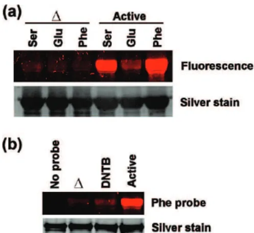
To further study and validate the probe labeling is specific to a wide range of enzyme classes, labeling experiments were performed with recombinant human PDI, an abundant chap-erone isomerase identified by our Ser, Thr and Phe substrate-based probes (Figure 2 and Supplemental Tables S1-S3). Analyses of activity with the serine, glutamate and phenylala-nine probes showed that PDI has substrate preferences. PDI strongly reacted with the Ser and Phe probes and was virtually not labeled by Glu (Figure 4a). These results correlate with the fluorescence 2D-GE analysis in hepatocytes, where PDI was not identified by the Glu probe, but was identified by the Ser and Phe probes (Figure 2 and Supplemental Tables S1, S3 and S4). Furthermore, the probe specific labeling of PDI isomerase was shown to be activity dependent, as heat denaturation (∆) and pretreatment with the PDI-specific inhibitor DNTB abolished the fluorescence signal (Figure 4b, Phe probe), while the protein levels remained constant (Figure 4b, silver stain). These results demonstrate that these probes can also interact specifically with
nonprotease enzymes in their active-state and are dependent on the probe structure.
Substrate-Based Activity Protein Profiling Changes dur-ing HCV Replication. To assess the protease and
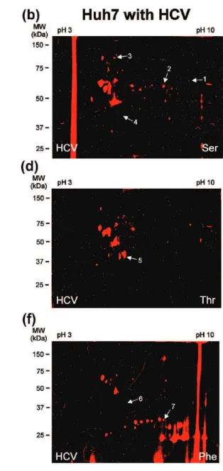
substrate-specific activities during HCV replication, we performed pro-tease substrate-based profiling on proteome extracts of naı¨ve Huh7 cells and a Huh7 cell line stably replicating the pFK-I389neo/NS3-3′/5.1 subgenomic HCV replicon35-41 at high levels (Figure 7a) and known to have distinct hepatic metabolic characteristics.42 Of the five substrate probes tested, three revealed some differential activity between the naı¨ve cell line and Huh7 highly replicating HCV (Figure 5). On the basis of the inclusion criteria described above, tandem mass spectro-metry identified seven proteins responsible for the differential
fluorescence signals observed during HCV replication (Supple-mental Table S6 and Figure 5g). The differential hits were mainly composed of hydrolases (including cathepsin D, an aspartic protease), as well as a couple chaperone proteins and an isomerase. Interestingly, the NS3/4A HCV protease was not detected by the probes.
The potential labeling of HCV NS3 protease was further examined in vitro using purified recombinant NS3/4A protease, which showed little or no reactivity with several substrate probes (Ser, Thr and Phe) (Figure 6c), when compared to PDI (Figure 4) or CES1 (Figure 7). Even the fluorophosphonate probe (FP), used as a positive control as it efficiently targets serine hydrolases,42yielded a very faint labeling with NS3/4A (Figure 6c). The low labeling efficiency of NS3/4A by ABPP
Figure 2.Substrate-based activity protein profiling in hepatoma Huh7 cells. The active proteome isolated from Huh7 cells was labeled
in vitrofor 1 h at 37 °C with 5 µM Phe (d) or 10 µM Ser (b), Thr (c), Glu (e) or His (f) probe. A denaturing preheating step of 100 °C for
5 min (∆) was performed as a control (a) to confirm the Ser probe specificity toward the active enzymes. The denatured blot in panel a is representative of the other four probes when exposed to the heat-denatured Huh7 proteome. After separation of the proteomes by two-dimensional gel electrophoresis, the fluorescently tagged proteins, indicated with arrows, were identified by LC-MS/MS (Supplemental Tables S1-S5, respectively). The other unmarked fluorescent spots were considered as nonspecific background as they did not meet the inclusion criteria of minimum two matching peptides, a MASCOT score above 100, a corresponding pI and molecular weight as well as being identified in at least two independent experiments.
probes could be attributed to several factors, such as its inherent reactivity, low abundance of NS3/4A relative to other host cell hydrolases or a product inhibition of NS3/4A.43
To examine if the fluorescence intensity difference observed during HCV replication could solely be attributed to differential enzymatic activity or could reflect differential gene expression, the expression level of the seven candidates influenced during HCV replication was analyzed by RT-PCR and Western blotting (Figure 6a,b). Both CES1 and NY-CO-25 activity levels (Figure 5g) and mRNA/protein levels (Figure 6a,b) were higher in Huh7 cells replicating HCV, thus, suggesting that the expression level of these genes is positively regulated in this cell line. Although the transcript levels of cathepsin D were observed to be similar (Figure 6a), the levels of inactive zymogen proprotein were
observed to be significantly higher and the active form of cathepsin D (50 kDa) was slightly lower in the HCV replicating cell line (Figure 6b), supporting the reduced fluorescence intensity observed from the phenylalanine probe (Figure 5e-g). The absence of any differential expression levels in the four remaining candidates (CCT4, PDI, PDIA6 and CGI-111) could be explained by the fact that these enzymes harbor a differential activity, while having unchanged protein levels. The zymogen form of these enzymes (such as the case for cathepsin D) or the HCV-mediated modulation of endogenous inhibitors/ activators could explain this observation.
We previously showed that CES1 harbored differential activ-ity with a fluorophosphonate (FP) activactiv-ity-based probe target-ing serine hydrolases.42 Known to exhibit a broad substrate specificity in the liver,44CES1 did show specific reactivity to the Ser probe (Figure 7b), supporting the 2D-GE LC-MS/MS analysis (Figure 5a,b and Supplemental Table 6) that substrate probes can specifically label a wider range of hydrolases besides proteases (Figure 3c). CES1 mRNA and protein levels measured by Northern and Western blotting (Figure 7a) further validated the differential expression of CES1 observed by RT-PCR in Huh7 cells replicating HCV (Figure 6a). The Ser probe labeling of CES1 was shown to be activity-dependent, as a predenaturation step (∆) or pretreatment with paraoxon, a serine hydrolase inhibitor, abolished the fluorescence signal, while the CES1 protein levels remained constant (Figure 7c). These results are consistent with the FP labeling of CES1, as previously re-ported.42These observations further demonstrate the applica-tion of ABPP substrate probes for screening the specificity and any potential off-target effects of novel enzyme inhibitors on the activity of complex cellular proteomes. The information can help to better understand proteome-drug interactions.
Discussion
We have previously reported the synthesis and characteriza-tion of a group of substrate-based quinolimine methide probes31,32and their usage in microarray-based activity-profil-Figure 3.Protein classes identified by substrate base activity
protein profiling during HCV replication. (a) Schematic repre-sentations of the HCV genome and the bicistronic (pFK-I389neo/ NS3-3′/5.1) subgenomic replicon used in this study (NS, non-structural proteins). (b) Labeled proteins identified by LC-MS/ MS were considered true substrate-based probe targets (54 proteins) in comparison to background proteome (150 proteins), based on how many identifications (minimum two) that were made with the following criteria: a matching of at least two peptides per protein, a MASCOT score above 100 and a molec-ular mass and pI corresponding to their migrating position during 2D gel electrophoresis. (c) Compilation of the 54 true probe targets from (b) and classification into their corresponding protein family (data from Supplemental Tables S1-S5).
Figure 4.Protein disulfide isomerase (PDI) reacts specifically to substrate probes in an activity-based manner. (a) Active recom-binant human PDI was treated with serine, glutamate and phenylalanine substrate-based probes (37 °C, 60 min) either with or without a denaturing preheating step (∆ ) 100 °C, 5 min), and subjected to SDS/PAGE electrophoresis and silver staining (as a loading control). (b) The phenylalanine probe specificity toward active PDI was analyzed with or without a denaturing preheating step (∆ ) 100 °C, 5 min) or a pretreatement with 0.7 mM dinitrothiocyanobenzene (DNTB) PDI inhibitor, followed by an incubation with 5 µM Phe probe for 1 h at 37 °C.
Figure 5.Substrate-based protein profiling during HCV replication identifies seven differentially active proteins. The active proteome isolated from naı¨ve Huh7 cells (a, c and e) or Huh7 stably expressing the HCV replicon (b, d and f) was labeled in vitro for 1 h at 37 °C with 10 µM Ser (a and b), Thr (c and d), His (not shown), Glu (not shown) probe or 5 µM Phe (e and f). After separation of the proteomes by two-dimensional gel electrophoresis, the fluorescently tagged proteins with differential activity, indicated with arrows, were identified by LC-MS/MS (Supplemental Table S6 and panel g). Gels labeled with the Glu and His probes are not shown due to the lack of differential activity (the labeling being identical to Figure 2e,f for both naı¨ve and HCV replicating Huh7 cells). (g) The differential activity of fluorescently labeled proteins between naı¨ve Huh7 cells and HCV replicating Huh7 cells was quantified with the ImageJ software (NIH) by measuring the fluorescence intensity of the corresponding protein spot (no. 1-7) tagged in panels a-f.
ing of proteases.32 These probes contain a reactive group composed of an N-acetylated amino acid that mimics the P1 position in a protease peptide substrate.31 Upon catalytic cleavage of the scissile peptide bond by the targeted enzyme, the probe generates a highly reactive quinolimine methide intermediate which reacts covalently with the protease, making its detection possible by fluorescence (Figure 8a). The unique-ness of these substrate-based probes is that their selectivity is not defined by the cleavage mechanism itself, but rather by the P1 subsite residue, making them capable of virtually targeting all classes of proteases. Herein, we demonstrate for the first time the efficacy of these substrate-based probes to target active enzymes in the complex proteome of Huh7 hepatoma cell line.
The five different substrate-based probes identified over 50 differentially active enzymes in the proteome of Huh7 cells. The four protease families (aspartic, threonine, serine and cysteine) that were positively identified by ABPP illustrate the high substrate selectivity of the probes. This approach appears to be suitable for profiling proteases that possess P1 site specificities, as the labeling pattern of the enzyme by the probe correlated with its known P1residue preferences. Cathepsin D,
who was strongly labeled by the phenylalanine probe, is known to prefer phenylalanine P1residues.45 The ubiquitin specific protease 14, the proteasome alpha 2 and beta 7 subunits and the endopeptidase Clp, known by their broad substrate speci-ficity46or preference toward hydrophobic residues,47,48were other phenylalanine probe targets. Cathepsin B favors nonbulky hydrophilic residues, such as arginine, at P1,49explaining its identification from the hydrophilic glutamate probe activity profile. These results support the previous in vitro character-ization of these probes and confirmed their capability of broadly targeting protease classes (serine, cysteine, metallo and aspartic) based on their P1preferences.31,32
Several other enzyme families were also targeted by the substrate-based probes, such as hydrolases (other than pro-teases), oxidoreductases, isomerase, chaperone, transferase and liase. These nonprotease hits could originate from either: (i) the substrate probes having a broader specificity than protease targets, (ii) sample contamination by background comigrating
Figure 6.Expression levels of some differentially active candi-dates are influenced under HCV replication. (a and b) Expression analysis of the seven differentially active protein candidates: chaperonin containing TCP1 (CCT4), carboxylesterase 1 (CES1), antigen NY-CO-25, protein disulfide isomerase-associated 6 (PDIA6), protein disulfide isomerase (PDI), cathepsin D (CTSD), isochorismatase domain containing 1 (CGI-111) (Figure 5 and Supplemental Table S6) was analyzed by RT-PCR (a) and Western blotting (b) in the naı¨ve Huh7 cell line and Huh7 stably expressing the bicistronic pFK-I389neo/NS3-3′/5.1 subgenomic replicon (HCV). (a) As a control to confirm the mRNA origin of the amplification, the cDNA was omitted in some samples (no cDNA). (b) β-Tubulin was used as a loading control for Western blotting. (c) The recombinant HCV NS3/4a protease produced and purified from
Escherichia coliwas tested for its probe reactivity at 37 °C for 60 min with fluorophosphonate (FP), serine, threonine and pheny-lalanine substrate-based probes either with or without a denatur-ing preheatdenatur-ing step (∆ ) 100 °C, 5 min), and subjected to SDS/ PAGE electrophoresis and silver staining.
Figure 7.Nonprotease carboxylesterase 1 enzyme is targeted by the substrate-based probes in an activity and substrate-prefer-ence based manner. (a) Northern analysis of CES1, HCV and β-actin (as a loading control) RNA (upper three blots) and detection of CES1, HCV NS3, HCV NS5A and β-tubulin (as a loading control) proteins with Ser probe labeling (upper blot) and Western blotting (lower four blots) in naı¨ve Huh7 human hepato-ma cell lines and in Huh7 cells stably replicating HCV. (b) Active proteome from Huh7 cells stably replicating HCV was treated with fluorophosphonate (FP), serine, threonine and phenylalanine substrate-based probes (37 °C, 60 min) either with or without a denaturing preheating step (∆ ) 100 °C, 5 min), and subjected to SDS/PAGE electrophoresis and CES1 Western blotting (WB) (as a loading control). (c) The serine probe specificity toward active CES1 was analyzed with or without a denaturing preheat-ing step (∆ ) 100 °C, 5 min) or a pretreatement with 60 µM of a serine hydrolase specific inhibitor (paraoxon), followed by an incubation with 10 nM FP probe as a positive control,42or 10
µM Ser probe for 1 h at 37 °C.
Figure 8.Proposed reaction mechanisms of the substrate based probes. (a) For proteases, the specific pocket (in blue) recognizes the unique P1amino acid (AA) of the substrate based probe. This translocates the probe reactive group to the enzyme active site
where a basic amino acid (B:) cleaves the scissile bond in proximity of the R lateral chain of P1generating a negatively charged
nitrogen group. Delocalization of the negative charge results in the release of the fluoride leaving group and generates a highly reactive electrophilic quinolimine methide intermediate. A nucleophile residue (Nu) within the enzyme active site attacks the electrophile quinolimine methide intermediate to covalently bind the probe to the enzyme. (b) With other enzymes, such as isomerases, the thiol-specific reactive group on cysteine attacks the scissile bond of the probe to form an acyl-enzyme intermediate. Delocalization of the negative charge releases the fluoride leaving group and creates a quinolimine methide intermediate with an electrophilic carbon that can be attacked either by a nucleophile residue (upper right panel) or by a second free thiol (lower right panel) generating a covalent bond with the enzyme. (c) Alternatively, for enzymes that used nucleophilic attack mechanisms, such as isomerases or hydrolases, the free thiol on cysteine residue or the hydroxyl on serine residue can directly attack the electropositive carbon of a reactive group that mimics the enzyme substrate.50
proteins after 2D-GE separation, or (iii) some of these enzymes have previously unknown or uncharacterized protease activity. The broader specificity of the substrate labeling could be explained by the substrate-based targeting nature of the probes instead of being mechanism-based like other ABPP probes.16 The selectivity of the cleavage reaction is defined by the P1 subsite residue having proper access to the catalytic site. Once properly fitted in the active site, the electrophilic carbon could be subject to attack by nucleophilic groups from other enzymes with active site nucleophiles, such as thiols from isomerases or hydroxyls from serine hydrolases (Figure 8b,c).50Although enzyme families other than proteases were detected by the probes, not all enzyme members of these families were active targets for these probes. For example, the serine hydrolase CES1 was positively targeted by the serine probe (Figure 7b), while other hydrolases, such as horseradish peroxidase (data not shown), alkaline phosphatase, lyzozyme and lipase,31,32did not show any reactivity toward the probes. This selectivity within nonprotease hydrolases could be attributed to unique proper-ties of the probe amino acyl P1lateral chain capable of specific ionic interactions with the enzyme’s catalytic pocket.
Probe specificity was further demonstrated with the isomerase PDI, which showed strong reactivity toward both the serine and phenylalanine probes, but virtually no reactivity against the glutamate probe (Figure 4a). Even with broader targets, the substrate-based probe labeling is nevertheless activity-based and appears to occur within the enzyme active site, as both heat denaturation and irreversible inhibitors (e.g., DNTB and paraoxon) abolished the covalent binding of the probe to the enzyme. This suggests that the nucleophile attack from the enzyme must originate from a catalytic residue and not from other nucleophile residues at the surface of the protein. These observations also eliminate prior speculations that the quino-limine methide intermediate may diffuse away from the protease active site and label neighboring proteins,16 as this intermediate might not even be produced under the new suggested nonprotease mechanisms (Figure 8c). Further evi-dence that the quinolimine methide intermediate diffusion and nonspecific binding to PDI is unlikely was confirmed in vitro with the purified 2D-GE identified enzyme target PDI. PDI showed strong probe reactivity, while no labeling was observed on the BSA carrier protein. BSA was previously shown not to be a substrate probe target.31 Although alternate labeling mechanisms are proposed (Figure 8b,c), further studies will be needed to provide a deeper insight into the labeling mechanism of nonprotease targets by the substrate probes.
To further increase the specificity of the probes toward their protease targets, polypeptide-based probes will likely be more selective compared to their amino acid-based counterparts.31 Background comigrating proteins could also be greatly reduced by employing MudPIT analysis18combined with a photocleav-able linker probe51instead of 2D-GE, although the topological protein information and high-throughput quantitative advan-tages of the 2D-GE technique would be sacrificed.
The unique substrate-targeting properties of these probes were used to provide a glimpse into the differential enzymatic activity during HCV replication. Seven enzymes were observed to be differentially active in the Huh7 cell line stably replicating the pFK-I389neo/NS3-3′/5.1 subgenomic replicon. Although, the HCV NS3/4A proteinase favors cysteine and threonine at the P1position of the cleavage site,52the protein could not be detected from the cell extract with the threonine probe, nor with the fluorophosphonate (FP) probe.42 Even purified
re-combinant NS3/4A showed poor reactivity toward ABPP probe labeling. The lack of ABPP detection of NS3/4A could be attributed to several factors, such as the low abundance of NS3/ 4A relative to other host cell hydrolases, low reactivity of NS3/ 4A for probes toward NS3/4A particularly with probes with a P1Thr residue compare to Cys,52 or a product inhibition of NS3/4A.43 Further analyses of the labeled candidates may suggest that some post-translational modifications are respon-sible for the differential activity of CTSD, as the levels of zymogen and active protein were shown to vary in HCV replication cells, whereas the transcript levels were found to be fairly constant. Similarly for CCT4, PDI, PDIA6 and CGI-111, post-translational modification or inhibitor/activator modu-lation by HCV could explain their differential activity as no changes were observed at the transcript and protein levels of these candidates. For CES1 and NY-CO-25, however, the differential activity observed likely originates from differential expression. These differentially active enzymes could either be attributed to HCV replication or could be cell line specific. The differential expression of CES1 in the pFK-I389neo/NS3-3′/5.1 HCV cell line was proven to be likely cell line specific with a fluorophosphonate ABPP probe targeting serine hydrolases.42 These observations with both mechanism-based (FP) and substrate-based probes further validate the usage of the latter in specifically targeting a broad range of enzymes to give a better understanding of the fundamental processes occurring during viral replication.
From the differentially active candidates identified during HCV replication, CES1 has been previously shown to have a specific role during HCV replication by influencing the cellular lipid metabolism.42 The differential activity of cathepsin D could link this protease to a few potential roles in HCV replicating Huh7 cells. Cathepsin D has been shown to act as a mitogen for cancer cells and stimulates their proinvasive and pro-metastatic properties, and therefore could be a result of the hepatoma origin of the Huh7 cell line.53By mainly being present in the lysosomes, Cathepsin D could have a similar role to cathepsin B and L for HCV. Cathepsin B and L were shown to activate the Ebola virus and other retroviruses during viral entry.54Although cathepsin B and L did not appear to be necessary for HCV low-pH-triggered entry,54to our knowledge, the role of cathepsin D during HCV entry or replication has not been studied so far. Similarly, the chaperone NY-CO-25 (i.e., HSP105) has also been shown to be overexpressed in cancer cells and in cells under severe stress (i.e., viral infection) and could help the pFK-I389neo/NS3-3′/5.1 cell line to cope with higher HCV viral titers.55Further characterization of these enzymes will be required to confirm their differential activities and to reveal their potential roles during HCV replication and infectivity.
Acknowledgment.
This work was supported by the Canadian Liver Foundation, the Canadian Institutes for Health Research and NRC’s Genomics and Health Initiative. We thank Dr. Ralf Bartenschlager for materials for replicon generation and Dr. John Kelly for mass spectrometry. The anti-CES1 serum was a kind gift from Dr. Lance Pohl.Supporting Information Available:
Supplemental Figure S1, substrate-based probe condition optimization for labeling the active proteomes from naı¨ve Huh7 cells and HCV replicon model. Supplemental Tables S1-S5, identification of labeled proteins with the five different substrate based probes. Supplemental Table S6, labeled protein candidates withferential activity during HCV replication. This material is available free of charge via the Internet at http://pubs.acs.org.
References
(1) Lavanchy, D. The global burden of hepatitis C. Liver Int. 2009, 29
Suppl 1, 74–81.
(2) Lindenbach, B. D.; Rice, C. M. Unravelling hepatitis C virus replication from genome to function. Nature 2005, 436 (7053), 933–8.
(3) Goffard, A.; Callens, N.; Bartosch, B.; Wychowski, C.; Cosset, F. L.; Montpellier, C.; Dubuisson, J. Role of N-linked glycans in the functions of hepatitis C virus envelope glycoproteins. J. Virol. 2005,
79 (13), 8400–9.
(4) Reed, K. E.; Xu, J.; Rice, C. M. Phosphorylation of the hepatitis C virus NS5A protein in vitro and in vivo: properties of the NS5A-associated kinase. J. Virol. 1997, 71 (10), 7187–97.
(5) Wang, C.; Gale, M., Jr.; Keller, B. C.; Huang, H.; Brown, M. S.; Goldstein, J. L.; Ye, J. Identification of FBL2 as a geranylgeranylated cellular protein required for hepatitis C virus RNA replication. Mol.
Cell 2005, 18 (4), 425–34.
(6) Jopling, C. L.; Yi, M.; Lancaster, A. M.; Lemon, S. M.; Sarnow, P. Modulation of hepatitis C virus RNA abundance by a liver-specific MicroRNA. Science 2005, 309 (5740), 1577–81.
(7) Jopling, C. L.; Schutz, S.; Sarnow, P. Position-dependent function for a tandem microRNA miR-122-binding site located in the hepatitis C virus RNA genome. Cell Host Microbe 2008, 4 (1), 77– 85.
(8) Hijikata, M.; Kato, N.; Ootsuyama, Y.; Nakagawa, M.; Shimotohno, K. Gene mapping of the putative structural region of the hepatitis C virus genome by in vitro processing analysis. Proc. Natl. Acad.
Sci. U.S.A. 1991, 88 (13), 5547–51.
(9) Hussy, P.; Langen, H.; Mous, J.; Jacobsen, H. Hepatitis C virus core protein: carboxy-terminal boundaries of two processed species suggest cleavage by a signal peptide peptidase. Virology 1996, 224 (1), 93–104.
(10) Moradpour, D.; Penin, F.; Rice, C. M. Replication of hepatitis C virus. Nat. Rev. Microbiol. 2007, 5 (6), 453–63.
(11) Okamoto, K.; Mori, Y.; Komoda, Y.; Okamoto, T.; Okochi, M.; Takeda, M.; Suzuki, T.; Moriishi, K.; Matsuura, Y. Intramembrane processing by signal peptide peptidase regulates the membrane localization of hepatitis C virus core protein and viral propagation.
J. Virol. 2008, 82 (17), 8349–61.
(12) Bartenschlager, R.; Ahlborn-Laake, L.; Mous, J.; Jacobsen, H. Nonstructural protein 3 of the hepatitis C virus encodes a serine-type proteinase required for cleavage at the NS3/4 and NS4/5 junctions. J. Virol. 1993, 67 (7), 3835–44.
(13) Hijikata, M.; Mizushima, H.; Akagi, T.; Mori, S.; Kakiuchi, N.; Kato, N.; Tanaka, T.; Kimura, K.; Shimotohno, K. Two distinct proteinase activities required for the processing of a putative nonstructural precursor protein of hepatitis C virus. J. Virol. 1993, 67 (8), 4665– 75.
(14) Lin, C.; Lindenbach, B. D.; Pragai, B. M.; McCourt, D. W.; Rice, C. M. Processing in the hepatitis C virus E2-NS2 region: identifica-tion of p7 and two distinct E2-specific products with different C termini. J. Virol. 1994, 68 (8), 5063–73.
(15) Puente, X. S.; Sanchez, L. M.; Overall, C. M.; Lopez-Otin, C. Human and mouse proteases: a comparative genomic approach. Nat. Rev.
Genet. 2003, 4 (7), 544–58.
(16) Evans, M. J.; Cravatt, B. F. Mechanism-based profiling of enzyme families. Chem. Rev. 2006, 106 (8), 3279–301.
(17) Kocks, C.; Maehr, R.; Overkleeft, H. S.; Wang, E. W.; Iyer, L. K.; Lennon-Dumenil, A. M.; Ploegh, H. L.; Kessler, B. M. Functional proteomics of the active cysteine protease content in Drosophila S2 cells. Mol. Cell. Proteomics 2003, 2 (11), 1188–97.
(18) Kaschani, F.; Gu, C.; Niessen, S.; Hoover, H.; Cravatt, B. F.; van der Hoorn, R. A. Diversity of serine hydrolase activities of unchal-lenged and botrytis-infected Arabidopsis thaliana. Mol. Cell.
Proteomics 2009, 8 (5), 1082–93.
(19) Barglow, K. T.; Cravatt, B. F. Activity-based protein profiling for the functional annotation of enzymes. Nat. Methods 2007, 4 (10), 822–7.
(20) Fonovic, M.; Bogyo, M. Activity-based probes as a tool for functional proteomic analysis of proteases. Expert Rev. Proteomic
2008, 5 (5), 721–30.
(21) Uttamchandani, M.; Li, J.; Sun, H.; Yao, S. Q. Activity-based protein profiling: new developments and directions in functional pro-teomics. ChemBioChem 2008, 9 (5), 667–75.
(22) Liu, Y.; Patricelli, M. P.; Cravatt, B. F. Activity-based protein profiling: the serine hydrolases. Proc. Natl. Acad. Sci. U.S.A. 1999,
96 (26), 14694–9.
(23) Marnett, A. B.; Nomura, A. M.; Shimba, N.; Ortiz de Montellano, P. R.; Craik, C. S. Communication between the active sites and dimer interface of a herpesvirus protease revealed by a transi-tion-state inhibitor. Proc. Natl. Acad. Sci. U.S.A. 2004, 101 (18), 6870–5.
(24) Kato, D.; Boatright, K. M.; Berger, A. B.; Nazif, T.; Blum, G.; Ryan, C.; Chehade, K. A. H.; Salvesen, G. S.; Bogyo, M. Activity-based probes that target diverse cysteine protease families. Nat. Chem.
Biol. 2005, 1 (1), 33–8.
(25) Chattopadhaya, S.; Chan, E. W. S.; Yao, S. Q. An affinity-based probe for the proteomic profiling of aspartic proteases.
Tetrahe-dron Lett. 2005, 46, 4053–6.
(26) Chan, E. W.; Chattopadhaya, S.; Panicker, R. C.; Huang, X.; Yao, S. Q. Developing photoactive affinity probes for proteomic profil-ing: hydroxamate-based probes for metalloproteases. J. Am. Chem.
Soc. 2004, 126 (44), 14435–46.
(27) Sieber, S. A.; Niessen, S.; Hoover, H. S.; Cravatt, B. F. Proteomic profiling of metalloprotease activities with cocktails of active-site probes. Nat. Chem. Biol. 2006, 2 (5), 274–81.
(28) Adam, G. C.; Cravatt, B. F.; Sorensen, E. J. Profiling the specific reactivity of the proteome with non-directed activity-based probes.
Chem. Biol. 2001, 8 (1), 81–95.
(29) Barglow, K. T.; Cravatt, B. F. Discovering disease-associated enzymes by proteome reactivity profiling. Chem. Biol. 2004, 11 (11), 1523–31.
(30) Adam, G. C.; Burbaum, J.; Kozarich, J. W.; Patricelli, M. P.; Cravatt, B. F. Mapping enzyme active sites in complex proteomes. J. Am.
Chem. Soc. 2004, 126 (5), 1363–8.
(31) Zhu, Q.; Girish, A.; Chattopadhaya, S.; Yao, S. Q. Developing novel activity-based fluorescent probes that target different classes of proteases. Chem. Commun. 2004, 7 (13), 1512–3.
(32) Srinivasan, R.; Huang, X.; Ng, S. L.; Yao, S. Q. Activity-based fingerprinting of proteases. ChemBioChem 2006, 7 (1), 32–6. (33) Taremi, S. S.; Beyer, B.; Maher, M.; Yao, N.; Prosise, W.; Weber,
P. C.; Malcolm, B. A. Construction, expression, and characteriza-tion of a novel fully activated recombinant single-chain hepatitis C virus protease. Protein Sci. 1998, 7 (10), 2143–9.
(34) Satoh, H.; Martin, B. M.; Schulick, A. H.; Christ, D. D.; Kenna, J. G.; Pohl, L. R. Human anti-endoplasmic reticulum antibodies in sera of patients with halothane-induced hepatitis are directed against a trifluoroacetylated carboxylesterase. Proc. Natl. Acad. Sci. U.S.A.
1989, 86 (1), 322–6.
(35) Lohmann, V.; Korner, F.; Koch, J.; Herian, U.; Theilmann, L.; Bartenschlager, R. Replication of subgenomic hepatitis C virus RNAs in a hepatoma cell line. Science 1999, 285 (5424), 110–3. (36) Supekova, L.; Supek, F.; Lee, J.; Chen, S.; Gray, N.; Pezacki, J. P.;
Schlapbach, A.; Schultz, P. G. Identification of human kinases involved in hepatitis C virus replication by small interference RNA library screening. J. Biol. Chem. 2008, 283 (1), 29–36.
(37) Rakic, B.; Clarke, J.; Tremblay, T. L.; Taylor, J.; Schreiber, K.; Nelson, K. M.; Abrams, S. R.; Pezacki, J. P. A small-molecule probe for hepatitis C virus replication that blocks protein folding. Chem. Biol.
2006, 13 (10), 1051–60.
(38) Rakic, B.; Brulotte, M.; Rouleau, Y.; Belanger, S.; Pezacki, J. P. Bleomycin is a potent small-molecule inhibitor of hepatitis C virus replication. ChemBioChem 2006, 7 (9), 1330–3.
(39) Tonary, A. M.; Pezacki, J. P. Simultaneous quantitative measure-ment of luciferase reporter activity and cell number in two- and three-dimensional cultures of hepatitis C virus replicons. Anal.
Biochem. 2006, 350 (2), 239–48.
(40) Sagan, S. M.; Rouleau, Y.; Leggiadro, C.; Supekova, L.; Schultz, P. G.; Su, A. I.; Pezacki, J. P. The influence of cholesterol and lipid metabolism on host cell structure and hepatitis C virus replication.
Biochem. Cell. Biol. 2006, 84 (1), 67–79.
(41) Rakic, B.; Sagan, S. M.; Noestheden, M.; Belanger, S.; Nan, X. L.; Evans, C. L.; Xie, X. S.; Pezacki, J. P. Peroxisome proliferator-activated receptor alpha antagonism inhibits hepatitis C virus replication. Chem. Biol. 2006, 13 (1), 23–30.
(42) Blais, D. R.; Lyn, R. K.; Joyce, M.; Rouleau, Y.; Pegoraro, A.; Stolow, A.; Tyrrell, D. L.; Pezacki, J. P., Carboxylesterase 1 enzyme on lipid droplets aids hepatitis C virus propagation. Submitted for publica-tion, 2009.
(43) Steinkuhler, C.; Biasiol, G.; Brunetti, M.; Urbani, A.; Koch, U.; Cortese, R.; Pessi, A.; De Francesco, R. Product inhibition of the hepatitis C virus NS3 protease. Biochemistry 1998, 37 (25), 8899– 905.
(44) Bencharit, S.; Edwards, C. C.; Morton, C. L.; Howard-Williams, E. L.; Kuhn, P.; Potter, P. M.; Redinbo, M. R. Multisite promiscuity in the processing of endogenous substrates by human carboxy-lesterase 1. J. Mol. Biol. 2006, 363 (1), 201–14.
(45) Scarborough, P. E.; Guruprasad, K.; Topham, C.; Richo, G. R.; Conner, G. E.; Blundell, T. L.; Dunn, B. M. Exploration of subsite binding specificity of human cathepsin D through kinetics and rule-based molecular modeling. Protein Sci. 1993, 2 (2), 264–76. (46) Groll, M.; Huber, R. Substrate access and processing by the 20S proteasome core particle. Int. J. Biochem. Cell Biol. 2003, 35 (5), 606–16.
(47) Hu, M.; Li, P.; Song, L.; Jeffrey, P. D.; Chenova, T. A.; Wilkinson, K. D.; Cohen, R. E.; Shi, Y. Structure and mechanisms of the proteasome-associated deubiquitinating enzyme USP14. EMBO J.
2005, 24 (21), 3747–56.
(48) Kang, S. G.; Ortega, J.; Singh, S. K.; Wang, N.; Huang, N. N.; Steven, A. C.; Maurizi, M. R. Functional proteolytic complexes of the human mitochondrial ATP-dependent protease, hClpXP. J. Biol.
Chem. 2002, 277 (23), 21095–102.
(49) Musil, D.; Zucic, D.; Turk, D.; Engh, R. A.; Mayr, I.; Huber, R.; Popovic, T.; Turk, V.; Towatari, T.; Katunuma, N.; Bode, W. The refined 2.15-A X-Ray crystal-structure of human liver cathepsin B - The structural basis for its specificity. EMBO J. 1991, 10 (9), 2321– 2330.
(50) Jeffery, D. A.; Bogyo, M. Chemical proteomics and its application to drug discovery. Curr. Opin. Biotechnol. 2003, 14 (1), 87–95. (51) Fonovic, M.; Verhelst, S. H. L.; Sorum, M. T.; Bogyo, M. Proteomics
evaluation of chemically cleavable activity-based probes. Mol. Cell.
Proteomics 2007, 6 (10), 1761–70.
(52) Bartenschlager, R.; Ahlborn-Laake, L.; Yasargil, K.; Mous, J.; Jacobsen, H. Substrate determinants for cleavage in cis and in trans by the hepatitis C virus NS3 proteinase. J. Virol. 1995, 69 (1), 198– 205.
(53) Benes, P.; Vetvicka, V.; Fusek, M. Cathepsin D-Many functions of one aspartic protease. Crit. Rev. Oncol. Hemat. 2008, 68 (1), 12– 28.
(54) Tscherne, D. M.; Jones, C. T.; Evans, M. J.; Lindenbach, B. D.; McKeating, J. A.; Rice, C. M. Time- and temperature-dependent activation of hepatitis C virus for low-pH-triggered entry. J. Virol.
2006, 80 (4), 1734–41.
(55) Saito, Y.; Yamagishi, N.; Hatayama, T. Nuclear localization mech-anism of Hsp105 and its possible function in mammalian cells.
J. Biochem. 2009, 145 (2), 185–191.
PR900788A