HAL Id: tel-02917932
https://tel.archives-ouvertes.fr/tel-02917932
Submitted on 20 Aug 2020HAL is a multi-disciplinary open access archive for the deposit and dissemination of sci-entific research documents, whether they are pub-lished or not. The documents may come from teaching and research institutions in France or abroad, or from public or private research centers.
L’archive ouverte pluridisciplinaire HAL, est destinée au dépôt et à la diffusion de documents scientifiques de niveau recherche, publiés ou non, émanant des établissements d’enseignement et de recherche français ou étrangers, des laboratoires publics ou privés.
Exploring Transcriptional Heterogeneity in the
Postnatal SVZ
Stefan Zweifel
To cite this version:
Stefan Zweifel. Exploring Transcriptional Heterogeneity in the Postnatal SVZ. Neurobiology. Univer-sité de Lyon, 2018. English. �NNT : 2018LYSE1047�. �tel-02917932�
N°d’ordre NNT: 2018LYSE1047
THESE de DOCTORAT DE L’UNIVERSITE DE LYON
opérée au sein de
l’Université Claude Bernard Lyon 1 Ecole DoctoraleN° ED 340
Biologie Moléculaire Intégrative et Cellulaire
Spécialité de doctorat: Biology Discipline: Neurosciences
Soutenue publiquement le 29/03/2018, par:
Stefan ZWEIFEL
Exploring Transcriptional Heterogeneity in the
Postnatal SVZ
Devant le jury composé de :
Bénédicte Durand Professeur Université Lyon 1 Présidente Nathalie Spassky Directrice de recherche Université Paris VI Rapporteure Antoine De Chevigny Chercheur Université Aix-Marseille Rapporteur Olivier Raineteau Directeur de recherche Université Lyon 1 Directeur de thèse
UNIVERSITE CLAUDE BERNARD - LYON 1
Président de l’Université M. le Professeur Frédéric FLEURY Président du Conseil Académique M. le Professeur Hamda BEN HADID Vice-président du Conseil d’Administration M. le Professeur Didier REVEL Vice-président du Conseil Formation et Vie
Universitaire
M. le Professeur Philippe CHEVALIER Vice-président de la Commission Recherche M. Fabrice VALLÉE
Directrice Générale des Services Mme Dominique MARCHAND
COMPOSANTES SANTE
Faculté de Médecine Lyon Est – Claude Bernard Directeur : M. le Professeur G.RODE Faculté de Médecine et de Maïeutique Lyon Sud –
Charles Mérieux
Directrice : Mme la Professeure C. BURILLON Faculté d’Odontologie Directeur : M. le Professeur D. BOURGEOIS Institut des Sciences Pharmaceutiques et Biologiques Directrice : Mme la Professeure C.
VINCIGUERRA
Institut des Sciences et Techniques de la Réadaptation Directeur : M. X. PERROT Département de formation et Centre de Recherche en
Biologie Humaine
Directrice : Mme la Professeure A-M. SCHOTT
COMPOSANTES ET DEPARTEMENTS DE SCIENCES ET
TECHNOLOGIE
Faculté des Sciences et Technologies Directeur : M. F. DE MARCHI
Département Biologie Directeur : M. le Professeur F. THEVENARD Département Chimie Biochimie Directrice : Mme C. FELIX
Département GEP Directeur : M. Hassan HAMMOURI
Département Informatique Directeur : M. le Professeur S. AKKOUCHE Département Mathématiques Directeur : M. le Professeur G. TOMANOV Département Mécanique Directeur : M. le Professeur H. BEN HADID Département Physique Directeur : M. le Professeur J-C PLENET UFR Sciences et Techniques des Activités Physiques
et Sportives
Directeur : M. Y.VANPOULLE Observatoire des Sciences de l’Univers de Lyon Directeur : M. B. GUIDERDONI Polytech Lyon Directeur : M. le Professeur E.PERRIN Ecole Supérieure de Chimie Physique Electronique Directeur : M. G. PIGNAULT
Institut Universitaire de Technologie de Lyon 1 Directeur : M. le Professeur C. VITON
Ecole Supérieure du Professorat et de l’Education Directeur : M. le Professeur A. MOUGNIOTTE Institut de Science Financière et d'Assurances Directeur : M. N. LEBOISNE
“Life is a Sisyphean race, run ever faster toward a finish line that is merely the start of the next race”
Matt Ridley, The Red Queen: Sex and the Evolution of Human Nature
“Success is peace of mind, which is a direct result of
self-satisfaction in knowing you made the effort to become the best of which you are capable.”
John Robert Wooden, 10 times NCAA Basketball Champion Coach
Abstract
Germinal activity persists in the postnatal mammalian brain in specialized niches, namely the dentate gyrus of the hippocampus and the subventricular zone (SVZ) surrounding the lateral ventricle. Neural stem cells (NSCs) of the postnatal SVZ differentiate into transient amplifying progenitors that will generate neuroblasts migrating through the rostral migratory stream, into the olfactory bulb, where they differentiate into neurons. The SVZ additionally generates glial progenitors that invade the nearby parenchyma. Recent work to which I have participated, highlights the heterogeneous nature of the postnatal SVZ in respect to different microdomains generating distinct neural lineages.
The objectives of my PhD work were twice: 1) to develop new means to explore the heterogeneity of the SVZ; and 2) to identify transcription factors expressed by subpopulations of NSCs of the SVZ and acting in their differential specification.
Objective 1: The SVZ is a highly complex and irregular region of ongoing postnatal germinal
activity. The heterogeneous character of the SVZ is evident and recent studies generated enormous datasets of transcripts, which are differentially expressed between divergent microdomains. However, an appropriate tool for fast analysis of the protein level along the full rostro-caudal and dorso-ventral extend of the SVZ is still missing. Therefore, I developed “FlashMap”, a semi-automatic software that allows rapid analysis of protein levels in the full SVZ, based on optical density measurements after immunohistochemistry. “FlashMap” generates easy readable heatmaps in two dimensions, which can be accurately superimposed on three-dimensional reconstructions of the ventricular system for rapid spatial visualization and analysis. This new approach will fasten research onto SVZ regionalization, by guiding the identification of markers, such as transcription factors expressed in specific SVZ microdomains.
Objective 2: I used transcriptomic as well as fate mapping approaches to investigate the
relation between regional expression of transcription factors by NSCs and their acquisition of distinct neural lineage fates. Our results support an early priming of NSCs to produce defined
cell types depending of their spatial location in the SVZ and identify Hopx as a marker of a subpopulation biased to generate astrocytes. Interestingly, manipulation of Hopx expression showed minor effects on astrogenesis, but resulted in marked changes in the number of NSCs and of their progenies. Taken together, our results highlight transcriptional and spatial
heterogeneity of postnatal NSCs, as well as their early priming toward specific lineages and suggest a role for Hopx in the evolution of SVZ germinal activity.
Résumé
Une activité activité germinale persiste après la naissance dans des niches spécialisées du cerveau des mammifères, à savoir le gyrus denté de l'hippocampe et la zone sous-ventriculaire (SVZ) bordant le ventricule latéral. Les cellules souches neurales (NSC) de la SVZ postnatale se différencient en progéniteurs transitoires qui vont générer des neuroblastes migrant à travers la voie de migration rostrale vers le bulbe olfactif, où ils se différencient en neurones. La SVZ génère également des progéniteurs gliaux qui se dispersent dans le parenchyme voisin. Les travaux récents auxquels j'ai participé soulignent la nature hétérogène de la SVZ postnatale, composée de différents microdomaines générant des lignées neurales distinctes.
Les objectifs de mon travail de thèse ont permis de : 1) développer de nouveaux moyens pour explorer l'hétérogénéité de la SVZ; et 2) d’identifier et d’étudier le rôle d’un facteur de transcription exprimé par une sous population des NSCs de la SVZ.
Objectif 1: La SVZ est une région hautement complexe et irrégulière dans laquelle une forte
activité germinale persiste après la naissance. Le caractère hétérogène de la SVZ est évident et des études récentes ont généré une très grande base de données de transcrits, qui sont différentiellement exprimés entre les microdomaines. Cependant, un outil approprié pour l'analyse rapide du niveau d’expression d’une protéine d’intérêt, le long des axes rostro-caudal et dorso-ventral de la SVZ est toujours manquant et nécessaire. Par conséquent, j’ai développé "FlashMap", un logiciel semi-automatique qui permet une analyse rapide des niveaux d’expression de protéines dans le SVZ, basé sur des mesures de densité optique après immunohistochimie. "FlashMap" génère des cartes thermiques facilement lisibles en deux dimensions, qui peuvent être superposées avec précision aux reconstructions tridimensionnelles du système ventriculaire pour une visualisation spatiale fine et rapide. Cette nouvelle approche accélérera la recherche sur la régionalisation de la SVZ, en permettant l'identification de marqueurs (e.g. facteurs de transcription) exprimés dans des régions discrètes de la SVZ.
Objectif 2: J’ai utilisé des approches de transcriptomique et de « fate mapping » des NSCs
pour étudier la relation entre l'expression régionale de facteurs de transcription et leur différenciation dans des lignées neurales distinctes. Mes résultats supportent un amorçage précoce des NSCs à produire différents types cellulaires en fonction de leur localisation spatiale dans la SVZ. Nos données identifient Hopx comme un marqueur d'une sous population de NSCs qui génère principalement des astrocytes. De façon intéressante, la manipulation de l'expression de Hopx montre des effets mineurs sur l'astrogénèse, mais entraîne des changements marqués quant au nombre de NSCs et de leur descendance. Dans son ensemble, Mes résultats mettent en évidence à la fois une hétérogénéité spatiale des NSCs postnatales ainsi que leur amorçage précoce à produire des types cellulaires distincts.
List of Abbreviations (Abbr.)
2D 2-dimensional
3D 3-dimensional
Aldh1l1 aldehyde dehydrogenase 1 family member L1
BF1 brain factor 1
BrdU 5-bromo-2'-deoxyuridin
CalB calbindin
CalR calretinin
CC corpus callosum
cKO conditional knock-out
CLoNE clonal labeling of neural progenies
CNS central nervous system
CSF cerebrospinal fluid
Ctrl. control
Cx cortex
DCX doublecortin
dEPO dorsal electroporation
DG dentate gyrus
DL dorso-lateral corner
dlEPO dorso-lateral electroporation
dlNSC dorso-lateral neural stem cell
dlSVZ dorso-lateral subventricular zone microdomain
DM dorso-medial corner
dmEPO dorso-medial electroporation
dmNSC dorso-medial neural stem cell
dmSVZ dorso-medial subventricular zone microdomain
dNSC dorsal neural stem cell
dpe days post electroporation
dSVZ dorsal subventricular zone microdomain
E11 embryonic day 11
EPO electroporation
FACS fluorescence activated cell sorting
GCL granule cell layer
GFAP glial fibrillary acidic protein
GFP green fluoresent protein
GL glomerular layer
GoF gain of function
Hopx hpe
Homeodomain-only protein hours post electroporation
JSdiv Jensen-Shannon divergence
KI knock-in
LGE lateral ganglionic eminence
lNSC lateral neural stem cell
LoF loss of function
lSVZ lateral subventricular zone microdomain
LV lateral ventricle
MADM mosaic analysis with double markers
MAGIC multiaddressable genome-integrative color
MAP2 microtubule-associated protein-2
MCL mitral cell layer
MGE medial ganglionic eminence
mSVZ medial subventricular zone microdomain
NeuN neuronal nuclei
NSC neural stem cell
NSCs Neural stem cells
NSE neuron specific enolase
OB olfactory bulb
OD optical density
Olig1/2 oligodendrocyte transcription factor 1/2
OPC oligodendrocyte precursor cell
oRG outre radial glia
P3 postnatal day 3
PSA-NCAM polysialylated-neural cell adhesion molecule
qPCR quantitative polymerase chain reaction
RG radial glia
RMS rostral migratory stream
ROI region of interest
S100β s100 calcium-binding protein B
SGZ subgranular zone
St striatum
SVZ subventricular zone
Tam tamoxifen
TAP transient amplifying progenitor
Tbr2 T-box brain protein 2
TF transcription factor
TH tyrosine hydroxylase
TOAD-64 Turned-On-After-Division 64-kDA
Contents
1. Introduction – Technique Dependent Progression in the Field of
Neuroscience………... 1
1.1. Nucleoside Analogs Shaped our Understanding of Germinal Regions……….... 1
1.1.1. Nucleoside Analogs………... 2
1.1.2. Nucleoside Analogs Give Insight into Embryonic Germinal Activity by Interkinetic Nuclear Migration……….... 6
1.1.3. Nucleoside Analogs reveal Two Sites of persisting Postnatal Germinal Activity……… 7
1.1.4. Nucleoside Analogs Give Insights into the Migration Patterns of Cells Generated at Embryonic and Postnatal Times………... 11
1.1.5. The SVZ Has a Complex Cytoarchitecture and Consists of Different Cell Types with Distinct Cycling Behaviors………... 15
1.2. Transgenesis Revealed Early Regionalization of Germinal Regions……… 17
1.2.1. The Development of Transgenesis Approaches Represents a Further Milestone in the History of Neuroscience………... 18
1.2.2. Expression Analyses and Fate Mapping Approaches Highlight the Regionalized Organization of the VZ/SVZ………. 21
Regionalization is Observed During the Period of Neurogenesis………... 21
Regionalization is Observed During the Period of Gliogenesis………….... 24
Regionalization of the SVZ is Retained After Birth………. 25
1.2.3. Function of Regionally Enriched TFs in Lineage Specification………. 28
Null Mutants Reveal the Importance of Single Regionalized Genes in Forebrain Development………... 28
Advanced Transgenic Approaches Allow more Specific Gene Manipulations……….... 31
1.3. Advanced Approaches to Explore the Transcriptional Correlates of Postnatal Heterogeneity……….... 32
1.3.1. Electroporation Allows Efficient Targeting of Different SVZ Microdomains……….. 32
1.3.2. Large Scale Transcriptional Profiling Reveals the Full Extent of Postnatal SVZ Heterogeneity……….. 35
1.4. Objectives of the PhD Thesis……….... 35
2. Chapter 1: “FlashMap” - A Fast and Semi-Automatic Tool for Accurate Spatial Analysis of Protein Expression in the Subventricular Zone………….. 37
2.1. Abstract………. 37
2.2. Introduction………... 38
2.3. Experimental Procedures………... 39
2.3.1. Animals and Ethics……….. 39
2.3.2. Tissue Processing and Immunohistochemistry………... 40
2.3.3. Image Acquisition and 3D Reconstruction……….. 40
2.3.4. Software Generation and Analysis……….. 41
2.4.1. The Subventricular Zone is a Poorly Defined Region of the Postnatal
Forebrain……….. 41
2.4.2. “FlashMap” Allows Rapid Investigation of Gene Expression along the Full Extent of the SVZ………... 43
2.4.3. Step by Step Walkthrough for an Optimal Use of “FlashMap”……….. 45
2.4.4. “FlashMapping” of the Full SVZ Highlights Regions of Maximal Germinal Activity………... 49
2.4.5. Subsampling of the Region of Interest Results in a Minimal Loss of Spatial Information……….. 52
2.4.6. “FlashMap” Allows a Rapid Analysis of SVZ Transcriptional Heterogeneity……….. 54
2.5. Discussion………. 57
2.6. Acknowledgments………. 59
2.7. Supplementary Figures……….. 60
3. Chapter 2: Hopx Defines Heterogeneity of Postnatal Subventricular Zone Neural Stem Cells……….... 65
3.1. Abstract………. 65
3.2. Introduction………... 66
3.3. Experimental Procedures………... 67
3.3.1. Animals and Ethics……….. 67
3.3.2. Plasmids Preparation and Electroporation………... 67
3.3.3. Immunohistochemistry……….... 68
3.3.4. FACsorting and qPCR……….... 68
3.3.5. Meta-analysis of Transcriptional Profiles………... 70
3.3.6. Quantifications and Statistics……….. 70
3.4. Results………... 71
3.4.1. Hopx is Enriched in NSCs of the dSVZ, as well as in the Astrocytic Lineage……….... 71
3.4.2. Hopx Expression Reveals Intraregional Heterogeneity within the dSVZ…... 74
3.4.3. Hopx Defines Dorsal SVZ Microdomains with Distinct Lineage Outputs... 76
3.4.4. Hopx Expressing NSCs are Biased to Acquire an Astroglial Fate………….. 79
3.4.5. Expression of Hopx is Partly Dispensable During Astrogenesis………....… 81
3.4.6. Hopx Overexpression Regulates the Appearance of Outer Radial Glial Cells………... 83
3.5. Discussion………. 85
3.6. Acknowledgments………. 88
3.7. Supplementary Figures……….. 90
4. General Discussion……….. 97
4.1. Summary and Open Questions……….. 97
4.2. The Potency of Postnatal NSCs: From a Population of Multipotent NSCs to Populations of Restricted NSCs……….... 98
4.2.1. Drosophila Neuroblasts as a Model of Progressive Restriction……….. 98
4.2.2. In Vitro and In Vivo Evidences Suggest that Cortical Progenitors Progressively Lose Their Potency to Produce Early Born Neurons………… 99
4.3. Clonal Approaches in Histology………... 101
4.3.1. Sparse Labeling as a First Approach for Clonal Analyses……….. 101
4.3.2. Bar Coding of Progenitors Exhibits a Subpopulation of Embryonal NSCs Entering Quiescence for Postnatal Preservation……….. 103
4.4. Development of Multicolor Approaches for High Throughput Clonal Analysis.. 104
4.4.1. Brainbow Inaugurated the Era of Multicolor Clonal Approaches…………... 104
4.4.2. StarTrack Gives Insights into Restricted Potency of NSC Populations…….. 105
4.4.3. CLoNE – an Inducible Multicolor Approach……….. 108
4.5. Clonal Approaches in Transcriptomic………... 108
4.5.1. Bulk Analysis Reveals Transcriptional Specificities of Postnatal Germinal Regions……… 109
4.5.2. Single Cells Analysis Refines Current Transcriptional Knowledge………... 110
4.6. Perspectives – Push Technical Advances Further……….……… 111
4.6.1. “FlashMap” Evolves into “FlashMap 2.0”……….. 111
4.6.2. Hopx as a First Marker of a Subpopulation of Committed NSCs…………... 112
4.6.3. Exploring NSCs Heterogeneity Beyond Hopx……… 115
5. Bibliography……… 117
6. List of Publications………. 133
1
1. Introduction – Technique Dependent Progression in the
Field of Neuroscience
The central nervous system (CNS) is a very complex, highly organized structure. It is composed of three major cell types, the neurons and the glia cells (i.e. astrocytes and the oligodendrocytes). It was originally proposed by Rudolf Virchow (1846) that glia had a mesenchymal origin, like other supporter cells in the body. This hypothesis was later dismissed by Wilhelm, who demonstrated a CNS origin for glial cells (summarized in Jacobson, 1991). It is now well accepted that both neurons and glial cells originate from so called “neural stem cells” (NSCs). It also became apparent that their production does not stop at birth, but that a germinal activity persists in the postnatal CNS.
1.1. Nucleoside Analogs Shaped our Understanding of Germinal
Regions
In this first part of my introduction, I would like to focus on how the use of nucleoside analogs shaped our current understanding of a persistent germinal activity in the postnatal brain. Prior to the “nucleoside analog age”, the identification of mitotic activity relied on pure observations of cell morphology using ancient staining techniques (e.g. by the use of thionin followed by eosin or erythrosin). Using these approaches, mitotic features in the CNS of rats, were reported as early as in 1912, by Ezra Allen (Allen, 1912). While many of those early observations have greatly influenced the emerging field of neurosciences, the introduction of nucleoside analogs to study proliferation represents a major step forward. Indeed, these new techniques allowed proving or rejecting early made hypotheses in a convincing manner and opened the era towards modern neuroscience.
Nucleoside analogs shaped our understanding of niches with postnatally persisting germinal activity. In addition, because they allow fate mapping of cells that have proliferated at the time of nucleoside administration, they can give insights into embryonal and postnatal migration patterns. Further, they helped to make conclusions about the subventricular zone (SVZ) architecture when combined with immunohistochemistry and electron microscopy. While (especially in the 90’s) many researchers added significant knowledge to the field using more
2
advanced techniques (e.g. virus approaches), this part of my thesis introduction focuses mainly on results achieved using nucleoside analogs in combination with cell specific markers. Most of the results discussed below are achieved using the radiolabeled nucleoside analog [H3]thymidine and the halogen-based nucleoside analog 5-bromo-2’-deoxyuridin (BrdU). These nucleosides respectively can be visualized by autoradiography or immunohistochemistry using specific antibodies.
1.1.1. Nucleoside Analogs
A key challenge in the development of nucleoside analogs was to develop a detectable component, which is incorporated into DNA, but not into RNA. While the bases adenine, guanine and cytosine are found in DNA and RNA, thymine is only found in DNA and was therefore chosen. Thymine joins with deoxyribose to create the nucleoside deoxythymidine, which is also named thymidine (Figure 1A). Initial trials were made with N15 radiolabeled thymidine. [N15]thymidine was shown to be efficiently incorporated into the DNA in rat tissue and was confirmed to be absent from RNA (Reichard and Estborn, 1951). Later studies used radioactive C14 to confirm its efficient uptake into synthesized DNA of proliferating cells (Friedkin et al., 1956), as well as in bacteria (Downing and Schweigert, 1956). However, the microscopic visualization by autoradiography (Pelc, 1956; Quastler and Sherman, 1959) appeared rather poor for [N15]thymidine and [C14]thymidine. This technical limitation was rapidly overcome by the use of [H3]thymidine. Tritium-labeled thymidine ([H3]thymidine) was used for the first time as a nucleoside analog in 1957 by Taylor, Woods and Hughes in broad beans. Briefly, root tips of the plant were placed into an [H3]thymidine containing medium, transferred into a nonradioactive colchicine containing medium (to prevent anaphase) and stained by the Feulgen reaction. Then radioactive versus native sister-chromatids could be quantified following autoradiographic visualization (Taylor et al., 1957) (Figure 1B). This innovative approach allowed them to make important conclusions about the fundamental rules of mitosis, and was therefore later used in other organisms. It should however be noted that although [H3]thymidine leaded to many hallmark findings, it is a time-consuming procedure and lacks spatial resolution due to its revelation by autoradiography. This is a particular issue in the central nervous system, where cells are densely organized. A later developed thymidine
3
analog, BrdU, had the potential to close this gap. It allows detection of newly synthetized DNA by immunohistochemistry.
Figure 1. Nucleoside analogs as a tool to detect germinal activity in the central nervous system
(A): Thymidine is identified as the nucleoside of choice for detection of DNA synthesis. (B): Tritiated thymidine harbors radioactive hydrogen isotopes. Cells incorporate it efficiently
into their DNA during cell division and it can be readily detected by autoradiography.
(C): BrdU is a halogen-based nucleoside analog harboring a bromine atom (blue) at the place
of the methyl group. After incorporation into DNA it can be detected by immunohistochemistry using specific antibodies. Other halogen-based nucleoside analogs are IdU and CldU with iodine (orange) or chlorine (green) atoms replacing the methyl group.
(D): EdU has an alkyne group replacing the methyl. After incorporation into the DNA the azide
5
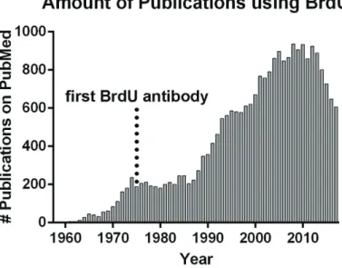
BrdU became popular by the development of antibodies having both high affinity and specificity. It overcomes the time-consuming procedure of autoradiography necessary for [H3]thymidine detection (Gratzner et al., 1975; Gratzner, 1982; Figure 1C). Finally, it allows concomitant immunodetection of cell type specific markers, allowing solid conclusions to be made regarding the identity and fate of BrdU+ cells. The increasing popularity of BrdU is reflected by the huge amount of scientific articles referenced on PubMed (24375 by end of 2017; Figure 2).
Figure 2. Timeline of BrdU publications
The first scientific article concerning BrdU was published in 1958. After the development of the first specific BrdU antibody in 1975 it became increasingly used for observations of diverse aspects of DNA synthesis and mitosis. By the end of 2017, 24375 research articles had been published by the scientific community.
Other halogen-based nucleoside analogs have appeared more lately, namely CldU and IdU (Figure 1C), as well as the “new generation” nucleoside analog EdU (Figure 1D), which can be chemically detected by the more advanced “click it” chemistry (for review Cavanagh et al., 2011). Those can be administrated at different time points. Their detections allow to address the long-term proliferative behavior of a cell, i.e. if it reenter cell cycle and therefore incorporate the second administrated nucleoside. Although they allow multiplex administration, the antibodies used to detect them need to be carefully chosen. Indeed, IdU and CldU have been shown to be specifically detected by two separate antibodies initially
6
generated for BrdU detection (Yokochi and Gilbert, 2007). The development of EdU circumvent the use of an antibody and allows a faster detection of DNA synthesis by “click it” chemistry, which can be accomplished within a few hours. In addition, the reaction component (fluorescent azides) have a high penetration capacity allowing efficient labeling of thick sections and even whole-mount tissues or organisms (Salic and Mitchison, 2008; Figure 1D). A drawback of nucleoside analogs are their potential toxic effects, which have been widely analyzed and discussed. Administration of BrdU in neonates has been reported to shorten their life span in a dose dependent manner (Craddock, 1981). It induces mutations (Kaufman, 1988) resulting in alterations in differentiation (Tapscott et al., 1989) and proliferation (Weghorst et al., 1991). This adverse effects are explained by the preferential pairing of BrdU with guanine instead of adenine (Kaufman, 1988), which can cause problems in subsequent divisions. Thus, doses superior to 60 mg/kg have been suggested to induce such cytotoxic effects (for review Cavanagh et al., 2011). Similarly, EdU administration leads to long-term toxic effects, while no significant alterations in cell proliferation and survival were observed for short survival times (Ponti et al., 2013).
1.1.2. Nucleoside Analogs Give Insight into Embryonic Germinal Activity by Interkinetic Nuclear Migration
An important step in using [H3]thymidine for studies in neurosciences, was the finding that radioactivity can be found in the brain as early as 15 minutes after intravenous injection into the young adult mouse (Hughes et al., 1958). Although this study focused on the gastrointestinal tract and did not make a thoughtful analysis of the brain, it demonstrated that [H3]thymidine rapidly distributes throughout the mouse body, including the brain, following intravenous injection. It therefore opened the way to study proliferation in this tissue. Thus, [H3]thymidine administration in mouse at embryonic day 11 (E11) revealed that the external half of the primitive ependymal layer of the cerebral vesicles is a region of intense DNA synthesis (Figures 3A and 3B). A time course analysis further revealed that [H3]thymidine can be detected in the ventricular (inner) half of the primitive ependymal layer, 6 hours after its injection (Figure 3C). At 48 hours, some labeled cells were observed in regions located away from the ependymal layer (Figure 3D). Taken together, these observations demonstrate
7
that regions of DNA synthesis and cell division are distinct. They also suggest that newly generated cells migrate away from its region of origin (Sidman et al., 1959). Using the same method in the early chick neural tube, the cell cycle dependent movement of the cell body was described in more details and referred as interkinetic nuclear migration (Sauer and Walker, 1959). Injections into pregnant female mice at different gestational time points (E11-E17) identified the primitive ependyma of the lateral ventricles (LV) as source of cortical cells along the entire embryonal period. This experiment also revealed the inside-out formation of the cortex, as described in more details below (Angevine and Sidman, 1961).
Figure 3. Embryonic NSCs undergo interkinetic nuclear migration.
(A): Representative coronal section from an early embryonal brain section (© 2008 Springer
Science+Business Media, LLC) illustrates [H3]thymidine administration.
(B-D): Micrographs highlight the position of labeled cells ( ) in the pallium at different time
points after [H3]thymidine administration. One hour after administration the labeled cells were found in the external half of the primitive ependymal layer (B). After 6 hours they were located in the inner half (C) and after 48 hours in the surrounding parenchyma.
Scale bars: A = 1 mm; D = 200 μm.
1.1.3. Nucleoside Analogs reveal Two Sites of persisting Postnatal Germinal Activity
[H3]thymidine experiments confirmed the original observations made years before by Ezra Allen (Allen, 1912). Indeed, proliferation was shown to persist postnatally in the SVZ at postnatal day 3 (P3), as well as in young adult and adult mice (Messier et al., 1958; Smart, 1961; Smart and Leblond, 1961). This demonstrates that the potential to produce new cells does not abruptly stop after birth or after completing the development. Therefore, the SVZ is a niche of ongoing postnatal proliferation (Figures 4A and 4B). Subsequent [H3]thymidine studies identified the dentate gyrus (DG) of the hippocampus as a second niche of ongoing
8
postnatal proliferation in the mammalian forebrain (Figures 4A and 4C). Importantly, the germinal potential of both the DG and SVZ was shown to decrease substantially during aging (Altman, 1963; Altman and Das, 1965). Major observations were made by Altman and Das from 1965 on. While already proposed earlier (e.g. Smart, 1961), their studies suggested the persistence of neurogenesis in the postnatal brain. They indeed observed germinal activity in the DG and SVZ of postnatal rats and guinea pigs, and presented data suggesting that it contributed to the generation of new neurons (Altman and Das, 1965; Altman and Das, 1967). Their claims were however examined by the scientific community with skepticism. In particular, technical limitations prevented them to unambiguously demonstrate the neuronal nature of newborn cells. Therefore Kaplan and Hinds reassessed their conclusions by combining [H3]thymidine labeling and electron microscopy of ultrathin (1 μm) sections. The existence of neurogenesis in those two regions was confirmed in the adult rat brain, by identifying labeled cells for neuron specific traits like long microtubule filled processes and a smooth contoured cell body (Kaplan and Hinds, 1977). Evidences for postnatal neurogenesis in mammals accumulated rapidly. Beside adult mice (Smart, 1961) and rats, germinal activity was demonstrated in adult cats (Altman, 1963), guinea pigs (Altman and Das, 1967), and dogs up to an age of 17 years (Fischer, 1967).
While those findings became widely accepted, postnatal mitotic activity in the primate brain remained controversial, and a number of conflicting findings were published. Noetzel and Rox analyzed adult mice and rhesus monkey brains in parallel. While they succeeded in detecting [H3]thymidine mitotic profiles in the mouse SVZ, they obtained negative results in rhesus monkeys. They concluded that the mitotic SVZ in mice is a leftover from the embryonic development and that “the absence of a SVZ” in rhesus monkeys is a sign of a higher developmental level and increased differentiation of the ape forebrain (Noetzel and Rox, 1964). Meanwhile others reported mitotic activity in the young and adult primate SVZ by revealing mitotic features (Lewis, 1968) or by [H3]thymidine administration (Kaplan, 1983). The number of proliferative cells described by Kaplan appeared however anecdotic, as only 14 labeled cells were observed within 48 analyzed sections. Pasko Rakic undertook a large-scale experiment to address this unresolved issue as well as of a possible postnatal cortical neurogenesis. His study included twelve rhesus monkeys of different ages treated with different doses of [H3]thymidine. While proliferation and some degree of neurogenesis was
9
confirmed in the DG during the first few postnatal months, [H3]thymidine+ cells in the cortex were identified as astrocytes and oligodendrocytes. Evidences for postnatal generated neurons in the cortex were not found (Rakic, 1985). Subsequent experiments by the Gould laboratory, confirmed germinal activity in the DG of old world monkeys (Macaca fascicularis and Macaca mulatta). They confirmed persisting granule cell neurogenesis by using mature and immature neuronal markers. They found a substantial number of cells being positive for NSE (neuron specific enolase), NeuN (neuronal nuclei), TOAD-64 (Turned-On-After-Division 64-kDa) and CalB (calbindin) in individuals of all ages with an age dependent reduction in neurogenesis (Gould et al., 1999). Another report from this group was far more controversial. They found evidences of neurogenesis in the adult SVZ (Macaca fasciculari), but also described migration of newborn neurons into the neocortex (prefrontal, inferior temporal and parietal cortex). While 2 hours after BrdU injection positive cells were located in the SVZ, they observed at longer time points cells with elongated nuclei in the white matter, classified as migrating cells, and in the neocortex. A fraction of these cells was confirmed to express the neuronal markers TOAD-64, NeuN, NSE and MAP2 (microtubule-associated protein-2), while others express the astrocytic marker GFAP (glial fibrillary acidic protein). Interestingly, survival of these adult born cells was found to decrease with age (Gould et al., 2001).
In parallel to these studies in old world monkeys, germinal activity and neurogenesis was also investigated in new world monkeys. Studies in the common marmoset C.jacchus demonstrated substantial mitotic activity in the postnatal SVZ using both [H3]thymidine and BrdU labeling. A high labeling index was observed at early postnatal time points, which dropped remarkably with progressing age to be very low by two years of age. The fate of the postnatal born cells was not analyzed (McDermott and Lantos, 1990). Similar observations were made in the dentate gyrus, were new born neurons acquired the morphological characteristics of granule neurons and expressed the neuronal marker NSE 3 weeks after BrdU injection (Gould et al., 1998). I confirmed these findings in common marmosets during my master thesis project, by using the proliferation marker Ki67. We found a similar age-dependent decrease in proliferation. We observed a large number of doublecortin (DCX) expressing neuroblasts, supporting a persistent neurogenesis. Finally, we demonstrated the presence of progenitors of defined neuronal subtypes by immunodetection of the early GABAergic marker Dlx2 and the glutamatergic marker Tbr2. While Dlx2+ progenitors persisted until adulthood, the pool of
10
Tbr2 expressing glutamatergic progenitors in the SVZ was found to be depleted early after birth (Azim et al., 2013).
A much-awaited study concerned the demonstration of such germinal activity and neurogenesis in humans. Eriksson et al., were the first to present evidences for neurogenesis in the aged human DG. They made use of postmortem brain biopsies from aged human patients that suffered from squamous cell carcinoma. These patients had been treated with BrdU at different time points during the course of their cancer for diagnostic purposes. Eriksson et al. described a persisting germinal activity in the human DG, as well as SVZ. First, they demonstrated a substantial germinal activity in the DG in all patients, although a large inter-individual variability was observed for the number of BrdU+ cells in both the subgranular zone (SGZ) and the granule cell layer (GCL). This variability can be explained by the different ages of the patients, as well as by differences in the time span between BrdU treatment and death. Therefore, although not quantitative, these results concluded for a significant germinal activity in humans. They also provided evidence for neurogenesis, based on BrdU co-expression with the neuronal markers NeuN, CalB and NSE in the DG (Eriksson et al., 1998). Due to the sparsity of tissues from BrdU treated patients, other approaches had to be developed in order to more systematically quantify neurogenesis in humans. Thus, an elegant study introduced the method of radiolabeled C14 for retrospective birth dating of human cells. Analysis of biopsies issued from individuals of different ages confirmed the occurrence of an extensive hippocampal neurogenesis and neuronal turnover throughout life. Thus, every day, 700 new neurons are generated per hippocampus resulting in an impressive yearly turnover of ~2% of the dentate granule cells. An age dependent decline was observed, but was estimated to be less dramatic compared to mice (Spalding et al., 2013). Using the same method persistence of germinal activity in the adult human SVZ was confirmed (Ernst et al., 2014). Parallel histological studies revealed persistent neurogenesis during infancy (Sanai et al., 2011) but also adulthood (Ernst et al., 2014).
Taken together nucleoside analogs reveal that germinal activity and neurogenesis is a landmark of the mammalian brain, including humans. These studies however highlight that variation in the intensity of this process, as well as in the fate and distribution of newborn neurons may exist.
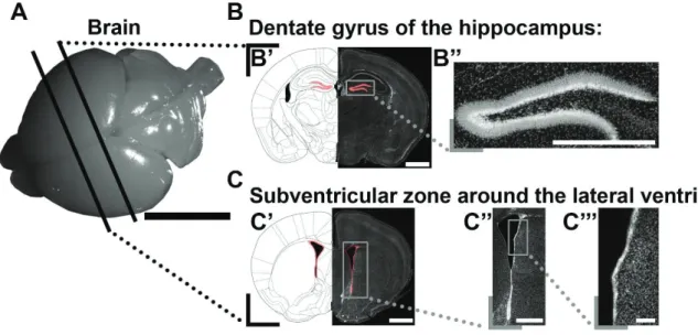
11
Figure 4. Nucleoside analogs reveal two sites of persistent postnatal germinal activity. (A): Representative picture of a P10 mouse brain illustrates virtual cuts at the level of the DG
and SVZ.
(B): The DG (highlighted in red) of the hippocampus is a niche of ongoing postnatal activity
(B’). Higher magnification micrograph shows the shape of the DG (B’’).
(C): The SVZ (highlighted in red) surrounds the LVs (C’). The micrograph illustrates that it
consists of 3 regionally distinct microdomains (lateral, dorsal, medial; C’’). The lateral microdomain is shown in higher magnification (C’’’).
Scale bars: A = 5 mm; B’, C’ = 1 mm; B’’, C’’ = 500 μm; C’’’ = 100 μm. Abbreviations: DG, dentate gyrus; SVZ, subventricular zone; LV, lateral ventricle.
1.1.4. Nucleoside Analogs Give Insights into the Migration Patterns of Cells Generated at Embryonic and Postnatal Times
Cells generated at proximity of the ventricular system rapidly migrate away to invade the surrounding parenchyma. Their pattern of migration however substantially differs at embryonic and postnatal time points. Analysis of cell distribution 10 days after birth, following [H3]thymidine administration at various gestational days (E11, E13, E15 and E17), revealed migration of embryonal born cells into the cortex. Interestingly, they found evidences for an inside-out development, challenging the prevailing view that newborn cells peripherally displace older cells (Tilney, 1934). Thus, cells born at E11 reside in the deepest layer of the cortex. Cells born two days later (E13) were found in the middle third and cells born 4 days later (E15) in the outer third of the cortex. Finally, cells born at E17 were located in the most superficial cell layer at P10 (Angevine and Sidman, 1961; Figures 5A and 5B).
12
The mode of migration and therefore the final destination of newborn cells of the SVZ changes fundamentally during the transition from embryonal to postnatal development. Temporal analysis of newborn cells migration in the postnatal forebrain was first investigated by injecting a group of rats of the same age (P30) and sacrificing them from 1 hour to 180 days after injection. Systematic analysis of cell distribution, allowed to identify the olfactory bulb (OB) as the major final destination of cells originating from the postnatal SVZ. Up to 24 hours [H3]thymidine labeled cells were found close to the LVs, while the OB was largely free of positive cells at this early time point. After 3 days, labeled cells were observed in the middle caudo-rostral portion of the rostral migratory stream (RMS) and after 6 days they arrived in the OB, where they distributed into both the granule cell and glomerular layers (GCL and GL, respectively) thereafter (Altman, 1969; Figure 5A and 5C).
13
Figure 5. Migration pattern in the prenatal and postnatal rodent forebrain.
(A): Representative picture of a P10 mouse brain illustrates virtual cuts at the level of the SVZ,
RMS and OB.
(B): The cortex develops in an inside-out fashion. Colored syringes in the timeline represent
different [H3]thymidine injection time points into pregnant females at E11 (red), E13 (orange), E15 (green) or E17 (blue).
Radiolabeled cells born at E11 reside in the deepest layer at P10. Cells born at E13 and E15 were found in the middle and outer third of the cortex, respectively. Cells that were generated at E17 were located in the most superficial layer.
(C): Postnatally born cells were found in the SVZ 1 day after injection, from where they invade
the RMS after 3 days and arrive in the OB after 6 days.
Scale bars: A = 5 mm; B, C = 1 mm. Abbreviations: OB, olfactory bulb; RMS, rostral migratory stream; SVZ, subventricular zone.
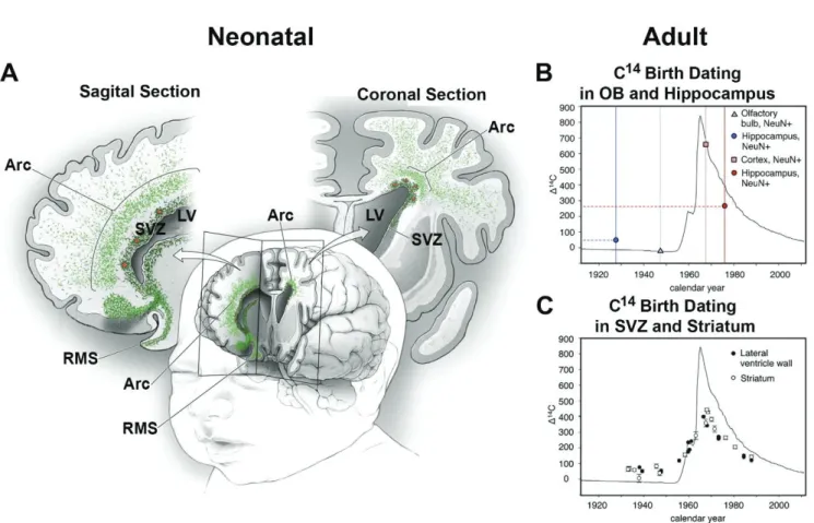
In humans the migration pattern is suggested to be remarkably different from rodents and obtained results are subject of controversial discussions. While in young individuals (up to 6 months) the RMS can be recognized by “broad streams” or chains of DCX and frequently co-expressing PSA-NCAM cells (Sanai et al., 2011), there are also extensive arc shaped migration features in the frontal lobe of infants (Paredes et al., 2016; Figure 6A). This situation changes dramatically in older individuals (from 2 years on). At these later stages, chains of migrating cells are not observed. Rather, rare single (or pairs of) migrating neuroblasts can be detected (Sanai et al., 2004; Sanai et al., 2011). A subsequent study, using retrospective C14 birth dating, supports these findings by concluding that neurogenesis in the adult human OB is negligible. On the other hand, the neuronal turnover in the adult hippocampus is evident (Bergmann et al., 2012; Figure 6B). Interestingly, histological and C14 birth dating experiments rather suggest a dispersion of newborn neurons at proximity of their site of origin, i.e. they may participate to the cellular turnover in the striatum (Ernst et al., 2014; Figure 6C). The analysis of neurogenesis in the adult marmoset, which I performed during my Master, support these conclusions. I could indeed observe many DCX+ cells in the striatum, which increase proportionally with age. Thus, as much as 30% of DCX+ cells were found in the striatum of a 56 months old marmoset (Azim et al., 2013). Together, these observations suggest a progressive disappearance of cues required for rostral neuroblast migration, which progressively disperse to nearest regions.
14
Figure 6. Migration pattern in the neonatal and adult human forebrain.
(A): Illustration of the migration pattern in a neonatal human brain in 3D, as well as in a sagittal
and a coronal section. Chains of migrating neuroblasts (green) leave the SVZ ventrally and enter the RMS. Dorsally of the SVZ there is an additional arc shaped region of extensive migration towards the cortex (modified from Paredes et al., 2016).
(B+C): Graphs showing the measured C14 concentration as a function of the calendar year. Y-axis represents the difference of the C14 concentration in the air compared to the natural level. The solid black line shows the concentration before, during and after the majority of the nuclear bomb tests. Represented data of hippocampus and OB illustrate and summarize the results from different studies (Bhardwaj et al., 2006; Bergmann et al., 2012; Spalding et al., 2013). Circles illustrate C14 values of hippocampal neurons obtained from individuals born before (blue) and after (red) the peak of nuclear bomb tests. C14 values at death of the individuals were increased and decreased compared to their concentration at birth, respectively. This suggests a postnatal neuronal turnover in the hippocampus. For neurons of the OB (triangle and square) no such correlation was observed (B). Post mortem C14 measurements in cells of the SVZ (black circle) and the striatum (white circle) show substantial differences to the C14 concentrations compared to the date of birth of the individuals (C; both graphs were obtained from Ernst et al., 2014). Abbreviations: LV, lateral ventricle; OB, olfactory bulb; RMS, rostral migratory stream; SVZ, subventricular zone.
15
1.1.5. The SVZ Has a Complex Cytoarchitecture and Consists of Different Cell Types with Distinct Cycling Behaviors
The cytoarchitecture of the SVZ, as well as the identity of neural stem cells it harbors, remained elusive for several decades. This was changed in the 90s with a series of landmark publications from the group of Arturo Alvarez-Buylla at the Rockefeller University in New York. The SVZ is not a homogeneous pool of cells, it is rather a heterogeneous mix of different cell types and cells at different levels in their maturation process, which differ in their morphology, function and cycling behavior. NSCs in the SVZ of the postnatal and adult brain originate from radial glia (RG) cells of the embryonal and perinatal brain (for review Kriegstein and Alvarez-Buylla, 2009). NSCs give sequentially rise to distinct cell types of divergent cycling behavior (Doetsch et al., 1997; Ponti et al., 2013). Doetsch and collaborators proposed in 1997 (Doetsch et al., 1997) a revolutionary view on the cellular architecture of the SVZ. They were able to distinguish a number of unique cell types, as well as to provide a detailed description of their organization in the SVZ. They proposed a nomenclature, which is still in use (i.e. type A, B and C cells; see below for more details) as well as the term “neurogenic niche” to define their peculiar 3D organization. Further, they demonstrated the presence of an abundant population of ependymal cells in the SVZ (type E cells), which are ciliated, aligned to the ventricular lumen and non-cycling. These different cell types are readily distinguishable by electron microscopy, based on their diverse ultrastructural morphologies and marker expression. Type A cells express PSA-NCAM and correspond to chains of migrating neuroblasts previously described in the RMS (Rousselot et al., 1995; Lois et al., 1996). They cluster in the SVZ to form a complex network of chains before engaging rostral migration toward the OB (Doetsch and Alvarez-Buylla, 1996). Type B cells exhibit ultrastructural traits (Peters et al., 1991) and expression signature (GFAP, vimentin) of astrocytes (Bignami and Dahl, 1974; Schiffer et al., 1986; Cohen et al., 1994). B cells can be further subdivided into two distinct subtypes, which are distinguishable by their ultrastructure and their position within the SVZ (B1: close to the ependymal layer; B2: at the SVZ-striatum border) and their proliferative behavior (B1 cells were not found to incorporate [H3]thymidine). Type C cells represent the transition between type B and type A cells. They appear in clusters, which are closely associated to type A chains. They contribute to half of the proliferating population, while being negative for type A and type B markers, which also incorporate [H3]thymidine, although to lesser extent.
16
[H3]thymidine positive type B and C cells are distributed along the entire dorso-ventral axis of the lateral SVZ, while cycling type A cells appear to be largely located in the dorsal and ventral aspects only (Doetsch et al., 1997; Figure 7). A more recent study investigated the cycling behaviors of type A, B and C cells in more details. They made use of dual labeling techniques using the nucleoside analogs CldU and EdU. This method revealed that the cell cycle length (TC) in adult mice progressively increases throughout differentiation (B1 cells: 17 hrs; C cells: 18-25 hrs). Further, the time for DNA synthesis (TS) was found to be surprisingly short for type B1 cells compared to type C cells (B1 cells: 4.5 hrs; C cells: 12-17 hrs). When interpreting these data it needs to be considered that only actively cycling B1 cells, but not the quiescent fraction of the population, were included into the analysis (Ponti et al., 2013). Interestingly, the TC and TS of actively cycling NSCs appear almost identical to the one reported for RG cells during embryonic development at E16 (Takahashi et al., 1995). Further analysis showed that type B1 and type C cells cycle 3 times before generating type A cells, which make one or two additional divisions in the SVZ before entering the RMS (Ponti et al., 2013). Cells with a similar ultrastructure like the described type B cells can also be found in the RMS, where they enwrap the chains of migrating type A cells and separate them from the surrounding parenchyma (Lois et al., 1996). Similarly, type B1 and B2 cells appear to isolate migrating neuroblasts in the SVZ from the ependymal layer and the striatal parenchyma. Whereas neurogenesis has been the focus of most studies, it should however be mentioned that SVZ NSCs do not only generate neurons, but also produce astrocytes (Reynolds and Weiss, 1992) and oligodendrocytes (Kirschenbaum and Goldman, 1995) in vitro. The in vivo evidence for the generation of glia by the postnatal SVZ was given by retrovirus approaches (Levison et al., 1993).
Taken together, the SVZ harbors stem cells showing astrocytic traits (type B cells), which have the capacity to produce a glial and neuronal progeny. Type B cells can be further subdivided into an actively cycling and a quiescent subpopulation (Levison et al., 1993; Doetsch et al., 1997; Doetsch et al., 1999). Recent findings, using more advanced technical approaches, which will be discussed in the general discussion of this thesis manuscript, suggest that most adult NSCs arise from a pool of radial glial cells, which enter quiescence between E13.5 and E15.5 (Furutachi et al., 2015; Fuentealba et al., 2015) and gradually reactivates after birth.
17
Figure 7. The cytoarchitecture of the adult SVZ.
(A+B): Representative micrographs of the SVZ (A) and the higher magnification of the lateral
microdomain (B) show the zone of interest to illustrate the SVZ cytoarchitecture.
(C+D): Ciliated ependymal cells (E; yellow) align the LV and isolate the lumen from the
proliferative SVZ. NSC (type B1; blue) give rise to type C cells (green) which generate migrating neuroblasts (type A; red) (schemes were modified from Doetsch et al., 1997 and Tong and Alvarez-Buylla, 2014).
Scale bars: A = 500 μm; B = 100 μm; C = 20 μm. Abbreviations: BV, blood vessel; LV, lateral ventricle; SVZ, subventricular zone.
1.2. Transgenesis Revealed Early Regionalization of Germinal
Regions
Although nucleotide analogs represent a technical breakthrough in the study of germinal activity and have led to major advances in its understanding, they also present limitations. Indeed, they are incorporated in all cycling cells, thereby preventing the study of subpopulations of NSCs. Also, they don’t provide any information on the molecular mechanisms involved in NSCs biology. The emergence of advanced transgenic approaches have allowed circumventing these limitations, to shape our current understanding of postnatal germinal regions. These regions appear now highly heterogeneous, with NSCs located in different location generating distinct progenies. Here I will summarize the key methodological developments, which have led to these conclusions.
18
1.2.1. The Development of Transgenesis Approaches Represents a Further Milestone in the History of Neuroscience
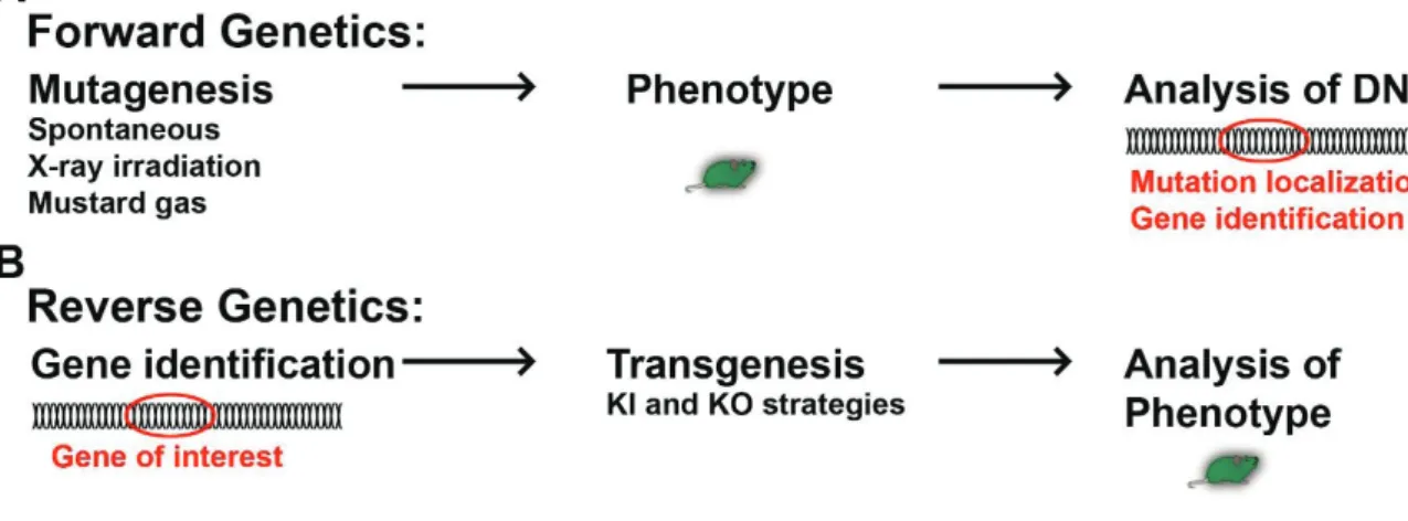
Domestication of animals and plants by selecting certain individuals for breeding, while excluding others from the line, can in the broader sense be defined as the slowest transgenic approach. The gene pool of a population gets artificially changed over many generations and new strains arise from the ancestor population. Spontaneous mutations might be kept in the population or rejected by excluding them from breeding. Approaches have been developed which allow direct genome editing. The first transgenic animals (fruit flys) were gained by random mutagenesis of the genome using different mutagens, e.g. by exposure to X-ray irradiation (Herman Joseph Muller, 20s) or mustard gas (Charlotte Auerbach, Alfred Joseph Clark, John Michael Robson, 40s). As insects have a segmental organized body plan (Lawrence, 1992), mutated larvae and flies were screened and those presenting identifiable phenotypes , such as the absence of certain segments, body parts or the poor development of them, were selected (for review McGinnis and Krumlauf, 1992). This method was used for early identification of key developmental regulator genes. Because it first identifies mutated phenotypes (induced or spontaneous), then the mutated gene, it is called forward genetic (for review St Johnston, 2002; Figure 8A). In Drosophila, segmental identity was found to depend on the segment position along the anterior-posterior axis. It led to the identification of homeobox genes, which expression follows a strict segmental code (for review McGinnis and Krumlauf, 1992). Many of the region specific transcription factors (homeobox and others) were discovered in Drosophila by spontaneous and induced mutagenesis approaches (forward genetic). Subsequently, their presence in mammals was confirmed by histological methods and their function resolved by transgenesis.
Later, reverse genetic approaches opened a new era in the field of neuroscience. They allowed to target specific genes to perform loss and gain of function experiments. The development of these refined transgenic strategies represents a milestone in the history of neurosciences. The capability to precisely target specific genes and loci in the genome allowed a wide range of powerful transgenic approaches (Figure 8B). This “transgenic era” in neurodevelopmental research began with the creation of null mutants for regionalized transcription factors (TF), for loss of function approaches.
19
Figure 8. Illustration of the principles of forward and reverse genetics.
(A): In forward genetic approaches mutations are induce or occur spontaneously. If a
phenotype was observed, the DNA was analyzed to identify the mutated gene.
(B): Reverse genetic approaches identify first the gene of interest. Targeted
mutations/modifications are induced by diverse transgenic approaches and the phenotype analyzed thereafter.
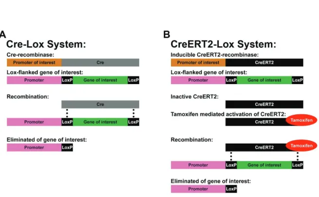
However, as many of these TFs are essential for the proper development, complete knock-out (KO) often leads to perinatal death (Qiu et al., 1995; Yoshida et al., 1997). Therefore, it is frequently not possible to investigate the function of these factors in a postnatal or adult context. The discovery of the Cre-Lox systems in combination with targeted knock-in (KI) strategies allowed to overcome this limitation. The Cre-Lox system can be defined as a technology for site-specific DNA recombination. Briefly, the gene of interest is flanked by Lox sites, which are recognized by a Cre-recombinase. This Cre-recombinase can be expressed by any cell type, based on its insertion into the genome, under the control of a well-chosen promoter. This allows controlling its spatial, and to a certain degree also its temporal expression (Sauer, 1998; Figure 9A). Newer generations of Cre-recombinases ensure a more advanced temporal control of recombination activity. By fusing genes coding for the Cre-recombinase and a fragment of the estrogen receptor, researcher have engineered several generations of Cre-recombinase (i.e. CreERT; CreERT2) that are sequestered into the cell cytoplasm. Administration of the chemical tamoxifen allows the nuclear translocation of the fusion protein, and therefore its activation (Feil et al., 1996; Figure 9B).
Taken together, the Cre-Lox systems allows conditional KO (cKO) experiments, which are crucial for investigating the function of certain genes at a given time of development and/or of
20
postnatal life. In addition, it allows the conditional expression of reporter genes for lineage fate mapping. In this context, several transgenic mouse lines have been generated over the years, which varies in the detectable marker they express (e.g. LacZ, GFP, tdTomato; Madisen et al., 2010). These new developments offered a powerful tool box to advance our understanding of SVZ heterogeneity and regionalized populations greatly.
Figure 9. Temporal and spatial genetic manipulations using the Cre-Lox system.
(A): The Cre-recombinase (gray) is under the control of a promoter of interest to ensure the
spatial and to a certain degree also temporal restricted expression. The gene of interest (green) is flanked by Lox sites (here LoxP; black), which are recognized by the Cre-recombinase. Expression of the Cre-recombinase leads automatically to recombination of the Lox sites and elimination of the gene of interest.
(B): Additional temporal control can be achieved by fusion of the Cre-recombinase with a
fragment of the estrogen receptor (here ERT2). CreERT2-recombinases (black) remain in the cytoplasm and therefore inactive until they are activated by tamoxifen (red) administration. Recombination of the Lox sites and elimination of the target gene occurs subsequently.
21
1.2.2. Expression Analyses and Fate Mapping Approaches Highlight the Regionalized Organization of the VZ/SVZ
One of the most important feature of multicellular organisms is the existence of a clearly defined rostro-caudal and dorso-ventral axis. This organization arises during early embryonic development and depends greatly on the regional expression of combinations of genes. In general, all cells are identical during the first embryonic days. However, cell fates start to diverge from each other already during blastocyst formation around E3.5, long before organogenesis (for review Saiz and Plusa, 2013; Kojima et al., 2014). Heterogeneous expressed cues are major players during embryogenesis and into adulthood. Much of the knowledge we have about these early patterning of the body plan originates from studies in insects, which have a segmental organized body (Lawrence, 1992). Like in Drosophila, the development of the mouse central nervous system is orchestrated by regionally expressed genes. Further, the developing forebrain, hindbrain and spinal cord resemble such segmental structures (reviewed in Rubenstein et al., 1994; Philippidou and Dasen, 2013). Focusing on the developing forebrain, we gained insight in these processes by discovering differentially expressed genes by initial immunohistochemistry. Such genes were closer investigated regarding their functional importance in fate decision of regionalized populations by transgenic approaches.
Regionalization is Observed During the Period of Neurogenesis
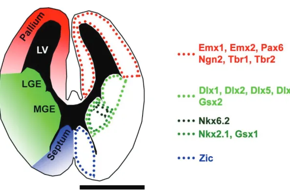
Several transcription factors have a clear regionalized expression pattern during early forebrain development. This is particularly apparent for markers of the pallium and the subpallium (i.e. lateral and medial ganglionic eminences, LGE, MGE; septum), some regions which contribute to the formation of specific SVZ microdomains at postnatal stages (reviewed in Fiorelli et al., 2015).
Many markers of the ganglionic eminences, belong to the ANTP class of the homeobox genes, which is the largest class of homeobox genes (Zhong et al., 2008; Zhong and Holland, 2011). This includes members of the Dlx family (Distal-less), namely Dlx1, Dlx2 (Bulfone et al., 1993), Dlx5 and Dlx6 (Simeone et al., 1994), which start to be highly expressed as early as E10. They are enriched in the ganglionic eminences and consistently absent from the pallial
22
domain. Other ventrally enriched genes of the homeobox familly are Nkx2.1 (also called TTF1), Nkx6.2, Gsx1 and Gsx2 (also called Gsh1 and Gsh2) of the Nk2.1, Nk6 and Gsx homeobox gene families (Zhong et al., 2008; Zhong and Holland, 2011). While Nkx2.1 was described to be partly overlapping to the Dlx genes (Price et al., 1992), Nkx6.2 was initially described as a ventral marker of the spinal cord and being largely absent of the developing forebrain (Qiu et al., 1998). However, later experiments revealed its restricted expression in a dorsal stripe of the MGE at E12.5 (Fogarty et al., 2007). Gsx1 and Gsx2 were both found to be expressed in the ganglionic eminences. While Gsx1 is restricted to the MGE (Valerius et al., 1995), Gsx2 is described to be expressed by both the LGE and MGE (Hsieh-Li et al., 1995;
Figure 10, green). Lineage tracing approaches have shown that subpallial progenitors give
rise to GABAergic interneurons. Different Cre reporter mouse lines were used to demonstrate that defined progenitor populations generate distinct lineages of interneurons, which invade the cortex tangentially (Fogarty et al., 2007).
Other homeobox genes show restricted expression to the dorsal forebrain regions, i.e. the pallium. The most studied ones are Emx1, Emx2 and Pax6 (Emx family of the ANTP class; Pax4/6 family of the PRD class; Zhong et al., 2008; Zhong and Holland, 2011). Emx1 and Emx2 start to be expressed in the mouse pallium at E9.5 and E8.5, respectively. While both genes are largely overlapping, Emx2 expression is more widespread than Emx1 (Simeone et al., 1992a). Similarly, the homeobox gene Pax6 is embryonal expressed in the pallium but absent from the ganglionic eminences (Stoykova and Gruss, 1994). Other transcription factors have been shown to act downstream of these homeobox genes, and as a result, keep a strict regional expression. Other highly specific pallial markers are the T-box transcription factors Tbr1 and Eomes (also referred as Tbr2; Bulfone et al., 1995; Bulfone et al., 1999), as well as the bHLH transcription factor Ngn2 (also referred as Neurog2; reviewed in Lee, 1997; Figure
10, red). Lineage tracing of the Tbr2 (Pimeisl et al., 2013) and Neurog2 (Berger et al., 2004;
Donega et al., 2018) lineages revealed their participation in producing cortical projection neurons (Mihalas et al., 2016). Expression of these TFs antagonize a subpallial expression of the bHLH transcription factor Mash1 (Casarosa et al., 1999).
A third region of the developing forebrain known to participate to the formation of the postnatal SVZ, is the most medial part of the developing forebrain, the septum (for review Fiorelli et al., 2015). Examples of medially enriched markers in the embryonal forebrain belong to the Zic
23
family of transcription factors (Aruga et al., 1994; Figure 10, blue). Zinc-finger TFs control various processes of animal development (for review Grinberg and Millen, 2005). In mammals, there are five Zic- related genes that share highly conserved zinc finger domains. In terms of neural development, Zic2 is particularly important for forebrain development. Thus, mice homozygous for the Zic2 hypomorphic allele (Zic2 kd/kd) show holoprosencephaly (HPE), in which the medial part of the forebrain is defective (Nagai et al., 2000).
Figure 10. Illustration of regionalized TFs expression in the embryonic forebrain.
Scheme of an E14 coronal section illustrates restricted expression in the pallium (red), the LGE and MGE (green) and the septum (blue). Restricted dorsal markers are Emx1, Emx2, Pax6, Ngn2, Tbr1 and Tbr2. The ganglionic eminences harbor some markers, which are expressed by both subdivisions (Dlx1, Dlx2, Dlx5, Dlx6 and Gsx2) and others that are restricted to the MGE (Nkx2.1 and Gsx2) or parts of it (Nkx6.2). Finally, markers showing restriction to the septum belong to the Zic family.
Scale bar = 1 mm. Abbreviations: LV, lateral ventricle; LGE, lateral ganglionic eminence; MGE, medial ganglionic eminence; TF, transcription factor.
24
Regionalization is Observed During the Period of Gliogenesis
Astrocytes are known to be produced at the end of the period of neurogenesis. It is classically accepted that RG cells switch fate and start to produce astrocytes that migrate to the cortex and associate with previously generated neurons in so called cortical columns (Magavi et al., 2012; for review Tabata, 2015). Recent studies indicate that their production is highly regionalized and newborn astrocyte precursors migrate radially away from their region of origin (Tsai et al., 2012). It has been shown that the major source of the cortical astrocyte population are astrocytes that proliferate and differentiate in the parenchyma (Ge et al., 2012; Figure 11A). TFs associated with astrogenesis are rare. In the developing spinal cord the TF NFIA has been proposed to inhibit neurogenesis and to trigger gliogenesis. Within the glia population, NFIA expression leads to migration and differentiation of astrocyte precursors. This function was found to be antagonized by the expression of Olig2 in oligodendrocyte precursors (Deneen et al., 2006). Despite the description of diverse subtypes of astrocytes (for review Tabata, 2015) there are, to my best knowledge, no TFs described for its association with the specification to these subtypes.
The other macroglia lineage, oligodendrocytes, are described to be produced in 3 different temporal and spatial waves. The first wave originates from the MGE and the medial part of the ventral forebrain, while the second is more laterally generated by the LGE. The final third wave appears postnatally in the dorsal SVZ microdomain. Ablation of a specific line does not result in a phenotype, as the missing “temporal lineage” is replaced by the remaining waves. However under physiological conditions, progenies of the first wave disappear postnatally (Kessaris et al., 2006; Figure 11B). While Olig1 and Olig2 are identified as general oligodendrocyte TFs (Zhou and Anderson, 2002), TFs specifying or differentiating oligodendrocytes from these three waves are still missing.
All together, these observations indicate that a regionalization of neurogenesis appears early in the developing forebrain. This regionalization relies on the regional expression of defined TFs and translate into the generation of different neuron subtypes. Similar principles apply to gliogenesis, although the exact transcriptional mechanisms involved in their regional production and its functional importance remains to be fully explored.
25
Figure 11. Illustration of astrogenesis and oligodendrogenesis.
(A): Astrocyte precursors that are generated in the SVZ, migrate radially away from their
region of origin and amplify subsequently. Cortical and white matter astrocytes (green) originate in the dorsal SVZ (dSVZ; modified from Bayraktar et al., 2015).
(B): Oligodendrogenesis occurs in 3 waves. The first wave (dark green) and the second wave
(light green) originate from the MGE and LGE, respectively, and invade the cortex tangentially. The postnatal third wave (red) of oligodendrogenesis occurs in the dSVZ and invades the CC and cortex radially (template used from Bayraktar et al., 2015).
Abbreviations: CC, corpus callosum; SVZ, subventricular zone; dSVZ, dorsal subventricular zone ; LGE, lateral ganglionic eminence ; MGE, medial ganglionic eminence.
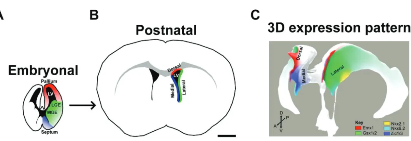
Regionalization of the SVZ is Retained After Birth
Both neurogenesis and gliogenesis persist in the postnatal SVZ (Levison and Goldman, 1993). Interestingly, the transcriptional regionalization of the developing forebrain is retained postnatally. This is reflected by the restricted expression of the same TFs within defined microdomains of the postnatal SVZ (for review Fiorelli et al., 2015). As described above, these distinct embryonic regions (pallial vs. ganglionic eminences vs. septal domains) are all contributing to the generation of the postnatal SVZ (dorsal vs. lateral vs. medial SVZ microdomains; Figure 12). The transcriptional analogies of the embryonic and postnatal/adult