ORIGINAL INVESTIGATION
The NMDA antagonist ketamine and the 5-HT agonist psilocybin
produce dissociable effects on structural encoding of emotional
face expressions
André Schmidt&Michael Kometer&Rosilla Bachmann&
Erich Seifritz&Franz Vollenweider
Received: 8 May 2012 / Accepted: 10 July 2012 / Published online: 27 July 2012 # Springer-Verlag 2012
Abstract
Rationale Both glutamate and serotonin (5-HT) play a key role in the pathophysiology of emotional biases. Recent studies indicate that the glutamate N-methyl-D-aspartate (NMDA) receptor antagonist ketamine and the 5-HT recep-tor agonist psilocybin are implicated in emotion processing. However, as yet, no study has systematically compared their contribution to emotional biases.
Objectives This study used event-related potentials (ERPs) and signal detection theory to compare the effects of the NMDA (via S-ketamine) and 5-HT (via psilocybin) receptor system on non-conscious or conscious emotional face pro-cessing biases.
Methods S-ketamine or psilocybin was administrated to two groups of healthy subjects in a double-blind within-subject placebo-controlled design. We behaviorally assessed objec-tive thresholds for non-conscious discrimination in all drug conditions. Electrophysiological responses to fearful, happy, and neutral faces were subsequently recorded with the face-specific P100 and N170 ERP.
Results Both S-ketamine and psilocybin impaired the encoding of fearful faces as expressed by a reduced N170 over parieto-occipital brain regions. In contrast, while S-ketamine also impaired the encoding of happy facial expres-sions, psilocybin had no effect on the N170 in response to happy faces.
Conclusion This study demonstrates that the NMDA and 5-HT receptor systems differentially contribute to the structur-al encoding of emotionstructur-al face expressions as expressed by the N170. These findings suggest that the assessment of early visual evoked responses might allow detecting phar-macologically induced changes in emotional processing biases and thus provides a framework to study the patho-physiology of dysfunctional emotional biases.
Keywords Glutamate . Serotonin . Ketamine . Psilocybin . Emotional processing biases . Event-related potential . Visual awareness . Non-conscious . Conscious
Introduction
Emotional processing including the recognition of other people’s feelings from their facial expression is fundamental to social interaction and behavior. The importance of face recognition in the human social functioning is shown by the fact that emotional faces increase neuronal activity relative to neutral faces in specific brain areas. For example, in-creased brain responses to emotional faces have been ob-served in visual face-selective areas of the brain, even when emotional faces were masked to prevent visual awareness (Pegna et al.2008; Smith2011). Thus, modulation of face-specific responses in the visual cortex by emotional expres-sions might correspond to a fundamental regulatory role of basic emotional signals associated with social appraisal and cognition (Schultz et al.2003; Singer et al.2004).
Emotional face processing can be modulated by seroto-nin (5-hydroxytryptamine, 5-HT). For example, acute ad-ministration of the selective serotonin reuptake inhibitor (SSRI) citalopram facilitated recognition of fear and happy facial expressions in the citalopram treated group relative to A. Schmidt (*)
:
M. Kometer:
R. Bachmann:
F. VollenweiderNeuropsychopharmacology and Brain Imaging, University Hospital of Psychiatry, University of Zurich, Zurich, Switzerland
e-mail: [email protected] E. Seifritz
Clinic of Affective Disorders and General Psychiatry, University Hospital of Psychiatry, University of Zurich, Zurich, Switzerland
the placebo group (Bhagwagar et al.2004; Browning et al.
2007; Harmer et al.2003). In a later study, repeated admin-istration of citalopram in healthy subjects increased the relative processing of positive to negative emotional faces in a manner directly opposite to the negative biases previ-ously described in depression (Harmer et al. 2004). Both findings suggest important neuropsychological evidence of a possible mechanism of action of antidepressant drugs (Nathan et al.2003). The findings are consistent with other studies examining the neurophysiological mechanisms un-derlying antidepressant effects on emotional processing. In these studies, acute citalopram administration was shown to inhibit visual evoked electrophysiological responses to un-pleasant stimuli, while it facilitates visual responses to pleasant stimuli (Kemp et al. 2003, 2004; Nathan et al.
2003). These studies suggest that this capacity to shift emotional biases—increasing the response to positive and decreasing the response to negative stimuli—seems not only to be characteristic for the action of SSRI on emotional pro-cessing but seems also to be highly relevant for the treatment of emotional biases found in depression (Harmer2008).
In addition, emotional face processing can also be affect-ed by glutamatergic manipulation. Specifically, a functional imaging study in healthy subjects showed that the visual activity in response to fearful faces is abolished under the influence of the glutamate N-methyl-D-aspartate (NMDA)
receptor antagonist ketamine (Abel et al.2003). Taken to-gether, these findings suggest that acute manipulations of the serotonergic and glutamatergic systems by psilocybin and ketamine may change emotional processing biases as indexed by the assessment of visual evoked responses to emotional expressions.
Recent studies used event-related potential (ERP) record-ing to investigate the time course of non-conscious and con-scious emotional face processing (Pegna et al.2011; Smith
2011). Studies of ERPs generated by faces often focus on key components such as the P100 and the face-sensitive N170. The P100 is an early positive occipital potential, peaking at around 80–120 ms post-stimulus, and reflects rapid extraction of information related to emotion or salience that occurs before more fine-grained perceptual analyses are completed (Vuilleumier and Pourtois2007). Modulation of the P100 has been shown with fearful (Fichtenholtz et al.2009; Pourtois and Vuilleumier 2006), angry (Santesso et al. 2008) and positive expressions (Batty and Taylor 2003; Brosch et al.
2008). The N170 is a negative occipitotemporal potential at approximately 170 ms post-stimulus and is associated with structural encoding of facial configurations (Itier and Taylor
2004; Rossion and Jacques2008). The view that the N170 ERP is not modulated by the emotional content of faces (Ashley et al. 2004; Eimer and Holmes 2002; Krolak-Salmon et al.2001) has been challenged by other findings (Batty and Taylor2003; Blau et al.2007; Jaworska et al.2010;
Krombholz et al. 2007; Schyns et al.2007; Sprengelmeyer and Jentzsch2006; Stekelenburg and de Gelder2004), even when faces are non-consciously processed (Pegna et al.2008; Smith2011). Thus, the N170 reflects not only face sensitivity but also emotion sensitivity during conscious as well as non-conscious face processing. Furthermore, it has been shown that the N170 amplitude correlates with the severity of de-pressive symptoms (Noll et al.2012), while both the P100 and N170 amplitudes are significantly differed between healthy volunteers and subjects with bipolar disorder (Degabriele et al. 2011; Sokhadze et al. 2011). These studies together demonstrate the potential of these ERPs to study pharmacological mechanisms underlying emo-tional biases and their pathophysiology.
In this study, we assessed these ERPs to compare the effects of the glutamate NMDA receptor antagonist ket-amine and the 5-HT receptor agonist psilocybin on visual processing stages during emotional face processing. Nota-bly, both ketamine and psilocybin are suggested to modulate neuronal activity in circuits implicated in emotion regulation and to have implications for the treatment of affective dis-orders. While it has repeatedly been pointed out that acute ketamine administration ameliorates depressive symptoms in treatment-resistant depression within a few hours persist-ing for several days (Diazgranados et al.2010; Zarate et al.
2006), a gradual reduction in trait anxiety at 1 and 3 months, as well as in depressive symptoms at 6 months, was ob-served after a single dose of psilocybin in terminal cancer and anxiety patients (Grob et al. 2011). Based on these findings, in this paper we hypothesized that manipulations of both the NMDA and 5-HT receptor system using ket-amine and psilocybin may inhibit negative face processing and facilitate the processing of positive faces as expressed by modulations of the P100 and N170 ERP. Given that visual responses are modulated by face visibility (Pessoa et al.2006), we further predicted that both drug effects on the visual ERPs might vary between non-conscious and conscious face processing. Thus, to test our hypothesis, we examined on the one hand whether psilocybin and ketamine affect visual ERP responses to emotional faces in a valence specific manner and on the other hand whether these drug effects depend on visual awareness.
Method Participants
Healthy subjects were recruited through advertisement from the local universities and were then separated into two groups (S-ketamine group: N021 [male, 12], mean age026±5.39 years; psilocybin group: N021 [male, 13], mean age023±2.22 years, all were students). Subjects
were healthy according to medical history, clinical ex-amination, electrocardiography, and blood analysis. Sub-jects were screened by the DIA-X diagnostic expert system (Wittchen and Pfister 1997), a semi-structured psychiatric interview to exclude those with personal or family (first-degrees relatives) histories of major psychi-atric disorders, and by the Symptom Checklist (SCL-90–R) (Derogatis 1994). Furthermore, subjects replied to the Mini-International Neuropsychiatric Interview, a brief, structured psychiatric interview (Sheehan et al. 1998). No subjects had to be excluded using these criteria. We verified the absence of a history of drug dependence by urine drug-screening and a self-made consumption questionnaire. In the S-ketamine group, seven subjects were occasional smokers (<10 cigarettes/day), eight subjects reported a sporadic or rare cannabis use in the past (<3 joints/month), two subjects reported experiences with MDMA (three pills lifetime), two subjects had previous LSD experiences (5 lifetime experiences), and one subject reported having previous experiences with ketamine (one occasions lifetime). In the psilocybin group, eight subjects were occasional smok-ers (<6 cigarettes/day), eight subjects reported a sporadic or rare cannabis use in the past (<2 joints/month), one subject reported experiences with MDMA (two pills lifetime), two subjects had prior use histories of ingesting psilocybin containing mushrooms (two lifetime experiences), and one subject reported experiences with LSD (two lifetime experiences). All subjects were free of any medication for at least 3 weeks before the experiment.
This study was approved by the Ethics Committee of the University Hospital of Psychiatry in Zurich. After receiving a written and oral description of the aim of this study, all participants gave written informed consent statements be-fore inclusion. The use of psilocybin was authorized by the Swiss Federal Office for Public Health, Department of Phar-macology and Narcotics, Berne, Switzerland.
Drug administration
In both groups, subjects underwent two sessions (placebo/ active drug) in a counterbalanced and double-blind fashion at an interval of at least 2 weeks. Subjects were monitored until all drug effects had worn off and were then released into the custody of a partner. For the S-ketamine/placebo infusion, an in-dwelling catheter was placed in the antecu-bital vein of the nondominant arm. Once the subject was ready, a bolus injection of 10 mg over 5 min was given. Following a 1-min break, a continuous infusion with 0.006 mg/kg/min was administered over 80 min. To keep S-ketamine’s plasma level fairly constant, the dose was reduced every 10 min by 10 % as previously described ((Feng et al.1995; Vollenweider et al.1997). In the placebo session, the same procedure was followed and an infusion of
physiological sodium chloride solution and 5 % glucose was given. Psilocybin (115 μg/kg) and lactose placebo were orally administered in gelatin capsules of identical number and appearance as previously described in studies assessing the acute psychological and physiological effects of psilo-cybin in healthy humans (Hasler et al.2004; Studerus et al.
2011). Psilocybin was orally given not only to ensure direct comparability with these previous studies and external va-lidity but also to induce stabile psychological effects after 60–90 min post-treatment, lasting for 60–120 min (Hasler et al. 2004). Furthermore, given the putative therapeutic po-tential of psilocybin (Kometer et al.2012; Vollenweider and Kometer 2010; Grob et al. 2011), an oral dosing regimen provides a more personable setup than utilizing an intravenous dosing regime for psilocybin.
Psychological assessment
The Altered State of Consciousness (ASC) questionnaire, a visual analog and self-rating scale, was used to assess the subjective psychological effects induced by S-ketamine and psilocybin (Dittrich1975,1998). A recent evaluation study of the ASC questionnaires has constructed 11 new lower-order scales (Studerus et al. 2010), which were assessed in this study. After the acute effects of S-ketamine (about 240 min post-treatment) and psilocybin (about 360 min after treatment) had subsided, the ASC questionnaire was given to retrospec-tively rate their introspective experiences since drug intake. Stimuli and backward masking procedure
As stimulus material, we took black and white photographs taken from the Ekman–Friesen series (Ekman and Friesen
1976). In order to exclude the hair and non-facial contours and further to match for luminance and contrast, each face was modified using Adobe Photoshop as previously done in other studies (Blau et al.2007; Pegna et al.2008; Pourtois et al.
2004). The final facial images subtended a visual angle of 3° horizontally and 4.4° vertically and were displayed in the center of a CRT monitor (refresh rate of 100 Hz). Subjects first underwent a mismatch negativity event-related paradigm for 15 min, which has been published elsewhere (Schmidt et al.2011). Emotional measures using backward masking para-digms were started 25 min after S-ketamine infusion and 110 min following psilocybin administration during the known plateau (Hasler et al.2004; Passie et al.2002).
Backward masking paradigms (facial affect discrimina-tion and EEG/ERP recording) were generated by a software dedicated to psychological testing (E-prime; Schneider et al.
2002). Backward masking is a key experimental paradigm for investigating sensory unawareness because this method inter-feres with the activity in the ventral occipitotemporal cortex, an area which is highly relevant for visual awareness
(Tamietto and de Gelder 2010). The accuracy of stimulus duration was confirmed using an oscilloscope. Figure1shows the backward making procedures for the facial discrimination task (A) and for the subsequent EEG/ERP recording (B). Facial affect discrimination Two discrimination tasks were conducted to establish thresholds for visual awareness in all drug conditions, i.e., to determine the time point, at which subjects can distinguish emotional from neutral expression above chance level. In a first task, subjects had to discrim-inate fearful from neutral faces, while in a second task they had to discriminate happy from neutral faces. For each discrimination task, target faces consisted of neutral and fearful/happy faces and were presented for 20, 30, 50, 90, or 170 ms (Williams et al.2004). Target faces were imme-diately followed by a neutral mask of the same person lasting for 150 ms. Participants performed five blocks of 40 trials (target–mask pairs) for each of both tasks, in which target faces were randomly presented with equal probability. Before the presentation of target–mask pairs, a fixation cross was presented for 1,000 ms. Subjects had to make a forced-choice decision about the valence of the target face (fearful/ happy vs. neutral) via button-press.
EEG/ERP recording During subsequent ERP recording, stimuli were identical to those used during the facial affect discrimination. Target faces comprised neutral, fearful, and happy faces and were immediately followed by a neutral mask of the same person for 150 ms. Each trial began with a fixation cross that lasted for 2,000 ms. According to the results of the discrimination tasks, target faces were 10 ms presented during non-conscious processing and 200 ms during conscious
processing. No subject response (button-press) was required. However, participants were given instructions that pairs of target–mask faces would be presented and that they would be asked questions about the first faces after testing.
Event-related potential recording
EEG recordings were made from 64 scalp electrodes using the ActiveTwo system (Biosemi, the Netherlands). The horizontal electroocculogram (EOG) was recorded from electrodes at-tached on the outer canthus of each eye. Similarly, vertical EOG was recorded from electrodes attached infraorbitally and supraorbitally to the left eye. All electrodes were active silver/ silver chloride electrodes and the offset of all electrodes was below 25 mV. Data were recorded at a sampling rate of 512 Hz. The common mode sense active electrode and the driven right leg passive electrode were used as reference and ground electrodes, respectively (seehttp://www.biosemi.com/ faq/cms&drl.htmfor more details on this setup).
For ERP analysis, independent component analysis was used to remove artifacts due to eye movements and blinks (Lee et al. 1999). The EEG data were recalculated offline against average reference. Then, epochs with a 200-ms pres-timulus baseline and a 500-ms post-spres-timulus interval were constructed. Epochs with amplitudes that exceeded ±100μV at any electrode were excluded from further averaging. After artifact rejection, epochs were averaged for each subject and were digitally filtered with a band-pass filter (1–30 Hz). P100 and N170 ERPs were computed at electrodes P08/P8/P10/O2 (right hemisphere) and PO7/P7/P9/O1 (left hemisphere) as peak positivity/negativity relative to baseline within the 130–200- and 150–250-ms window latency, respectively, as Fig. 1 Schematic of the
backward masking paradigms. During the discrimination threshold tasks (a), a fixation cross was first presented for 1,000 ms, followed by the target face, which lasted for 20, 30, 50, 90, or 170 ms, respectively. Finally, a neutral mask of the same person was presented for 150 ms. After each target–mask pair, subjects were asked to answer via key press. During ERP recording (b), the fixation cross was presented for 2,000 ms. The presentation time for the target faces was 10 ms for non-conscious processing and 200 ms for conscious processing
previously described (Frühholz et al. 2011; Jaworska et al.
2010; Wronka and Walentowska2011). Statistical analysis
Discrimination performances were analyzed according to sig-nal detection theory, which provides a measure of sensitivity that is independent of subject’s response bias (Macmillan and Creelman1991). This procedure avoid potential limitations of subjective self-report performances, in which subjects report no visual awareness but may nonetheless experience some level of awareness (Bernat et al. 2001). Threshold settings were determined by Student’s t tests against chance level (sensitivity indexes of d′00). D’s were further subjected to a repeated measurement analysis of variance (ANOVA) with the within-subject factors target duration (20, 30, 50, 90, 170 ms), valence (fearful, happy), and treatment (placebo, drug), as well as with the between-subject factor group (S-ketamine, psilo-cybin). In a first step, P100 and N170 ERP data were sepa-rately analyzed for the S-ketamine and psilocybin group by repeated measurement ANOVAs with the within-subject fac-tors treatment (placebo, S-ketamine, or psilocybin), target du-ration (non-conscious, conscious), valence (fearful, happy, and neutral), and laterality (right, left). To further compare both drug effects on the specific ERP, in a second step, we comput-ed the change scores between the placebo and both drug conditions (placebo−drug). Change scores were subjected to a repeated-measures ANOVA with the within-subject factors target duration (non-conscious, conscious), valence (fearful, happy, and neutral), and laterality (right, left) and with the between-subject factor group (S-ketamine, psilocybin). Re-peated measurement ANOVA on the ASC data with treatment and scale as within-subject factors and group as between-subject factor was used to examine drug-induced psychologi-cal effects. Where the ANOVA null hypotheses of equal means were rejected, we used Fisher’s least significant difference tests (LSD) for post hoc testing. For further analysis, we computed the average of the d′ change score (d′ pla−d′ drug) over all target durations as indexed by“d′ fear” and “d′ happy.” Given that the activity in face-sensitive areas within the visual cortex increases gradually with subjective rating of recognition suc-cess (Bar et al.2001), we used linear regression analysis to examine the relationship between discrimination success (i.e., d′ fear and d′ happy) and N170 changes scores.
Results
Facial affect discrimination
Student’s t tests against d′00 revealed that for the discrim-ination of fearful relative to neutral faces (Fig.2), d′ at 20 ms
under placebo did not significantly differ from chance level
(S-ketamine group: mean d′00.05, SD00.31, p00.5; psilocybin group: mean d′00.1, SD00.35, p00.19), while d′ at 30 ms became significantly above chance level (S-ketamine group: mean d′00.30, SD00.29) (p< 0.0001; psilocybin group: mean: d′00.36, SD00.63, p< 0.05). In contrast, under the influence of both drugs, d′s at 30 ms were still not above chance level (S-ketamine group: mean d′00.12, SD00.67, p00.42; psilocybin group: mean d′00.04, SD00.7, p00.81), whereas per-formances at 50 ms reached significance (S-ketamine group: mean d′00.36, SD00.37, p’s<0.001; psilocybin group: mean d′00.20, SD00.33, p<0.05). During the discrimination of happy faces (Fig. 3), d′ values for
each duration time in all drug conditions significantly varied from chance level.
Independent of threshold setting, repeated-measures ANOVA over both groups revealed that d′ significantly increased across target duration [F(4, 160)0195.19; p< 0.00001; η2p00.83]. In general, d′ values for happy faces were significantly higher than for fearful faces (p<0.05) as indicated by a significant main effect for valence [F(1,40)0 4.15; p<0.05; η2
p00.09]. Furthermore, a significant main
effect for treatment was found [F(1,40)036.04; p<0.00001; η2
p00.47]. Particularly, a treatment×valence×group
interac-tion [F(1, 40)04.11; p<0.05; η2
p00.09] revealed that
S-ketamine significantly reduced d′ for both fearful (p< 0.001) and happy faces (p<0.001) relative to placebo, while psilocybin exclusively reduced d′ for fearful (p<0.001) but not for happy faces (p00.87).
Event-related potential responses
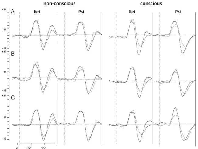
Mean of grand averages over both hemisphere of the P100 and N170 ERP during non-conscious and conscious pro-cessing are shown in Fig.4.
P100 event-related potential
Repeated-measures ANOVA on the P100 amplitudes revealed generally more pronounced P100 amplitudes over right compared to left electrodes in both the S-ketamine [F (1,20)018.23; p<0.001; η2
p00.48] and the psilocybin group
[F(1,20)022.22; p<0.001; η2p00.53]. However, no treat-ment effects were observed for the P100 amplitudes in the psilocybin [F(1,20)00.099; p00.13; η2
p03.00] and-ketamine
group F(1,20)00.011; p<0.917; η2
p00.001]. Comparing the
effect of S-ketamine and psilocybin, repeated-measures ANOVA on the change scores for the P100 amplitude showed no main effects and interactions, reflecting an equal influence of S-ketamine and psilocybin on the P100 amplitude.
N170 event-related potential
Repeated-measures ANOVA on the S-ketamine data revealed that N170 amplitudes were generally more pro-nounced over right compared to left electrodes and more pronounced for emotional relative to neutral faces, as indi-cated by a significant main effect for laterality (F(1,20)0 25.97; p<0.0001;η2
p00.56) and valence (F(2,40)05.47; p<
0.01;η2p00.21). Furthermore, a main effect of treatment was
found (F(1, 20)08.73; p<0.01; η200.30), reflecting the overall N170 reduction under S-ketamine. The treatment× laterality interaction (F(1,20)046.70; p<0.00001; η2
p00.71)
showed that the treatment effect occurred only over right electrodes (p<0.000001) (Fig.5, left), but not over left elec-trodes (p00.14). In general, the S-ketamine effect on the N170 amplitude was more pronounced during conscious (p < 0.00001) (mean difference−1.1 μV) than non-conscious pro-cessing (p<0.001) (mean difference−0.6 μV), indicated by Fig. 2 Sensitivity indices (d′)±
SE represented as a function of target duration during fear discrimination. Notably, both S-ketamine and psilocybin signif-icantly reduced d′ values for fearful relative to neutral faces
Fig. 3 Sensitivity indices (d′)± SE represented as a function of target duration during the discrimination of happy faces. Notably, S-ketamine but not psilocybin significantly reduced d′ values for happy relative to neutral faces
treatment×target duration interaction (F(1,20)04.75; p<0.05; η2
p00.19). No treatment×valence interaction was found for
S-ketamine on the N170 (F(1,20)01.88; p00.1.66; η2
p00.086).
Repeated-measures ANOVA on the psilocybin data revealed significant main effects for laterality (F(1, 20)0 39.43; p<0.00001;η2
p00.66) and treatment (F(1,20)014.21; Fig. 4 Mean of grand averages over both hemispheres of the P100 and
N170 ERP for fearful (a), neutral (b), and happy faces (c) during non-conscious (left) and non-conscious processing (right) following placebo
(solid line), S-ketamine (Ket: dashed line), and psilocybin (Psi: dotted line) administration, respectively
Fig. 5 Mean N170 amplitudes± SE over right electrodes for fearful, neutral, and happy faces under S-ketamine (red line), psi-locybin (blue), and placebo (green). Note: Significant differ-ences between treatment condi-tions at *p<0.01 and at **p<0.00001
p < 0.01; η2
p00.42), reflecting the more pronounced
re-sponse over right relative to left electrodes (p<0.00001) and the general N170 reduction induced by psilocybin (p<0.01). This treatment effect was found only over right electrodes (F (1,20)06.61; p<0.05; η2
p00.25). The main effect of valence
indicated the more pronounced N170 amplitude for emotional compared to neutral faces (F(2, 40)09.79; p< 0.001;η2
p00.33). Furthermore, the laterality×treatment×target
duration interaction (F(1, 20)04.52; p<0.05; η2 p00.18)
revealed that the N170 effect over right electrodes was more pronounced during conscious (p < 0.000001) (mean difference−1.38 μV) than non-conscious processing (p< 0.01) (mean difference−0.71 μV). Finally, as shown by a significant treatment × valence interaction (F(2, 40)05.92; p < 0.01; η2
p00.23), psilocybin significantly reduced the
N170 amplitudes in response to fearful (p < 0.000001) and neutral faces (p < 0.01), but not to happy faces (p00.1) (Fig. 5, right).
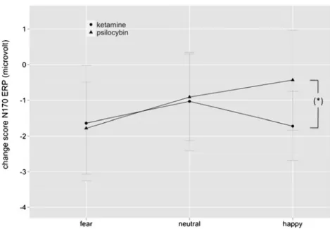
Comparing the S-ketamine and psilocybin effects on the N170, repeated-measures ANOVA on the change scores revealed significant main effects for target duration (F(1, 40)06.53; p<0.05; η2 p00.14) (F(1,40)06.53; p<0.05; η2p0 0.14), laterality (F(1,40)034.29; p<0.00001; η2 p00.46), and valence (F(2,80)05.15; p<0.01; η2 p00.11). LSD post hoc
testing revealed that in general both drug effects were more pronounced over right than left electrodes (p<0.00001) and more pronounced during conscious than non-conscious pro-cessing (p<0.05). Most intriguingly in regard to the contri-bution of glutamate and serotonin to emotional processing, a laterality×valence×group interaction was found (F(2,80)0 4.02; p<0.05; η2
p00.09). Particularly, although S-ketamine
and psilocybin equally modulated the N170 amplitudes in response to fearful (p00.78) and neutral faces (p00.82) over right electrodes, happy faces were differentially modulated by S-ketamine and psilocybin (p<0.05) (Fig.6).
Finally, we asked whether the drug-induced changes in the behavioral performances (d′ fear and d′ happy) might be explained through the drug effects on the N170 over right electrodes using linear regression analysis. For both groups, we found no significant relations between d′ fear and N170 change score for fearful faces (psilocybin group: F00.28, p00.757; S-ketamine group: F02.23, p00.136), as well as between d′ happy and N170 change score for happy faces (psilocybin group: F01.25, p00.311; S-ketamine group: F00.974, p00.397).
Psychological assessment
Both S-ketamine and psilocybin produced similar altera-tions on the global ASC scores (Fig. 7). Repeated-measures ANOVA on the ASC data showed significant main
effects for treatment (F(1,40)080.99; p<0.00001; η2 p00.67)
and scale (F(11,440)09.73; p<0.000001; η2
p00.20). A triple
treatment×scale×group interaction indicated significant dif-ferences between both drug effects on specific scales [F(11, 440)05.35; p<0.00001; η2
p00.12]. Post hoc testing showed
that S-ketamine increased all scales relative to placebo (p’s< 0.01), expect for anxiety (p00.09), while psilocybin increased all scales (p’s<0.01), expect for auditory alterations (p00.42) and anxiety (p00.36). Moreover, LSD post hoc analysis showed that S-ketamine produced significantly higher scores than psilocybin for disembodiment (p<0.000001), auditory alterations (p<0.05), and for impaired control and cognition (p<0.05). Otherwise, psilocybin produced more pronounced visual illusions and elementary hallucinations than S-ketamine as indexed by the elementary imagery score (p<0.01).
Discussion
In this paper, we present a suitable approach to compare glutamatergic and serotonergic effects on emotional face processing. Specifically, according to the firstly assessed discrimination thresholds, we used event-related potential recording to investigate the effect of the NMDA receptor antagonist S-ketamine and the 5-HT receptor agonist psilo-cybin either on conscious or non-conscious emotional face processing. Our study provide three major results: Firstly, the structural encoding of facial configurations as expressed by the N170 ERP is impaired by S-ketamine and psilocybin, while the fast extraction of visual emotion-related informa-tion (i.e., P100 ERP) is not affected by both drugs. Second-ly, the N170 ERP is differentially modulated by S-ketamine and psilocybin. Although both drugs reduce the N170 ERP responses to fearful faces, the structural encoding of happy faces is only impaired by S-ketamine. Finally, both drug effects on the N170 ERP are more pronounced during conscious than non-conscious processing. In the follow-ing, we discuss our results and consider their potential implications.
On the behavioral level, regardless of threshold setting, S-ketamine and psilocybin differentially affected facial dis-crimination performances. Particularly, the subjective ability to discriminate fearful from neutral face identities was im-paired following both S-ketamine and psilocybin adminis-tration. In contrast, the discrimination performance of happy relative to neutral faces was only affected by S-ketamine but not by psilocybin, suggesting that the effect of psilocybin is valence specific on the behavioral level.
On the electrophysiological level, the N170 amplitude was more pronounced for emotional relative to neutral faces in both groups without any drug intake (i.e., under placebo),
reflecting an emotional face processing bias as previously reported (Blau et al.2007; Jaworska et al.2010; Krombholz et al.2007; Pegna et al. 2008; Schyns et al. 2007; Smith
2011). Psilocybin impaired the structural encoding of fearful faces as expressed by reduced N170 responses to fearful faces, while no psilocybin-induced N170 alteration in re-sponse to happy faces was found in this study. Thus, psilo-cybin shifted the negative N170 processing bias seen under placebo. These findings differ from previous studies report-ing that the SSRI citalopram does not acutely modulate the N170 ERP in response to emotional faces in healthy sub-jects (Kerestes et al.2009; Labuschagne et al.2010), where-as later ERPs such where-as the N250 or the LPP, which have been
associated with “expression decoding,” are modulated by citalopram (Kerestes et al.2009; Labuschagne et al.2010). Beyond methodological differences across these studies such as the use of the reference electrodes, the differential effect of psilocybin and citalopram on the N170 ERP may be due to the different pharmacological effects of psilocybin and citalopram on the 5-HT system. While citalopram over-all increases 5-HT brain levels (Elliott et al.2011; Nutt et al.
1999), psilocin, the active metabolite of psilocybin, is an agonists at 5-HT receptors which binds with high affinity specifically at 5-HT1, HT2, and 5-HT6 receptors (Nichols
2004). That the different pharmacological profiles of psilo-cybin and citalopram on the 5-HT system may be most Fig. 7 Effects of S-ketamine
(dashed line) and psilocybin (dotted line) on the ASC scales. Mean scores and ±SE (both n021). Note: *p<0.05, **p< 0.01, and ***p<0.000001 indi-cate significant differences be-tween drugs. Symptoms scores were expressed as percent of scale maximum
Fig. 6 Mean change scores of N170 ERP±SE as a function of face valence over right parieto-occipital electrodes. Notably, the N170 ERP reduction for fearful and neutral faces was compara-ble among both drugs, but the N170 ERP for happy faces was significantly more reduced after S-ketamine (circle) than psilocy-bin exposure (triangle). *p<0.05 indicates a significant difference between the effect of S-ketamine and psilocybin on happy faces
critical in mediating their N170 effects on emotional face processing is further supported by the finding that decreas-ing brain 5-HT levels by acute tryptophan depletion (ATD) did not affect the N170 during face processing as well (Jaworska et al.2010). Along this line, the P100 ERP is also differently altered by psilocybin and ATD. Particularly, while ATD enhances the P100 for sad versus joyful faces (Jaworska et al.2010), in this study no P100 alteration to facial expressions was found under psilocybin. Thus, the sensitivity of the P100 and N170 ERP may differentially depend on 5-HT brain levels and the activation of a set of different 5-HT receptors subtypes.
Discussing the effect of S-ketamine on the visual ERPs, the only work with reference to our result is a previous fMRI study, which explored the functional network following ket-amine administration during emotional face processing (Abel et al.2003). The key finding of this study was that the amygdala and fusiform gyrus (FG) activity in response to fearful faces under placebo was abolished following ket-amine administration. The authors suggested that this ketamine-induced effect on limbic and visual regions is associated with the emotional blunting and depersonaliza-tion phenomena that are evident in ketamine states (Krystal et al.1994; Vollenweider et al.1997). Such an interpretation is consistent with the present finding that S-ketamine re-duced the N170 ERP not only in response to fearful but also to neutral and happy faces, reflecting an overall emotional blunting of visually induced neural responses.
According to several source localization studies (Deffke et al. 2007; Rossion et al. 2003; Sadeh et al. 2010), the generators of the N170 ERP have been localized to the FG, which encodes the structural configuration of facial features. The significance of structural information encoding in visual perception is shown by a functional relationship between object discrimination performance and FG activity. In par-ticular, a previous study found that FG activity increases gradually with subjective rating of recognition success (Bar et al. 2001). An identical relationship was also suggested following citalopram administration in healthy subjects (Harmer et al.2003). It has been proposed that the enhanced fear detection in healthy subjects treated with citalopram (Harmer et al. 2003) may be partly due to an increased activity in the FG (Del-Ben et al.2005). Albeit not statisti-cally underpinned, we observed that both drug effects on the subjective discrimination performances correspond approx-imately to the electrophysiological changes on the N170 following drug administration (cf. Figs. 2, 3, and 5). These findings suggest that the relationship between discrimination success and FG activity/N170 amplitude might remain following manipulation of the 5-HT and NMDAR system. However, the lack of a statistical significance warrants further investigations to strengthen this relationship.
Neurofunctionally, increased visual evoked responses to relevant emotional expressions are likely mediated via rich interconnections between the FG and the amygdala (Amaral et al.2003; Freese and Amaral2005), the coupling of which is additionally strengthened during attentive viewing of affective faces (Fairhall and Ishai 2007; Herrington et al.
2011). Furthermore, emotional face processing also involves prefrontal areas, which are functionally connected with the FG and the amygdala (Dima et al.2011). Critically, both S-ketamine and psilocybin were found to deactivate limbic and to increase prefrontal neural activity during resting states in healthy subjects (Vollenweider and Kometer
2010). Thus, it is arguable that the psilocybin- and S-ketamine-induced N170 effects in response to fearful faces may be due to functional alterations in the amygdala-prefrontal network. However, why psilocybin and S-ketamine had dissociable effects on happy face processing is difficult to derive from the present data. A possible explanation could be that S-ketamine and psilocybin differ-entially modulate circuitries responsible for the processing of positive expressions because the processing of positive information such as happy faces also involves reward-related areas (Adolphs 2003; Ishai 2007; Singer et al.
2004) relative to the processing of negative information and further because the N170 showed priming effects as a function of reward (Marini et al. 2011). However, this is highly speculative at the present time.
Another key finding of this study was further that the psilocybin- and S-ketamine-induced N170 effect was more pronounced during conscious than non-conscious visual processing independent of the face expression. This finding fits with the assumption that the N170 is associated with perceptual consciousness of the face (Rossion and Jacques
2008) and that FG responses are modulated by the level of target visibility (Pessoa et al. 2006; Pessoa and Padmala
2005). Furthermore, numerous studies have described an increase of the N170 ERP with selective attention (Gazzaley et al. 2005; Wronka and Walentowska 2011), suggesting top-down attentional control. In particular, the visual cortex receives top-down modulation from frontal and parietal areas in relation to visual attention (Bressler et al.2008) in the time range of the N170 ERP (Rose et al.2005). In this view, several studies reported that psilocybin attenuates attentional performances (Carter et al. 2005; Gouzoulis-Mayfrank et al.2002; Quednow et al.2011). A recent study examining the influence of psilocybin on the spatiotemporal dynamics of object completion and found a dose-dependent reduction of the N170 ERP response (Kometer et al.2011). The authors suggested that this reduction might reflect a psilocybin-induced failure to allocate attention. Similarly, previous evidence revealed that ketamine produce cognitive deficits including impairments of attention (Morgan et al.
more pronounced effects of psilocybin and S-ketamine on the N170 during conscious relative to non-conscious pro-cessing indicate a drug-induced reduction of attentional resources. One point of contention, however, may be that there is some evidence of conscious perception of happy faces with 20 ms in this study. This means that we cannot infer from the discrimination threshold task that 10 ms is truly non-conscious for the processing of happy faces. How-ever, recent ERP studies confirmed presentation times be-low 20 ms for non-conscious processing of happy faces (Balconi and Lucchiari 2008; Pegna et al. 2011; Smith
2011). Therefore, it is conceivable to assume that presenta-tion times of 10 ms as used in our ERP experiment are really non-conscious also for the processing of happy faces.
Summarized, this study demonstrated that the NMDA and 5-HT receptor systems differentially contribute to the structural encoding of emotional face expressions as expressed by the N170 event-related potential. Our findings confirm the emotion sensitivity of the N170 ERP during conscious and non-conscious face processing as recently reported (Pegna et al.2008; Smith2011) and further suggest that the assessment of early visual evoked responses might allow detecting pharmacologically induced change in emo-tional processing biases and provides thus a suitable frame-work to study pathophysiological mechanisms underlying dysfunctional emotional biases.
Acknowledgments The present study was supported by the Swiss Neuromatrix Foundation (AS, MK, RB, FXV) and the Heffter Research Center Zurich (FXV).
Conflict of interest None of the authors have any financial, personal, or organizational conflicts of interest to report in relation to this manuscript.
References
Abel KM, Allin MP, Kucharska-Pietura K, David A, Andrew C, Williams S, Brammer MJ, Phillips ML (2003) Ketamine alters neural processing of facial emotion recognition in healthy men: an fMRI study. Neuroreport 14:387–391
Adolphs R (2003) Cognitive neuroscience of human social behaviour. Nat Rev Neurosci 4:165–178
Amaral DG, Behniea H, Kelly JL (2003) Topographic organization of projections from the amygdala to the visual cortex in the macaque monkey. Neuroscience 118:1099–1120
Ashley V, Vuilleumier P, Swick D (2004) Time course and specificity of event-related potentials to emotional expressions. Neuroreport 15:211–216
Balconi M, Lucchiari C (2008) Consciousness and arousal effects on emotional face processing as revealed by brain oscillations. A gamma band analysis. Int J Psychophysiol 67:41–46
Bar M, Tootell RB, Schacter DL, Greve DN, Fischl B, Mendola JD, Rosen BR, Dale AM (2001) Cortical mechanisms specific to explicit visual object recognition. Neuron 29:529–535
Batty M, Taylor MJ (2003) Early processing of the six basic facial emotional expressions. Brain Res Cogn Brain Res 17:613–620
Bernat E, Bunce S, Shevrin H (2001) Event-related brain potentials differentiate positive and negative mood adjectives during both supraliminal and subliminal visual processing. Int J Psychophy-siol 42:11–34
Bhagwagar Z, Cowen PJ, Goodwin GM, Harmer CJ (2004) Normali-zation of enhanced fear recognition by acute SSRI treatment in subjects with a previous history of depression. Am J Psychiatry 161:166–168
Blau VC, Maurer U, Tottenham N, McCandliss BD (2007) The face-specific N170 component is modulated by emotional facial ex-pression. Behav Brain Funct 3:7
Bressler SL, Tang W, Sylvester CM, Shulman GL, Corbetta M (2008) Top-down control of human visual cortex by frontal and parietal cortex in anticipatory visual spatial attention. J Neurosci 28:10056–10061
Brosch T, Sander D, Pourtois G, Scherer KR (2008) Beyond fear: rapid spatial orienting toward positive emotional stimuli. Psychol Sci 19:362–370
Browning M, Reid C, Cowen PJ, Goodwin GM, Harmer CJ (2007) A single dose of citalopram increases fear recognition in healthy subjects. J Psychopharmacol 21:684–690
Carter OL, Burr DC, Pettigrew JD, Wallis GM, Hasler F, Vollenweider FX (2005) Using psilocybin to investigate the relationship be-tween attention, working memory, and the serotonin 1A and 2A receptors. J Cogn Neurosci 17:1497–1508
Deffke I, Sander T, Heidenreich J, Sommer W, Curio G, Trahms L, Lueschow A (2007) MEG/EEG sources of the 170-ms response to faces are co-localized in the fusiform gyrus. NeuroImage 35:1495–1501
Degabriele R, Lagopoulos J, Malhi G (2011) Neural correlates of emotional face processing in bipolar disorder: an event-related potential study. J Affect Disord 133:212–220
Del-Ben CM, Deakin JF, McKie S, Delvai NA, Williams SR, Elliott R, Dolan M, Anderson IM (2005) The effect of citalopram pretreat-ment on neuronal responses to neuropsychological tasks in nor-mal volunteers: an FMRI study. Neuropsychopharmacology 30:1724–1734
Derogatis L (1994) SCL-90-R: Symptom Checklist-90-R. Administra-tion, scoring and procedures manual. National Computer Sys-tems, Minneapolis
Diazgranados N, Ibrahim L, Brutsche NE, Newberg A, Kronstein P, Khalife S, Kammerer WA, Quezado Z, Luckenbaugh DA, Salvadore G, Machado-Vieira R, Manji HK, Zarate CA (2010) A randomized add-on trial of an N-methyl-D-aspartate antagonist in treatment-resistant bipolar depression. Arch Gen Psychiatry 67:793–802
Dima D, Stephan KE, Roiser JP, Friston KJ, Frangou S (2011) Effec-tive connectivity during processing of facial affect: evidence for multiple parallel pathways. J Neurosci 31:14378–14385 Dittrich A (1975) Zusammenstellung eines Fragebogens (APZ) zur
Erfassung abnormer psychischer Zustände [Construction of a questionnaire (APZ) for assessing abnormal mental states]. Z Klin Psychol Psychiatr Psychother 23:12–20
Dittrich A (1998) The standardized psychometric assessment of altered states of consciousness (ASCs) in humans. Pharmacopsychiatry 31:80–84 Eimer M, Holmes A (2002) An ERP study on the time course of
emotional face processing. Neuroreport 13:427–431
Ekman P, Friesen W (1976) Pictures of facial affect. Consulting Psy-chologists, Palo Alto
Elliott R, Zahn R, Deakin JF, Anderson IM (2011) Affective cognition and its disruption in mood disorders. Neuropsychopharmacology 36:153–182
Fairhall SL, Ishai A (2007) Effective connectivity within the distributed cortical network for face perception. Cereb Cortex 17:2400–2406 Feng N, Vollenweider FX, Minder EI, Rentsch K, Grampp T,
mass spectrometry method for determination of ketamine in plas-ma and its application to huplas-man samples. Ther Drug Monit 17:95–100
Fichtenholtz HM, Hopfinger JB, Graham R, Detwiler JM, LaBar KS (2009) Event-related potentials reveal temporal staging of dynam-ic facial expression and gaze shift effects on attentional orienting. Soc Neurosci 4:317–331
Freese JL, Amaral DG (2005) The organization of projections from the amygdala to visual cortical areas TE and V1 in the macaque monkey. J Comp Neurol 486:295–317
Frühholz S, Jellinghaus A, Herrmann M (2011) Time course of implicit processing and explicit processing of emotional faces and emo-tional words. Biol Psychol 87:265–274
Gazzaley A, Cooney JW, McEvoy K, Knight RT, D’Esposito M (2005) Top-down enhancement and suppression of the magnitude and speed of neural activity. J Cogn Neurosci 17:507–517
Gouzoulis-Mayfrank E, Thelen B, Maier S, Heekeren K, Kovar KA, Sass H, Spitzer M (2002) Effects of the hallucinogen psilocybin on covert orienting of visual attention in humans. Neuropsychobi-ology 45:205–212
Grob CS, Danforth AL, Chopra GS, Hagerty M, McKay CR, Halberstadt AL, Greer GR (2011) Pilot study of psilocybin treatment for anxiety in patients with advanced-stage cancer. Arch Gen Psychiatry 68:71–78
Harmer CJ (2008) Serotonin and emotional processing: does it help explain antidepressant drug action? Neuropharmacology 55:1023–1028
Harmer CJ, Bhagwagar Z, Perrett DI, Völlm BA, Cowen PJ, Goodwin GM (2003) Acute SSRI administration affects the processing of social cues in healthy volunteers. Neuropsychopharmacology 28:148–152
Harmer CJ, Shelley NC, Cowen PJ, Goodwin GM (2004) Increased positive versus negative affective perception and memory in healthy volunteers following selective serotonin and norepineph-rine reuptake inhibition. Am J Psychiatry 161:1256–1263 Hasler F, Bourquin D, Brenneisen R, Vollenweider FX (2002) Renal
excretion profiles of psilocin following oral administration of psilocybin: a controlled study in man. J Pharm Biomed Anal 30:331–339
Hasler F, Grimberg U, Benz M, Huber T, Vollenweider F (2004) Acute psychological and physiological effects of psilocybin in healthy humans: a double-blind, placebo-controlled dose–effect study. Psychopharmacology (Berl) 172:145–156
Herrington JD, Taylor JM, Grupe DW, Curby KM, Schultz RT (2011) Bidirectional communication between amygdala and fusiform gyrus during facial recognition. NeuroImage 56:2348–2355 Ishai A (2007) Sex, beauty and the orbitofrontal cortex. Int J
Psycho-physiol 63:181–185
Itier RJ, Taylor MJ (2004) N170 or N1? Spatiotemporal differences between object and face processing using ERPs. Cereb Cortex 14:132–142
Jaworska N, Thompson A, Shah D, Fisher D, Ilivitsky V, Knott V (2010) Electrocortical effects of acute tryptophan depletion on emotive facial processing in depression-prone individuals. Eur Neuropsychopharmacol 20:473–486
Kemp AH, Gray MA, Line P, Silberstein RB, Nathan PJ (2003) Prelim-inary electrophysiological evidence for modulation of the process-ing of negative effect by serotonin. Brain Cogn 51:198–200 Kemp AH, Gray MA, Silberstein RB, Armstrong SM, Nathan PJ
(2004) Augmentation of serotonin enhances pleasant and suppresses unpleasant cortical electrophysiological responses to visual emotional stimuli in humans. NeuroImage 22:1084– 1096
Kerestes R, Labuschagne I, Croft RJ, O’Neill BV, Bhagwagar Z, Phan KL, Nathan PJ (2009) Evidence for modulation of facial emo-tional processing bias during emoemo-tional expression decoding by
serotonergic and noradrenergic antidepressants: an event-related potential (ERP) study. Psychopharmacology (Berl) 202:621–634 Kometer M, Schmidt A, Bachmann R, Studerus E, Seifritz E, Vollenweider FX (2012) Psilocybin Biases Facial Recognition, Goal-Directed Behavior, and Mood State Toward Positive Relative to Negative Emotions Through Different Serotonergic Subreceptors. Biol Psychiatry. doi:10.1016/j.biopsych.2012.04.005
Krolak-Salmon P, Fischer C, Vighetto A, Mauguière F (2001) Process-ing of facial emotional expression: spatio-temporal data as assessed by scalp event-related potentials. Eur J Neurosci 13:987–994
Krombholz A, Schaefer F, Boucsein W (2007) Modification of N170 by different emotional expression of schematic faces. Biol Psy-chol 76:156–162
Krystal J, Karper L, Seibyl J, Freeman G, Delaney R, Bremner J, Heninger G, Bowers MJ, Charney D (1994) Subanesthetic effects of the noncompetitive NMDA antagonist, ketamine, in humans. Psychotomimetic, perceptual, cognitive, and neuroendocrine responses. Arch Gen Psychiatry 51:199–214
Labuschagne I, Croft RJ, Phan KL, Nathan PJ (2010) Augmenting serotonin neurotransmission with citalopram modulates emotional expression decoding but not structural encoding of moderate intensity sad facial emotional stimuli: an event-related potential (ERP) investigation. J Psychopharmacol 24:1153–1164
Lee TW, Girolami M, Sejnowski TJ (1999) Independent component analysis using an extended infomax algorithm for mixed subgaus-sian and supergaussubgaus-sian sources. Neural Comput 11:417–441 Macmillan N, Creelman C (1991) Detection theory: a user’s guide.
Cambridge University Press, Cambridge
Marini F, Marzi T, Viggiano MP (2011) “Wanted!” The effects of reward on face recognition: electrophysiological correlates. Cogn Affect Behav Neurosci 11:627–643
Morgan CJ, Mofeez A, Brandner B, Bromley L, Curran HV (2004) Acute effects of ketamine on memory systems and psychotic symptoms in healthy volunteers. Neuropsychopharmacology 29:208–218 Nathan PJ, Kemp AH, Harrison BJ (2003) Antidepressants and
emo-tional processing. Neuropsychopharmacology 28:1383, author reply 1384–5
Newcomer JW, Farber NB, Jevtovic-Todorovic V, Selke G, Melson AK, Hershey T, Craft S, Olney JW (1999) Ketamine-induced NMDA receptor hypofunction as a model of memory impairment and psychosis. Neuropsychopharmacology 20:106–118
Nichols DE (2004) Hallucinogens. Pharmacol Ther 101:131–181 Noll LK, Mayes LC, Rutherford HJ (2012) Investigating the impact of
parental status and depression symptoms on the early perceptual coding of infant faces: an event-related potential study. Soc Neu-rosci. doi:10.1080/17470919.2012.672457
Nutt DJ, Forshall S, Bell C, Rich A, Sandford J, Nash J, Argyropoulos S (1999) Mechanisms of action of selective serotonin reuptake inhibitors in the treatment of psychiatric disorders. Eur Neuro-psychopharmacol 9(Suppl 3):S81–S86
Passie T, Seifert J, Schneider U, Emrich HM (2002) The pharmacology of psilocybin. Addict Biol 7:357–364
Pegna AJ, Landis T, Khateb A (2008) Electrophysiological evidence for early non-conscious processing of fearful facial expressions. Int J Psychophysiol 70:127–136
Pegna AJ, Darque A, Berrut C, Khateb A (2011) Early ERP modula-tion for task-irrelevant subliminal faces. Front Psychol 2:88 Pessoa L, Padmala S (2005) Quantitative prediction of perceptual
decisions during near-threshold fear detection. Proc Natl Acad Sci U S A 102:5612–5617
Pessoa L, Japee S, Sturman D, Ungerleider LG (2006) Target visibility and visual awareness modulate amygdala responses to fearful faces. Cereb Cortex 16:366–375
Pourtois G, Vuilleumier P (2006) Dynamics of emotional effects on spatial attention in the human visual cortex. Prog Brain Res 156:67–91
Pourtois G, Grandjean D, Sander D, Vuilleumier P (2004) Electrophys-iological correlates of rapid spatial orienting towards fearful faces. Cereb Cortex 14:619–633
Quednow BB, Kometer M, Geyer MA, Vollenweider FX (2011) Psilocybin-induced deficits in automatic and controlled inhibition are attenuated by ketanserin in healthy human volunteers. Neuro-psychopharmacology 37:630–640
Rose M, Schmid C, Winzen A, Sommer T, Büchel C (2005) The functional and temporal characteristics of top-down modulation in visual selection. Cereb Cortex 15:1290–1298
Rossion B, Jacques C (2008) Does physical interstimulus variance ac-count for early electrophysiological face sensitive responses in the human brain? Ten lessons on the N170. NeuroImage 39:1959–1979 Rossion B, Joyce CA, Cottrell GW, Tarr MJ (2003) Early lateralization and orientation tuning for face, word, and object processing in the visual cortex. NeuroImage 20:1609–1624
Sadeh B, Podlipsky I, Zhdanov A, Yovel G (2010) Event-related potential and functional MRI measures of face-selectivity are highly correlated: a simultaneous ERP-fMRI investigation. Hum Brain Mapp 31:1490–1501
Santesso DL, Meuret AE, Hofmann SG, Mueller EM, Ratner KG, Roesch EB, Pizzagalli DA (2008) Electrophysiological correlates of spatial orienting towards angry faces: a source localization study. Neuropsychologia 46:1338–1348
Schmidt A, Bachmann R, Kometer M, Csomor PA, Stephan KE, Seifritz E, Vollenweider FX (2011) Mismatch negativity encoding of prediction errors predicts S-ketamine-induced cognitive impair-ments. Neuropsychopharmacology 37:865–875
Schneider W, Eschman A, Zuccolotto A (2002) E-prime reference guide. Psychology Software Tools, Pittsburgh
Schultz RT, Grelotti DJ, Klin A, Kleinman J, Van der Gaag C, Marois R, Skudlarski P (2003) The role of the fusiform face area in social cognition: implications for the pathobiology of autism. Philos Trans R Soc Lond B Biol Sci 358:415–427
Schyns PG, Petro LS, Smith ML (2007) Dynamics of visual informa-tion integrainforma-tion in the brain for categorizing facial expressions. Curr Biol 17:1580–1585
Sheehan D, Lecrubier Y, Sheehan K, Amorim P, Janavs J, Weiller E, Hergueta T, Baker R, Dunbar G (1998) The Mini-International Neuropsychiatric Interview (M.I.N.I.): the development and val-idation of a structured diagnostic psychiatric interview for DSM-IV and ICD-10. J Clin Psychiatry 59:22–33
Singer T, Kiebel SJ, Winston JS, Dolan RJ, Frith CD (2004) Brain responses to the acquired moral status of faces. Neuron 41:653–662
Smith ML (2011) Rapid processing of emotional expressions without conscious awareness. Cereb Cortex 22:1748–1760
Sokhadze EM, Tasman A, Tamas R, El-Mallakh RS (2011) Event-related potential study of the effects of emotional facial expres-sions on task performance in euthymic bipolar patients. Appl Psychophysiol Biofeedback 36:1–13
Sprengelmeyer R, Jentzsch I (2006) Event related potentials and the perception of intensity in facial expressions. Neuropsychologia 44:2899–2906
Stekelenburg JJ, de Gelder B (2004) The neural correlates of perceiv-ing human bodies: an ERP study on the body-inversion effect. Neuroreport 15:777–780
Studerus E, Gamma A, Vollenweider F (2010) Psychometric evalua-tion of the altered states of consciousness rating scale (OAV). PLoS One 5:e12412
Studerus E, Kometer M, Hasler F, Vollenweider FX (2011) Acute, subacute and long-term subjective effects of psilocybin in healthy humans: a pooled analysis of experimental studies. J Psychophar-macol 25:1434–1452
Tamietto M, de Gelder B (2010) Neural bases of the non-conscious perception of emotional signals. Nat Rev Neurosci 11:697–709 Vollenweider F, Kometer M (2010) The neurobiology of psychedelic
drugs: implications for the treatment of mood disorders. Nat Rev Neurosci 11:642–651
Vollenweider FX, Leenders KL, Scharfetter C, Antonini A, Maguire P, Missimer J, Angst J (1997) Metabolic hyperfrontality and psy-chopathology in the ketamine model of psychosis using positron emission tomography (PET) and [18F]fluorodeoxyglucose (FDG). Eur Neuropsychopharmacol 7:9–24
Vuilleumier P, Pourtois G (2007) Distributed and interactive brain mechanisms during emotion face perception: evidence from func-tional neuroimaging. Neuropsychologia 45:174–194
Williams LM, Liddell BJ, Rathjen J, Brown KJ, Gray J, Phillips M, Young A, Gordon E (2004) Mapping the time course of noncon-scious and connoncon-scious perception of fear: an integration of central and peripheral measures. Hum Brain Mapp 21:64–74
Wittchen HU, Pfister H (1997) DIA-X-Interviews: Manual für Screening-Verfahren und Interview. Swets & Zeitlinger, Frankfurt
Wronka E, Walentowska W (2011) Attention modulates emotional expression processing. Psychophysiology 48:1047–1056 Zarate CA, Singh JB, Carlson PJ, Brutsche NE, Ameli R, Luckenbaugh
DA, Charney DS, Manji HK (2006) A randomized trial of an N-methyl-D-aspartate antagonist in treatment-resistant major de-pression. Arch Gen Psychiatry 63:856–864