O R I G I N A L A R T I C L E
Hassan Sadri Æ Thai Nguyen-Tang Æ Richard Stern Pierre Hoffmeyer Æ Robin Peter
Control of severe hemorrhage using C-clamp and arterial embolization
in hemodynamically unstable patients with pelvic ring disruption
Received: 17 January 2004 / Published online: 24 June 2005
Ó Springer-Verlag 2005
Abstract Introduction Hemorrhage is the leading cause of death in patients with a pelvic fracture. The majority of blood loss derives from injured retroperitoneal veins and broad cancellous bone surfaces. The emergency management of multiply injured patients with pelvic ring disruption and severe hemorrhage remains controver-sial. Although it is well accepted that the displaced pelvic ring injury must be rapidly reduced and stabilized, the methods by which control of hemorrhagic shock is achieved remain under discussion. It has been proposed to exclusively use external pelvic ring stabilization for control of hemorrhage by producing a ‘tamponade effect’ of the pelvis. However, the frequency of clinically important arterial bleeding after external fixation of the pelvic ring remains unclear. We therefore undertook this retrospective review to attempt to answer this one important question: How frequently is arterial emboli-zation necessary to control hemorrhage and restore hemodynamic stability after external pelvic ring fixa-tion? Materials and methods We performed a retro-spective review of 55 consecutive patients who presented with unstable types B and C pelvic ring fractures. Those patients designated as being in hemorrhagic shock (defined as a systolic blood pressure less than 90 mmHg after receiving 2 L of intravenous crystalloid) were treated by application of the pelvic C-clamp. Patients who remained in hemorrhagic shock, or were deter-mined to be in severe shock (defined as mandatory catecholamines or more than 12 blood transfusions over
2 h), underwent therapeutic angiography within 24 h in order to control bleeding. Results Fourteen patients were identified as being hemodynamically unstable (ISS 30.1±11.3 points) and were treated with a C-clamp. In those patients with persistent hemodynamic instability, arterial embolization was performed. After C-clamp application, 5 of 14 patients required therapeutic angi-ography to control bleeding. Two patients died, one from multiple sources of bleeding and the other from an open pelvic fracture (total mortality 2/14, 14%). Conclusions Although the C-clamp is effective in con-trolling hemorrhage, one must be aware of the need for arterial embolization to restore hemodynamic stability in a select subgroup of patients.
Keywords Pelvic fracture Æ Pelvic hemorrhage Æ C-clamp Æ Pelvic angiography Æ Arterial embolization
Introduction
Hemorrhage associated with pelvic fractures presents a therapeutic challenge. While it is the leading cause of death (between 30% and 58%) [15, 23, 26] in patients with a pelvic fracture, it has been reported that among those who die, the pelvic fracture is the actual cause of death in less than 50% of cases [4]. The majority of blood loss is from injured retroperitoneal veins and bone, the latter accounted for by the broad cancellous surfaces at the fracture site. Only 20% of the deaths are associated with major arterial injury [3]. The search for the source of bleeding includes the thoracic cavity, intraperitoneal cavity, the retroperitoneal space, as well as the extremities.
The emergency management of multiply injured patients with pelvic ring disruption and severe hemor-rhage remains controversial. Although it is well accepted that the displaced pelvic ring injury must be rapidly re-duced and stabilized [10, 24], the methods by which control of hemorrhagic shock is achieved remain under
No benefits in any form have been received or will be received from a commercial party related directly or indirectly to the subject of this article. No funds were received in support of this study. H. Sadri Æ T. Nguyen-Tang Æ R. Stern (&) Æ P. Hoffmeyer R. Peter
Orthopaedic Surgery Service, University Hospital of Geneva, 24, rue Micheli-du-Crest, 1211 Geneva 14, Switzerland E-mail: [email protected] Tel.: +41-22-3727849 Fax: +41-22-3727799 DOI 10.1007/s00402-005-0821-7
discussion. It has been proposed to exclusively use external pelvic ring stabilization for control of hemor-rhage by producing a ‘tamponade effect’ of the pelvis [11,
24], and this can be obtained with the C-clamp [10,15]. Ertel et al. [9] have proposed pelvic packing when the C-clamp proves insufficient to control bleeding. It has been reported that 2–11% of all patients with unstable pelvic fractures require embolization due to persistent arterial bleeding [1, 2], and there are recommendations for vascular embolization to restore hemodynamic sta-bility without the use of external pelvic fixation [1, 19,
20]. However, the frequency of clinically important arterial bleeding after external fixation of the pelvic ring remains unclear. We therefore undertook this retro-spective review to attempt to answer this one important question: How frequently is arterial embolization nec-essary to control hemorrhage and restore hemodynamic stability after external pelvic ring fixation?
Patients and methods
From January 1, 1991, to December 31, 1999, 55 con-secutive patients with unstable pelvic fractures classified as subgroups of types B and C injuries according to Tile and Pennal [28] were admitted to our level I hospital for surgical management. Thirty-five patients were classified as polytrauma, defined as having at least 2 different injuries with one of these being life-threatening [7], and an Injury Severity Score (ISS) equal to or greater than 20 points [13]. The evaluation of the patient and resusci-tation protocol were performed according to ATLS guidelines [5]. All patients underwent ultrasound of the abdomen as part of our standard protocol. Further assessment included plain radiographs of the chest, pelvis, cervical and thoracolumbar spine.
Fourteen of these patients had hemorrhagic shock, defined as a systolic blood pressure less than 90 mmHg after receiving 2 L of intravenous crystalloid [1, 2]. In these patients, the pelvic ring was reduced and
main-tained by external fixation using a pelvic anti-shock C-clamp within 2 h of arrival at our hospital. In all pa-tients, the C-clamp was positioned according to the method of Ganz et al. [10] with the use of the image intensifier, providing anteroposterior, inlet and outlet views. In patients with a diastasis of the pubic symphysis, C-clamp application reduced this to less than 1 cm. In those patients with vertical and/or posterior displacement of the hemipelvis, C-clamp application was preceded by traction on the involved side lower extremity which was successful in achieving a satisfactory reduction within 1 cm of displacement. Patients who remained in hemor-rhagic shock, or were determined to be in severe shock (defined as mandatory catecholamines or more than 12 blood transfusions over 2 h [17]), underwent therapeutic angiography within 24 h in order to control bleeding. Our protocol did not change during the period of evalu-ation during this retrospective study. Patients were fol-lowed for a minimum of 2 weeks after admission to the hospital. These patients were out of the intensive care unit and either on the general orthopaedic ward or had been transferred to a rehabilitation facility.
Statistical analysis
Statistical analysis was made using SPSS software, ver-sion 8.0.1 (SPSS, Chicago, IL, USA). Descriptive statis-tics were used, and ISS scores are presented as mean and standard deviation. Wilcox signed rank test was used to compare the data before and after C-clamp application. The p-value reported is two-sided. The results were considered statistically signficant if p<0.05.
Results
The average age of the 55 patients was 36 years (range 16–66 years). There were 42 men and 13 women. The mechanisms of injury included motorcycle accidents
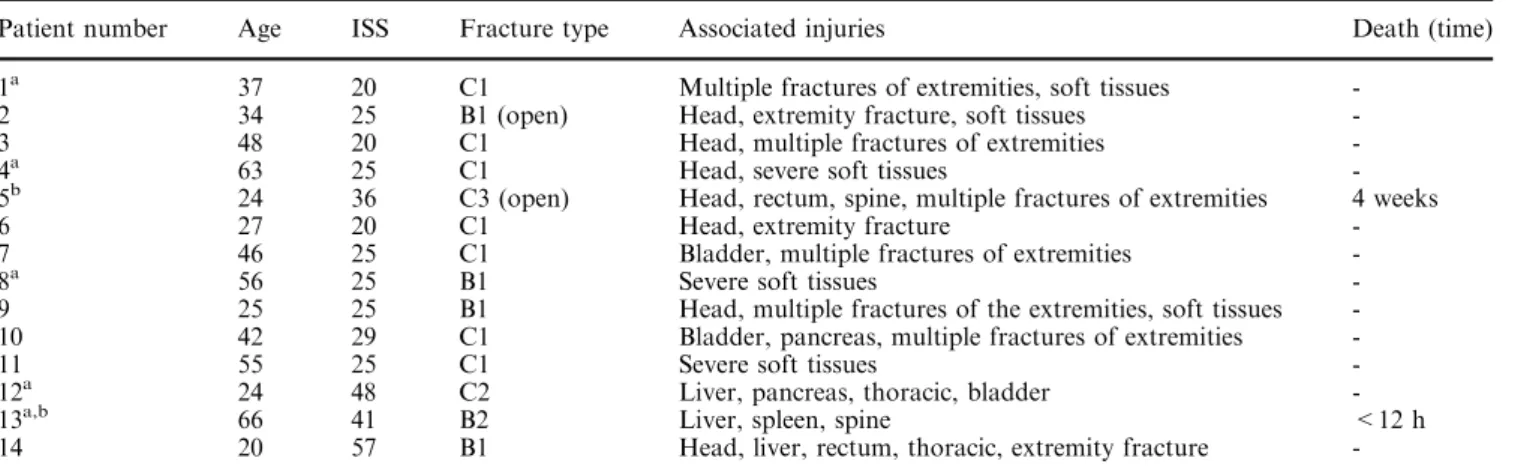
Table 1 Details of patients treated with C-clamp (ISS Injury Severity Score)
Patient number Age ISS Fracture type Associated injuries Death (time) 1a 37 20 C1 Multiple fractures of extremities, soft tissues
-2 34 25 B1 (open) Head, extremity fracture, soft tissues
-3 48 20 C1 Head, multiple fractures of extremities
-4a 63 25 C1 Head, severe soft tissues
-5b 24 36 C3 (open) Head, rectum, spine, multiple fractures of extremities 4 weeks
6 27 20 C1 Head, extremity fracture
-7 46 25 C1 Bladder, multiple fractures of extremities
-8a 56 25 B1 Severe soft tissues
-9 25 25 B1 Head, multiple fractures of the extremities, soft tissues -10 42 29 C1 Bladder, pancreas, multiple fractures of extremities
-11 55 25 C1 Severe soft tissues
-12a 24 48 C2 Liver, pancreas, thoracic, bladder
-13a,b 66 41 B2 Liver, spleen, spine <12 h
14 20 57 B1 Head, liver, rectum, thoracic, extremity fracture
-a
Arterial embolization
b
(n=20), falls from a great height (n=17), and motor vehicle accidents (n=12). Other patients were injured as pedestrians (n=3), in a tram (n=1) or bicycle accident (n=1), or following a crush injury (n=1). Analysis of the pelvic fractures showed 25 type B fractures (B1=16; B2=9) and 30 type C (C1=26, C2=2, C3=2). Mo-torcycle accidents were responsible for all the open fractures in this series (3 type B and 2 type C). Associ-ated injuries included: central nervous system (n=16), chest (n= 10), urogenital (n=13), spleen (n=5), liver (n=8), pancreas (n=7), rectal (n=3), sacral plexus (n=8) and injuries of the extremities (n=44).
Of these 55 consecutive patients, 14 with pelvic ring disruption and hemodynamic instability were treated with pelvic stabilization utilizing the C-clamp (Table1). The mean ISS of the entire patient population was 25.2±7.1 points. The mean ISS of this group of 14 pa-tients was 30.1±11.3 points. There were no C-clamp-related complications. These 14 patients received an average of 17 units of blood (range 6–50 units). Details concerning blood transfusions before and after C-clamp application are summarized in Table2. Although the C-clamp was efficient in most cases in stabilizing the pa-tients, the number of blood units transfused before and after C-clamp application was not statistically significant (p=0.308).
Seven patients underwent angiography. Two patients were excluded from our evaluation of angiography in controlling acute hemorrhage. One was a patient who
required angiography 28 days post-injury to control hemorrhage from the obturator artery after removal of a perineal drain which had been placed following drainage of a perineal abscess. An additional patient underwent angiography for evaluation of a cold lower extremity which was due to a dissection of the superficial femoral artery. In the remaining 5 patients, emergent arterial embolization within 24 h was required to control bleeding and restore hemodynamic stability. The char-acteristics of these patients, including time to emboli-zation, length of time for the procedure, and transfusion before and after angiography, are summarized in Table3. Therapeutic angiography was efficient in con-trolling bleeding in 4 out of 5 patients, but one patient (#13) who had sustained liver and spleen injuries and a mesenteric artery injury died within 12 h after arrival from multifocal exsanguinating hemorrhage. He was the only patient to have undergone a laparotomy. Another patient with an open type C pelvic fracture who had been initially stabilized with a pelvic clamp and con-comitant colostomy died 4 weeks after admission from septic shock. Global mortality overall was 7% (4/55), and among the patients treated with the C-clamp it was 14% (2/14 patients).
Discussion
Exsanguinating hemorrhage represents the major cause of death within the first 24 h after injury in multiply injured patients with pelvic ring fractures [12,21,25,27]. An average blood loss of 9.2 to 14.8 units has been recorded in patients with unstable pelvic ring fractures [4], with Dalal et al. [6] noting lower blood loss and transfusion requirements in patients sustaining lateral compression injuries compared with other types. A 3% mortality rate was reported in patients with normal hemodynamics, compared with a 38% mortality rate for those admitted in a hypotensive state [18]. For this reason, the immediate recognition of hemorrhagic shock and effective control of bleeding play a crucial role in patient survival.
The treatment of exsanguinating hemorrhage in multiply injured patients with pelvic ring disruption is controversial. Although acute reduction and external fixation of the disrupted pelvis are well accepted meth-ods [10, 24], which specific technique and device to use
Table 2 Details of blood units administered before and after C-clamp application
Patient Number of blood units before C-clamp application
Number of blood units after C-clamp application
1 4 10 2 8 9 3 3 2 4 5 10 5 14 14 6 3 2 7 9 8 8 4 6 9 7 10 10 10 7 11 4 3 12 7 11 13 7 43 14 15 9
Table 3 Details of patients requiring arterial embolization for hemodynamic instability Patient number Time to embolization (h) Number of units transfused before embolization Number of units transfused after embolization Arteries embolized Reason for angiography Death (time)
1 18 14 0 Obturator Hemorrhagic shock
-4 12 15 0 Lateral sacral Hemorrhagic shock
-8 1 9 1 Superior gluteal Severe shock
-12 2 16 2 Small branch of external iliac Severe shock
-13b 3 10 40 Superior gluteal; bilateral lumbar L3 Hemorrhagic shock <12 h
b
give rise to debate. Although we have not used this in the emergency setting, ORIF, such as bridge plating of the symphysis pubis of open book lesions, may also be useful in diminishing pelvic volume and thus controlling hemorrhage. Because of the critical hemodynamic con-dition of these patients, we favor only external devices that are easily applicable and can be used effectively. We prefer the C-clamp because of its mechanical superiority over an anterior frame, particularly in cases of type C injuries [22]. Although an anterior external fixator pro-vides sufficient stability in type B1 pelvic ring injuries (open book), the anteroposterior view of the pelvis obtained in the emergency room often does not permit one to distinguish it from a type C injury. In this study, hemodynamically unstable patients were rapidly trans-ferred to the operating room, where the C-clamp was assembled and applied within 20 min in all patients, thus achieving adequate stability of the pelvic ring quickly. This review, similar to others [9, 10, 15, 23, 26, 29], confirms the effectiveness of the C-clamp in the emer-gency situation in controlling the pelvic ring in patients with severe hemorrhage.
Control of retroperitoneal bleeding may be achieved in three ways, namely by: (1) decreasing blood loss from broad cancellous surfaces at fracture sites and lacerated veins and soft tissues by stabilizing the fracture and the forming hematoma; (2) limiting low pressure bleeding by normalization of pelvic volume and a retroperitoneal tamponade effect; and (3) localizing and controlling arterial bleeding sites via therapeutic angiography or surgical exploration. External pelvic fixation is effective in controlling hemorrhage by the first two mechanisms cited [11, 16, 24], while a recent study suggests that although low-pressure venous hemorrhage may be tamponaded by an external fixator, it is unlikely that such a fixator can generate sufficient pressure to stop arterial bleeding [14]. In a recent study, Ertel [9] ques-tioned the efficiency of external fixation in controlling retroperitoneal hemorrhage by the tamponade effect. Based on intraoperative findings, he pointed out that the retroperitoneal hematoma can drain not only through the ruptured pelvic floor, but also into the abdomen or chest, and thus one does not achieve a tamponade effect. Therefore, laparotomy and pelvic packing were recom-mended. The mortality in this group of patients was 25% (5/20 patients). Although exploration of the hematoma, ligation of obvious bleeding sites, and packing of the retroperitoneal space have been largely abandoned in North America, such an approach has its ardent supporters [8,9,23].
Angiography with embolization has been described as an alternative for controlling pelvic fracture hemorrhage [1,2,19,20]. Agolini et al. [1,2,19,20] reported that only 1.9% of patients (15/806 patients) with pelvic ring injury required embolization. Although the authors could suc-cessfully stop bleeding in all patients, the mortality was 47%. Our experience in type B and C fractures revealed 14 of 55 patients (26%) to be hemodynamically unstable, requiring C-clamp pelvic stabilization. In 9 patients, no
further treatment was required to maintain hemody-namic stability. But in 5 patients (9%), therapeutic angiography was necessary to control bleeding. The most frequent artery requiring embolization was the superior gluteal (3 out of 7 vessels), and this vessel seems to be more frequently injured in posterior pelvic fractures [19]. Similar results have been reported by Burgess et al., where 11 of 162 patients (7%) required angiography, but this number increased to 20% of the total in those with unstable fracture patterns [4]. In our study, four B1 le-sions were treated with the C-clamp (patients 2, 8, 9, and 14). The C-clamp was effective in stabilizing three of these patients, but one patient required embolization (patient 4). The C-clamp seems efficient in controlling hemorrage in this type of lesion, although our numbers are too small to achieve statistical significance. Only one patient with a B2 lesion was treated with the clamp and embolization, but unfortunately died. We cannot there-fore draw conclusions about the usefulness of the C-clamp in this lesion pattern.
In a report of 20 patients treated with the C-clamp [9], 14 (70%) underwent pelvic packing to control bleeding. The global mortality rate was 25%, and the mortality rate of patients who required both a C-clamp and pelvic packing was 29% (4/14 patients). In our study, the global mortality rate in a similar group of patients was 14% (2/14 patients), and in the subgroup who needed embolization, it was 20% (1/5 patients). However, the number of patients in both of these studies is small and does not permit statistical analysis. Never-theless, it is clear that in some patients external fixation by itself for unstable pelvic ring disruptions is not suf-ficient for effective control of bleeding. The C-clamp and adjunctive embolization appears to be as efficient in controlling hemorrhage as the C-clamp associated with laparotomy and pelvic packing. In those hospitals where angiography facilities are available, it seems reasonable to recommend that patients who remain hemodynami-cally unstable after rapid application of the C-clamp be treated with arterial embolization. Although the C-clamp was effective in controlling hemorrhage in the majority of patients in this review, 36% required arterial embolization to control hemorrhage.
References
1. Agolini SF, Shah K, Jaffe J, Newcomb J, Rhodes M, Reed JF, 3rd (1997) Arterial embolization is a rapid and effective tech-nique for controlling pelvic fracture hemorrhage. J Trauma 43:395–399
2. Ben-Menachem Y, Coldwell DM, Young JW, Burgess AR (1991) Hemorrhage associated with pelvic fractures: causes, diagnosis, and emergent management. Am J Roentgenol 157:1005–1014
3. Brown JJ, Greene FL, McMillin RD (1984) Vascular injuries associated with pelvic fractures. Am Surg 50:150–154 4. Burgess AR, Eastridge BJ, Young JW, Ellison TS, Ellison PS,
Jr., Poka A, Bathon GH, Brumback RJ (1990) Pelvic ring disruptions: effective classification system and treatment protocols. J Trauma 30:848–856
5. Collicott PE, Hughes I (1980) Training in advanced trauma life support. Jama 243:1156–1159
6. Dalal SA, Burgess AR, Siegel JH, Young JW, Brumback RJ, Poka A, Dunham CM, Gens D, Bathon H (1989) Pelvic frac-ture in multiple trauma: classification by mechanism is key to pattern of organ injury, resuscitative requirements, and out-come. J Trauma 29:981–1000; discussion 1000–1002
7. Ertel W, Friedl HP, Trentz O (1994) Multiple organ dysfunc-tion syndrome (MODS) following multiple trauma: radysfunc-tionale and concept of therapeutic approach. Eur J Pediatr Surg 4:243– 248
8. Ertel W, Oberholzer A, Platz A, Stocker R, Trentz O (2000) Incidence and clinical pattern of the abdominal compartment syndrome after ‘damage-control’ laparotomy in 311 patients with severe abdominal and/or pelvic trauma. Crit Care Med 28:1747–1753
9. Ertel W, Keel M, Eid K, Platz A, Trentz O (2001) Control of severe hemorrhage using C-clamp and pelvic packing in mul-tiply injured patients with pelvic ring disruption. J Orthop Trauma 15:468–474
10. Ganz R, Krushell RJ, Jakob RP, Kuffer J (1991) The antishock pelvic clamp. Clin Orthop 267:71–78
11. Ghanayem AJ, Wilber JH, Lieberman JM, Motta AO (1995) The effect of laparotomy and external fixator stabilization on pelvic volume in an unstable pelvic injury. J Trauma 38:396– 400
12. Gilliland MD, Ward RE, Barton RM, Miller PW, Duke JH (1982) Factors affecting mortality in pelvic fractures. J Trauma 22:691–693
13. Greenspan L, McLellan BA, Greig H (1985) Abbreviated In-jury Scale and InIn-jury Severity Score: a scoring chart. J Trauma 25:60–64
14. Grimm MR, Vrahas MS, Thomas KA (1998) Pressure-volume characteristics of the intact and disrupted pelvic retroperito-neum. J Trauma 44:454–459
15. Heini PF, Witt J, Ganz R (1996) The pelvic C-clamp for the emergency treatment of unstable pelvic ring injuries. A report on clinical experience of 30 cases. Injury 27:S-A38–45 16. Kellam JF (1989) The role of external fixation in pelvic
dis-ruptions. Clin Orthop 241:66–82
17. McMurtry R, Walton D, Dickinson D, Kellam J, Tile M (1980) Pelvic disruption in the polytraumatized patient: a management protocol. Clin Orthop 151:22–30
18. Naam NH, Brown WH, Hurd R, Burdge RE, Kaminski DL (1983) Major pelvic fractures. Arch Surg 118:610–616 19. O’Neill PA, Riina J, Sclafani S, Tornetta P 3rd (1996)
Angio-graphic findings in pelvic fractures. Clin Orthop 329:60–67 20. Panetta T, Sclafani SJ, Goldstein AS, Phillips TF, Shaftan GW
(1985) Percutaneous transcatheter embolization for massive bleeding from pelvic fractures. J Trauma 25:1021–1029 21. Patterson FP, Morton KS (1973) The cause of death in
frac-tures of the pelvis: with a note on treatment by ligation of the hypogastric (internal iliac) artery. J Trauma 13:849–856 22. Pohlemann T, Krettek C, Hoffmann R, Culemann U, Gansslen
A (1994) Biomechanical comparison of various emergency stabilization measures of the pelvic ring (in German). Unfall-chirurg 97:503–510
23. Pohlemann T, Culemann U, Gansslen A, Tscherne H (1996) Severe pelvic injury with pelvic mass hemorrhage: determining severity of hemorrhage and clinical experience with emergency stabilization. Unfallchirurg 99:734–743
24. Riemer BL, Butterfield SL, Diamond DL, Young JC, Raves JJ, Cottington E, Kislan K (1993) Acute mortality associated with injuries to the pelvic ring: the role of early patient mobilization and external fixation. J Trauma 35:671–675; discussion 676–677 25. Rothenberger DA, Fischer RP, Strate RG, Velasco R, Perry JF Jr (1978) The mortality associated with pelvic fractures. Sur-gery 84:356–361
26. Schutz M, Stockle U, Hoffmann R, Sudkamp N, Haas N (1996) Clinical experience with two types of pelvic C-clamps for unstable pelvic ring injuries. Injury 27:S-A46–50
27. Sevitt S (1968) Fatal road accidents. Injuries, complications, and causes of death in 250 subjects. Br J Surg 55:481–505 28. Tile M, Pennal GF (1980) Pelvic disruption: principles of
management. Clin Orthop 151:56–64
29. Witschger P, Heini P, Ganz R (1992) Pelvic clamps for con-trolling shock in posterior pelvic ring injuries. Application, biomechanical aspects and initial clinical results. Orthopade 21:393–399