Quantitative Structure–Permeation
Relationships for Solute Transport
across Silicone Membranes
Sandrine Geinoz,1Sebastien Rey,1Gilles Boss,1 Annette L. Bunge,2Richard H. Guy,3
Pierre-Alain Carrupt,1Marianne Reist,1and Bernard Testa1,4
Received April 15, 2002; accepted July 30, 2002
Purpose. The purpose of this work was to assess the molecular
prop-erties that influence solute permeation across silicone membranes and to compare the results with transport across human skin.
Methods. The permeability coefficients (log Kp) of a series of model
solutes across silicone membranes were determined from the analysis of simple transport experiments using a pseudosteady-state math-ematical model of the diffusion process. Subsequently, structure– permeation relationships were constructed and examined, focusing in particular on the difference between solute octanol/water and 1,2-dichloroethane/water partition coefficients (⌬log Poct-dce), which
re-ported upon H-bond donor activity, and the computationally derived molecular hydrogen-bonding potential.
Results. The hydrogen-bond donor acidity and the lipophilicity of the
compounds examined greatly influenced their permeation across sili-cone membranes. Furthermore, for a limited dataset, a significant correlation was identified between solute permeation across silicone membranes and that through human epidermis.
Conclusion. The key molecular properties that control solute
perme-ation across silicone membranes have been identified. For the set of substituted phenols and other unrelated compounds examined here, a similar structure-permeation relationship has been derived for their transport through human epidermis, suggesting application of the results to the prediction of flux across biological barriers.
KEY WORDS: Silicone membrane permeability; phenols;
hydro-gen-bonding capacity; lipophilicity; skin transport.
INTRODUCTION
Transdermal drug delivery is an important area of phar-maceutical and toxicologic research. The skin acts as a highly efficient barrier preventing the ingress of xenobiotics and re-ducing water loss from the body. Notwithstanding this barrier property, the topical application of drugs is a promising route of administration whose potential advantages are well docu-mented (1).
Understanding the physicochemical factors that control passive percutaneous absorption is a topic of current interest (2–4). Knowledge of these factors may allow one to predict
the absorption characteristics of candidates for transdermal delivery. In this respect, many in vivo and in vitro experimen-tal techniques have been used to elucidate the underlying diffusion mechanisms (5). However, the ultimate outcome of any model system is obviously its ability to yield observations in agreement with the more complex process it is meant to mimic. For percutaneous penetration, this means in vivo ex-periments in humans, which are often ethically unacceptable (e.g., during early-stage drug development), expensive, and time-consuming. Among the various models used to mimic in
vivo experiments in humans, a wide variety of synthetic
mem-branes may be identified (6).
The skin is a heterogeneous membrane, and it is recog-nized that the superficial stratum corneum most frequently controls percutaneous absorption, i.e., this layer represents the rate-determining step for diffusion (7). The lipoidal na-ture of the stratum corneum diffusion pathway suggests that artificial lipophilic membranes may provide useful in vitro models for permeation studies (8). Silicone membranes are of particular interest in this context (3,9,10).
The purpose of the present study was to characterize the mechanisms of permeation across silicone (polydimethylsilox-ane) membranes by emphasizing the most distinctive struc-tural parameters in a series of permeants. As previously de-scribed, the hydrogen-bonding capacity of compounds re-stricts their skin permeation (11). An initial set of substituted phenols was therefore chosen to evaluate how H-bonding in-fluences permeation across such membranes, since in such compounds simple substitutions produce significant varia-tions in H-bonding properties with only little change in other molecular parameters such as size and shape. In addition to these phenols, a small additional set of four drugs was also examined.
Experimental and computational parameters were used to quantify the H-bonding capacity. The experimental param-eter was the difference between solute partition coefficients measured in two solvent systems (⌬log Poct-dce; 12). The com-putational parameter was calculated by the molecular hydro-gen-bonding potentials (MHBPs), which was recently devel-oped in our laboratory (13). Permeation experiments were performed in a simple diffusion cell, and a mathematical model of pseudosteady-state diffusion was developed to de-termine the permeability coefficients.
MATHEMATICAL MODEL
The most commonly used method to analyze in vitro permeation data obtained by an infinite dose technique is the lag time method of the steady-state results (14). Whereas this approach is readily applicable, it is often difficult to determine when steady state is achieved, a serious shortcoming because a misleading interpretation at this stage can lead to large errors in permeation values (15). Furthermore, for this analy-sis to apply, concentrations in the receptor solution must be kept constant (usually at nearly zero) throughout the experi-ment. The pseudosteady-state method described here by-passes such a problem. Taking into account drug concentra-tion buildup in the receiving chamber, the mathematical model removes the experimental constraint of maintaining sink conditions.
The experimental permeation method (Fig. 1) involved
1Institut de Chimie The´rapeutique, BEP, Universite´ de Lausanne,
CH-1015, Lausanne, Switzerland.
2Department of Chemical Engineering and Petroleum Refining,
Col-orado School of Mines, Golden, ColCol-orado 80401.
3Centre Interuniversitaire de Recherche et d’Enseignement,
Univer-sities of Geneva and Lyon, (“Pharmapeptides”), F-74166 Archamps, France; University of Geneva, Faculty of Sciences, CH-1211 Geneva 4, Switzerland.
4To whom correspondence should be addressed. (e-mail: Bernard.
1622
placing a dilute solution of the test compound in the donor chamber (D) and monitoring the accumulation of that com-pound in the initially solute-free solution of the receiving chamber (R). A membrane of thickness L and area A sepa-rated the two well-stirred compartments (of volumes VDand
VR, respectively) containing the same solvent. Assuming a
one-directional flux, the solute diffused from the donor cham-ber (CD), shown on the left-hand side of the membrane, into the less concentrated solution (CR), shown on the right. This
in vitro permeation system is described mathematically with a
pseudosteady-state model as outlined in the Appendix (16) with the result:
ln
冉
CD− CR CD 0冊
= − ␥ ⭈ t (1) in which ␥ = A⭈ Kp VD⭈ VR⭈ 共VD+ VR 兲 (2)and Kp, the permeability coefficient, is defined as:
Kp= DM⭈ P
L (3)
where DMis the diffusion coefficient in the membrane and P is the partition coefficient of the solute between the mem-brane and solvent. Plotting the negative natural logarithm of the concentration ratio against time, a straight line with a slope equal to␥ is observed from which Kp can be deter-mined as
Kp=
␥ ⭈ VD
2⭈ A (4)
in which it was assumed that VD⳱ VR.
MATERIALS AND METHODS Materials
2,6-Difluorophenol, 2,6-dichlorophenol, 2,6-dibromo-phenol, 2,5-dinitro2,6-dibromo-phenol, and orphenadrine were purchased from Aldrich (Buchs, Switzerland). Phenol, 2-bromophenol, 4-bromophenol, 2-nitrophenol, 3-nitrophenol, 4-nitrophenol, 2,4-dinitrophenol, and nitrobenzene were obtained from Fluka (Buchs, Switzerland). Lidocaine and (S)-nicotine were provided by Sigma (Buchs, Switzerland) and diazepam by Lipomed (Arlesheim, Switzerland). The Silatos™ silicone
sheeting (150 × 200 × 0.12 mm, d⳱ 1.33 g/cm3), a
medical-grade dimethylsiloxane polymer, was purchased from Atos M e d i c a l ( H o¨ r b y , S w e d e n ) . A n a l y t i c a l g r a d e 1 , 2 -dichloroethane (DCE) and n-octanol were obtained from Fluka (Buchs, Switzerland). All other reagents were of ana-lytical grade and were used as received. Distilled water was used throughout.
pKaMeasurements
The protonation constants were determined by potentio-metric titration using the GLpKaapparatus (Sirius Analytical
Instruments Ltd, Forrest Row, East Sussex, UK) as previ-ously described (17). The low aqueous solubility of com-pounds 3, 4, 5, 6, and 13 (Table I) required pKameasurements in the presence of methanol as co-solvent. For acidic com-pounds, at least five separate 20-mL semi-aqueous solutions of ca. 1 mM, in 20–40% (w/w) methanol, were initially alka-linized to an appropriately high pH with standardized KOH. The solutions were then titrated with HCl 0.5 M to low pH (minimum 2.0). The same procedure was applied for basic compounds but the analysis ran from low to high pH (maxi-mum 12.0) with standardized KOH. The titrations were con-ducted under an inert gas atmosphere (Ar) at 25.0 ± 0.1°C. The initial estimates of psKavalues (the apparent ionization
constants in the H2O/co-solvent mixture) were obtained from Bjerrum plots. These values were refined by a weighted non-linear least-squares procedure. The refined values were then extrapolated to zero percent of co-solvent by the Yasuda-Shedlovsky procedure (18).
Partition Coefficients Measurements
The partition coefficients in octanol/H2O and DCE/H2O
were determined by the pH-metric method with the GLpKa apparatus. The principle of the pH-metric method for pKa
and log P measurements has been explained in detail else-where (17,18). At least three separate titrations of compounds
1–15 (ca. 1 mM) were performed in the pH range 1.8 to 12.2
using various volumes of octanol or DCE (volume ratios of organic solvent/H2O ranging from 0.3 to 0.8). All experiments were performed under Ar at 25.0 ± 0.1°C.
Assessment of Hydrogen-Bonding Capacity
⌬log Poct-alk has been shown to express essentially the
capacity of solutes to donate hydrogen bonds (19). However, the determination of partition coefficients in alkane/water systems is often difficult because of the low solubility of many compounds. The DCE/water system appears to be a promis-ing alternative by which to overcome these experimental con-straints (12). Hence, the H-bond donor acidity of each drug was assessed by the difference (log Poct–log Pdce).
In addition to experimental approaches to quantify a molecule’s capacity to form hydrogen bonds, a computational tool—molecular hydrogen-bonding potentials (MHBPs)— has been recently described (13), composed of a H-bonding donor potential (MHBPdo) and a H-bonding acceptor
poten-tial (MHBPac), which are calculated in a stepwise procedure. First, a H-bonding fragmental system containing literature donor (␣) and acceptor () values (20,21) was developed, as well as geometric functions relating the variations in potential with distance and angle. The fragmental system and the
geo-Fig. 1. Schematic diagram of the in vitro permeation experiment.
metric functions were then combined to generate the H-bonding potentials. These are calculated at each point of the molecular surface and the sums of the donor and acceptor potentials are the two parameters which characterize the mo-lecular H-bonding capacity (∑MHBP).
Permeation Experiments
A wide variety of diffusion cell systems have been devel-oped for use with rate-limiting membranes, but many show relatively poor mixing hydrodynamics and lack the possibility of automation (22). In this study, all in vitro experiments were performed with a specially designed diffusion cell consisting of two half compartments of 9 mL volume and an effective diffusion area of 2.0 cm2. Silicone membranes were cut into
round pieces of 24 mm in diameter and clamped between the two glass chambers using a Teflon joint. The membranes were immersed in distilled water for 1 h before use. The donor chamber was filled with a dilute drug solution (ca. 1 mM) and buffered to pH 4.0 (50 mM citrate-phosphate salts) for phe-nols or pH 7.4 (50 mM phosphate salts) for basic compounds. All buffers contained 5% EtOH (23,24) although the pres-ence of the solvent was necessary only for the less soluble compounds. Both compartments were stirred with teflon-coated magnetic bars at 150 rpm; two other stirrers, con-nected to a motor, were positioned below the cell system to produce synchronous stirring in both cells. This entire device was fastened in a plexiglas cage and temperature controlled by immersion in a water bath at 37°C. Finally, the solution in each compartment was separately circulated through a
flow-through cell (Hellma, type 176.700-QS, Mu¨llheim, Germany) mounted in a spectrophotometer (Perkin Elmer, Lambda 11, Ueberlingen, Germany) using Teflon tubing (1/30 inches in-ternal diameter, Zeus Industrial Products, Raritan, NJ, USA). A peristaltic pump set at 9 mL/min (Ismatec, Reglo FMI 005, Glattbrugg, Switzerland) allowed on-line measurements in the chambers and data collection by a computer connected to the spectrophotometer.
It must be noted, however, that the low permeation of relatively hydrophilic solutes (log Poct< 1) precluded the
ac-curate determination of their Kp.
Adjustment for Ionization
Permeability coefficients were measured for chemicals with different ionization behavior. Some of the analyzed com-pounds were essentially neutral at the experimental pH, whereas others were partly or mostly ionized. As a first ap-proximation, penetration can be attributed to the neutral spe-cies alone, particularly when the percent unionized is greater than 10%. Consequently, the permeation coefficients of unionized species were calculated by dividing the observed permeability coefficient, based on the total concentration, by the unionized fraction (fui). For compounds with a single acid-base reaction, this parameter is correlated to the dissociation constant (pKa) and the pH of the donor chamber by the fol-lowing equation:
fui=
1
共1 + 10g兲 (5)
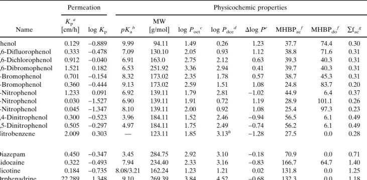
Table I. Experimental and Computational Physicochemic Parameters for the Compounds Studied
Name
Permeation Physicochemic properties
Kp a
[cm/h] log Kp pKa b
MW
[g/mol] log Poct c log P dce d ⌬log Pe MHBP ac f MHBP do f ∑f ac g ∑f do g 1 Phenol 0.129 −0.889 9.99 94.11 1.49 0.26 1.23 37.7 74.4 0.30 0.60 2 2,6-Difluorophenol 0.333 −0.478 7.09 130.10 2.05 0.93 1.12 38.8 71.6 0.31 0.60 3 2,6-Dichlorophenol 0.912 −0.040 6.91 163.0 2.75 2.12 0.63 39.3 40.3 0.31 0.36 4 2,6-Dibromophenol 1.521 0.182 6.53 251.92 3.36 2.94 0.41 39.7 40.3 0.31 0.36 5 2-Bromophenol 0.701 −0.154 8.32 173.02 2.35 1.78 0.57 38.7 45.3 0.31 0.36 6 4-Bromophenol 0.360 −0.444 9.13 173.02 2.59 1.51 1.08 24.8 83.7 0.20 0.67 7 2-Nitrophenol 1.233 0.091 6.92 139.11 1.79 2.81 −1.02 44.9 6.4 0.37 0.05 8 4-Nitrophenol 0.030 −1.527 6.90 139.11 1.91 0.72 1.19 28.9 101.1 0.26 0.82 9 3-Nitrophenol 0.045 −1.347 8.10 139.11 2.00 0.92 1.08 25.4 97.3 0.23 0.79 10 2,4-Dinitrophenol 0.300 −0.523 3.96 184.11 1.52 2.46 −0.94 56.5 6.1 0.49 0.05 11 2,5-Dinitrophenol 0.505 −0.297 4.97 184.11 1.75 2.49 −0.74 56.2 6.1 0.49 0.05 12 Nitrobenzene 2.009 0.303 — 123.11 1.85 3.13h −1.28 27.5 0.0 0.28 0.00 13 Diazepam 0.450 −0.347 3.45 284.75 2.92 3.10 −0.18 70.9 0.0 0.71 0.00 14 Lidocaine 0.322 −0.493 7.94 234.40 2.33 3.16 −0.83 166.7 64.7 1.40 0.50 15 Nicotine 0.184 −0.735 8.08/3.21 162.24 1.23 1.21 0.02 131.8 0.0 1.25 0.00 16 Orphenadrine 22.289 1.348 9.10 269.39 3.84 4.52 −0.68 132.3 0.0 1.18 0.00
aCalculated permeation coefficient using the non-steady-state diffusion model and adjustment for ionization (see Eq. 5); n⳱ 3; SD < 0.02. bDeterminated by potentiometry; n⳱ 3; SD < 0.1.
cLogarithm of n-octanol/water partition coefficient determined by potentiometry; n⳱ 3; SD < 0.02. dLogarithm of 1,2-dichloroethane/water partition coefficient determined by potentiometry; n⳱ 3; SD < 0.02. eDifference between the log P
octand log Pdceof a solute.
fMHBP calculated on the molecular surface for an acceptor (ac) or a donor (do) solute. gSum of the fragmental values of polar atoms (ac) or polar hydrogen atoms (do) in a solute. hMeasured by centrifugal partition chromatography, with pH 4.6 buffer as the stationary phase (12).
where the exponent g⳱ (pH − pKa) for acids and (pKa− pH)
for bases.
RESULTS AND DISCUSSION
The experimental and computational physicochemic pa-rameters for the two sets of compounds are summarized in Table I. All experiments were performed at a fixed pH value of 4.0 or 7.4 for the initial or additional set, respectively, and the calculated permeation coefficients were adjusted for ion-ization according to Eq. 5.
Permeability of Phenolic Compounds across Silicone Membranes
Effect of Lipophilicity
The actual partition coefficient between a membrane and the bathing solution is a key determinant of permeability. In this study, the amphiprotic n-octanol and the inert (DCE) were chosen as surrogate models. As revealed by Fig. 2(A and B) log Pdcedescribes solute permeation across silicone mem-branes better than log Poct. The reason may be that the
poly-dimethylsiloxane membrane, like DCE but unlike octanol, has little ability to form hydrogen bonds (see below). More-over, although there is a general trend of increasing perme-ability with lipophilicity in DCE as depicted in Fig. 2B, two distinct groups of compound are apparent in the graph. The first represents the halophenols, the second the nitro-derivates. As is readily appreciated, lipophilicity is strongly influenced by the nature of the substituents and by their po-sition around the aromatic ring.
Effect of Hydrogen-Bonding Capacity
This property has often been used in structure-permeation relationships and is related to skin structure-permeation (11,25). Different experimental approaches can be used to assess H-bonding capacity; e.g., the solvatochromic param-eters␣ (H-bond donor acidity) and  (H-bond acceptor ba-sicity) (26–28). The⌬log Poct-dce(log Poct− log Pdce)
param-eter expresses mainly the hydrogen-bond donor acidity of a solute (12). Indeed, log Poctcontains no contribution from the
solute H-bond donor capacity, unlike log Pdceof which it is a major component.
The separation between the two groups of compounds seen in Fig. 2B (the halophenols and the nitro-derivates) is clearer in Fig. 2C. For the halophenols, the⌬log Poct-dce pa-rameter decreases when the substituent is located close to the hydroxy group; compare, for example, 2-bromophenol (5) and 4-bromophenol (6). The presence of a halo-atom in the ortho- rather than para-position weakens the H-bond donor acidity of the −OH moiety due to proximity effects, and fa-cilitates permeability across the polymer membrane (see later Table I).
For the nitrophenols, the influence of the substitution pattern on membrane permeability can be summarized as fol-lows. First, the presence of a single phenolic group produces a positive⌬log P value relative to non-H-bond forming sol-utes. The lower log Pdcerelative to log Poctis consistent with
the fact that DCE does not form H-bonds with solutes. The presence of nitro substituents in the para- or metaposition means that the phenolic group predominates over the nitro
group in terms of ⌬log Poct-dce value. The hydrogen-bond
donor acidity of para-nitrophenol is slightly reinforced com-pared to meta-nitrophenol due to an inductive effect, thereby decreasing its permeability. Finally, the⌬log Poct-dcevalues of ortho-substituted phenols are significantly reduced due to their high log Pdcevalues, indicating that this H-bond donor capacity is not expressed in the DCE/water system. A strong
Fig. 2. Permeability of the initial set of compounds across silicone
membranes as a function of lipophilicity measured in terms of n-octanol/water partitioning (A), 1,2-dichloroethane/water distribution (B), and by the difference between these two solvent systems (C).䊉 halophenols, dihalophenols and phenol, 䊊 nitrophenols, ❋ di-nitrophenols,䊐 nitrobenzene. Compound numbering follows that in Table I.
intramolecular hydrogen bond between the nitro group and the phenolic function accounts for this behavior.
The strong stabilization of this intramolecular H-bond in nonpolar solvents (29) renders these compounds much more lipophilic than other nitrophenols in the DCE/water system. In the octanol/water system, the large amount of water in the organic phase offers additional possibilities for the formation of intermolecular H-bonds, which compete with intramolecu-lar H-bonds and affect the lipophilicity ranking of these com-pounds (Table I).
In addition to experimental approaches to quantify a molecule’s capacity to form hydrogen bonds, a computational tool—MHBPS—has been developed (13). Examples of such calculated potentials are illustrated in Fig. 3 for ortho and para-nitrophenol. The H-bonding acceptor potential on the molecular surface of 4-nitrophenol (Fig. 3A) and 2-nitrophe-nol (Fig. 3C) are similar, in stark contrast to their H-bonding donor potential (Fig. 3B) and (Fig. 3D), respectively. Indeed, the presence of an intramolecular interaction greatly de-creases or abolishes the H-bonding donor capacity of a solute. As depicted in Fig. 3B and the corresponding parameters of the linear regression (n⳱ 12, r2⳱ 0.60, q2⳱ 0.40), a major
determinant of diffusion across silicone membranes is clearly the H-bond donor acidity but not the H-bond acceptor ba-sicity (Fig. 4A). This observation agrees with the permeation results from human skin experiments (3). Moreover, Fig. 4B illustrates the influence of an ortho-nitro group on the MHBPs. As can be seen, the dinitrophenols (10, 11) deviate from the linear relationships between H-bonding capacity and permeation through silicone membranes, suggesting that other factors beside H-bonding influence permeation. Several studies based on silicone membrane permeation support this observation.
The results obtained support, complement and extend the extensive body of work by Matheson et al. (30,31) on the transport of a considerable number of aromatic and
hetero-cyclic compounds across silicone membranes. These earlier publications highlighted the use of physicochemic properties, such as hydrophobic fragmental constants, molar refractivity (chosen as a volume parameter), Hammett’s constants taking into account substituent electronic effects, mole fraction solu-bility in isopropyl alcohol and melting point, to develop pre-dictive models. It was also shown using comparative molecu-lar field analysis and atomic charge calculations that other parameters, including molecular weight and intramolecular hydrogen bonding potential, for example, contributed to the determination of permeation flux (32–34).
Permeability of Selected Drugs across Silicone Membranes
A set of four additional drugs was investigated to assess the generality of the previous observations. The influence of physicochemical and structural variability was therefore ana-lyzed over an extended range of lipophilicity and molecular weight compared to the initial set.
As seen in Fig. 5, log Pdceis a significant parameter for the prediction of solute permeability across silicone mem-branes. As mentioned above, the major contributions to log
Pdce are hydrophobicity and H-bond donor acidity (12).
Equation 6 illustrates the linear relationship between perme-ability coefficient and partition coefficient in DCE/water; that is, a single parameter allows a fair prediction of permeability:
Fig. 3. Calculated molecular H-bonding acceptor and H-bonding
do-nor potentials of 4-nitrophenol (A and B, respectively) and 2-nitro-phenol (C and D, respectively).
Fig. 4. Permeability of the initial set of compounds across silicone
membranes as a function of their molecular H-bonding acceptor (A) and donor (B) potential.䊉 halophenols, dihalophenols and phenol, 䊊 nitrophenols, ❋ di-nitrophenols, 䊐 nitrobenzene.
log Kp= 0.49 共±0.26兲 ⭈ log Pdce− 1.36 共±0.55兲
n= 16; r2= 0.70; q2= 0.56; s = 0.38; F = 32 (6)
In this and the following equations, 95% confidence limits are given in parentheses; n is the number of compounds, r2the
squared correlation coefficient, q2the cross-validated
corre-lation coefficient, s the standard deviation, and F the Fischer’s test.
The hydrogen-bonding donor acidity is a relevant param-eter which greatly influences permeation across polydimeth-ylsiloxane membranes. Adding the four drugs to the linear regression between log Kpand MHBPdoyields a poorer cor-relation (n⳱ 16, r2 ⳱ 0.42, q2 ⳱ 0.22) because diazepam,
nicotine, and orphenadrine have no H-bond donor capacity. However, a better relationship is recovered by adding log Poct in the multilinear Eq. 7:
log Kp= 0.56 共±0.37兲 ⭈ log Poct− 0.0108 (±0.0064) ⭈ ∑MHBPdo− 1.16 (±0.72)
n= 16; r2= 0.77; q2= 0.61; s = 0.35; F = 21 (7) Statistically, Eq. 7 is comparable with Eq. 6. This shows that predictions of silicone membrane permeation can be based on the single log Pdceparameter (Eq. 6), which
ex-presses the same intermolecular forces as the combined log
Poctand a H-bond donor parameter (e.g.,∑MHBPdoin Eq. 7, or⌬log P; 12,28).
Comparison with Human Skin Permeation
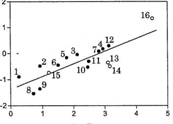
As silicone membranes are artificial barriers used to model skin lipids (3,6), the membrane permeability coeffi-cients (Kp(sil)) of seven compounds were compared to their corresponding in vitro values across human epidermis (Kp(epid); Fig. 6) The correlation was good (Eq. 8):
log Kp(sil)= 1.15 共±0.36兲 ⭈ log Kp(epid)+ 1.29 (±0.58) n= 7; r2= 0.90; q2= 0.83; s = 0.19; F = 46 (8) As indicated by the positive intercept in Eq. 8, the per-meability through silicone membrane is about 10 times higher than that through excised human skin (Table II). The rela-tionship indicates that silicone membranes may be a useful trend-predictive model for skin permeation.
CONCLUSION
This study demonstrates that the permeation of xenobi-otics across polydimethylsiloxane membranes is controlled primarily by their H-bond donor capacity, in turn strongly influenced by intramolecular interactions. Lipophilicity also plays a role. Thus, a single H-bond donor parameter is shown to correlate with silicone membrane permeability for a con-generic set of phenols, whereas a lipophilicity term must be added when heterogeneous drugs are included in the regres-sion (Eq. 7). Interestingly, the permeation of the extended set is also well described by lipophilicity in the dichloroethane/ water system (log Pdce, Eq. 6), which encodes a strong con-tribution from H-bond donor capacity (12), in contrast to log
Poctwhich does not.
ACKNOWLEDGMENTS
Financial support was provided by the Fonds national suisse de la recherche scientifique, and by the Programme commun de recherche en ge´nie biome´dicale (Universities of Geneva and Lausanne, and the Ecole polytechnique fe´de´rale, Lausanne).
APPENDIX
Development of the Mathematical Model
The key assumptions are the following: (1) depletion of the donor chamber; (2) accumulation in the receiving
cham-Table II. Experimental Permeability Coefficients of Selected
Com-pounds through Silicone Membranes and across Human Epidermis
Chemical
Silicone membrane Human epidermisa Kp [cm/h] log Kp Kp [cm/h] log Kp 1 Phenol 0.129 −0.889 0.008 −2.090 6 4-Bromophenol 0.360 −0.444 0.036 −1.440 7 2-Nitrophenol 1.233 0.091 0.100 −1.000 8 4-Nitrophenol 0.030 −1.527 0.006 −2.250 9 3-Nitrophenol 0.045 −1.347 0.006 −2.250 14 Lidocaine 0.322 −0.493 0.017 −1.770b 15 Nicotine 0.184 −0.735 0.019 −1.710
aData from Flynn et al. (36). bData from Johnson et al. (37). Fig. 5. Permeability of the initial set (䊉) and additional set of
com-pounds (䊊) through silicone membranes as a function of lipophilicity in the 1,2-dichloroethane/water system (Eq. 6).
Fig. 6. Correlation of solute permeabilities through human epidermis
ber; (3) instantaneous equilibrium at the skin-solution inter-faces; (4) a homogeneous membrane as the barrier to diffu-sion (5) a constant diffudiffu-sion coefficient; (6) no binding or metabolism within the membrane; (7) no solvent diffusion.
To determine the solute concentration profile and the flux across the membrane, it is necessary to write three un-steady-state solute mass balances:
membrane M: ⭸CM ⭸t =DM⭈ ⭸2 CM ⭸x2 (A1) donor chamber D: VD A ⭈ dCD dt = DM⭈ ⭸CM ⭸x
冏
x=0 (A2) receptor chamber R: VR A ⭈ dCR dt = − DM⭈ ⭸CM ⭸x冏
x=L (A3) where C is the concentration of the permeant expressed in g/cm3, V is the volume in cm3of each chamber, DM is the
diffusion coefficient in the membrane in cm2/s, A is the area
of the membrane in cm2, and x is the depth in membrane M.
These differential equations are subject to the following specific conditions:
at t= 0, CD= CD0, C
R= 0, CM= 0 (A4)
at x= 0, CM= PMⲐD⭈ CD (A5)
at x= L, CM= PMⲐR⭈ CR (A6)
If the two compartments contain the same solvent con-ditions, the following simplification is allowed: PM/D⳱ PM/R ⳱ P. Described as the partition coefficient of the solute be-tween the membrane and solvent, this latter parameter is assumed not to vary with solute concentration.
Moreover, if P⭈ L ⭈ A << V, the amount of solute in the membrane will be always negligible compared with the amount of solute in the two compartments. As a result, the variation in concentration in the donor and receptor solutions will be slow compared to the diffusion rate across the mem-brane. In other words, the concentration profile across the membrane will always be close to its steady-state value, even though the compartment concentrations are time-dependent. This suggests that a pseudosteady-state solution strategy is appropriate.
Assuming pseudosteady state, where the rate of change of concentration, ⭸CM/⭸t, will be zero, Eq. A1 is first inte-grated twice and combined with Eqs. A5 and A6, to obtain the following expression for the concentration profile
CM= P ⭈ CD− P
L⭈ x ⭈ 共CD− CR兲 (A7)
Eq. A7 is differentiated and evaluated at x⳱ 0 and x ⳱ L to obtain: ⭸CM ⭸x
冏
x=0 =⭸CM ⭸x冏
x=L= − P L共CD− CR兲 (A8)It has been shown that it is experimentally more robust to analyze the data as the difference [CD(t) − CR(t)] rather
than either CD(t) or CR(t) individually (14). Subtracting Eq. A3 from Eq. A2 and substituting for Eq. A8 yields the fol-lowing:
d
dt共CD− CR兲 = − ␥ ⭈ 共CD− CR兲 (A9)
in which␥ is defined by Eq. 2.
Eq. A9 is solved subject to the initial condition Eq. A4 to give:
ln
冉
CD− CRCD
0
冊
= − ␥ ⭈ t (A10)This pseudosteady-state analysis holds as long as
P⭈ L ⭈ A/VD<∼0.1, assuming that VD⳱ VR(35).
REFERENCES
1. J. Hadgraft and R. H. Guy. Transdermal Drug
Delivery-Development Issues and Research Initiatives. Marcel Dekker,
New York, 1989.
2. R. O. Potts and R. H. Guy. Predicting skin permeability. Pharm.
Res. 9:663–669 (1992).
3. M. T. D. Cronin, J. C. Dearden, G. P. Moss, and G. Murray-Dickson. Investigation of the mechanism of flux across human skin in vitro by quantitative structure–permeability relationships.
Eur. J. Pharm. Sci. 7:325–330 (1999).
4. L. A. Kirchner, R. P. Moody, E. Doyle, R. Bose, J. Jeffery, and I. Chu. The prediction of skin permeability by using physicochem-ical data. ATLA 25:359–370 (1997).
5. N. Sekkat and R. H. Guy. Biological models to study skin per-meation. In: B. Testa, H. van de Waterbeemd, G. Folkers, and R. H. Guy (eds.), Pharmacokinetic Optimization in Drug
Re-search: Biological, Physicochemical and Computational Strategies.
Wiley-VCH, Zurich, 2001 pp. 155–172.
6. J. Houk and R. H. Guy. Membrane models for skin penetration studies. Chem. Rev. 88:455–471 (1988).
7. J. Hadgraft and W. J. Pugh. The selection and design of topical and transdermal agents: A review. J. Invest. Dermatol. 3:131–135 (1998).
8. G. Ridout, J. Houk, R. H. Guy, G. C. Santus, J. Hadgraft, and L. L. Hall. An evaluation of structure-penetration relationships in percutaneous absorption. Farmaco 47:869–892 (1992). 9. E. R. Garrett and P. B. Chemburkar. Evaluation, control, and
prediction of drug diffusion through polymeric membranes. J.
Pharm. Sci. 57:944–948 (1968).
10. J. du Plessis, W. J. Pugh, A. Judefeind, and J. Hadgraft. The effect of hydrogen bonding on diffusion across model membranes: con-sideration of the number of H-bonding groups. Eur. J. Pharm.
Sci. 13:135–141 (2001).
11. R. O. Potts and R. H. Guy. A predictive algorithm for skin per-meability: The effects of molecular size and hydrogen bond ac-tivity. Pharm. Res. 12:1628–1633 (1995).
12. G. Steyaert, G. Lisa, P. Gaillard, G. Boss, F. Reymond, H. H. Girault, P. A. Carrupt, and B. Testa. Intermolecular forces ex-pressed in 1,2-dichloroethane/water partition coefficient: A sol-vatochromic analysis. J. Chem. Soc. Faraday Trans. 93:401–406 (1997).
13. S. Rey, G. Caron, G. Ermondi, P. Gaillard, A. Pagliara, P. A. Carrupt, and B. Testa. Development of molecular hydrogen bonding potentials (MHBPs) and their application to structure permeation relations. J. Mol. Graphics Model. 19:521–535 (2001). 14. B. W. Barry. Dermatological Formulations. Dekker, New York,
1983.
15. J. C. Shah. Analysis of permeation data: evaluation of the lag time method. Int. J. Pharm. 90:161–169 (1993).
16. E. L. Cussler. Diffusion: Mass Transfer in Fluid Systems, 2nd Ed. Cambridge University Press, New York, 1997.
17. A. Avdeef. pH-Metric log P. Part 1. Difference plots for deter-mining ion-pair octanol-water partition coefficients of multiprotic substances. Quant. Struct.-Act. Relat. 11:510–517 (1992). 18. A. Avdeef, J. E. A. Comer, and S. J. Thomson. pH-Metric log P.
3. Glass electrode calibration in methanol-water, applied to pKa
determination of water-insoluble substances. Anal. Chem. 65:42– 49 (1993).
Partitioning of solutes in different solvent systems: the contribu-tion of hydrogen-bonding capacity and polarity. J. Pharm. Sci.
80:590–598 (1991).
20. M. H. Abraham. Scales of solute hydrogen-bonding: Their con-struction and application to physicochemical and biochemical processes. Chem. Soc. Rev. 22:73–83 (1993).
21. M. H. Abraham. Hydrogen bonding. 31. Construction of a scale of solute effective or summation hydrogen-bond basicity. J. Phys.
Org. Chem. 6:660–684 (1993).
22. D. R. Friend. In vitro skin permeation techniques. J. Control.
Release 18:235–248 (1992).
23. E. R. Garrett and P. B. Chemburkar. Evaluation, control, and prediction of drug diffusion through polymeric membranes. II. J.
Pharm. Sci. 57:949–959 (1968).
24. R. E. Kasting. Synthetic Polymeric Membranes. A structural
per-spective, 2nd Ed. Wiley, New York, 1985.
25. M. H. Abraham, H. S. Chadha, and R. C. Mitchell. The factors that influence skin penetration of solutes. J. Pharm. Pharmacol.
47:8–16 (1995).
26. R. W. Taft, J. L. M. Abboud, M. J. Kamlet, and M. H. Abraham. Linear solvation energy relations. J. Sol. Chem. 14:153–186 (1985).
27. R. W. Taft, M. H. Abraham, R. M. Doherty, and M. J. Kamlet. The molecular properties governing solubilities of organic non-electrolytes in water. Nature 313:304–306 (1985).
28. N. El Tayar, R. S. Tsai, B. Testa, P. A. Carrupt, C. Hansch, and A. Leo. Percutaneous penetration of drugs: A quantitative struc-ture-permeability relationship study. J. Pharm. Sci. 80:744–749 (1991).
29. H. van de Waterbeemd. Hydrophobicity of Organic Compounds. CompuDrug International, Vienna, 1986.
30. M. W. Hu and L. E. Matheson. The development of predictive method for the estimation of flux through polydimethylsiloxane membranes. III. Application to a series of substituted pyridines.
Pharm. Res. 10:732–736 (1993).
31. L. E. Matheson and M. W. Hu. The development of a predictive method for the estimation of flux through polydimethylsiloxane membranes. IV. Application to a series of substituted quinolines.
Pharm. Res. 10:839–842 (1993).
32. Y. Chen, W-L. Yang, and L. E. Metheson. Prediction of flux through polydimethylsiloxane membranes using atomic charge calculations. Int. J. Pharm. 94:81–88 (1993).
33. R. Liu and L. E. Matheson. Comparative molecular field analysis combined with physicochemical parameters for prediction of polydimethylsiloxane membrane flux in isopropanol. Pharm. Res.
11:257–266 (1994).
34. Y. Chen, P. Vayumhasuwan, and L. E. Matheson. Prediction of flux through polydimethylsiloxane membranes using atomic charge calculations: Application to an extended data set. Int. J.
Pharm. 137:149–158 (1996).
35. R. Mills, L. A. Woolf, and R. O. Watts. Simplified procedures for diaphragm-cell diffusion studies. AlChE J. 14:671–673 (1968). 36. G. L. Flynn. Physicochemical determinants of skin absorption. In
T. R. Gerrity and C. J. Henry (eds.), Principles of Route-to-Route
Extrapolation for Risk Assessment. Elsevier, Amsterdam, 1990
pp. 93–127.
37. J. E. Johnson, D. Blankstein, and R. Langer. Evaluation of solute permeation through the stratum corneum: lateral bilayer diffu-sion as the primary transport mechanism. J. Pharm. Sci. 86:1162– 1172 (1997).