Sylvain Terraz
Christophe Constantin Pietro Edoardo Majno Laurent Spahr Gilles Mentha Christoph D. Becker Received: 9 October 2006 Revised: 3 February 2007 Accepted: 23 February 2007 Published online: 21 March 2007 # Springer-Verlag 2007
Image-guided multipolar radiofrequency
ablation of liver tumours: initial clinical results
Abstract The local effectiveness and clinical usefulness of multipolar radiofrequency (RF) ablation of liver tumours was evaluated. Sixty-eight image-guided RF sessions were per-formed using a multipolar device with bipolar electrodes in 53 patients. There were 45 hepatocellular carci-nomas (HCC) and 42 metastases with a diameter≤3 cm (n=55), 3.1–5 cm (n=29) and >5 cm (n=3); 26 nodules were within 5 mm from large vessels. Local effectiveness and complications were evaluated after RF procedures. Mean follow-up was 17±10 months. Recurrence and survival rates were analysed by the Kaplan-Meier
meth-od. The primary and secondary tech-nical effectiveness rate was 82% and 95%, respectively. The major and minor complication rate was 2.9%, respectively. The local tumour pro-gression at 1- and 2-years was 5% and 9% for HCC nodules and 17% and 31% for metastases, respectively; four of 26 nodules (15%) close to vessels showed local progression. The survi-val at 1 year and 2 years was 97% and 90% for HCC and 84% and 68% for metastases, respectively. Multipolar RF technique creates ablation zones of adequate size and tailored shape and is effective to treat most liver tumours, including those close to major hepatic vessels.
Keywords Radiofrequency ablation . Liver . Hepatocellular carcinoma . Metastasis
Introduction
Evidence is building up that image-guided radiofrequency (RF) ablation has a place in the treatment of limited hepatocellular carcinoma (HCC) in cirrhosis because it may offer effective primary local tumour control and long-term survival comparable with surgery in over 90% of cases [1–3]. In more advanced tumours, RF can be used alone or in conjunction with transarterial chemoembolisa-tion (TACE) in order to prevent local tumour progression [4]. Currently, available data suggest that successful ablation of liver metastases, notably of colorectal cancer, is more difficult to achieve than with HCC [5–9]. This is probably due to differences in tumour conductivity and perfusion-mediated tissue cooling of the surrounding liver
[10]. Hence, technical improvements of RF ablation are aiming at increasing ablation zones in order to provide adequate tumour coverage, particularly in the tumours close to large vessels.
All major published clinical series of RF ablation of liver tumours are based on monopolar energy deposition. Different systems are currently available using single, cluster or multitined expandable designs with either internal cooling or open circuit perfusion [11]. Experi-mental comparative studies have shown that their ability to create large ablation zones may vary considerably [12,13]. Bipolar energy deposition has the advantage of being more reproducible and has been proposed to obtain large ablation zones [14–16]. Recently, a new bipolar system has become available for clinical use that offers bipolar and multipolar S. Terraz (*) . C. Constantin .
C. D. Becker
Department of Radiology, Geneva University Hospital, rue Micheli-du-Crest 24, 1211 Geneva 14, Switzerland e-mail: [email protected] Tel.: +41-22-3727094 Fax: +41-22-3727072 P. E. Majno . G. Mentha Department of Surgery, Geneva University Hospital, rue Micheli-du-Crest 24, 1211 Geneva 14, Switzerland
L. Spahr
Department of Gastroenterology, Geneva University Hospital, rue Micheli-du-Crest 24, 1211 Geneva 14, Switzerland
settings. Preliminary reports have addressed technical feasibility and safety of this device [17,18].
The purpose of the present study was to evaluate the local effectiveness and the clinical usefulness of image-guided RF ablation with this multipolar technique in primary and secondary liver malignancies.
Materials and methods
Patients and lesions
The present study was approved by the institutional ethics committee and was based on clinical and radiological data of a consecutive series of 68 multipolar RF sessions for 87 malignant hepatic nodules in 53 patients, between July 2003 and June 2006. All patients were included after informed consent was obtained. General contraindications included prothrombin time ratio <50%, platelet count <50,000/mm3, cirrhosis of Child class C, encephalopathy, major ascites, bacterial infection, vascular invasion and extrahepatic metastases.
Thirty-one patients (24 men, seven women; mean age, 65 years; range, 15–82 years) had HCC, including 19 patients (61%) with Child A cirrhosis, nine patients (29%) with Child B and three patients (10%) without underlying liver disease. In 24 patients, multipolar RF ablation was the first treatment, whereas seven patients had undergone prior treatment; hepatic resection (n=4), TACE (n=2) and monopolar RF ablation (n=1). Forty-five HCC nodules were diagnosed according to the European Association for the Study of the Liver; 27 hypervascular nodules≥2 cm by dynamic imaging techniques and 18 hypovascular and/ or <2 cm nodules by ferucarbotran-enhanced MR imaging or coaxial core biopsy [19]. The long-axis diameter of index tumours was≤3.0 cm in 27 nodules, 3.1–5.0 cm in 15 nodules and >5.0 cm in three nodules (Table1). Eight nodules were located within 5 mm from vessels≥3 mm in diameter (hepatic vein, n=5; portal branch, n=2; inferior vena cava, n=1) [20]. Eighteen nodules were located
within 5 mm from adjacent organs (gastrointestinal tract, n=2; gallbladder, n=2; kidney, n=1) or from the liver capsule (n=13).
Twenty-two patients (13 men, nine women; mean age, 64 years; range, 39–88 years) had liver metastases. Sixteen patients had 28 metastases from colorectal carcinoma; 13 patients had received systemic chemotherapy previously. One patient had eight metastases from a gastric stromal tumour. The five other patients had metastases from breast cancer (n=2), lung cancer (n=1), bladder cancer (n=1) and cholangiocarcinoma (n=2). Forty-two liver metastases were diagnosed, either by US, CT and Mn-DPDP-enhanced MR (n=32) or by coaxial core biopsy (n=10). The long-axis diameter of index tumours was≤3.0 cm in 28 nodules and 3.1–5.0 cm in 14 nodules. Eighteen nodules were situated within 5 mm from major vessels (hepatic vein, n=9; portal branch, n=6; inferior vena cava, n=3), whereas ten others were located within 5 mm from the gallbladder (n=2), the gastrointestinal tract (n=3) or the liver capsule (n=5).
RF ablation procedures
The 68 RF treatments were performed by four experienced radiologists in liver interventions under general anaesthesia or short-acting intravenous sedation. Prophylactic ceftri-axone was intravenously administered before electrode insertion and repeated twice over 48 h. US guidance was chosen in 56 procedures (82%) and 16-detector CT was used in 11 procedures (16%). One small HCC nodule was treated under MRI guidance (2%) using a 0.23-T open-magnet and a MR-compatible electrode [21].
The bipolar electrodes (Celon ProSurge; Celon Medical Instruments, Teltow, Germany) had a diameter of 1.8 mm, an active tip length of 20, 30, or 40 mm and a shaft length of 10–25 cm, and were internally cooled by means of a pump (Celon Aquaflow III) [18]. For small nodules, one bipolar electrode with an appropriate active tip length was placed centrally within the tumour. For larger tumours, two
Table 1 Size of HCC nodules and metastases before RF ablation (index tumours) and corresponding ablation zone 24 h and 1month after RF ablation. Data are mean value ± standard deviation, with ranges in parentheses; n number of nodules
Parameters Index tumour Ablation zone (24 h) Ablation zone (1 month)
HCC (n=45) long-axis [cm] 2.4±1.0 (0.9–5.4) 4.7±1.4 (2.4–8.6) 3.7±1.2 (1.6–6.4) short-axis [cm] 1.8±0.8 (0.7–4.8) 3.2±1.0 (1.8–5.8) 2.6±1.2 (0.9–6.9) volume [cm3] 7.3±11.7 (0.2–68.0) 38.0±35.9 (5.5–188.0) 18.6±23.4 (0.7–120.2) Metastases (n=42) long-axis [cm] 2.2±0.9 (0.7–5.0) 4.9±1.3 (1.6–8.0) 3.8±1.3 (1.4–6.5) short-axis [cm] 1.7±0.8 (0.6–4.3) 3.3±0.9 (0.9–5.0) 2.5±0.9 (0.9–4.2) volume [cm3] 6.3±9.7 (0.1–55.2) 41.1±25.7 (0.9–108.9) 17.8±13.0 (0.9–48.0)
or three electrodes were placed in parallel in the outer third of the nodule (Fig. 1). The mean distance between the electrodes was 1.6±0.7 cm (range, 0.9–2.6 cm). One electrode was used in 24 (28%) of the 87 ablations, two electrodes in 42 ablations (48%) and three electrodes in 21 ablations (24%). Whenever a tumour was in the vicinity of a large vessel or an extrahepatic organ, at least one electrode was placed between the tumour and related structure (Fig. 2). The puncture time was defined as the time from the skin puncture to the final evaluation of electrodes positioning.
RF ablations were performed with a 470-kHz RF generator (CelonLab Power), which can manage simulta-neously up to three bipolar electrodes by automatic impedance-feedback [18]. The initial power output was chosen between 20 and 150 W (mean, 65±34 W), depending on tumour size and number of electrodes. The endpoint of energy deposition was based on empirical parameters, taking into account the tumour type, size and geometry. These parameters were monitored by a dedicated software (CelonPower Monitor). In a large HCC nodule with mixed vascularity, selective TACE of the hypervas-cular portion was done before RF ablation of the
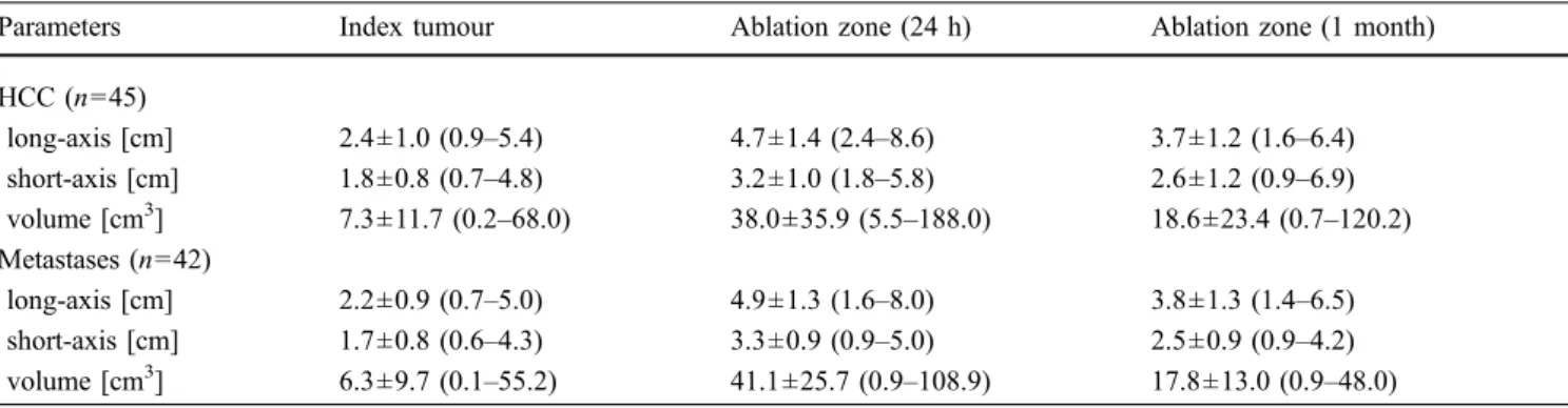
Fig. 1 Large hepatic metastasis from bronchogenic carcinoma close to the right kidney in a 53-year-old male after left pneumectomy. The patient refused hepatic resection. a Fast spin-echo T2-weighted MR image obtained before RF ablation shows a large (long-axis diameter, 5.0 cm; volume, 55 cm3) and heterogeneously hyperintense mass in segment 6 close to median portion of the right kidney (white arrows). b Ultrasound scan obtained during the placement of three bipolar RF electrodes with 4-cm active tips. The first and second electrodes are positioned at the lateral border of the tumour, in an anterior and posterior fashion respectively. A third electrode (not
shown) was placed between the medial border of the tumour and the right kidney. c Contrast-enhanced CT scan obtained during portal phase 24 h after RF ablation (power output, 100 W; duration, 45 min; total applied energy, 235 kJ) shows that the index tumour is completely covered by the ablation zone (long-axis diameter, 6.8 cm; volume 105 cm3) with an ablative margin larger than 5 mm. No lesion
of the right kidney is observed. d Fast spin-echo T2-weighted MR image obtained at 7 months after RF ablation shows the ablation zone with low signal intensity (white arrows) without detectable residual tumour
hypovascular zone [22]. Intraductal cooling by cold saline through a percutaneously placed 20G Chiba needle was performed during RF ablation of a HCC nodule close to left hepatic duct [23]. Gray-scale US imaging was used for monitoring of the transient hyperechoic zone. Contrast-enhancement with microbubbles was performed immedi-ately after six procedures. Three CT-guided procedures were done with intravenous contrast media. For the small nodule ablated under MRI guidance, T2-weighted images were used to evaluate treatment response. A second exposure was achieved in two ablations after electrodes repositioning.
Track coagulation was performed with each electrode separately. Post-procedural imaging was achieved in order to detect immediate complications. The procedure time, including electrodes placement and removal, RF ablation and imaging control, was recorded. After RF ablation, all patients stayed in the hospital overnight for medical observation and laboratory; they were discharged after recovery from sedation or any complication. Symptoms of post-ablation syndrome were recorded during the hospital stay and at the one month visit.
Follow-up and image analysis
Description of the results of RF ablation was based on recommendations by the International Working Group on Image-Guided Tumor Ablation [24]. Twenty-four hours after RF procedure, the technical success and early complications were assessed by a triphasic contrast-enhanced CT with the following parameters: 2 ml/kg of iodinated contrast agent, rate of 3–5 ml/s, section thickness 3.0 mm, reconstruction interval 1.5 mm. Any preexisting tumour region not covered by the ablation zone was considered as residual tumour. If the ablation zone was ellipsoid, its volume was derived from its three orthogonal diameters along the electrodes axis on the portal phase images. If the ablation zone was irregular, an institutional software calculated the volume by three-dimensional watershed algorithm [25]. Ablative margins were evaluated by image fusion in the axial and coronal planes.
One month after treatment, technical effectiveness was assessed by using contrast-enhanced CT for hypervascular nodules and hepatospecific contrast-enhanced MRI for Fig. 2 Small hepatic metastasis
from colorectal carcinoma close to the inferior vena cava (IVC) in a 57-year-old male after right hepatectomy. a Fast spin-echo T2-weighted MR image ob-tained before RF ablation shows a mildly hyperintense nodule (long-axis diameter, 1.9 cm; volume, 2.9 cm3) in contact with the medial border of the IVC (white arrows). b Unenhanced CT scan obtained just before RF ablation (power output, 35 W; duration, 27 min; total applied energy, 51 kJ) shows the place-ment of two bipolar electrodes with a 3-cm active tip. The first electrode was positioned be-tween the IVC and the tumour and the second one was posi-tioned on the medial side of the tumour. c Contrast-enhanced CT scan obtained during portal phase immediately after RF ablation shows that the index tumour is completely covered by the ablation zone (maximal diameter, 5.3 cm; volume 52 cm3) with an ablative margin
larger than 10 mm. The IVC is still patent. d Contrast-enhanced CT scan obtained during portal phase 17 months after RF ablation shows that the ablation zone has undergone involution, without evidence of local tumour progression
hypovascular nodules. Any contrast-enhanced foci on CT and hyperintensity on T2-weighted images or non-enhanc-ing area on contrast-enhanced MR images at the bound-aries of the treated area was considered as incomplete ablation. Follow-up consisted in monitoring of tumour markers and CT or MRI scans every 3–4 months, and results were recorded according to standard criteria [24,
26]. Recurrence rates were calculated from the first RF ablation to the imaging follow-up that revealed tumour progression. Survival data were calculated from the first RF treatment to the time of death or analysis (September 2006). Local tumour progression and new tumours nodules prompted additional RF ablation, if the patient still met the requirements.
Statistical analysis
Descriptive statistical measures, including mean values, standard deviations and ranges, were calculated for the ablation zone sizes. The relationship between ablation zone volume and applied energy was analysed by Pearson correlation analysis. The difference between ablative margins between HCC and metastases were analysed by The Mann-Whitney U-test. Local tumour progression and survival rates were estimated by the Kaplan-Meier method and the curves were compared with the log-rank test. A P value of less than 0.05 was considered as a statistically significant difference. Statistical analysis was performed with a commercial software (JMP version 6.0 for MacOsX; SAS Institute, Cary, USA).
Results
RF ablation procedures
RF ablation with complete tumour coverage was achieved in 86 of the 87 malignant nodules for a technical success rate of 99%. In one patient with advanced macronodular cirrhosis, one of the multiple benign nodules had been ablated under US guidance, instead of the malignant HCC nodule. Complete ablation was obtained after a second RF session. The mean puncture time per tumour was 15±6 min (range, 3–36 min) using US, 31±9 min (12–58 min) using CT (P=0.009), and 73 min for the nodule treated under MR guidance. The mean ablation time per tumour was 29± 10 min (10–50 min). The mean procedure time was 67± 34 min (39–132 min) with US, 128±51 min (54–221 min) with CT (P=0.031) and 202 min with MR guidance. A mean total energy of 61±46 kJ (range, 11–215 kJ) was applied per HCC nodule and 75±44 kJ (13–235 kJ) per metastasis.
No procedure-related death was observed. Two major complications (2.9%) occurred among the 68 procedures. In a patient with a subcapsular HCC of the hepatic dome, a
right pneumothorax occurred during electrodes position-ing. A chest tube was immediately placed, the RF procedure was continued and CT showed complete reso-lution of the pneumothorax after 48 h. One case of electrode tract seeding was identified after treatment of a subcapsular HCC, although a route through liver paren-chyma had been chosen. A 4.2-cm hypervascular nodule was found on 6-month CT in the greater omentum and resected surgically. Minor complications were observed in two (2.9%) of 68 procedures. A partial right portal vein thrombosis was successfully treated with oral acenocou-marol during 4 weeks. A small second-degree skin burn at the puncture site completely resolved after application of silver sulphadiazine cream. Side effects were observed in 13 (19%) of the 68 procedures, including asymptomatic pleural or peritoneal effusion (n=8), and self-limited subcapsular or peritoneal bleeding (n=5). Postablation syndrome occurred after 19 (28%) of 68 RF sessions; symptoms peaked on day 2 and always resolved within 8 days with nonsteroidal anti-inflammatory drugs. The mean duration of hospitalisation was 2.1±1.2 days (range, 1–5 days). Forty-four patients (83%) were discharged after overnight observation. Six patients (11%) stayed an additional night for observation of post-ablation syndrome and three patients (6%) stayed longer for treatment of complications (n=3).
Ablation zone size and shape
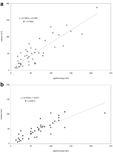
The size of the ablation zone 24 h and one month after RF ablation is presented in Table 1. At 24 h, the difference between the mean long-axis and short-axis diameters of ablation zone and the index tumour was on average 2.3 cm and 1.4 cm for HCC nodules, and 2.7 cm and 1.6 cm for metastases, respectively. The ablation zone volume 24 h after RF ablation was five- to six-times larger than the index tumour volume and there was substantial involution 1 month after treatment. The maximum ablation zone achieved was 8.6×7.2×5.8 cm (188 cm3) in a HCC nodule and 8.0×5.2×5.0 cm (109 cm3) in a metastasis. The mean ablation zone volume achieved with one, two and three electrodes was 15.0±11.5 cm3, 34.1±18.0 cm3and 75.6± 37.1 cm3, respectively. In the HCC group, the ablation zone volume showed a close correlation with the applied energy (Fig.3a). In the metastasis group, this correlation was less strong, which is probably due to the outlier metastasis to the right of the graph (Fig.3b). Sixty-eight (78%) of the 87 ablation zones were concentric with regard to index tumour, whereas 19 ablation zones (22%) were eccentric but completely covering the tumour. Ablative margins were ≥5 mm in 78 (90%) of 87 ablation zones and ≥10 mm in 51 (59%) of 87 ablation zones (Table2). No statistically significant difference was found between ablative margins of ≤3 cm and >3 cm nodules, and between HCC and metastatic nodules.
Follow-up
At 1-month imaging, complete tumour ablation was depicted in 85(98%) of 87 nodules. The extra-hepatic portion of a subcapsular HCC was not totally ablated and, in another patient, the ablation zone of a metastasis showed residual tumour. On subsequent follow-up, local tumour progression was observed in four HCC nodules and in ten
metastases. It is noteworthy that none of the eight HCC nodules close to a major vessel showed local tumour progression. In metastases, the rate of local tumour progression was 22% (4/18) in nodules adjacent to large vessels and 25% (6/24) at distance from them. Complete tumour destruction after one RF session was achieved in 71 of 87 malignancies for a primary effectiveness rate of 82%. Since 12 of the 16 tumours with local tumour progression were retreated with RF and resulted in complete ablation, the secondary technique effectiveness rate was 95%. Two patients with residual hypervascular HCC underwent selective TACE. One patient with two local and multiple new recurrences was treated by chemotherapy.
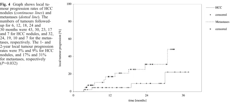
The mean follow-up period was 17.1±10.0 months (range, 3.3–37.7 months). The 1-and 2-year local tumour progression rates were 5% and 9% for HCC nodules, and 17% and 31% for metastases, respectively (P=0.032) (Fig. 4). The recurrence rates of new tumours at 1 and 2 years were 16% and 29% for HCC, and 33% and 58% for metastases, respectively (P=0.047). The overall recurrence rates were 20% and 36% for HCC, and 42% and 79% for metastases, respectively (P=0.041).
At the end of the study, three of 31 patients of the HCC group had died from liver failure with tumour progression, liver failure without tumour progression and myocardial infarction. At the end of the study, eight of 22 patients with liver metastases had died. Six patients died from metastatic disease and two patients died from urosepsis and hemor-rhagic stroke. The overall survival rates for the patients with HCC were 97% at 1 year and 90% at 2 years; the median follow-up was 18.1 months (Table3). The overall survival rates for the patients with metastases were 84% at 1 year and 68% at 2 years; the median follow-up was 20.8 months. The difference between the survival curve for patients with HCC and patients with metastases was statistically significant (P=0.038).
Discussion
Multipolar RF ablation with the simultaneous use of one or more bipolar electrodes has the potential advantage to create larger ablation zones compared with sequential use of monopolar electrodes with repositioning. Since the alter-nating current is confined to the area under treatment, this
Table 2 Ablative margins of the ablation zones after RF ablation for HCC nodules and metastases (n number of nodules) n Ablative margin <5 mm Ablative margin 5–10 mm Ablative margin >10 mm
HCC≤3 cm 27 4 (15%) 9 (33%) 14 (52%)
HCC>3 cm 18 2 (11%) 7 (39%) 9 (50%)
Metastases≤3 cm 28 3 (11%) 7 (25%) 18 (64%)
Metastases >3 cm 14 0 (0%) 4 (29%) 10 (71%)
All 87 9 (10%) 27 (31%) 51 (59%)
Fig. 3 Graphs show relationship between the volume of the ablation zone in cm3and applied energy in kJ. a In the HCC group, an increase in applied energy produced an increase in volume (R2= 0.747), as determined by V=0.760XE+6.157, where V is volume and E is applied energy. b In the metastases group, the regression line equation was V=0.552XE+10.071 (R2=0.687). If the outlier to the right of the graph is eliminated, the correlation is better (R2= 0.718)
approach also permits to avoid adverse events inherent to the monopolar technique, such as unpredictable electrical current paths, heterogeneous energy deposition and skin burns at the grounding pad [15, 27–29]. Although the technical aspects of multipolar RF ablation have been addressed, published clinical experience with percutaneous use of this technology is still limited [18,30]. The data of our study confirm that the multipolar technique is feasible in a clinical procedure of reasonable duration using either US or CT targeting. Although MR guidance is also feasible, the procedural time is still quite excessive. Despite the use of multiple electrodes in the majority of procedures, the rate of complications was no higher than from series with mono-polar devices [1,2,7,8,31–33]. It is noteworthy that most adverse events had occurred in lesions protruding from the liver contour, which are recognised as a risk factor for complications, including electrode track seeding [34].
The size of the ablation zone correlated with the amount of deposited energy for both HCC and metastases. Although the difference was not significant, total energy deposition was greater in metastases than in HCC, depending on tissue impedance and perfusion-mediated
tissue cooling. Therefore, applied energy in metastases should be about 40% greater to obtain the same volume than HCC nodules. The creation of ablation zones with at least a 5 mm margin was possible without electrodes repositioning in the vast majority of cases. A single bipolar electrode is sufficient for complete coverage of nodules less than 1.5 cm, but we recommend two or more electrodes for larger nodules. When multiple electrodes are used, they should be placed in a parallel fashion, in order to obtain an ovoid ablation zone. When treating tumours that are adjacent to major vessels or extrahepatic organs, at least one electrode should be placed between the involved structure and the tumour periphery. In that configuration, the tumour are “in brackets” between the electrodes. It is noteworthy that 22 (85%) of the 26 tumour nodules close to large vessels were ablated without local progression. In an animal experiment using bipolar RF ablation, it had been demonstrated that heat sink effect was decreased and tumour cell survival next to vessels was reduced [35].
Published results from ex-vivo studies indicated that multipolar RF ablation enables larger ablation zones to be Fig. 4 Graph shows local
tu-mour progression rates of HCC nodules (continuous lines) and metastases (dotted line). The numbers of tumours followed-up for 6, 12, 18, 24 and 30 months were 43, 30, 23, 17 and 7 for HCC nodules, and 32, 24, 19, 10 and 7 for the metas-tases, respectively. The 1- and 2-year local tumour progression rates were 5% and 9% for HCC nodules, and 17% and 31% for metastases, respectively (P=0.032)
Table 3 Overall survival rates for patients with HCC and
patients with liver metastases (n number of patients followed-up)
Time (months) HCC Metastases
n Survival (%) n Survival (%) 0 31 100 22 100 6 30 97 19 95 12 18 97 17 84 18 14 90 15 74 24 12 90 8 68 30 6 79 5 60 36 3 79 0 –
obtained than with monopolar devices [12, 13, 36, 37]. This observation was confirmed by other in-vivo experi-ments in pig liver, especially with regard to the heat sink effect [35, 38,39]. Although a direct clinical comparison between multipolar and monopolar RF techniques is crucial, it was beyond the scope of the present study. The limitations of our study were the small number of histologically confirmed metastases, as well as the limited follow-up time with regard to tumour progression and survival. Nonetheless, our current data regarding treatment of HCC compare favourably with other published series when comparing tumours of similar size [2, 40–43]. The results of RF treatment of metastatic nodules are much more difficult to compare because of histologic stratifica-tion, tumour size, concomitant chemotherapy regimens and
previous surgical therapy [5–9]. While almost one-third of nodules treated in our series were within 5 mm from large vessels, most clinical reports on liver RF ablation have failed to mention the anatomic vicinity of hepatic vessels. In conclusion, the multipolar technique is effective in producing ablation zones of adequate size and according to individual tumour geometry in the majority of primary and secondary liver tumours, including tumours close to major hepatic vessels. The use of multiple electrodes does not seem to increase the risk of complications compared with previously reported series.
Acknowledgements The authors would like to thank André Roggan, PhD, for his excellent advice and technical support.
References
1. Livraghi T, Meloni F, Morabito A, Vettori C (2004) Multimodal image-guided tailored therapy of early and intermediate hepatocellular carcinoma: long-term survival in the experience of a single radiologic referral center. Liver Transpl 10(2 Suppl 1):98–106 2. Lencioni R, Cioni D, Crocetti L,
Franchini C, Pina CD, Lera J, Bartolozzi C (2005) Early-stage hepatocellular carcinoma in patients with cirrhosis: long-term results of percutaneous image-guided radio-frequency ablation. Radiology 234:961–967
3. Lu DS, Yu NC, Raman SS, Lassman C, Tong MJ, Britten C, Durazo F, Saab S, Han S, Finn R, Hiatt JR, Busuttil RW (2005) Radiofrequency ablation of he-patocellular carcinoma: treatment success as defined by histologic examination of the explanted liver. Radiology 234:954–960
4. Bruix J, Sherman M (2005) Manage-ment of hepatocellular carcinoma. Hepatology 42:1208–1236
5. Livraghi T, Solbiati L, Meloni F, Ierace T, Goldberg SN, Gazelle GS (2003) Percutaneous radiofrequency ablation of liver metastases in potential candi-dates for resection: the“test-of-time approach”. Cancer 97:3027–3035 6. Poston GJ (2005) Radiofrequency
ab-lation of colorectal liver metastases: where are we really going? J Clin Oncol 23:1342–1344
7. Solbiati L, Livraghi T, Goldberg SN, Ierace T, Meloni F, Dellanoce M, Cova L, Halpern EF, Gazelle GS (2001) Percutaneous radio-frequency ablation of hepatic metastases from colorectal cancer: long-term results in 117 patients. Radiology 221:159–166
8. Gillams AR, Lees WR (2004) Radio-frequency ablation of colorectal liver metastases in 167 patients. Eur Radiol 14:2261–2267
9. Berber E, Pelley R, Siperstein AE (2005) Predictors of survival after radiofrequency thermal ablation of co-lorectal cancer metastases to the liver: a prospective study. J Clin Oncol 23:1358–1364
10. Goldberg SN, Hahn PF, Tanabe KK, Mueller PR, Schima W, Athanasoulis CA, Compton CC, Solbiati L, Gazelle GS (1998) Percutaneous radiofrequency tissue ablation: does perfusion-mediated tissue cooling limit coagulation necrosis? J Vasc Interv Radiol 9:101–111
11. Mulier S, Miao Y, Mulier P, Dupas B, Pereira P, de Baere T, Lencioni R, Leveillee R, Marchal G, Michel L, Ni Y (2005) Electrodes and multiple electrode systems for radiofrequency ablation: a proposal for updated terminology. Eur Radiol 15:798–808 12. Pereira PL, Trubenbach J, Schenk M,
Subke J, Kroeber S, Schaefer I, Remy CT, Schmidt D, Brieger J, Claussen CD (2004) Radiofrequency ablation: in vivo comparison of four commercially available devices in pig livers. Radiology 232:482–490
13. Denys AL, De Baere T, Kuoch V, Dupas B, Chevallier P, Madoff DC, Schnyder P, Doenz F (2003) Radio-frequency tissue ablation in the liver: in vivo and in vitro experiments with four different systems. Eur Radiol 13:2346–2352
14. McGahan JP, Gu WZ, Brock JM, Tesluk H, Jones CD (1996) Hepatic ablation using bipolar radiofrequency electrocautery. Acad Radiol
3:418–422
15. Burdio F, Guemes A, Burdio JM, Navarro A, Sousa R, Castiella T, Cruz I, Burzaco O, Lozano R (2003) Bipolar saline-enhanced electrode for radiofrequency ablation: results of experimental study of in vivo porcine liver. Radiology 229:447–456 16. Lee FT Jr, Haemmerich D, Wright AS,
Mahvi DM, Sampson LA, Webster JG (2003) Multiple probe radiofrequency ablation: pilot study in an animal model. J Vasc Interv Radiol 14:1437–1442
17. Tacke J, Mahnken A, Roggan A, Gunther RW (2004) Multipolar radiofrequency ablation: first clinical results. Rofo 176:324–329
18. Frericks BB, Ritz JP, Roggan A, Wolf KJ, Albrecht T (2005) Multipolar radiofrequency ablation of hepatic tumors: initial experience. Radiology 237:1056–1062 19. Bruix J, Sherman M, Llovet JM,
Beaugrand M, Lencioni R, Burroughs AK, Christensen E, Pagliaro L, Colombo M, Rodes J, EASL Panel of Experts on HCC (2001) Clinical management of hepatocellular carcinoma. Conclusions of the Barcelona-2000 EASL conference. European Association for the Study of the Liver. J Hepatol 35:421–430 20. Lu DS, Raman SS, Vodopich DJ, Wang
M, Sayre J, Lassman C (2002) Effect of vessel size on creation of hepatic radiofrequency lesions in pigs: assessment of the“heat sink” effect. AJR Am J Roentgenol 178:47–51
21. Kelekis AD, Terraz S, Roggan A, Terrier F, Majno P, Mentha G, Roth A, Becker CD (2003) Percutaneous treatment of liver tumors with an adapted probe for cooled-tip, impedance-controlled radio-frequency ablation under open-magnet MR guidance: initial results. Eur Radiol 13:1100–1105
22. Veltri A, Moretto P, Doriguzzi A, Pagano E, Carrara G, Gandini G (2006) Radiofrequency thermal ablation (RFA) after transarterial chemoembolization (TACE) as a combined therapy for unresectable non-early hepatocellular carcinoma (HCC). Eur Radiol 16:661–669
23. Elias D, Sideris L, Pocard M, Dromain C, De Baere T (2004) Intraductal cooling of the main bile ducts during radiofrequency ablation prevents biliary stenosis. J Am Coll Surg 198:717–721 24. Goldberg SN, Grassi CJ, Cardella JF,
Charboneau JW, Dodd GD 3rd, Dupuy DE, Gervais D, Gillams AR, Kane RA, Lee FT Jr, Livraghi T, McGahan J, Phillips DA, Rhim H, Silverman SG, Society of Interventional Radiology Technology Assessment Committee; International Working Group on Guided Tumor Ablation (2005) Image-guided tumor ablation: standardization of terminology and reporting criteria. Radiology 235:728–739
25. Rosset A, Spadola L, Ratib O (2004) OsiriX: an open-source software for navigating in multidimensional DICOM images. J Digit Imaging 17:205–216
26. Leoni CJ, Potter JE, Rosen MP, Brophy DP, Lang EV (2001) Classifying com-plications of interventional procedures: a survey of practicing radiologists. J Vasc Interv Radiol 12:55–59 27. Goldberg SN, Solbiati L, Halpern EF,
Gazelle GS (2000) Variables affecting proper system grounding for radio-frequency ablation in an animal model. J Vasc Interv Radiol 11:1069–1075
28. Desinger K, Stein T, Tschepe J, Mueller G (1996) Investigations on radio-frequency current applications in bipolar technique for interstitial therotherapy (RF-ITT). Minim Invasive Med 7:92–97 29. Haemmerich D, Staelin ST,
Tungjitkusolmun S, Lee FT Jr, Mahvi DM, Webster JG (2001) Hepatic bipolar radio-frequency ablation between separated multipronge electrodes. IEEE Trans Biomed Eng 48:1145–1152
30. Veenendaal LM, Borel Rinkes IH, van Hillegersberg R (2006) Multipolar radiofrequency ablation of large hepatic metastases of endocrine tumours. Eur J Gastroenterol Hepatol 18:89–92 31. de Baere T, Risse O, Kuoch V, Dromain
C, Sengel C, Smayra T, Gamal El Din M, Letoublon C, Elias D (2003) Adverse events during radiofrequency treatment of 582 hepatic tumors. Am J Roentgenol 181:695–700
32. Livraghi T, Solbiati L, Meloni MF, Gazelle GS, Halpern EF, Goldberg SN (2003) Treatment of focal liver tumors with percutaneous radio-frequency ablation: complications encountered in a multicenter study. Radiology 226:441–451
33. Teratani T, Yoshida H, Shiina S, Obi S, Sato S, Tateishi R, Mine N, Kondo Y, Kawabe T, Omata M (2006) Radio-frequency ablation for hepatocellular carcinoma in so-called high-risk locations. Hepatology 43:1101–1108 34. Jaskolka JD, Asch MR, Kachura JR,
Ho CS, Ossip M, Wong F, Sherman M, Grant DR, Greig PD, Gallinger S (2005) Needle tract seeding after radiofrequency ablation of hepatic tumors. J Vasc Interv Radiol 16:485–491
35. Haemmerich D, Wright AW, Mahvi DM, Lee FT Jr, Webster JG (2003) Hepatic bipolar radiofrequency ablation creates coagulation zones close to blood vessels: a finite element study. Med Biol Eng Comput 41:317–323 36. Clasen S, Schmidt D, Boss A, Dietz K,
Krober SM, Claussen CD, Pereira PL (2006) Multipolar radiofrequency ab-lation with internally cooled electrodes: experimental study in ex vivo bovine liver with mathematic modeling. Radiology 238:881–890
37. Lee JM, Han JK, Lee JY, Kim SH, Choi JY, Lee MW, Choi SH, Eo H, Choi BI (2006) Hepatic radiofrequency ablation using multiple probes: ex vivo and in vivo comparative studies of monopolar versus multipolar modes. Korean J Radiol 7:106–117
38. Laeseke PF, Sampson LA, Haemmerich D, Brace CL, Fine JP, Frey TM, Winter TC 3rd, Lee FT Jr (2006) Multiple-electrode radiofrequency ablation creates confluent areas of necrosis: in vivo porcine liver results. Radiology 241:116–124
39. Burdio F, Navarro A, Sousa R, Burdio JM, Guemes A, Gonzalez A, Cruz I, Castiella T, Lozano R, Berjano E, Figueras J, de Gregorio MA (2006) Evolving technology in bipolar perfused radiofrequency ablation: assessment of efficacy, predictability and safety in a pig liver model. Eur Radiol 16:1826–1834
40. Tateishi R, Shiina S, Teratani T, Obi S, Sato S, Koike Y, Fujishima T, Yoshida H, Kawabe T, Omata M (2005) Percu-taneous radiofrequency ablation for hepatocellular carcinoma. An analysis of 1000 cases. Cancer 103:1201–1209 41. Raut CP, Izzo F, Marra P, Ellis LM,
Vauthey JN, Cremona F, Vallone P, Mastro A, Fornage BD, Curley SA (2005) Significant long-term survival after radiofrequency ablation of unresectable hepatocellular carcinoma in patients with cirrhosis. Ann Surg Oncol 12:616–628
42. Shibata T, Shibata T, Maetani Y, Isoda H, Hiraoka M (2006) Radiofrequency ablation for small hepatocellular carcinoma: prospective comparison of internally cooled electrode and expandable electrode. Radiology 238:346–353
43. Lin SM, Lin CJ, Lin CC, Hsu CW, Chen YC (2005) Randomised con-trolled trial comparing percutaneous radiofrequency thermal ablation, percutaneous ethanol injection, and percutaneous acetic acid injection to treat hepatocellular carcinoma of 3 cm or less. Gut 54:1151–1156