HAL Id: hal-00172107
https://hal.archives-ouvertes.fr/hal-00172107
Submitted on 14 Sep 2007HAL is a multi-disciplinary open access archive for the deposit and dissemination of sci-entific research documents, whether they are pub-lished or not. The documents may come from teaching and research institutions in France or abroad, or from public or private research centers.
L’archive ouverte pluridisciplinaire HAL, est destinée au dépôt et à la diffusion de documents scientifiques de niveau recherche, publiés ou non, émanant des établissements d’enseignement et de recherche français ou étrangers, des laboratoires publics ou privés.
DNA renaturation at the water-phenol interface
A. Goldar, J.-L. Sikorav
To cite this version:
A. Goldar, J.-L. Sikorav. DNA renaturation at the water-phenol interface. European Physical Journal E: Soft matter and biological physics, EDP Sciences: EPJ, 2004, 14, pp.211-239. �10.1140/epje/i2004-10011-7�. �hal-00172107�
DNA RENATURATION AT THE WATER-PHENOL INTERFACE
Running title: Interfacial DNA Renaturation
Arach Goldar§, ‡
*
and Jean-Louis Sikorav§*
§
Laboratoire de Biophysique de l’ADN, CEA/Saclay, DBJC/SBGM, 91191 Gif-sur-Yvette Cedex, France
‡
Service de Physique de l’Etat Condensé, CEA/Saclay, DRECAM, 91191 Gif-sur-Yvette Cedex, France
*
Address correspondence to A. Goldar (Telephone: 33 1 69 08 75 98. FAX: 33 1 69 08 43 89. E-mail: [email protected]) or J.-L. Sikorav (Telephone: 33 1 69 08 66 43. FAX: 33 1 69 08Abbreviations: PERT, phenol emulsion reassociation technique; EDTA,
ethylenediaminetetraacetic acid; Tris, tris(hydroxymethl)aminomethane; ssDNA, single-stranded DNA; dsDNA, double-single-stranded DNA; SSBP, single-single-stranded DNA binding protein; T4 gp32, gene 32 protein of bacteriophage T4; Eco SSB, Escherichia coli single-stranded binding protein
Abstract
We study DNA adsorption and renaturation in a water-phenol two-phase system, with or without shaking. In very dilute solutions, single-stranded DNA is adsorbed at the interface in a salt-dependent manner. At high salt concentrations the adsorption is irreversible. The adsorption of the single-stranded DNA is specific to phenol and relies on stacking and hydrogen bonding. We establish the interfacial nature of a DNA renaturation at a high salt concentration. In the absence of shaking, this reaction involves an efficient surface diffusion of the single-stranded DNA chains. In the presence of a vigorous shaking, the bimolecular rate of the reaction exceeds the Smoluchowski limit for a three-dimensional diffusion-controlled reaction. DNA renaturation in these conditions is known as the Phenol Emulsion Reassociation Technique or PERT. Our results establish the interfacial nature of PERT. A comparison of this interfacial reaction with other approaches shows that PERT is the most efficient technique and reveals similarities between PERT and the renaturation performed by single-stranded nucleic acid binding proteins. Our results lead to a better understanding of the partitioning of nucleic acids in two-phase systems, and should help design improved extraction procedures for damaged nucleic acids. We present arguments in favor of a role of phenol and water-phenol interface in prebiotic chemistry.
The most efficient renaturation reactions (in the presence of condensing agents or with PERT) occur in heterogeneous systems. This reveals the limitations of homogeneous approaches to the biochemistry of nucleic acids. We propose a heterogeneous approach to overcome the limitations of the homogeneous viewpoint.
Introduction
In 1977 Kohne, Levison and Byers (1) reported a striking experiment allowing a fast renaturation of complementary nucleic acids. They put denatured nucleic acids in an aqueous solution containing monovalent salts. Enough phenol was then added to obtain a second liquid phase. The constant and vigorous shaking of this biphasic system at room temperature created an instable emulsion in which the fast renaturation of the nucleic acids took place. They called this method the phenol emulsion reassociation technique or PERT. PERT accelerates RNA-RNA as well as RNA-RNA-DNA reactions, but is most efficient with DNA-DNA reactions at very low concentrations (1, 2). Under these conditions, the rate of DNA renaturation is independent of the length of the complementary strands, and is “many thousand times faster than under the standard conditions” (1). The rate shows a weak temperature dependence: the rate of renaturation is only 3-4 times faster at 56° C than at room temperature (1). In addition the rate depends on a critical manner on the salt concentration (3). PERT has been used with profit in many laboratories; it has allowed for instance Kunkel and coworkers (4) to isolate the gene responsible for the muscular dystrophy of Duchenne de Boulogne. The mechanism of PERT is still poorly understood. Kohne et al. (1) realized the possible physiological interest of such a system as well as its plausible prebiotic significance, but the lack of understanding of its basic mechanism seems to have precluded further investigations along these lines.
We have undertaken an experimental study of the behavior of nucleic acids in water-phenol two-phase systems, with the initial goal of clarifying the mechanism of PERT. We have hypothesized that PERT involves the coupling between two processes, single-stranded DNA (ssDNA) adsorption and annealing initiated at the water-phenol interface. We have therefore decided to study this reaction by using a decoupling scheme: instead of examining solely the behavior of the two complementary ssDNA chains obtained through the denaturation of a
native double-stranded DNA (dsDNA) (1-3), we have chosen to study 1) the behavior of each ssDNA molecule in the absence of their complementary strand (with or without shaking) and 2) the renaturation of the two complementary strands with various stoichiometries (again with or without shaking). Similar decoupling approaches have already been employed for other systems: DNA renaturation coupled with DNA aggregation in the presence of RecA protein (5); DNA renaturation coupled with ssDNA folding (6, 7) and DNA renaturation coupled with DNA aggregation in the presence of spermine (8).
In this work we use this decoupling scheme to investigate the partitioning and renaturation of very dilute ssDNA solutions in a water-phenol two-phase system. We first demonstrate that there is a salt-dependent adsorption of a single-stranded DNA molecule in the absence of its complementary strand at the water-phenol interface. The adsorption is irreversible at high monovalent salt concentrations. In addition to phenol, we test thirteen other organic solvents for their ability to confine the single-stranded DNA at the water-solvent interface. Phenol turns out to be the most efficient solvent. We study the consequences of the gradual addition of an ssDNA in a high salt concentration water-phenol two-phase system where the complementary strand has been previously irreversibly adsorbed. We observe the progressive release of the adsorbed complementary strand in the aqueous phase, and show that this strand has renatured with the added ssDNA. This establishes the interfacial nature of the renaturation reaction in these conditions. We further investigate the effect of shaking on the rate of adsorption and renaturation. In the absence of shaking, adsorption is a slow, monomolecular diffusion-controlled process. The adsorption is completed within a few seconds if the solution is vigorously shaken. The study of DNA renaturation in the absence of shaking shows the existence of a regime where the rate of the reaction depends on the two-dimensional diffusion of the two complementary single-strands. We finally establish the interfacial nature of PERT, and determine the bimolecular rate of renaturation obtained with this technique. The measured
value (4.2 × 10¹⁰ M-1 s-1) exceeds the rates obtained using either condensing agents or single-stranded DNA binding proteins (SSBPs).
These experimental studies have progressively led us to ponder on general questions dealing with fundamental and applied biochemistry of nucleic acids. We will list briefly these questions, and examine them in the discussion section:
1) Adsorption and renaturation of DNA in the water-phenol two-phase system. What is the origin of the efficiency of phenol as a single-stranded nucleic acid binding and confining compound? What are the respective roles of hydrophobicity and aromaticity in the interaction of phenol and other simple organic compounds with nucleic acids (9-19)? What is the mechanism of the interfacial renaturation, with or without shaking?
2) Comparison with other renaturing approaches. How does the mechanism of this interfacial renaturation (with or without shaking) compare with the standard thermal renaturation approach (20-24), and with approaches involving condensing agents (3, 8, 25) or single-stranded binding proteins (SSBPs, often referred to as nucleic acid chaperones) (26-34)? What are the consequences of the coupling of the adsorption process with the coil-helix transition for the mechanism of this interfacial renaturation (35, 36)? How does the interaction of phenol compare with the interaction of tyrosine and other aromatic amino acids of SSBPs with single-strand nucleic acids? In the absence of shaking, our results show the existence an efficient two-dimensional diffusion process of the irreversibly adsorbed ssDNA chains. In a general manner, how can one obtain an efficient surface diffusion for nucleic acids?
3) Implications for the partitioning of nucleic acids in water-phenol and other two-phase systems. The purification of nucleic acids in two-phase systems (chloroform, water-phenol or water two-phase systems) is a common technique of molecular biology (37-45). The partitioning of nucleic acids in water-phenol systems depends on numerous parameters: nucleic acid structure and conformation (double-stranded, single-stranded, denatured,
supercoiled), nucleic acid concentration and sequence, pH, temperature, and salt concentration (40, 46-51). Losses of nucleic acids due to an “aggregation” at the water-chloroform or water-phenol interface or a transfer to the organic phase have been reported (51-55): what is the role of surface phenomena in the partitioning of single and double-stranded nucleic acids?
4) Implications for the evolution of biological chemistry. Phenol is known to be a plausible prebiotic compound (see for instance (56) and further references therein). Furthermore, phenol is a very efficient nucleic acid binding and confining compound. This raises the possibility that phenol was an active compound in an early nucleic acid world (57-59). What could have been the role of phenol and phenolic compounds in prebiotic chemistry and in the early stages of life? Surface phenomena, in particular adsorption on solid surfaces such as clays or other mineral surfaces are thought to have had an important role in prebiotic chemistry (60-64). Can mechanisms involving an adsorption at a liquid-liquid interface have also contributed to prebiotic chemistry?
Materials and methods
Materials
DNA
Two synthetic complementary 118 base-long single-stranded DNAs denoted 118+ (MW = 36318.3 Da) and 118- (MW = 36477.4 Da) were obtained from Eurogentec. The sequence of 118+ corresponds to the sequence of the nucleotides 4760-4877 of the plus strand of φX 174 (65) (positions 4758 and 4876 correspond to restriction sites for the enzyme Hae III).
Phenol and other organic solvents
Phenol (Molecular Biology grade) was obtained from Sigma and stored at –20 °C. We used a modification of the protocol described in (43) to prepare a water-phenol solution with a phenolic pH greater than 7.8. Phenol was melted at 68 °C, and an equal volume of 0.5 M Tris-HCl (pH = 8) at room temperature was added to it. The mixture was shaken at room temperature for 15 minutes and left at rest to separate the two phases. The organic phase was then collected using a separating funnel. This process was repeated with an equal volume of 0.1 M Tris-HCl (pH = 8), until the pH of the phenolic phase (measured using a pH meter calibrated for aqueous solutions) exceeds 7.8 (this required at least two more extractions). When the proper pH was obtained (usually 7.85), the aqueous phase was removed and replaced by one volume of 0.1 M Tris-HCl (pH = 8). This solution was kept at 4 °C and used within a month. We did not add 8-hydroxyquinoline (39), because the presence of this compound modifies the partitioning of ssDNA in the water-phenol two-phase system (data not shown).
Dodecane, carbon tetrachloride, chloroform and benzylalcohol were obtained from Merck; Chlorobenzene, fluorobenzene, iodobenzene and guaiacol (2-methoxyphenol) from Aldrich;
Aniline and bromobenzene from Fluka; 1-Butanol from Prolabo; dichloromethane from SdS, and toluene from Sigma.
Plasticware
The experiments described in this work were performed at extremely low ssDNA concentrations (typically with a chain concentration in the 1-100 picomolar range). Low DNA concentrations together with high salt concentrations can lead to a significant adsorption on the walls of the tube if no precautions are taken. To prevent this effect, all experiments were done in 1.7 ml low-binding microcentrifuge tubes (Marsh Biomedical Products) coated with a methyl brush as follows: the tubes were filled with a solution of 2% dimethyldichlorosilane in (1, 1, 1)-trichloroethane (BDH). After at least 6 hours of incubation, the silane solution was removed and the tubes were thoroughly rinsed with a TE buffer (10 mM Tris HCl (pH = 7.5), 1 mM EDTA). In the water-phenol system, the adsorption of ssDNA on the walls of the tubes (determined as explained below) is always less than 6 % of the total amount of ssDNA present in the tube at all salt concentrations between 0 and 3 M.
DNA labeling
The ssDNA 118+ and 118- were 5’-end labeled with T4 polynucleotide kinase using γ32
P-ATP (> 5000 Ci/mmol, AmershamPharmaciaBiotech) to a specific radioactivity of about 108 cpm/µg. The labeled ssDNAs were separated from unincorporated γ32P-ATP by gel filtration
on a Sephadex-G50 column (Nick Column, Pharmacia) equilibrated in a TE buffer (10 mM Tris HCl (pH = 7.5), 1 mM EDTA). The amount of unincorporated γ32P-ATP still present in
the solution of labeled ssDNA after the gel filtration was determined by an autoradiography of a sample submitted to a separation by gel electrophoresis as described below. The unincorporated γ32P-ATP accounted for 5 % or less of the total radioactivity present in the
solution of labeled ssDNA.
Unless stated otherwise, all partitioning experiments involving vortexing were performed as follows. The labeled ssDNA (160 µl containing the labeled ssDNA at a chain concentration of 125 pM (about 0.58 ng) in a TE buffer with various concentrations of NaCl) was vortexed for 20 s at 2400 rpm (rounds per minute). The emulsion was then centrifuged at 15000 × g for 1 min. After centrifugation, 100 µl of the aqueous phase and 10 µl of the organic phase were removed and their radioactivity was determined by scintillation counting. Direct Cerenkov counting was generally used for both types of samples (aliquots of aqueous or organic phases). We checked the reliability of these measurements for the organic phases by adding a scintillation cocktail (Pico-Fluor 40, Packard) to the aliquot and counting again. For phenol only, we also determined the amount of radioactivity adsorbed on the tube, by adding 1 ml of a TE solution containing the same NaCl concentration to the tube containing the rest of the two-phase mixture (60 µl of aqueous phase and 30 µl of organic phase): this yields a homogeneous liquid phase. The tube was vortexed for 20 s, the liquid phase removed and the radioactivity still bound to the walls of the tube was determined.
Renaturation experiments
The products of the renaturation experiments were separated by gel electrophoresis on a 15 % polyacrylamide gel. The gel was dried and the quantities of single and double-stranded DNA as well as of unincorporated γ32P-ATP were determined using a Phosphor Imager (Molecular
Results
Partitioning of ssDNA in the water-phenol system
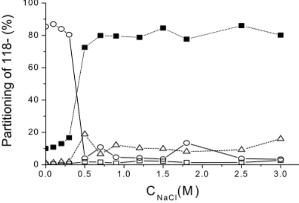
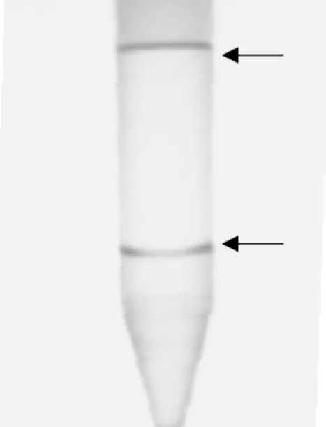
Figure 1 shows the partitioning of the 32P labeled ssDNA 118- in the water-phenol two-phase system as function of NaCl concentration. Below 0.3 M NaCl the ssDNA remains in the aqueous phase; it is almost completely removed from this phase at higher salt concentrations. Only a small amount of ssDNA is transferred to the organic phase (typically 10 % or less of the radioactive material) or is found adsorbed on the walls of the tubes (typically 6 % or less of the radioactive material). The amount of radioactivity present in these three phases (aqueous phase, organic phase and surface of the tube) represents only 20% of the total radioactivity above 0.5 M NaCl. Conversely, the difference between the total radioactivity and this amount (represented by full squares in figure 1 accounts for 80% of the total radioactivity. To localize this missing radioactivity we performed a similar partitioning experiment at 0.85 M NaCl in a bigger (15 ml) tube also treated with dimethyldichlorosilane beforehand, and obtained an overnight autoradiography of the tube (an experiment suggested to us by Gilbert Zalcser). Figure 2 shows a picture of the tube, and the autoradiography of a portion of it. There is a peak located at the water-phenol interface (Figure. 2b), which accounts for the missing radioactivity (there is no radioactivity at the air-water interface). We conclude that above 0.5 M NaCl the missing radioactivity is located at the water-phenol interface.
The experiments described below in the water-phenol system have been performed at 0.85 M NaCl. We therefore characterized in greater detail the partitioning of ssDNA at this concentration by repeating this experiment 10 times: 1) 80 ± 8% of the ssDNA is adsorbed the water-phenol interface; 2) 6 ± 1% of the ssDNA is transferred in the phenolic phase; 3) 3-5 %
the ssDNA is adsorbed on the walls of the tubes; 4) 3-5% of the total radioactivity is present in the aqueous phase. This radioactivity does not correspond to ssDNA but to γ32P-ATP
(aliquots of the aqueous phase run on a polyacrylamide gel never show the ssDNA band but only the γ32P-ATP band; data not shown). We also obtained similar results in experiments
with the complementary ssDNA 118+ (data not shown).
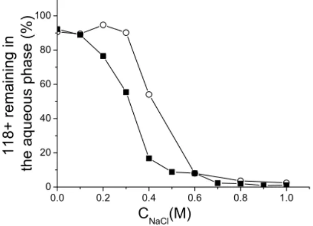
The reversibility of the adsorption as a function of NaCl concentration was determined in the following manner. We first adsorbed the 32P labeled 118+ by vortexing and centrifugation as described above in the presence of different NaCl concentration between 0 and 1M NaCl. We measured the amount of radioactivity present in the aqueous phase (figure 3, open circles). We then prepare 10 different tubes where the 32P labeled 118+ was adsorbed in the presence of 1 M NaCl. We removed 155 µl of the aqueous phase, and replace it by 155 µl of a phenol-saturated TE solution containing different NaCl concentrations. We again vortexed and centrifuged the tubes and determined the amount of radioactivity present in the aqueous phase. Figure 3 (full square) shows the result of this desorption experiment. The adsorbed 118+ is completed desorbed at low salt (0.1 M and below). One observes a hysteresis between 0.2 and 0.6 M NaCl. The differential partitioning of the ions between the two phases could account for this hysteresis. The adsorption is irreversible at high salt above 0.6 M. We have checked this irreversibility at 0.85 M by repeating several times the desorption experiment at this salt concentration: we observed no release of the adsorbed DNA.
Partitioning of ssDNA using other organic solvents
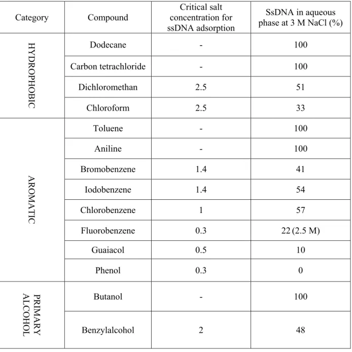
We performed similar partitioning experiments using 13 other organic solvents that are only partially miscible with water. The results are summarized in Table 1. The solvents were divided into three classes: simple hydrophobic solvents, aromatic solvents and primary alcohols. Five solvents are unable to remove the ssDNA from the aqueous phase, even at 3 M
NaCl (dodecane, toluene, carbon tetrachloride, butanol and aniline). Dichloromethane and chloroform begin to adsorb ssDNA above 2.5 M NaCl only. The other solvents are all planar aromatic compounds. The best solvents are fluorobenzene, guaiacol and phenol. Figure 4 shows that they all start to remove the ssDNA from the aqueous phase at low NaCl concentrations (above 0.3 M NaCl for fluorobenzene and phenol, and 0.5 for guaiacol). However, phenol is the most efficient solvent, allowing a complete adsorption above 0.5 M, a salt concentration where about 80% of the ssDNA is still in the aqueous phase in the presence of fluorobenzene or guaiacol.
Renaturation and transfer to the aqueous phase of an adsorbed ssDNA in the presence of its complementary strand
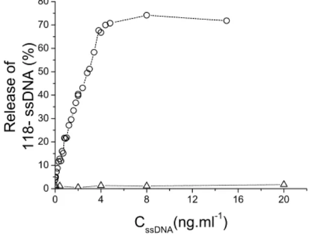
As shown before, at 0.85 M NaCl, 80 % of the ssDNA (118+ or 118-) is irreversibly adsorbed at the water-phenol interface. We performed the following experiment at this salt concentration. We first adsorbed the 32P labeled 118- by vortexing and centrifugation as described above, and then removed 155 µl of the aqueous phase. We replaced it by 155 µl of a TE solution containing the same concentrations of phenol and NaCl, and various amounts of the unlabeled complementary strand 118+. We again vortexed and centrifuged the tube, and then determined the amount of radioactivity present in the aqueous phase. Figure 5a shows that the addition of increasing amounts of the complementary strand 118+ leads to the progressive release of the adsorbed 118- in the aqueous phase. Up to 70 ± 10 % of the 118- can be released; this plateau value is obtained when a concentration of 4 ng/ml of 118+ is reached, corresponding to the amount of 118- present in the tube. The release therefore involves the formation of a 1-1 complex. The release is specific to the complementary strand, since it is not observed when similar amounts of the same strand 118- (instead of the complementary strand) are added in the aqueous phase (Figure 3, open triangles). We
analyzed the released material by gel electrophoresis: it consists solely of a renatured double-stranded DNA (data not shown). Since the adsorption at the water-phenol interface is irreversible for each of the two complementary ssDNA taken separately in our experimental conditions, the renaturation reaction must have been initiated at the interface. Furthermore, the double-stranded DNA that is the outcome of this reaction is not adsorbed at the interface (it partitions entirely in the aqueous phase in our experimental conditions). Thus, as soon as a double-stranded DNA segment is nucleated, it is expelled from the interfacial region. Figure 5b shows that there is initially a strict linear relation between the amount of added 118+ and the amount of released 118-, which confirms that the release involves a stoichiometric process. The slope of the curve in figure 5b is equal to 0.63 ± 0.01. This value corresponds to the product of the two fractions of adsorbed ssDNA (0.8 × 0.8 = 0.64). This confirms the strict interfacial nature of the reaction: only the adsorbed chains can participate in the reaction. The height of the plateau (70 ± 10 %) corresponds to the complete release of 118- by the renaturation reaction.
Effects of vortexing on adsorption and renaturation a. Adsorption without vortexing
Figure 6a shows the percentage of radioactive material removed from the aqueous phase as a function of time in the absence of vortexing. The inset in figure 6a further shows that this percentage increases at first linearly with the square root of time, up to about 1.5 × 10⁴ s. This
dependency is suggestive of an irreversible, diffusion-controlled process (66-68). We will model the kinetic of ssDNA adsorption assuming that the adsorption is diffusion limited. At time t = 0 the bulk number of ssDNA chains is N0 (N0 = C0 × V). The chain concentration C0 is supposed to remain constant close to the air-water interface. This assumption, known as a semi-infinite approximation, will be justified a posteriori. The number N(t) of ssDNA chains
adsorbed at time t at the water-phenol interface is obtained by solving a one-dimensional diffusion equation with boundary conditions pertaining to the conical geometry of our system (figure 6b). Under these conditions the rate of adsorption is given by:
( )
t D C h ah dt z t dN d z 1 3 12 sin 4 , 3 0 2 0 + = = π π 1Where D3d is the three-dimensional diffusion coefficient of the ssDNA chain 118-, h is the
height of the water column, a is the distance between the water-phenol interface and the apex of the cone, and the interface lies at z = 0 (see figure 6b). By integrating both sides of this equation and using the boundary condition N(t = 0) = 0, we obtain the following expression for the number of adsorbed ssDNA chains as a function of time.
t D C h ah t N 0 3d 2 3 12 sin 8 ) ( + = π π 2
In our experiments, 5% of the total amount of radioactivity corresponds to γ32P-ATP, which
always remains in the aqueous phase. In order to model the experimental results, a constant A should therefore be added to the right hand side of equation 2.
A t V D h ah N t N d + + = 12 3 sin 8 ) ( 3 2 0 π π 3
Equation 3 has been used to fit the first regime of adsorption (Figure 4.a). The values of the fitting parameters are:
Parameters D3d (cm
2.s-1)
A
The value of the constant A is in good agreement with the value of 5% determined above. From the value obtained for D3d we can estimate the characteristic time th required to explore
the height h of the aqueous phase using the relation h2 = 6D3dth. We obtain a time th of about
4.4 ×10⁵ s. The time t
h corresponds to time required to deplete the air-water interface. Since
the fitted experimental data have been obtained on a much shorter time scale, the semi-infinite approximation is indeed valid.
b. Adsorption with vortexing
The effects of the duration of vortexing and of the vortexing velocity on the kinetics of adsorption of the 32P labeled ssDNA 118- are shown in figure 7. At the highest vortexing velocity (2400 rpm), a complete adsorption is achieved immediately (that is within the first five seconds). The rate of adsorption increases with the vortexing velocity for lower velocities. The curves suggest the existence of two regimes. At first there is a roughly linear increase of the amount of adsorbed DNA with time. In the second regime the increase is much reduced; the amount of adsorbed ssDNA at this quasi-plateau increases with vortexing velocity.
c. Renaturation without vortexing
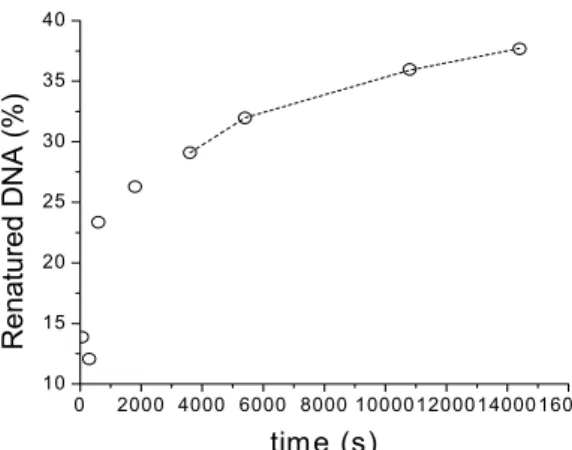
Figure 8a shows the percentage of renatured DNA as a function of time obtained in the two-phase system in the absence of vortexing (during the renaturation stage). In this experiment,
32P labeled ssDNA 118- (125 pM in the aqueous phase) was first adsorbed at the water-phenol
interface by vortexing and centrifugation. We then removed 155 µl of the aqueous phase, and replaced it by 155 µl of a TE solution containing the same concentrations of phenol and NaCl, and the 32P labeled complementary strand 118+ (also at 125 pM). The two-phase system was left to react without agitation. The reaction was quenched by the addition of enough TE buffer
(600 µl TE buffer) to destroy the two-phase system, and an aliquot was analyzed by gel electrophoresis. The rate renaturation is slow, less than 40% of the DNA having renatured within three hours. A comparison of the rate of this reaction with the rate of adsorption observed without agitation (figure 8b) shows that initially the two processes occur at similar rates; later on renaturation becomes the slower of the two processes. This is fully consistent with the conclusion drawn above that renaturation takes place at the interface: the complementary strand must first be adsorbed before it renature. Later on the percentage of renatured DNA increases linearly with the square root of time (Figure 6.b). We now propose a model to explain this dependency.
The renaturation reaction has two kinetics components (Figure 7): The ssDNA must be adsorbed and then it has to search the interface for its complementary strand to renature. At the beginning of the reaction, adsorption and renaturation occur at similar rates. Renaturation is therefore not rate limiting: the concentration of adsorbed 118- is high enough, and when the 118+ reaches the interface it encounters quickly its complementary and forms a dsDNA which is expelled into the aqueous phase. As the reaction proceeds, the concentration of available adsorbed 118- decreases and therefore the rate of the bimolecular reaction between the two complementary strands slows down. In this second regime the rate of the reaction depends on a combination of the diffusion-controlled rate of adsorption and the interfacial recombination. According to this picture, at still longer times, there is a third regime where the rate of reaction is given solely by the rate of recombination at the interface (we do not reach this regime in the present study). The transition between the first and the second regime can be described using the standard terminology of surface chemistry (69, 70). When a reaction between two compounds is catalyzed by a surface, it is possible to distinguish two types of processes. In the first process, the reaction occurs between the two adsorbed compounds, and involves a surface diffusion of at least one of them. This is known as a
Langmuir-Hinshelwood mechanism. In the second process, there is no need for a surface diffusion: the reaction occurs between the adsorbed compound and the second compound coming from the volume above the surface. The encounter between the two compounds involves only a 3-dimensional diffusion. This is known as an Eley-Rideal mechanism. In our case, the first regime is compatible with an Eley-Rideal mechanism: where 118+ encounters the adsorbed 118- through a 3-dimensional diffusion (path A in figure 9). A surface diffusion step (path B in Figure 9) may be present, but is too fast to be detected. In the second regime, the rate of renaturation becomes slower than the rate of adsorption. This corresponds to a Langmuir-Hinshelwood mechanism (path B in Figure 7).
We will model the recombination reaction at the interface assuming a diffusion-controlled annealing process. We further assume that the 118- and the 118+ ssDNA have the same shape and two-dimensional diffusion coefficient D2d. Let us call the 118- the target and the 118+ the
probe. We describe the target molecule as a disc of radius R1 corresponding to half the
end-to-end distance of the 118- chain. A given time t, Ntarget(t) target molecules are embedded in the water-phenol interface, and the average area per target molecule can be estimated assuming that the distribution of target molecules on the interface of area S is homogeneous. The average area per target molecule is equal to:
( )
( )
( )
( )
2 2 target target R t t N S t A = =π 4where R2(t) is the radius. The problem of the capture of a probe molecule by a target molecule
corresponds therefore to a diffusion in a hollow cylinder of inner radius R1 and outer radius
R2. The diffusion in a hollow cylinder has attracted considerable attention over time, and
several solutions corresponding each to a particular boundary initial value have been proposed (67, 71-73). To our knowledge, these solutions deal only with situations where the radii R1
and R2 are independent of time. In these cases, the diffusion equation in cylindrical
(67). In the present situation, when a probe encounters a target molecule, the formed product is expelled from the interface into the bulk. Therefore, this reaction is an interfacial annihilation reaction. As the reaction between the target and probe molecules proceeds, the water-phenol interface is depleted from target molecules and therefore the average area per target molecule increases. The present problem is that of the diffusion in a hollow cylinder with a moving boundary R2(t). Under these circumstances it is not possible to solve the
diffusion equation by separating time and space variables. To overcome this mathematical problem we use a steady-state approximation expected to be valid at long times, when the concentration of target molecules becomes low enough. Under this assumption, the rate of capture of a probe molecule by a target molecule is such that the flux of probe molecules injected at r (the in-plane polar coordinate) equals the flux kcapture of probe molecules which
disappear at 2R1: 1 2 2 1 capture 8 R r d dr dP D R k = = π 5
Where P(r) is the probability to find a probe molecule at position r (in plane polar coordinate) away from a target molecule. In a cylindrical symmetry the diffusion equation under steady state condition is written as:
0 ) ( = dr r dP r dr d , 2R1 <r< R2 6
with boundary conditions P(2R1) = 0, P(R2) = 1. Such conditions imply that 1) the target surface concentration is very dilute and 2) that there is a reservoir of probe molecules.
Berg and Purcell (74) studied a similar problem for a plane with fixed boundary conditions. They concluded that the two-dimensional capture rate will dominate solely if the two dimensional rate of capture is much bigger than the three-dimensional rate. In our case the situation is somewhat different. Using the same approach as Berg and Purcell, we assume that the steady-state rate at the surface is reached when the rate of capture kcapture at two
dimensions is equal to the rate of adsorption at three dimensions. Using this assumption and the fact that the variation of the number of probe molecules Nprobe(z, t) in the z (vertical)
direction is independent of the in plane variations of the number Nprobe(r, t) of probe
molecules, we can write an additional boundary condition to equation 6:
( )
,( )
( )
probe(
0)
probe( )
0 0 probe probe 0 probe = ∇ + = ∇ = ∇N r z = N r N z = N z N r z zUsing the above considerations, the number of probes N(r) for 2R1< r < R2 is therefore given by:
( )
( )
= = 1 2 1 0 probe probe 2 ln 2 ln , , R R R r dz z t dN t r N z 7where the first term in the bracket in the right hand side corresponds to the gradient of concentration of probes molecule in the direction normal to the interface. As the probability
P(r) = Nprobe (r,t)/Nprobe (r, t= 0), the rate of capture is equal to
t D D R R V h ah k d d 1 2 ln 32 3 12 sin 3 2 1 2 2 3 2 capture + = π π 8
The rate of capture depends on time and on the number of target molecules. Two extreme cases can be considered. 1) If R2 = 2R1 the value of the capture rate diverges and goes to
infinity. This condition corresponds to the close packing of target molecules at the interface. This situation corresponds to a very concentrated interface and is beyond the range of validity of the calculated rate of capture. However, the divergence of the two dimensional capture rate indicates that for a concentrated interface the two dimensional diffusion process is not the rate limiting step. 2) If R2 is sufficiently larger than R1, the situation corresponds to a dilute
interface, where the steady-state approximation can be applied. Under this condition the two-dimensional rate of capture contributes to the overall rate of the reaction.
The variation of number of target molecule at the interface is equal to:
( )
(
)
( )
0 ) ( , target target capture target N t N t k dt t dN − = 9where Ntarget(0) is the initial number of target molecules at the interface. Combining equations
8 and 9, we obtain after integration:
( )
(
)
( )
(
( )
)
( )
( )
t D D V h ah N N N R R t N t N R R d d s s 3 12 sin 4 6 0 2 0 1 0 ln 2 ln 2 2 1 ln 2 ln 2 3 2 2 2 3 target target target 1 target target 1 + − = + − − + − π π 10Where Rs= πS is the radius of the interface. Under the close packing condition the number of
target molecules lying on the interface is equal to the square of the ratio of the radius of the interface and the radius of the target (Nclose packing =
2 1 RR s
). Under the hypothesis that the
concentration of target molecule is very dilute at the interface (Ntarget(t) << Nclose packing at any time) equation 10 can be approximated to:
( )
( )
( )
0 1 2 ln 2 3 12 sin 0 128 target 3 2 1 2 target 2 3 target N V t D D R R h ah N t N d d s + + + − ≈ π π 11As pointed out above the recombination reaction at the interface can be considered as an annihilation reaction. Therefore the fraction of renatured ssDNA is equal to:
( )
( )
( )
( )
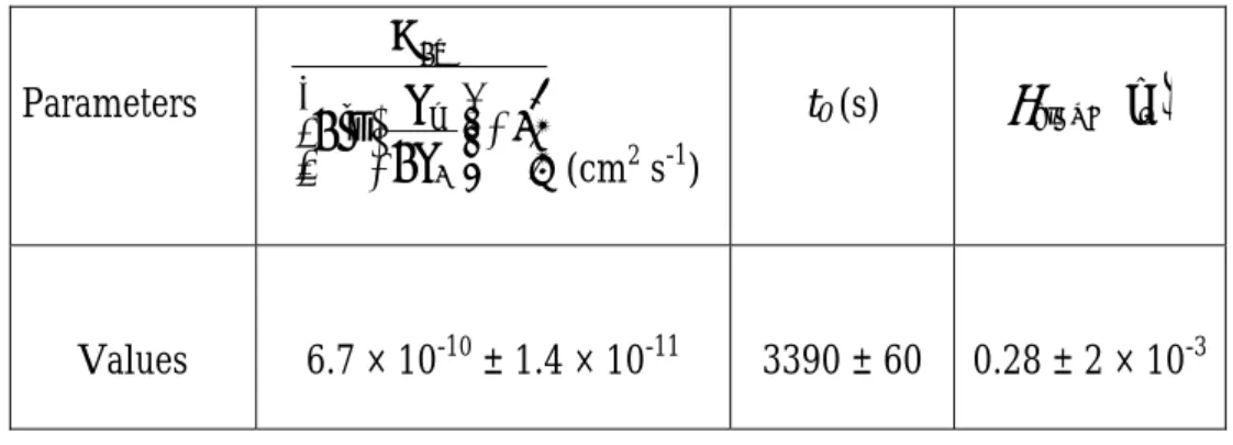
t D D V h ah R R N t N N t d d s 3 12 sin 1 2 ln 2 128 0 0 3 2 2 1 2 3 target target target dsDNA + + = − = π π θ 12 and therefore the fraction of renatured ssDNA should change as the square root of time. As explained above the considered reaction has two kinetics components, the first one is the three-dimensional adsorption process and the second one is the two-dimensional process. These two processes correspond to the two asymptotic limits of the reaction (respectively at short and long times). In the intermediate regime the two kinetic components work cooperatively to form the dsDNA molecule. To model the data presented in figure 6.a, equation 12 should be modified. The two-dimensional steady-state can be considered to have been reached after a time t0 where the θdsDNA(t0) fraction of dsDNA has already been formed. Under these conditions equation 12 should be modified, replacing t by t-t0 and adding aconstant equal to θdsDNA(t0):
( )
( )
0 dsDNA( )
0 3 2 2 1 2 3 dsDNA 3 12 sin 1 2 ln 2 128 t t t D D V h ah R R t d d sθ
π
π
θ
− + + + = 13Equation 13 has been used to fit the last part of the experimental data presented on figure 8a. The value of the fitted parameters are presented below:
Parameters + 1 2 ln 2 1 2 R R D s d (cm2 s-1) t0 (s) θdsDNA t
( )
0 Values 6.7 × 10⁻¹⁰ ± 1.4 × 10⁻¹¹ 3390 ± 60 0.28 ± 2 × 10⁻³Considering that the end-to-end distance of the target molecule is given by:R bNν
2 1
1≈ , where
ν is the Flory exponent ( ½ ≤ ν ≤ 1 in two dimensions) and b is the size of a monomer (b = 3.5 × 10⁻⁸ cm in our case), one can obtain lower and upper estimates for R1 and from these bracket
the two dimensional diffusion coefficient:
3.2 × 10⁻⁹ ± 6.7 × 10⁻¹¹ cm2 s-1 ≤ D2d ≤ 3.8 × 10⁻⁹ ± 8.1 × 10⁻¹¹ cm2 s-1. The two-dimensional
diffusion coefficient depends weakly (logarithmically) on the conformation of the ssDNA chains, leading to a narrow range of possible values: 3.5 × 10⁻⁹ ± 3.7 × 10⁻¹⁰ cm2 s-1
d. Renaturation with vortexing: PERT
Figure 10a shows the kinetics of renaturation of the two complementary ssDNA 118+ and 118- (1.66 pM each) in a 100 µl mixture containing 90 µl of TE plus 0.85 M NaCl and 10 µl phenol vigorously shaken (2400 rpm). This corresponds precisely to the experimental conditions of PERT (1) (vigorous shaking of denatured DNA, equal amounts of complementary strands). The mixture was vortexed at 2400 rpm for various times, the reaction was quenched by the addition of enough TE buffer (100 µl TE buffer) to destroy the emulsion, and an aliquot was analyzed by gel electrophoresis. The reaction is quite fast: about two thirds of the DNA have renatured within 30 s. Nevertheless, by comparing the rates of adsorption and of renaturation (figure 7 and figure. 108.a), we see again that renaturation is the slower of the two processes. We know from the experiment on the rate of adsorption at this vortexing velocity that each complementary ssDNA is completely adsorbed within five
seconds. In the renaturation experiment, all the data obtained after 5 seconds therefore involve complementary strands that have been previously completely adsorbed. The renaturation process between 5 and 30 s takes place exclusively at the water-phenol interface. This establishes the interfacial nature of PERT in this experiment. Assuming that the reaction follows a standard bimolecular kinetics, the reciprocal of fss, the fraction of DNA remaining
single-stranded at time t is expected to grow linearly with t according to:1/ fss= k2C0t+ 1,
where k2 is the bimolecular rate constant, and C0 the molar concentration of single-strands
(here C0 = 1.66 pM). The reciprocal of fss has been plotted as a function of time in figure 10b.
The rate constant obtained by a linear fit yields k2 = 4.2 ± 0.4 × 10¹⁰ M-1s-1. This fit is only
valid for times t > 5 s. The exact meaning of this rate constant is complex: the solution is vigorously vortexed, and the three-dimensional diffusion process has become negligible. It is replaced by a convective flow that controls the arrival and the removal of material at the interface between water and phenol droplets. We calculated the rate of the reaction assuming a classical bimolecular process. The validity of this calculation can be questioned. The system is not under a steady state flow and the distribution of the reactants is not uniform in the solution. These inhomogeneities could lead to a hydrodynamic instability and in turn change radically the kinetics of the reaction (75).
Discussion
We have studied an interfacial DNA renaturation, taking place at a water-phenol interface. We have used a decoupling scheme to establish the interfacial nature of this reaction. The decoupling scheme has led to a characterization of a salt-dependent interfacial confinement of single-stranded DNA, which was studied with and without shaking. We have tested 13 other organic solvents for their capacity of adsorbing ssDNA at the water-organic solvent interface at room temperature in the presence of NaCl. Phenol is the most efficient of all tested solvents. The study of DNA renaturation in the absence of shaking shows the existence of a regime where the rate of the reaction depends on the two-dimensional diffusion of the complementary single-strands. We have determined both the three-dimensional and two-dimensional diffusion coefficients of the ssDNA. The two-two-dimensional coefficient D2d is 10
times smaller than the three-dimensional coefficient D3d. We have also investigated the effect
of shaking on the interfacial renaturation. We established the interfacial nature of PERT in extremely dilute DNA solutions and measured the bimolecular rate constant for the reaction in these conditions. We will now discuss these results following the plan outlined in the introduction.
1) Adsorption and renaturation of DNA in the water-phenol two-phase system
Water-phenol mixtures; salt effects on such mixtures.
We study the behavior of nucleic acids in a water-phenol two-phase system, at various salt concentrations. The discussion of our data requires some knowledge of water-phenol mixtures with and without added salt. Mixtures of water and phenol have been avidly studied over the last century and a wealth of experimental data is available both for bulk ((76, 77) and
further references therein, (78)) and interfacial properties (although only at the air-solution interface ((79, 80) and further references therein). At room temperature water and phenol are only partially miscible (9% phenol in water and 29% water in phenol in weight percent) (78). The two solvents become miscible above 66 °C (in other words the system has an upper critical solution temperature). The phase diagram has a complicated structure at low temperature (below 10 °C) (77, 81): it is closed by the formation of a phenol hydrate (with formula (C6H5OH)2H2O). The crystal structure of phenol hydrate has been determined (82).
Phenol and water molecules are connected by hydrogen bonds, each water molecule being surrounded by four phenol molecules with a distorted tetrahedral structure.
Both adsorption and interfacial renaturation requires the presence of monovalent salts (typically in the order of one mole per liter for PERT (1)). The addition of salt modifies both bulk and interfacial properties of water-phenol mixture. Indeed, the addition of salt greatly enlarges the two-phase region in the temperature versus concentration water-phenol phase diagram (76, 83-87). The miscibility of the two solvents is decreased at fixed temperature, and the upper critical solution temperature is increased. The mutual solubility of water and phenol is decreased because the salt is preferentially soluble in water; this rule is sometime called Timmermans rule (76) (see (88-90) for theoretical modeling). At a fixed temperature the addition of salt lowers the solubility of phenol in the aqueous phase, from 9% without salt to about 5-6% in the presence of molar concentrations of salt (see for instance table 2 in (1)).
Water-phenol nucleic acid mixtures
How can we understand the interaction between phenol and nucleic acids in our system? Clearly, the presence of salt plays a key role, and the polyelectrolyte nature of the nucleic acids must be taken into account (91, 92). Also, we must distinguish between ssDNA and dsDNA. It is well known that phenol interacts better with ssDNA or single-stranded
polynucleotides than with dsDNA: Leng et al. (18) studied the interaction between phenol and nucleic acids by mean of thermal stability, optical activity, viscosity and NMR measurements. They demonstrated that phenol interacts with denatured DNA, poly(A) and poly(U), but poorly or not at all with poly(C) and native DNA. The binding of phenol to single-stranded nucleic acids is therefore also sequence-dependent (we will not discuss this aspect in this work). The preferential binding of phenol to denatured DNA rather than to native DNA can also be inferred from studies of the DNA helix-coil transition in the presence of phenol: phenol lowers the melting temperature of dsDNA (10, 11, 18, 93, 94). Another argument is provided by our results: the dsDNA, once formed at the water-phenol interface is expelled in the aqueous phase. This shows that in contrast with the irreversibly adsorbed ssDNA, dsDNA does not interact efficiently with phenol. We will focus our discussion on the interaction of phenol with ssDNA. The behavior of nucleic acids in the presence of mixed solvents is often described using continuous approaches, (95, 96). We will examine the outcomes of continuous and discrete approaches for the understanding of phenol-ssDNA interactions, with or without salts. This will show the limitations of continuous approaches. We first briefly recall the polyelectrolyte properties of nucleic acids in aqueous solutions.
Electrostatics of nucleic acids in aqueous solutions
In neutral aqueous solutions each phosphate group of the backbone chain of dsDNA and ssDNA is dissociated and carries a negative charge. Nucleic acids are strongly negatively charged polyelectrolytes (see (91, 92) for reviews). In the absence of salt, the long-range electrostatic repulsion among the charged phosphate groups tends to destabilize the helical structure of the dilute dsDNA solutions (91) and to extend ssDNA. In the presence of moderate concentrations of added monovalent cations (typically between 10 mM to 1M), the range of the electrostatic interaction is screened, and the tertiary structure of dsDNA or ssDNA is altered. dsDNA has a local standard helical B structure (we neglect here sequence
specific effects) and adopts a random or swollen coil conformation at large scale. ssDNA is much more flexible than dsDNA (97) and can have different types of local structure (helical or disordered). ssDNA can fold upon itself in a salt dependent manner, which results in intra-strand imperfect base pairing (98-100).
SsDNA-Phenol interactions: continuous and discrete approaches
The helical structure of ssDNA is the result of a balance between a destabilizing long range electrostatic repulsion and stabilizing short range stacking attractions. This qualitative description of ssDNA structure suggests that the configuration and the properties of ssDNA are mainly influenced by electrostatic and polarization forces. Two approaches can be chosen to describe these effects.
a) Continuous approach
Without salt
In this approach the milieu surrounding ssDNA is consider to be homogeneous and continuous. The strength of electrostatic and polarizing forces depends strongly on the ability of the solvent to be polarized. This solvent property is expressed through the dielectric constant of the medium (101). Water has a high dielectric constant (εwater = 80) i.e. in an
external electric field water molecules are easily oriented in the direction of the field. In the close neighborhood of ssDNA the strength of the electric field is important and therefore there exists a shell of oriented water molecules around the DNA molecule (102-104). The hydration shell will interact with the ssDNA’s bases through hydrogen bonding and polarization interactions. Therefore the strength of stacking between bases should be affected by the quality of solvent-ssDNA interactions (98, 105). Phenol has a dielectric constant much lower than water (εphenol = 10 at 60° C (106)) and is partially miscible in water (9% in volume, at
room temperature in the absence of salt). The effective dielectric constant of a phenol saturated water phase in the absence of salt can be estimated using the Clausius-Mossotti
equation (also known in optics as the Lorentz-Lorenz equation (101)), and the values given above for εwater and εphenol. This yields an εsolution = 76.8 (without salt). Thus, the addition of
phenol to the solution decreases the effective dielectric constant of the aqueous phase. According to Mel’nikov et al. this should lead to a weak compaction of the chain (96).
With added salt
The addition of salt into the solution decreases the amount of dissolved phenol in aqueous phase. At high salt concentrations (in particular at 0.85 M NaCl), we estimate that the percentage of phenol in the aqueous phase has dropped to 5%. This corresponds to an εsolution
of about 77, close to the value of pure water. The increase of the dielectric permittivity induces a decrease of the local concentration of counterions surrounding ssDNA (92) and increases the Debye screening length. The addition of salt also decreases the range of electrostatic interactions. Under these circumstances, the destabilizing electrostatic repulsion should become less important than the stacking energy between bases that confers to ssDNA its local structure and its stability. Therefore one expects a folding of the ssDNA induced by the stacking the bases, which could involve imperfect intramolecular base-pairing as is observed in pure water (98, 100). To sum up, the continuous approach predicts a simple folding of the ssDNA molecule in the presence of phenol and salt, which would enhance intramolecular interactions (imperfect base-pairing).
b) Discrete approach
A discrete approach also takes into account forces at the molecular level (short range effect). We first briefly recall the role of short range interactions in the structure and stability of nucleic acids.
We will distinguish two types of short-range interactions: stacking and hydrogen bonding. DNA’s bases are planar aromatic molecules and can form hydrogen bonds. Bases can interact together by their mutual polarization through a Π-Π interaction. This interaction is called Π-stacking. The competition between the short-range attractive stacking interaction between two adjacent bases and the long-range repulsive electrostatic interaction among the charged phosphate group of the backbone confers their helical structure to ssDNA and dsDNA. Watson-Crick hydrogen bonding between complementary bases (adenine with thymine and guanine with cytosine) further stabilizes the double-helical structure of dsDNA. The origin of these two short-range interactions (stacking and hydrogen bonding) lies in the capacity of the DNA bases to be polarized. The ability of DNA bases to create hydrogen bonds and the hydrophilic character of the anionic phosphate oxygen imply that water is an integral part of nucleic acid structure (103). The polymorphism of DNA (ssDNA or dsDNA) is intimately linked to the activity of water (107).
Discrete approach without salt
The above description treats the solvent as a continuous medium and does not take into account explicitly the interactions between the water, phenol and the polyelectrolyte. To describe the interaction of ssDNA with phenol molecule it is necessary to take into account its molecular nature. Water is a structured liquid (108). In the absence of phenol, in the close vicinity of ssDNA, the water molecule network follows the geometry of the polyelectrolyte (109, 110). The ability of phenol to form hydrogen bonds and its aromatic character allow this molecule to interact with water and to disturb locally the hydrogen network formed among water molecules. The interaction between water and phenol molecules can involve the hydrogen bonding observed in phenol hydrate crystals (82). In the presence of phenol, the water molecules in the vicinity of ssDNA must now both follow the geometry of ssDNA and
interact with phenol molecules. This implies that phenol is in close vicinity of ssDNA. Therefore it is strongly favorable for phenol molecules to interact with ssDNA, either through a stacking interaction or hydrogen bonding. Phenol indeed increases the solubility of bases (13, 14): this shows the existence of a favorable interaction between phenol and the bases. On the other hand, dioxane and pyridine, two aromatic compounds with low dielectric constants have been shown by to decrease the solubility of pyrophosphate (10). This should also be the case for phenol: it should decrease the solubility of the phosphate backbone. As a result, the interaction of phenol with ssDNA is going to be a subtle balance of factors that either increase the solubility of the bases or decrease the solubility of the phosphate backbone.
The phenol molecule is repelled by the charged oxygen of the phosphate groups but can form a hydrogen bond with the oxygen linked to the phosphorus atom through a double bond. In the absence of salt, the stacking interaction between the bases in ssDNA is destabilized. This favors an intercalation of phenol between two adjacent bases. This could induce a change in the direction and the orientation of the bases with respect to the helical axis. In summary, in the absence of salt, phenol could form a complex with ssDNA involving a stacking interaction, occurring through an intercalation mechanism (111).
Discrete approach with added salt
The addition of monovalent counterions into the solution changes dramatically this picture. The concentration of dissolved phenol in the aqueous phase decreases as the concentration of added salt increases. SsDNA is a highly charged polyelectrolyte and the addition of cations into the solution screens the negative charge of the phosphate groups and induces the folding of ssDNA as discussed above. At low salt concentration (below the Manning’s threshold (92)), the counterions are trapped along the phosphate backbone of the ssDNA. For ssDNA in the absence of phenol Manning’s threshold (or local counterion concentration near the
ssDNA) is equal to 0.21 M (92) (a similar value can be obtained by solving the Poisson-Boltzmann equation). Above Manning’s threshold, electrostatic effects become less important. Both the repulsion between the charged phosphate backbone and the phenol molecule and the repulsion between two adjacent phosphates are decreased. Phenol molecules can more easily surround the ssDNA chain. The stacking energy between bases is on the order of a few kBT (Boltzmann’s constant kB times the temperature T). In the absence of an
electrostatic repulsion, bases can rotate freely around the phosphate backbone and can be stabilized by the phenol molecules present in the vicinity of the ssDNA chain. At this stage, we can only speculate on the conformation of this folded ssDNA: the helical structure of the DNA could be changed either by changing the direction of the helical turn or by having the phosphate backbone at the center with the bases completely exposed to the solvent and stacked with phenol molecules. In support of a stacking interaction, we can mention the results of Leng et al. (18). The authors have shown using proton magnetic resonance that phenol stacks with adenine bases of poly(A) and uracil bases of poly(U). Their experimental system and ours differ (they use a deuterated solution containing 0.11 M phenol and 0.25 sodium cacodylate), but their observation strongly favors a stacking mechanism in our system too. The presence or absence of an intercalating process cannot be inferred from their results. Another argument in favor of a stacking interaction between phenol and ssDNA comes from an analysis of the results obtained with the 13 other organic solvents (discussed below).
Figure 11 summarizes the two structures that we have discussed for a phenol-ssDNA complex: without salt or at low salt, the interaction would require an intercalation process, while at high salt (roughly above 0.2 M: see below), we expect stacking without intercalation. The existence of such complexes will have to be tested by spectroscopic of microscopic techniques. Remarkably, phenol has been shown to lower the viscosity of poly(A) in the
presence of 1 M NaCl (18): this observation is not in favor of a simple intercalation mechanism, which should lead to an increase of the viscosity.
Similar structures have been proposed to describe protein-ssDNA in some filamentous bacteriophages (112), and have been observed in various single-stranded nucleic acid-protein complexes (113). These two ssDNA-phenol complexes correspond to different distances between two adjacent bases. The intercalated complex leads to a distance of about 7 Å, while in the second structure this distance could be still that found in B DNA.
The further increase in salt concentration (from 0.3 to 1 M) has two effects: 1) ssDNA folding, 2) expulsion of phenol from the aqueous phase. Both phenol and bases can interact with Na+ through cation-π interactions (19). Phenol complexed through stacking to the bases becomes expelled from this phase, and this contributes to the transfer of the ssDNA from aqueous phase to the interfacial region between phenol and water.
Conformation of the ssDNA in the water-phenol mixture
a) conformation in the aqueous phase: an interpretation of the three-dimensional diffusion coefficient of ssDNA
Both fluorobenzene and phenol start to adsorb ssDNA at 0.3 M. This rules out a role for hydrogen bonding and suggests a simple electrostatic mechanism. According to this mechanism, the 0.3 M concentration corresponds to the local counterion concentration Cloc near the ssDNA. Using Manning’s equation (92), we calculate the distance d between two adjacent bases: = solution loc B C q T k d 24.3 4π2 ε 16
where q is the protonic charge. This yields d = 3.5 Å, close to the value found in B DNA. This is inconsistent with an intercalation mechanism (a distance of 7 Å would correspond to a
concentration Cloc = 0.08 M). This simple computation also supports a mechanism involving stacking without intercalation. Let us now try to understand the value of the diffusion coefficient in terms of a conformation of the ssDNA. We use a simple model where the ssDNA is approximated to a sphere of radius RG, the radius of gyration, (to be more exact we
should use the hydrodynamic radius of the ssDNA which is proportional to the radius of gyration;. however in a first approximation we consider both radii equal). Using the Stokes-Einstein relation, the diffusion coefficient is equal to:
G B R T k D πη 6 = 17
where η is the viscosity of the solvent. We consider that in the experimental condition the viscosity of the aqueous phase is the viscosity of water (η = 0.98 × 10⁻³ kg.m-1.s-1). From this
equation we obtain a radius of gyration of 93 Å. This value can be related to the chain conformation using a simple scaling relation, valid for a flexible chain (114):
ν
dN
RG ≈ 18
where d is the distance between two adjacent bases, N the number of monomers (118) and ν is the Flory exponent (1/3 ≤ ν ≤ 1) that defines the conformation of the chain. Combining equations 17 and 18, we derive a Flory’s exponent ν ~ 0.68. This value suggests that in the experimental conditions, the ssDNA chain has a swollen conformation. Our result is obviously crude and should only be viewed as indicative of the state of the chain.
b) Conformation of ssDNA chains at the water-phenol interface.
What is the conformation of ssDNA chains adsorbed at the water-phenol interface? Answering this question is important for understanding the mechanism of the interfacial renaturation. When a polymeric chain is adsorbed on a surface, it can retain its three-dimensional conformation if it becomes stuck on the surface. On the other hand, if it is able to
rearrange freely on this surface, it can then spread and become a two-dimensional polymer. These two possibilities have been described for dsDNA spread on electron micrograph grids (115). Here, the chains are adsorbed at a liquid-liquid interface, on which they are able to diffuse, and the adsorption is irreversible. These three facts suggest that the adsorbed ssDNA chain at the water-are flattened and can be considered as two-dimensional polymers. One can envision the chains with their bases immersed in the phenolic phase, while the phosphate backbone and the sugars remain in the aqueous phase. There exist several possible simple conformations for the ssDNA chain: globular (swelling exponent ν = ½), swollen coil (ν = 3/4), or extended rod (ν = 1). An adsorbed chain can be viewed as a disk of radius R1 and
occupies a surface π(R1)². From this surface one can define an overlap concentration C*
corresponding to the close packing of such disks. In the experiment where we renature DNA without shaking, renaturation becomes slower than adsorption when there remains about 0.47 ng of adsorbed 118- (corresponding to a release of about 20 % of the adsorbed material; see figure 8b). Now the onset of the decrease of the rate of renaturation should occur for a surface concentration close to the overlap concentration C*. We can therefore use this experiment to guess the conformation of the adsorbed chain. For a globular conformation, (R1 = 0.5 × 3.5 ×
(118)1/2 = 19 Å), we obtain C* = 580 ng/cm2, corresponding to an amount of chains of 58 ng on the interface. Assuming as swollen coil conformation (R1 = 0.5 × 3.5 × (118)3/4 = 62.6 Å),
we obtain C* = 50 ng/cm2, corresponding to an amount of chains of 5 ng. Finally, for an
extended rod (R1 = 206 Å), we obtain C* = 5 ng/cm2, corresponding to an amount of chains of
0.5 ng on the interface, very close to the value of 0.47 ng mentioned above. This simple reasoning suggests that the chains have an extended, rodlike structure at the interface. It is worth noting that a similar conclusion would still hold if we assume an intercalating mechanism (leading to a monomer size of 7 rather than 3. 5 Å). Indeed, with an intercalated
chain having a swelling exponent ν (R1 = 0.5 × 7 × (118)ν Å), the close packing of 0.47 ng of
ssDNA is obtained with ν ≈ 0.85 implying an extended structure.
Comparison of phenol with other simple organic compounds: roles of hydrophobicity and aromaticity
In addition to phenol, we have tested 13 organic solvents for their capacity of adsorbing ssDNA at the water-organic solvent interface at room temperature in the presence of NaCl. We will now compare our results with those reported in the literature, and discuss them in terms of hydrophobic and aromatic interactions. The interaction of simple organic compounds with DNA has been described by several authors (10, 12, 17). These authors have concluded that simple organic compounds interact with ssDNA mainly through a hydrophobic interaction, but the situation is actually more delicate.
According to Kauzmann (9), a hydrophobic interaction has a small and positive enthalpy variation and a large and positive entropy variation. In contrast, a stacking interaction between two planar aromatic compounds is not as a role entropically driven. Base stacking for instance is associated with a negative enthalpy and a negative entropy variation (15, 116). In other words, in water, it is more favorable for monomeric bases to form a stacked self-associated structure rather than to be excluded from water (13). This suggests that the description of ssDNA-organic compounds using a simple hydrophobic interaction must be carefully examined.
Levine, Gordon and Jencks (10) studied 54 organic compounds for their capacity to lower the melting temperature of dsDNA (destabilization of dsDNA in the presence of 0.04 M salt followed at 75°C). They came to the conclusion that the decrease of the melting temperature of dsDNA by these compounds is due to their preferential binding to ssDNA. The 6 most efficient compounds in their test were phenol, p-methoxyphenol, benzyl alcohol,
aniline (four organic solvents) plus pyridine and purine. These six compounds are polar, planar, aromatic molecules. Levine, Gordon and Jencks concluded that the most efficient interaction with single-stranded nucleic acids is a hydrophobic interaction. They did not rule out a special mechanism for the action of aromatic compounds in their assay, but considered that this mechanism would be a minor contribution to an overall hydrophobic effect. In a similar study Ts’o and coworkers (11) examined 16 organic compounds for their capacity to lower the melting temperature of dsDNA or poly(A) and obtained similar result: their most efficient compounds were polar, planar, aromatic molecules such as purine, purine derivatives or phenol. Helmer, Kiehs and Hansch (12) analyzed the data of Levine et al. (10) by looking for correlations between octanol-water partition coefficients P of the compounds and their capacity to lower the melting temperature of dsDNA. They found that for the 12 alcohols and phenols as well as for the 5 amides used by Levine et al. there exist a good linear relationship between log (C) (where C is the molar concentration of the compound required to obtain 50% denaturation of dsDNA at 73°C in the work of Levine et al.) and log (P). From these results they too concluded that these compounds act through hydrophobic interactions in the sense of Kauzmann.
The most efficient organic solvents in our adsorption assay are also all planar, aromatic, compounds. The three most efficient compounds are phenol, guaiacol (o-methoxyphenol) and fluorobenzene. Strikingly, phenol is the most efficient compound in these two very different assays. This underlines the efficiency of phenol as a ssDNA ligand. The most efficient compounds in the two assays are all planar, aromatic, polar molecules. The aromaticity of these compounds is therefore essential for an optimal interaction with single-stranded nucleic acids. Whether the interaction of ssDNA with these aromatic compounds is a typical (entropically driven) hydrophobic interaction will require further studies.