HAL Id: hal-02047418
https://hal.archives-ouvertes.fr/hal-02047418
Submitted on 17 Dec 2020
HAL is a multi-disciplinary open access archive for the deposit and dissemination of sci-entific research documents, whether they are pub-lished or not. The documents may come from teaching and research institutions in France or abroad, or from public or private research centers.
L’archive ouverte pluridisciplinaire HAL, est destinée au dépôt et à la diffusion de documents scientifiques de niveau recherche, publiés ou non, émanant des établissements d’enseignement et de recherche français ou étrangers, des laboratoires publics ou privés.
Harnessing γδ T cells in anticancer immunotherapy
Dalil Hannani, Yuting Ma, Takahiro Yamazaki, Julie Dechanet-Merville,
Guido Kroemer, Laurence Zitvogel
To cite this version:
Dalil Hannani, Yuting Ma, Takahiro Yamazaki, Julie Dechanet-Merville, Guido Kroemer, et al.. Har-nessing γδ T cells in anticancer immunotherapy. Trends in Immunology, Elsevier, 2012, 33 (5), pp.199-206. �10.1016/j.it.2012.01.006�. �hal-02047418�
Harnessing ɣδ T cells in anticancer
immunotherapy
Dalil Hannani1,2,4, Yuting Ma1,2,4, Takahiro Yamazaki1,2,4, Julie Dechanet-Merville5, Guido Kroemer1,3,6,7,8,9 and Laurence Zitvogel1,2,4
1Institut Gustave Roussy, Villejuif, France 2
INSERM, U1015, F-94805 Villejuif, France
3
INSERM, U848, F-94805 Villejuif, France
4
Universite´ Paris Sud-XI, F-94805 Villejuif, France
5CNRS UMR 5164, Universite´ Bordeaux 2, 33076 Bordeaux, France 6
Metabolomics Platform, Institut Gustave Roussy, Villejuif, France
7
Centre de Recherche des Cordeliers, Paris, France
8 Pôle de Biologie, Hôpital Europe´ en Georges Pompidou, AP-HP, Paris, France 9
Université Paris Descartes, Paris 5, Paris, France
ɣδ T lymphocytes are involved in the stress response to injured epithelia and in tissue homeostasis by limiting the dissemination of malignant or infected cells and by regulating the nature of the subsequent adaptive im-mune response. ɣδ T cells have potent MHC-unrestricted cytotoxicity, a high potential for cytokine release and broad-spectrum recognition of cancer cells, and as such, are attractive effectors for cancer immunotherapy. Cur-rent expectations are going beyond ex vivo manipulation of the Vɣ9Vδ2 T subset, and target novel ɣδ T cell subsets, properties or receptors, to harness these un-conventional T lymphocytes against cancer. This Opin-ion article discusses novel aspects of ɣδ T cell function during the course of anticancer therapies, as well as new avenues for their clinical implementation.
ɣδ T cell characteristics
ɣδ T cells have several innate cell-like attributes that allow their early and rapid activation following recognition of conserved stress-induced ligands [1,2]. For example, ɣδ T cells can be activated by non-self-ligands, such as viral glycoproteins derived from herpes viruses, and phosphoan-tigens generated either by the isoprenoid pathway used by microorganisms [3]
or by the mevalonate pathway in infected or transformed cells [4]. ɣδ T cells can also be indirectly activated by proinflammatory cytokines re-leased by Toll-like receptor (TLR)-induced dendritic cells (DCs) [interleukin (IL)-12, IL-23 and type-I interferons (IFNs)], with or without concomitant engagement of T cell receptor (TCR) and/or activating natural killer (NK) cell receptors [5–7]. Both human and mouse CD16-expressing ɣδ T cells can also recognize and be activated by antibody-opsonized cells or microorganisms through binding of IgGs, which mediates antibody-dependent cell cytotoxicity (ADCC). On activation, ɣδ T cells expand, acquire cytotoxic functions and secrete an array of proinflammatory (Th1) cytokines. Thus, these cells are attractive for cell therapy strategies against cancer.
In humans, ɣδ T cells represent 3–5% of blood lympho-cytes. Among this population, the Vɣ9Vδ2 T cell subset is predominant (>70%), whereas in other tissues such as gut epithelia, dermis, spleen and liver, ɣδ T cells mostly ex-press the Vδ1 TCR and to a lesser extent the Vδ3 TCR. The Vδ2– population has received less attention than Vɣ9Vδ2 cells, but recent description of their specific anti-cytomeg-alovirus (CMV) activity and antitumor
crossreactivity has raised a new interest in this subset. Here, we summarize recent advances in our understanding of how the different ɣδ T cell subsets contribute to antitumor responses during different cancer therapies, including treatment with bisphosphonates and immunogenic chemotherapy (Box 1). We highlight the potential therapeutic value of combin-ing immunotherapy to target ɣδ T cells with chemothera-peutic approaches.
ɣδ T cells in cancer immunosurveillance
ɣδ T cells can exert cytolytic functions against a variety of premalignant or malignant primary tumor cells and tumor stem cells in an MHC-dependent or -independent manner [8–10]. Activated Vɣ9Vδ2 T cells show in vitro cytolytic activity towards myeloma [11–13], lymphoma cell lines [14,15] and solid tumors [16–19]. Ex vivo expanded ɣδ T cells from patients with glioblastoma multiforme (GBM) harbor intact killing functions against GBM cells; however, both ɣδ T cell counts and the mitogen-stimulated prolifer-ative response of circulating ɣδ T cells are decreased when compared to healthy volunteers [20]. In immunocompro-mised patients, a large number of Vδ2– ɣδ T cells, which exert crossreactive recognition of both CMV-infected and transformed epithelia cells, are associated with reduced risk of developing cancer. In a longitudinal case–control study involving 18 immunosuppressed recipients who de-veloped cancer between 2 and 6 years after transplantation and 45 recipients who did not, the median percentage of ɣδ T cells among total lymphocytes in patients with malig-nancies was significantly lower (<4%) before the diagnosis of cancer compared with that in control patients. An in-crease in Vδ2– ɣδ T cell numbers correlates with a lower
incidence of cancer only in recipients who experienced pre-or post-graft CMV infection, suggesting a role for the CMV-induced Vδ2– ɣδ T cells (mainly Vδ1 ɣδT cells) in protection against tumors. Consistent with this, a retrospective fol-low-up of kidney recipients for 8 years revealed that CMV-naive recipients had a fivefold higher risk of developing cancer compared with CMV-seropositive patients [21]. CMV replication is also associated with a decrease in relapse risk in acute myeloid leukemia (AML) patients who undergo allogenic stem cell transplantation [22]. This might rely on the antileukemic activity capacity of Vδ1 ɣδT cells [23]. Indeed, after bone marrow transplantation in patients suffering from acute lymphoid leukemia (ALL), donor-derived Vδ1 ɣδT cells are activated and proliferate in response to primary recipient ALL blasts. In addition, these cells lyse ALL blasts. Importantly, Vδ1 ɣδT cells do not respond to HLA-mismatch nonleukemic cells, offer-ing promising perspectives for immunotherapy using allo-genic Vδ1 ɣδ T cells.
Whether the presence of ɣδ tumor-infiltrating lympho-cytes (TILs) reflects good or poor prognosis in cancer is still controversial. ɣδ TILs from epithelial tumors in breast, prostate or colon cancer belong to the resident Vδ2– popu-lation [10,24], whereas in other types of epithelial tumors (such as renal cell carcinoma; RCC) ɣδ T cell infiltrates are found to comprise mainly Vɣ9Vδ2 T cells [17]. ɣδ TILs isolated in various types of cancer, including colorectal, breast, prostate, ovarian and RCC [17,24,25], recognize and kill both the autologous tumor and a broad range of related tumors, presumably via the recognition of shared stress-related ligands, but do not kill nontransformed cells [17,25].The infiltration of Vɣ1 and Vδ1 ɣδ T cells into necrotizing melanomas (NMMs) significantly correlates with
increased survival [26], whereas intratumoral ɣδ T cell infiltration in RCC tumors does not
[27]. These studies suggest that ɣδ T cell infiltration might be beneficial; however, the immunoregulatory properties of ɣδ T cells should not be ignored [28]. Indeed, in breast and prostate cancer, the Vδ1+ ɣδ T subset of TILs potently suppresses naive and effector T cell responses and blocks the matura-tion and function of DCs. This suppressive function is reversed by addition of TLR8 ligands [29]. In addition, ɣδ T cells could prevent tumor elimination in an IL-10-dependent manner [30].
Mouse models suggest that the contribution of ɣδ T cells to anticancer surveillance is nonredundant with ab T cells. Mice lacking ɣδ T cells are vulnerable to cutaneous malig-nancy when inoculated with carcinoma cells, or when in-duced by chemical carcinogenesis, underlining their crucial role in cancer immunosurveillance [9,31]. ɣδ T-cell-deficient mice also show that ɣδ T cells strongly contribute to host protection against papillomas, whereas ab T cells have only a moderate effect or even promote the progression of papil-lomas to carcinomas [9,31]. ɣδ T cells that are probably responsible for restraining malignancy are the NKG2D-expressing, skin-associated NKG2D T cells, suggesting that these cells respond very rapidly to stress-ligand-expressing cells (i.e. transformed cells) [9,31]. Using the TRAMP trans-genic mouse model of prostate cancer, one group has provid-ed evidence that ɣδ T cells can mediate protective immunosurveillance against spontaneously arising cancer [8].In nude mice inoculated subcutaneously with nasopha-ryngeal carcinoma cell lines, infused human peripheral ɣδ T cells could temporarily accumulate in tumors, causing a transient reduction in tumor growth [32]. In a preclinical breast tumor model, infused ɣδ T cells traffic to breast tumors in a TCR-dependent manner and prevent tumor growth [33]. Another study using mice transplanted with HER-2+ human breast cancer cell lines has shown that in vivo activation of ɣδ T cells (through bisphosphonates) enhances the efficacy of standard immunotherapy (admin-istration of trastuzumab, a monoclonal antibody directed against HER2/neu receptors) [34]. Also, coadministration of zoledronic acid – a synthetic ɣδ T cell activator – together with intravesical infusion of ɣδ T cells induces antitumor effects against orthotopically inoculated human bladder cancer cells in immunodeficient mice and prolongs survival [35].CMV-specific Vδ2– ɣδ T cells can also target cancer cells in vivo. Prophylactic injections of Vδ2– ɣδ T cell clones in subcutaneous (s.c.) HT29 tumors established in immunode-ficient mice prevent the development of these colon tumors, whereas Vδ2Vɣ9 ɣδ T cells fail to do this. More importantly, systemic intraperitoneal (i.p.) treatment with Vδ2– ɣδ T cell clones delays the growth of HT29 s.c. tumors in a chemokine C-C motif receptor 3 (CCR3)-dependent manner. Vδ2–ɣδ T cell clones express CCR3 and migrate in vitro in response to chemokines (macrophage inflammatory protein-1 d and monocyte chemoattractant protein-4) secreted by HT29 cells [36]. Taken together, these findings suggest thatɣδT cellscan be harnessed for cancer immunotherapy and could synergize with other immunotherapy (such as monoclonal antibodies).
IL-17-producing ɣδ T cells (ɣδ T17) contribute to the efficacy of anticancer therapies
Bidirectional tumor–host interactions (i.e. tumor-induced immunosuppression vs antitumor immunity) during the course of certain anticancer therapies can influence thera-peutic success [37–39]. Indeed, some cytotoxic compounds are capable of triggering an ‘immunogenic’ cell death, in that chemotherapy-induced cell death elicits a tumor-spe-cific immune response that controls residual tumor cells instead of inducing tolerance [40–42]. Failure to activate an antitumor immune response compromises the success of conventional
anticancer chemotherapies. Anthracy-clines, oxaliplatin and X-rays can trigger immunogenic cell death. Four major checkpoints dictate the immunogenicity of cell death and how the host immune system recognizes dying tumor cells. First, optimal phagocytosis of dying tumor cells requires the translocation of endoplasmic re-ticulum (ER)-resident calreticulin (CRT)
[43] and disulfide isomerase ERp57 [44] to their plasma membrane. The ER stress response, initiated by the phosphorylation of eif2a and culminating in the combined exposure of CRT and ERp57 to the plasma membrane, dictates DC uptake of apoptotic bodies, a crucial step to elicit an immune re-sponse against cancer [41,45]. Second, the chromatin-bind-ing high mobility group box 1 protein (HMGB1) is released by dying tumor cells and binds to TLR4 to facilitate antigen processing and presentation to T cells [46]. Third, release of ATP by dying cells is required to engage the purinergic receptor P2RX7, which triggers IL-1b secretion through a cascade involving the Nlrp3 inflammasome, and culmi-nates with IL-1b-dependent ɣδ T cell activation and Tc1 polarization of antigen-specific T cell responses [47,48]. The antitumor effects of anthracyclines, oxaliplatin and X-rays are abrogated in mice devoid of T cells or Th1 cytokines (such as IFNg and the IFNg receptor, and IL-1b and IL-1 receptor 1)
[47–49].
These data suggest that immunogenic cell death in-duced by chemotherapy could influence the composition of the immune infiltrate present in tumors, which in turn could affect the therapeutic response. Supporting this, it has recently been reported that ɣδ T cells that produce IL-17 (ɣδT17), followed by IFNg-producing CD8+ T cells (CTLs), infiltrate tumor beds post-chemotherapy and both ɣδ T cells and CTLs, as well as IL-17 and IFNg, are essential for the therapeutic success of anthracyclines in several preclinical models [47,49] (Figure 1). The transcriptional analysis of the cytokine/chemokine profile in regressing tumor beds post-anthracycline treat-ment has revealed a four- to fivefold increase in the gene products associated with the typical Th1 signature [Eomes, T-bet, IFN-g, lymphotoxin-b, chemokine C-C motif ligand 5 (C-Ccl5), chemokine C-C-X-C-C motif ligand (C-Cxcl)10, C-Cxcl9, and tumor necrosis factor (TNF)-a], as well as gene products encoding IL-7 receptor, IL-21, aryl hydrocarbon receptor (AhR), Cxcl2 and forkhead box p3 (Foxp3). The mandatory role of the 17A and IL-17RA pathway has been demon-strated in several transplantable tumor models (EG7 thy-moma, MCA205 sarcoma, CT26 colon cancer, and TS/A mammary carcinomas) and in 3-methylcholanthrene (MCA)-induced spontaneous sarcoma [49]. Moreover, doxo-rubicin treatment increases the proportion of both IFN-g-and IL-17-producing TILs (which are CD44+ CD62L– CD69+ granzyme B+ CD27– NKG2D– Vɣ4+ or Vɣ6+). Eight days post-chemotherapy in MCA205 sarcomas, the major source of IFN-g is CD8+ T cells, whereas that of IL-17 is mostly TCR d+ T cells rather than CD4+ Th17 cells. Doxo-rubicin-based chemotherapy substantially enhances IFN-g production by CD8+ and CD4+ TILs, as well as IL-17 production by ɣδ TILs. ɣδ TILs invade MCA205 tumor beds within 3 days of treatment, rapidly divide and pro-duce IL-17 shortly after chemotherapy. The early induc-tion of IL-IL-17 contrasts with the later induction of IFN-g production by CD8+ T cells on day 8 post-chemotherapy.
The percentage of ɣδ T17 cells correlates with the frequen-cy of IFN-g-producing CD8+ T lymphocytes (Tc1) in the tumor microenvironment. Accordingly, in the absence of ɣδ T17 or IL-17, priming of adaptive anticancer Th1/Tc1 responses is hampered, suggesting a helper role of ɣδ T17 cells in the context of immunogenic cell death.
The absence of the TCR d chain, as well as Vɣ4 and Vɣ6 ɣδ T cells, has been found to reduce the efficacy of chemo-therapy. Adoptive transfer of ɣδ T cells from naı¨ve mice enhances chemotherapy efficacy and restores the sensitiv-ity of IL-17 knockout mice, provided that these cells ex-press IL-1 receptor 1 and are competent for IL-17 production. ɣδ T17 cells respond to ex vivo propagated bone-marrow-derived DCs (but not macrophages) loaded with dying tumor cells (but not live tumor cells) for the production of IL-17 and IL-22 in a manner depending on IL-1b [but not IL-23, transforming growth factor (TGF)-b or IL-6]
[47]. It is of note that neutralization of IL-23 fails to abrogate the efficacy of chemotherapy
[49]. A beneficial role of ɣδ T17 cells in cancer is supported by a study showing that ɣδ T17 cells are indispensable for the efficacy of Mycobacterium bovis bacillus Calmette–Guerin (BCG) against bladder cancer by recruiting neutrophils [50]. In contrast to these data, IL-17-producing ɣδ TILs could support tumor outgrowth by promoting angiogenesis. Such ɣδ T17 TILs exhibit low levels of perforin and are stimu-lated through TCR, NKG2D and receptors for IL-23, IL-6 and TGF-b to produce IL-17 and cyclooxygenase (COX)-2 in tumor beds [51]. In fact, ɣδ T17 cells from chemotherapy-treated tumor beds depend upon IL-1 receptor 1 to mediate their immunostimulatory effects (instead of TCR, NKG2D and IL-23, as described in
[51]).
These findings unravel the capacity of distinct subsets of ɣδT cells, presumably antigen-naı¨ve (as opposed to self-reactive) and IL-1b-responsive [52], to link innate signals to adaptive immunity during a sterile inflammation such as chemotherapy-induced cell death. Knowing whether human ɣδT cells invade tumor beds at the onset of chemo-therapy and radiotherapy may help in harnessing such effectors to improve conventional therapies of cancer.
Aminobisphosphonates and bromohydrin pyrophosphate: rationale for the utilization of ɣδ2 T cells as antitumor weapons. Synthetic aminobisphosphonates (pamidronate, zoledro-nate) are well known for treatment of Paget’s disease and bone metastases and are potent ɣδ T cell stimulatory compounds that induce cytokine secretion and cell-medi-ated cytotoxicity
[13]. The nitrogen-containing bisphospho-nate zoledronic acid (ZOL) blocks the mevalonate pathway, leading to intracellular accumulation of isopentenyl pyro-phosphate and triphosphoric acid I-adenosin-50 -yl ester 3-(3-methylbut-3-enyl) ester (IPP and ApppI) mevalonate metabolites. Vɣ9Vδ2 T cells appear to recognize IPP and ApppI in ZOL-treated cancer cells as tumor phosphoanti-gens in vitro. These findings may also be significant in vivo because there is a correlation between the ability of Vɣ9Vδ2 T cells to kill tumors and the intracellular IPP and ApppI concentrations in ZOL-treated breast cancer cells. In ZOL-treated mice bearing s.c. breast cancer xeno-grafts, Vɣ9Vδ2 T cells traffic to tumor beds containing high concentrations of IPP and ApppI, which lead to tumor regression. These findings suggest that cancers producing high amounts of IPP and ApppI after ZOL administration may benefit from Vɣ9Vδ2 T-cell-mediated immunotherapy [53].
The antitumor activity of ɣδ T cells in vivo was first tested in a clinical trial in 2003 in which recombinant IL-2 (rIL-2) was combined with pamidronate for the treatment of hematological malignancies (non-Hodgkin lymphoma and multiple myeloma) [54–56]. The combination of pami-dronate and low-dose IL-2 was well tolerated. Preselection of patients based on ‘positive in vitro proliferation of ɣδ T cells in response to pamidronate and IL-2’ was associated with significant activation/proliferation of ɣδ T cells (in 55% of cases) in vivo and
33% of objective responses (tumor regression). In end-stage breast cancer for which admin-istration of zoledronate and rIL-2 was combined, the number of peripheral Vɣ9Vδ2 T cells correlated signifi-cantly with clinical outcome. The seven patients who failed to sustain Vɣ9Vδ2 T cells showed progressive clini-cal deterioration (PD), whereas three patients who sus-tained robust peripheral Vɣ9Vδ2 cell populations showed declining serum concentrations of the glycoprotein Mucin-
1 (MUC-1) tumor-related marker (CA15.3); one patient showed partial remission (PR) and two exhibited stable disease (SD) [55]. In metastatic hormone-refractory pros-tate cancers treated with ZOL and rIL-2, the numbers of effector memory ɣδ T cells and serum levels of tumor necrosis factor–related apoptosis-inducing ligand (TRAIL) (produced by activated ɣδ T cells) correlated with declining prostate-specific antigen levels and objective clinical responses that comprised three PR and five SD. By contrast, most patients treated only with zoledronate (seven out of nine) failed to sustain either ɣδ cell numbers or serum TRAIL and showed PD [56]. This result is in line with a recent report showing no efficacy of ZOL alone in adjuvant breast cancer [57].
Adoptive transfer of ɣδ T cells for treatment of some cancers such as metastatic RCC or non-small cell lung carcinoma has been investigated in several clinical trials during the past decade (Table 1). ɣδ T cells can either be infused into patients after ex vivo activation and expansion with phosphoantigens, or without experimental manipula-tion, instead being directly activated in vivo by administra-tion of synthetic aminobisphosphates (such as bromohydrin pyrophosphate, pamidronate or ZOL) and exogenous T cell growth factors [54– 56]. These therapies are usually feasible and well tolerated (with the exception of risk for thrombo-embolic events in patients with multiple myeloma [58]), but therapeutic benefits seem only to occur when used in com-bination with other therapies [16,59], including cytokines (such as IL-2 and IL-21) [54–56,60], traditional chemother-apy or monoclonal antibodies, because ɣδ T cells mediate ADCC [61,62].
These studies examining the antitumor activity of ɣδ T cells (Table 1) highlight the therapeutic potential of ɣδ T cells in patients in whom Vɣ9Vδ2 T cells can expand and survive. However, more knowledge is needed of ɣδ T cell biology (survival, trafficking patterns, subset variability, ligands and/or TCR identifications) to further such trials [63]. Improving ɣδ T cell function for combinatorial regimens with immunotherapy and chemotherapy
Vɣ9Vδ2 ɣδ T cell-triggering may be ameliorated by com-bining TCR stimulation with engagement of NK receptors or TLRs [64]. In addition, because ɣδ T cells may be inhibited by T regulatory cells [65–67], any therapeutic intervention aimed at reducing T regulatory cell (Treg) immunosuppressive functions could be valuable for restor-ing ɣδ T cell activity.
Although Vδ1 ɣδ T cell ligands have not been identified yet, these cells can be triggered using a specific activating anti-Vδ1 TCR antibody coupled with activating signals such as NKG2D ligands (Figure 2). Using this strategy, one group has developed protocols to expand both human peripheral and tumor infiltrating ɣδ T cells, which show cytotoxicity and antitumor activity [68–70]. In addition, Vδ1 ɣδ T cells that are activated by certain CMV-infected cells or CMV-induced autoantigens display crossreactivity against tumor cells. Thus,
a protocol for Vδ1 ɣδ T cell amplification using autologous CMV-infected cells may favor in vivo Vδ1 ɣδ T cell antitumor activity [21,71]. In immunocompromised mice, sustained expansion of human peripheral blood ɣδ T cells is observed upon stimulation with anti-ɣδ TCR antibody (which expands both Vδ1 and Vδ2 subsets of ɣδ T cells). Compared to phosphoantigen-stimulated ɣδ T cells, the antibody-expanded cells manifest similar functional phenotypes and cytotoxic activity to-wards tumor cell lines between subsets. Vδ1 T cells are more prone to infiltrate CCL17- or CCL22-expressing lym-phomas than Vδ2 T cells because of high expression levels of CCR4 and CCR8 [68]. Vδ1 T cells are also preferentially expanded by extended exposure to concanavalin A in the presence of IL-2. These cells display potent cytotoxicity toward B cell chronic lymphocytic leukemia and represent a new perspective for the treatment of this disease [72].
Similar to conventional T cells, the polarization of ɣδ T cells crucially influences the nature of the downstream adaptive immune response. In the context of a combinato-rial regimen using both chemotherapy and immunothera-py, IL-1R1+ ɣδ T17 cells appear crucial in that they precede and correlate with trafficking of IFNg+ CD8+ T cells to the tumor site [47]. These cells depend on IL-1b for their triggering and therefore administration of an ‘immunogen-ic chemotherapy’ (such as oxaliplatin or anthracycline or an X-ray-based regimen) or local delivery of TLR agonists in the tumor microenvironment (which stimulate local DCs, a source of IL-1b) may be instrumental in polariza-tion of these ɣδ TILs. It is of note that targeting of TLR1 and TLR2 (by pathogens or synthetic ligands), as well as dectin-1 on ɣδ T cells bearing a CCR6+ IL-17+ phenotype, promotes (alone or in conjunction with rIL-23) their pro-liferation and increased Th17-like polarization [73]. Alter-natively, passive infusion of prepolarized ɣδ T17 cells is conceivable. Co-culture of naive Vɣ9Vδ2 T cells with phos-phoantigens and a cocktail of cytokines (IL-1b, TGF-b, IL-6, and IL-23) leads to selective induction of the transcrip-tion factor Retinoic acid receptor-related Orphan nuclear Receptor (RORgt) and polarization toward IL-17 produc-tion. IL-17+ Vɣ9Vδ2 T cells express granzyme B, TRAIL, FasL, and CD161 and upon activation, rapidly induce CXCL8-mediated migration of neutrophils in vitro. Inter-estingly, an increased percentage of circulating IL-17+ Vɣ9Vδ2 lymphocytes has been found in children with bacterial meningitis [74].
Combinations of cellular immune-based therapies with chemotherapy and other antitumor agents may be of clini-cal benefit in the treatment of malignancies. High cytotox-icity against solid-tumor-derived cell lines has been reported with combination treatment using Vɣ9Vδ2 T cells, chemotherapeutic agents and the bisphosphonate ZOL. Pretreatment with low concentrations of chemother-apeutic agents or ZOL has been shown to sensitize tumor cells to rapid killing by Vɣ9Vδ2 T cells. Vɣ9Vδ2 T cell cytotoxicity and IFNg secretion are mediated by perforin following TCR-dependent and isoprenoid-mediated recog-nition of tumor cells [18].
Concluding remarks
Abundant IFNg or IL-17 secretion, potent cytotoxicity and MHC-independent targeting of a broad spectrum of tumors make ɣδ T cells promising targets for cancer immunotherapy using high-affinity phosphoantigens and/or anti-Vδ1 (or Vδ2– TCR) antibodies. Should human data corroborate the findings in mice, the recent demon-stration that ɣδ T cells might participate in the efficacy of chemotherapy will open novel avenues of combinatorial
therapies for solid tumors. In the past decade, the safety profile of ɣδ T cells and the dose-limiting toxicity of phos-phoantigens and rIL-2 have been demonstrated. Ongoing clinical trials are examining the potential for exploiting the ADCC activity of ɣδ T cells. Manipulation of Vɣδ T cell functions by targeting their activating NKG2D receptor (i.e. by blocking soluble MHC class I chain-related genes (MIC)A/B) or co-stimulatory receptors (CD27, TLR3 or TLR7) or interfering with immunosuppressive check-points [Programmed Death-1 (PD-1) or cytotoxic T lym-phocyte antigen (CTLA)4, Tregs] may be of further interest. Finally, it would be interesting to examine the
prospect for predicting ‘ɣδ T cell susceptible’ hematologi-cal malignancies using specific gene signatures in future work [75].
Acknowledgments
D.H is supported by Association pour la Recherche sur le Cancer. Y.M is supported by the China Scholarship Council. T.Y is supported by INSERM. JDM is supported by Fondation pour la Recherche Medicale´ (Equipe labellise´e), Ligue Contre le Cancer, Association pour la Recherche contre le Cancer and INCA. GK is supported by Ligue Nationale contre le Cancer (Equipes labellise´e), Agence Nationale pour la Recherche, the AXA Chair for Longevity Research, European Commission (Apo-Sys, ArtForce, ChemoRes, ApopTrain), Fondation Bettencourt-Schueller, Fondation pour la Recherche Me´dicale, Institut National du Cancer and Cance´ropoˆle Ile-de-France. LZ is supported by Ligue contre le Cancer (e´quipe
labellise´e), Fondation pour la Recherche Me´dicale, INFLACARE EU grant 2008, INCa and Fondation de France (2009-2011).
References
1 Bonneville, M. et al. (2010) Gammadelta T cell effector functions: a blend of innate programming and
acquired plasticity. Nat. Rev. Immunol. 10, 467–478
2 Hayday, A.C. (2009) Gammadelta T cells and the lymphoid stress-surveillance response. Immunity 31, 184– 196
3 Hintz, M. et al. (2001) Identification of (E)-4-hydroxy-3-methyl-but-2-enyl pyrophosphate as a major activator for human gammadelta T cells in Escherichia coli. FEBS Lett. 509, 317–322
4 Gober, H.J. et al. (2003) Human T cell receptor gammadelta cells recognize endogenous mevalonate metabolites in tumor cells. J. Exp. Med. 197, 163–168
5 Devilder, M.C. et al. (2006) Potentiation of antigen-stimulated V gamma 9 V delta 2 T cell cytokine production by immature dendritic cells (DC) and reciprocal effect on DC maturation. J. Immunol. 176, 1386– 1393
6 Devilder, M.C. et al. (2009) Early triggering of exclusive IFN-gamma responses of human Vgamma9Vδelta2 T cells by TLR-activated myeloid and plasmacytoid dendritic cells. J. Immunol. 183, 3625–3633
7 Conti, L. et al. (2005) Reciprocal activating interaction between dendritic cells and pamidronate-stimulated gammadelta T cells: role of CD86 and inflammatory cytokines. J. Immunol. 174, 252–260
8 Liu, Z. et al. (2008) Protective immunosurveillance and therapeutic antitumor activity of gammadelta T cells demonstrated in a mouse model of prostate cancer. J. Immunol. 180, 6044–6053
9 Girardi, M. (2006) Immunosurveillance and immunoregulation by gammadelta T cells. J. Invest. Dermatol. 126, 25–31
10 Ferrarini, M. et al. (1996) Killing of laminin receptor-positive human lung cancers by tumor infiltrating lymphocytes bearing gammadelta(+) T-cell receptors. J. Natl. Cancer Inst. 88, 436–441
11 Ferrarini, M. et al. (2002) Human gammadelta T cells: a nonredundant system in the immune-surveillance against cancer. Trends Immunol. 23, 14–18
12 von Lilienfeld-Toal, M. et al. (2006) Activated gammadelta T cells express the natural cytotoxicity receptor natural killer p 44 and show cytotoxic activity against myeloma cells. Clin. Exp. Immunol. 144, 528–533 13 Kunzmann, V. et al. (2000) Stimulation of gammadelta T cells by aminobisphosphonates and induction of
antiplasma cell activity in multiple myeloma. Blood 96, 384–392
14 Fisch, P. et al. (2000) Inhibitory MHC class I receptors on gammadelta T cells in tumour immunity and autoimmunity. Immunol. Today 21, 187–191
15 Fisch, P. et al. (1997) Control of B cell lymphoma recognition via natural killer inhibitory receptors implies a role for human Vgamma9/Vdelta2 T cells in tumor immunity. Eur. J. Immunol. 27, 3368–3379
16 Viey, E. et al. (2005) Peripheral gammadelta T-lymphocytes as an innovative tool in immunotherapy for metastatic renal cell carcinoma. Expert Rev. Anticancer Ther. 5, 973–986
17 Viey, E. et al. (2005) Phosphostim-activated gamma delta T cells kill autologous metastatic renal cell carcinoma. J. Immunol. 174, 1338–1347
18 Mattarollo, S.R. et al. (2007) Chemotherapy and zoledronate sensitize solid tumour cells to Vgamma9Vdelta2 T cell cytotoxicity. Cancer Immunol. Immunother. 56, 1285–1297
19 Liu, Z. et al. (2005) Ex vivo expanded human Vgamma9Vdelta2+ gammadelta-T cells mediate innate antitumor activity against human prostate cancer cells in vitro. J. Urol. 173, 1552–1556
20 Bryant, N.L. et al. (2009) Characterization and immunotherapeutic potential of gammadelta T-cells in patients with glioblastoma. Neuro Oncol. 11, 357–367
21 Couzi, L. et al. (2010) Cytomegalovirus-induced gammadelta T cells associate with reduced cancer risk after kidney transplantation. J. Am. Soc. Nephrol. 21, 181–188
22 Elmaagacli, A.H. et al. (2011) Early human cytomegalovirus replication after transplantation is associated with a decreased relapse risk: evidence for a putative virus-versus-leukemia effect in acute myeloid leukemia patients. Blood 118, 1402–1412
23 Lamb, L.S., Jr et al. (2001) Human gammadelta(+) T lymphocytes have in vitro graft vs leukemia activity in the absence of an allogeneic response. Bone Marrow Transplant. 27, 601–606
24 Groh, V. et al. (1999) Broad tumor-associated expression and recognition by tumor-derived gamma delta T cells of MICA and MICB. Proc. Natl. Acad. Sci. U.S.A. 96, 6879–6884
25 Corvaisier, M. et al. (2005) V gamma 9 V delta 2 T cell response to colon carcinoma cells. J. Immunol. 175, 5481–5488
26 Bialasiewicz, A.A. et al. (1999) Alpha/beta- and gamma/delta TCR(+) lymphocyte infiltration in necrotising choroidal melanomas. Br. J. Ophthalmol. 83, 1069–1073
27 Inman, B.A. et al. (2008) Questionable relevance of gamma delta T lymphocytes in renal cell carcinoma. J. Immunol. 180, 3578–3584
28 Hayday, A. and Tigelaar, R. (2003) Immunoregulation in the tissues by gammadelta T cells. Nat. Rev. Immunol. 3, 233–242
29 Peng, G. et al. (2007) Tumor-infiltrating gammadelta T cells suppress T and dendritic cell function via mechanisms controlled by a unique toll-like receptor signaling pathway. Immunity 27, 334–348
30 Ke, Y. et al. (2003) Inhibition of tumor rejection by gammadelta T cells and IL-10. Cell. Immunol. 221, 107– 114
31 Girardi, M. et al. (2003) The distinct contributions of murine T cell receptor (TCR)gammadelta+ and TCRalphabeta+ T cells to different stages of chemically induced skin cancer. J. Exp. Med. 198, 747–755 32 Zheng, B.J. et al. (2001) Anti-tumor effects of human peripheral gammadelta T cells in a mouse tumor
33 Beck, B.H. et al. (2010) Adoptively transferred ex vivo expanded gammadelta-T cells mediate in vivo antitumor activity in preclinical mouse models of breast cancer. Breast Cancer Res. Treat. 122, 135–144 34 Capietto, A.H. et al. (2011) Stimulated gammadelta T cells increase the in vivo efficacy of trastuzumab in
HER-2+ breast cancer. J. Immunol. 187, 1031–1038
35 Yuasa, T. et al. (2009) Intravesical administration of gammadelta T cells successfully prevents the growth of bladder cancer in the murine model. Cancer Immunol. Immunother. 58, 493–502
36 Devaud, C. et al. (2009) Antitumor activity of gammadelta T cells reactive against cytomegalovirus-infected cells in a mouse xenograft tumor model. Cancer Res. 69, 3971–3978
37 Zitvogel, L. et al. (2006) Cancer despite immunosurveillance: immunoselection and immunosubversion. Nat. Rev. Immunol. 6, 715–727
38 Zitvogel, L. et al. (2011) Immune parameters affecting the efficacy of chemotherapeutic regimens. Nat. Rev. Clin. Oncol. 8, 151–160
39 Zitvogel, L. et al. (2008) Immunological aspects of cancer chemotherapy. Nat. Rev. Immunol. 8, 59–73 40 Green, D.R. et al. (2009) Immunogenic and tolerogenic cell death. Nat. Rev. Immunol. 9, 353–363
41 Obeid, M. et al. (2007) Calreticulin exposure dictates the immunogenicity of cancer cell death. Nat. Med. 13, 54–61
42 Casares, N. et al. (2005) Caspase-dependent immunogenicity of doxorubicin-induced tumor cell death. J. Exp. Med. 202, 1691–1701
43 Obeid, M. et al. (2007) Ecto-calreticulin in immunogenic chemotherapy. Immunol. Rev. 220, 22–34
44 Panaretakis, T. et al. (2008) The co-translocation of ERp57 and calreticulin determines the immunogenicity of cell death. Cell Death Differ. 15, 1499–1509
45 Panaretakis, T. et al. (2009) Mechanisms of pre-apoptotic calreticulin exposure in immunogenic cell death. EMBO J. 28, 578–590
46 Apetoh, L. et al. (2007) Toll-like receptor 4-dependent contribution of the immune system to anticancer chemotherapy and radiotherapy. Nat. Med. 13, 1050–1059
47 Ma, Y. et al. (2011) Contribution of IL-17-producing gamma delta T cells to the efficacy of anticancer chemotherapy. J. Exp. Med. 208, 491–503
48 Ghiringhelli, F. et al. (2009) Activation of the NLRP3 inflammasome in dendritic cells induces IL-1beta-dependent adaptive immunity against tumors. Nat. Med. 15, 1170–1178
49 Mattarollo, S.R. et al. (2011) Pivotal role of innate and adaptive immunity in anthracycline chemotherapy of established tumors. Cancer Res. 71, 4809–4820
50 Takeuchi, A. et al. (2011) IL-17 production by gammadelta T cells is important for the antitumor effect of Mycobacterium bovis bacillus Calmette–Guerin treatment against bladder cancer. Eur. J. Immunol. 41, 246– 251
51 Wakita, D. et al. (2010) Tumor-infiltrating IL-17-producing gammadelta T cells support the progression of tumor by promoting angiogenesis. Eur. J. Immunol. 40, 1927–1937
52 Jensen, K.D. et al. (2008) Thymic selection determines gammadelta T cell effector fate: antigen-naive cells make interleukin-17 and antigen-experienced cells make interferon gamma. Immunity 29, 90–100
53 Benzaid, I. et al. (2011) High phosphoantigen levels in bisphosphonate-treated human breast tumors promote Vgamma9Vdelta2 T-cell chemotaxis and cytotoxicity in vivo. Cancer Res. 71, 4562–4572
54 Wilhelm, M. et al. (2003) Gammadelta T cells for immune therapy of patients with lymphoid malignancies. Blood 102, 200–206
55 Meraviglia, S. et al. (2010) In vivo manipulation of Vgamma9Vdelta2 T cells with zoledronate and low-dose interleukin-2 for immunotherapy of advanced breast cancer patients. Clin. Exp. Immunol. 161, 290–297
56 Dieli, F. et al. (2007) Targeting human {gamma}delta} T cells with zoledronate and interleukin-2 for immunotherapy of hormone-refractory prostate cancer. Cancer Res. 67, 7450–7457
57 Coleman, R.E. et al. (2011) Breast-cancer adjuvant therapy with zoledronic acid. N. Engl. J. Med. 365, 1396–1405
58 Morgan, G.J. et al. (2010) First-line treatment with zoledronic acid as compared with clodronic acid in multiple myeloma (MRC Myeloma IX): a randomised controlled trial. Lancet 376, 1989–1999
59 Nicol, A.J. et al. (2011) Clinical evaluation of autologous gamma delta T cell-based immunotherapy for metastatic solid tumours. Br. J. Cancer 105, 778–786
60 Kobayashi, H. et al. (2010) Complete remission of lung metastasis following adoptive immunotherapy using activated autologous gammadelta T-cells in a patient with renal cell carcinoma. Anticancer Res. 30, 575– 579
61 Li, H. et al. (2010) Effect of ex vivo-expanded gammadelta-T cells combined with galectin-1 antibody on the growth of human cervical cancer xenografts in SCID mice. Clin. Invest. Med. 33, E280–E289
62 Gertner-Dardenne, J. et al. (2009) Bromohydrin pyrophosphate enhances antibody-dependent cell-mediated cytotoxicity induced by therapeutic antibodies. Blood 113, 4875–4884
63 Gomes, A.Q. et al. (2010) Targeting gammadelta T lymphocytes for cancer immunotherapy: from novel mechanistic insight to clinical application. Cancer Res. 70, 10024–10027
64 O’Brien, R.L. et al. (2007) gammadelta T-cell receptors: functional correlations. Immunol. Rev. 215, 77– 88
65 Mahan, C.S. et al. (2009) CD4+ CD25(high) Foxp3+ regulatory T cells downregulate human Vdelta2+ T-lymphocyte function triggered by anti-CD3 or phosphoantigen. Immunology 127, 398–407
66 Kunzmann, V. et al. (2009) Inhibition of phosphoantigen-mediated gammadelta T-cell proliferation by CD4+ CD25+ FoxP3+ regulatory T cells. Immunology 126, 256–267
67 Goncalves-Sousa, N. et al. (2010) Inhibition of murine gammadelta lymphocyte expansion and effector function by regulatory alphabeta T cells is cell-contact-dependent and sensitive to GITR modulation. Eur. J. Immunol. 40, 61–70
68 Zhou, J. et al. (2012) Anti-gammadelta TCR antibody-expanded gammadelta T cells: a better choice for the adoptive immunotherapy of lymphoid malignancies. Cell. Mol. Immunol. 9, 34–44
69 Kang, N. et al. (2009) Adoptive immunotherapy of lung cancer with immobilized anti-TCRgammadelta antibody-expanded human gammadelta T-cells in peripheral blood. Cancer Biol. Ther. 8, 1540–1549
70 Chen, J. et al. (2001) Antitumor activity of expanded human tumor-infiltrating gammadelta T lymphocytes. Int. Arch. Allergy Immunol. 125, 256–263
71 Halary, F. et al. (2005) Shared reactivity of V{delta}2(neg) {gamma}{delta} T cells against cytomegalovirus-infected cells and tumor intestinal epithelial cells. J. Exp. Med. 201, 1567–1578
72 Siegers, G.M. et al. (2011) Human Vdelta1 gammadelta T cells expanded from peripheral blood exhibit specific cytotoxicity against B-cell chronic lymphocytic leukemia-derived cells. Cytotherapy 13, 753–764
73 Martin, B. et al. (2009) Interleukin-17-producing gammadelta T cells selectively expand in response to pathogen products and environmental signals. Immunity 31, 321–330
74 Caccamo, N. et al. (2011) Differentiation, phenotype, and function of interleukin-17-producing human Vgamma9Vdelta2 T cells. Blood 118, 129–138
75 Gomes, A.Q. et al. (2010) Identification of a panel of ten cell surface protein antigens associated with immunotargeting of leukemias and lymphomas by peripheral blood gammadelta T cells. Haematologica 95, 1397–1404
76 Bennouna, J. et al. (2008) Phase-I study of Innacell gammadelta, an autologous cell-therapy product highly enriched in gamma9delta2 T lymphocytes, in combination with IL-2, in patients with metastatic renal cell carcinoma. Cancer Immunol. Immunother. 57, 1599–1609
77 Bennouna, J. et al. (2010) Phase I study of bromohydrin pyrophosphate (BrHPP, IPH 1101), a Vgamma9Vdelta2 T lymphocyte agonist in patients with solid tumors. Cancer Immunol. Immunother. 59, 1521– 1530
78 Nakajima, J. et al. (2010) A phase I study of adoptive immunotherapy for recurrent non-small-cell lung cancer patients with autologous gammadelta T cells. Eur. J. Cardiothorac. Surg. 37, 1191–1197
79 Sakamoto, M. et al. (2011) Adoptive immunotherapy for advanced non-small cell lung cancer using zoledronate-expanded gammadelta T cells: a phase I clinical study. J. Immunother. 34, 202–211
80 Noguchi, A. et al. (2011) Zoledronate-activated Vgamma9gammadelta T cell-based immunotherapy is feasible and restores the impairment of gammadelta T cells in patients with solid tumors. Cytotherapy 13, 92– 97
Figure 1. Contribution of IL-17-producing ɣδ T cells (ɣδ T17) to the efficacy of anticancer chemotherapy. (a)
Some chemotherapy regimens induce immunogenic cell death (with release of calreticulin exposure, HMGB1 and ATP by dying tumor cells) leading to (b) tumor cell engulfment by DCs and DC activation through a pathway involving the Nlrp3/Asc/casp-1 inflammasome platform, culminating in (c) IL-1b production by DCs. (d) Within 2–3 days, ɣδ T17 are recruited to the tumor bed and activated in an IL-1b-dependent manner. The IL-17-producing ɣδ T cells (e) directly contribute to tumor lysis and may help (f) conventional T cell priming by DCs and/or (g) T cell recruitment into the tumor bed because the accumulation of ɣδ T17 cells precedes and correlates with rapid IFNg-producing CTL infiltration, which is also crucial for tumor eradication.
Figure 2. Improving ɣδ T cell functions in combinatorial regimen against cancer. ɣδ T cells can be infused into
patients after ex vivo expansion, activation and polarization.
(a) IL-17-producing ɣδ T cells can be generated in co-culture with IL-1b-secreting DCs exposed to anthracycline-treated tumor cells [47]. (b) Vɣ9Vδ2 ɣδ T cells can be polarized into IL-17-producing cells using phosphoantigens in the presence of cytokines [74]. Vδ1 ɣδ T cells can be activated and expanded using either (c) autologous CMV-infected cells [21] or (d) anti-Vδ1 TcR antibody. (e) Patients can be treated either by infusion of ex vivo expanded ɣδ T cells (as delineated in a–d) or by direct in vivo ɣδ T cell activation using immunogenic chemotherapy followed by BrHPP + rIL-2 (to trigger Vɣ9Vδ2 ɣδ T cell activation), or anti-Vδ1 TcR antibody (to trigger Vδ1 ɣδ T cells) and/or NKG2D ligands or TLR agonists.
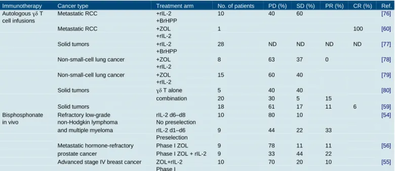
Table 1. Selection of clinical trials assessing safety and efficacy of ɣδ T cell infusions and their stimulatory factors.
Immunotherapy Cancer type Treatment arm No. of patients PD (%) SD (%) PR (%) CR (%) Ref.
Autologous ɣδ T Metastatic RCC +rIL-2 10 40 60 [76]
cell infusions +BrHPP
Metastatic RCC +ZOL 1 100 [60]
+rIL-2
Solid tumors +rIL-2 28 ND ND ND ND [77]
+BrHPP
Non-small-cell lung cancer +ZOL 8 63 37 0 [78]
+rIL-2
Non-small-cell lung cancer +ZOL 15 60 40 [79]
+rIL-2
Solid tumors ɣδ T alone 5 40 40 [80]
combination 20 30 5 15
Solid tumors 18 61 17 11 6 [59]
Bisphosphonate Refractory low-grade rIL-2 d6–d8 10 80 10 [54]
in vivo non-Hodgkin lymphoma No preselection
and multiple myeloma rIL-2 d1–d6 9 44 22 33 Preselection
Metastatic hormone-refractory Phase I ZOL 9 78 11 11 [56]
prostate cancer Phase I ZOL + rIL-2 9 33 44 22
Advanced stage IV breast cancer ZOL+rIL-2 10 70 20 10 [55]
Phase I
CR, complete response; PD, progressive disease; PR, partial response; SD, stable disease.
Box 1. Triggering ɣδ T-cell-mediated antitumor activity: focus on aminobiphosphonates and chemotherapy
Aminobiphosphonates are synthetic chemical compounds that potently activate ɣδ T cells and have shown promising efficacy in the treatment of several types of cancer (RCC, metastatic prostate cancer and breast cancer). So far, only a few drugs have been tested, in particular ZOL, pamidronate and bromohydine pyrophosphate (BrHPP; also known as IPH1101). All these synthetic compounds mimic natural ɣδ T cell activating ligands such as IPP or HM-BPP [(E)-4-hydroxy-3-methyl-but-2-enyl pyrophosphate].
Some chemotherapy agents (such as doxorubicin and oxaliplatin) have the capacity to induce an immunogenic tumor cell death, which elicits an antitumor immune response that is crucial for tumor eradication and long-term protection against relapse. ɣδ T cells are recruited to the tumor bed after immunogenic chemotherapy and appear as crucial contributors to chemotherapy efficacy. Doxorubi-cin is an anthracycline antibiotic that works by intercalating DNA. Doxorubicin is commonly used in the treatment of a wide range of cancers, including hematological malignancies, many types of carcinoma, and soft tissue sarcomas.
Oxaliplatin is a platinum-based anticancer agent. The cytotoxicity of platinum compounds results from inhibition of DNA synthesis. Oxaliplatin is mainly used in the treatment of colorectal carcinoma.