Nephrol Dial Transplant (2014) 29: 619–625 doi: 10.1093/ndt/gft452
Advance Access publication 8 November 2013
Long-term enzyme replacement therapy is associated with
reduced proteinuria and preserved proximal tubular
function in women with Fabry disease
Thaneas Prabakaran
1, Henrik Birn
1,2, Bo M. Bibby
3, Axel Regeniter
4, Søren S. Sørensen
5, Ulla
Feldt-Rasmussen
6, Rikke Nielsen
1and Erik I. Christensen
11Department of Biomedicine, Aarhus University, Aarhus, Denmark,2Department of Nephrology, Aarhus University Hospital, Aarhus,
Denmark,3Department of Biostatistics, Aarhus University, Aarhus, Denmark,4Laboratory Medicine, Basel University Hospital, Basel, Switzerland,5Department P, Rigshospitalet, Copenhagen, Denmark and6Department of Medical Endocrinology, Rigshospitalet, Copenhagen, Denmark
Correspondence and offprint requests to: Erik Ilsø Christensen; E-mail: [email protected]
A B S T R AC T
Background. Fabry disease is an X-linked lysosomal storage disorder caused by mutations in the GLA gene. Deficiency of α-galactosidase A (α-Gal A) causes intracellular accumu-lations of globotriaosylceramide (GL-3) and related glyco-sphingolipids in all organs, including the kidney, often leading to end-stage renal failure. In women with Fabry disease, accumulation of GL-3 in the glomerular podocytes and other renal cells induces progressive, proteinuric nephropathy, but not as severe as in men. Enzyme replacement therapy (ERT) with recombinant α-Gal A reduces cellular GL-3 deposits in podocytes and tubular epithelial cells. We have previously shown that α-Gal A is delivered to these cells by different pathways involving different receptors. This study investigated the long-term changes in albuminuria, estimated glomerular filtration rate (eGFR) and urinary markers of both glomerular and tubular dysfunction in women with Fabry disease treated with ERT.
Methods. A retrospective, single centre, cohort study evalu-ated the long-term association between ERT, albuminuria and eGFR in 13 women with Fabry disease and mild renal involve-ment. In particular, we analysed the changes in the proteinuric profile, including the glomerular marker IgG, the tubular markers α1-microglobulin and retinol-binding protein (RBP),
and the shared tubular and glomerular markers albumin and transferrin.
Results.ERT was associated with a significant reduction in al-buminuria and a relatively stable eGFR. The decrease in albu-minuria was paralleled by a decrease in both glomerular and tubular urine protein markers.
Conclusions.The data indicate that long-term ERT is associ-ated with a reduction in albuminuria and glomerular and tubular urinary protein markers in women with Fabry disease and mild renal manifestations.
Keywords: albuminuria, enzyme replacement therapy, Fabry disease, proteinuria
INTRODUCTIO N
Fabry disease is an X-linked lysosomal disorder that results from mutations of the gene (GLA) that encodes the lysosomal hydrolase α-galactosidase A (α-Gal A) [1]. The enzymatic defect leads to progressive lysosomal accumulation of globo-triaosylceramide (GL-3) and related glycosphingolipids in the kidney and other tissues [1, 2]. In classically affected males, clinical onset occurs in childhood or adolescence and is characterized by several symptoms [1,3,4]. Glycosphingolipid accumulates over time leading to kidney failure, cerebrovascu-lar manifestations, heart failure and eventually premature death [5].
Nephropathy is a dominant feature in Fabry disease and impairment of renal function occurs due to progressive accumulation of GL-3 in renal endothelial, interstitial, tubular epithelial and glomerular cells [1, 6–10]. Early findings in Fabry nephropathy include isosthenuria and tubular dysfunc-tion [1]. Progressive decline in the glomerular filtration rate (GFR) and proteinuria are detrimental signs of renal dysfunc-tion in Fabry disease that may eventually lead to end-stage renal failure [11–14]. This usually occurs in the third tofifth decade of life, when the lysosomal GL-3 accumulation is
ORIGINAL
irreversible [8,13]. Fabry nephropathy is generally less severe in women than in men [15]. However, many heterozygous female individuals may be affected similarly to hemizygous male individuals due to random X-chromosomal inactivation [5,16–18].
Currently, enzyme replacement therapy (ERT) is the only specific treatment for Fabry disease patients [9, 14, 19–23], which has been shown to reduce the GL-3 deposits from mul-tiple cell types. Several studies have suggested that ERT with recombinant α-Gal A stabilizes or slows the progression of Fabry nephropathy in Fabry disease patients [22,24–28]. We have previously demonstrated that recombinant α-Gal A is taken up by the endocytic receptors, megalin and mannose-6-phosphate receptor (M6PR) in the proximal tubule cells [29], and megalin, M6PR and sortilin in the podocytes [30], and by M6PR and sortilin in the glomerular endothelial cells [31], suggesting that ERT may interact with several different parts of the nephron.
In this study, we investigated the association between ERT in women with Fabry disease and the changes in albuminuria, estimated glomerular filtration rate (eGFR and urinary markers of both glomerular and tubular dysfunction during a mean follow-up period of 6 years.
M AT E R I A L S A N D M E T H O D S Ethics
The study was approved by the Regional Research Ethics Committee and conducted in accordance with the Helsinki Declaration. Informed consent was obtained from all participants.
Study population
This was a retrospective, single centre, cohort study of 17 Caucasian women with Fabry disease referred to the Danish Fabry Centre, Copenhagen University Hospital, Denmark, at which treatment of Fabry patients in Denmark has been centralised. The study included 13 female patients treated with ERT for >1 year and four untreated female patients, all older than 10 years, referred from 2003 to 2011. The study excluded 18 female patients followed at the centre during the same period, either untreated and/or lacking urine samples or on ERT for <1 year. Fabry disease was confirmed in all patients by GLA mutation analysis. All patients underwent regular and systematic examinations for manifestations of Fabry disease as a part of the normal follow-up procedure at the hospital. Patients received agalsidase beta (Fabrazyme®, Genzyme) 1 mg/kg intravenously every second week during the follow-up period. Because of a shortage of Fabrazyme® in 2010/2011, most patients subsequently received agalsidase alfa (Replagal®, Shire) 0.2 mg/kg for the remaining follow-up period.
Data and sample collection
Relevant clinical data were collected from patientfiles. Data and urine samples were collected over a period of up to 7 years. Plasma creatinine was measured in a certified laboratory using a standardized Roche Modular enzymatic creatinine
assay. The eGFR was calculated using the CKD-EPI equation [32,33]. Morning urine samples were collected at baseline and during follow-up. Urine samples were frozen at −80°C until processed.
Urine protein analysis
Frozen morning urine samples were analysed for total protein ( pyrogallol red method) and creatinine (standardized enzymatic method) on a Roche systems Modular clinical chemistry analyser. The following urinary proteins were measured on a Beckman Coulter Image nephelometry system: albumin, IgG, transferrin, retinol-binding protein (RBP) and α1-microglobulin (α1M) [34,35]. The urinary excretions of all
proteins were normalized according to the urine creatinine concentration. Normal reference ranges for IgG, transferrin, albumin, α1M, RBP and total protein excretion are listed in
Table1.
Statistical analyses
The continuous variables are presented by medians and ranges. The albuminuria and eGFR data were analysed using a random coefficient model with the time as a covariate and ERT as a treatment factor. The albuminuria data were log-transformed based on the inspection of residual plots. A P < 0.05 was considered statistically significant. Data analysis was performed using Stata version 12.1 and thefigures were prepared using Adobe Photoshop CS3.
R E S U LTS Patients
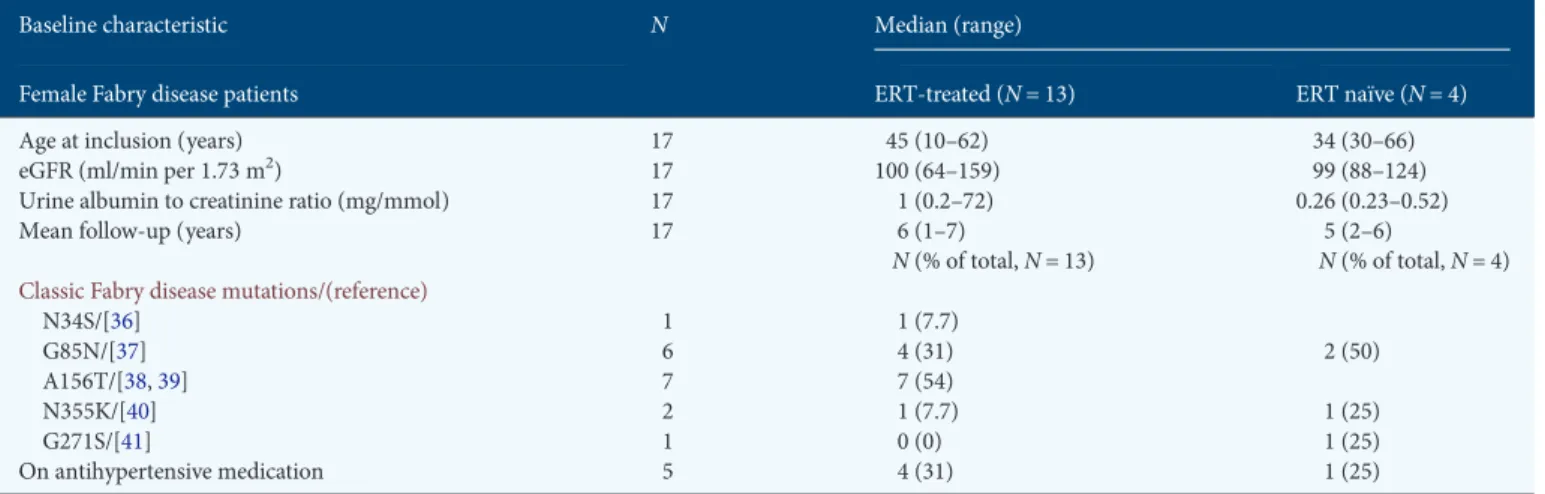
Thirteen ERT-treated and four ERT-naive female patients were included. Patient characteristics are given in Table2. The ERT treatment was initiated on the day of inclusion and con-tinued until the end of follow-up. The ERT-naive patients were not on ERT prior to inclusion or during follow-up. The included patients revealed little or no clinical signs of kidney disease (Table 2). Nevertheless, renal biopsies from three of the patients demonstrated GL-3 inclusions in the podocytes and tubule epithelial cells suggesting renal involvement (data not shown). No major side effects to ERT were reported and there were no deaths or loss in follow-up.
Albuminuria and eGFR
There was a slight decrease in eGFR during the follow-up period (Figure 1A). In ERT-treated patients, the average Table 1. References for established normal values for urinary protein excretion [34, 35]
Urinary protein Normal range (mg/mmol creatinine)
IgG <1.13 Transferrin <0.19 Albumin <2.26 α1M <1.36 RBP <0.08 Total protein <11.31 ORIGINAL ARTICLE
decline in eGFR was 1.45 mL/min/1.73 m2per year (95% CI: 0.31–2.58 mL/min/1.73 m2 per year, P = 0.012) compared with 0.89 mL/min/1.73 m2per year (95% CI:−1.51–3.30 mL/ min per 1.73 m2per year, P = 0.47) in ERT naive. The decline in the two groups was not significantly different, P = 0.68. During the same period, there was a clearly significant de-crease in the albumin/creatinine ratio in the ERT-treated patients (22% per year, 95% CI: 11–34%, P < 0.0001) (Figures1B and2). In the ERT-naive patients, a significant in-crease was observed (39% per year, 95% CI: 15–67%, P < 0.0001) (Figures 1B and 2). The difference between the two groups was significantly different, P < 0.0001.
Other urine protein markers
To examine the association between ERT and selective markers of glomerular protein leakage as well as proximal tubule reabsorptive dysfunction, we examined the urinary excretion of IgG, RBP, a1M and transferrin, the latter being a
marker of both glomerular and tubular dysfunction. Treat-ment with ERT was associated with a significant decrease in the IgG/creatinine ratio (8% per year, 95% CI: 2–13% per year, P = 0.010) andα1M/creatinine ratio (9% per year, 95% CI: 4–
14% per year, P < 0.0001, Figure 3). A trend towards lower protein excretion was also observed for the RBP/creatinine ratio (1% per year, 95% CI:−6–7% per year, P = 0.827), trans-ferrin/creatinine ratio (6% per year, 95% CI:−2–13% per year, P = 0.143) and total protein/creatinine ratio (5% per year, 95% CI: −8–19% per year, P = 0.469) in the ERT-treated patients (Figure 3). ERT-naive patients revealed no significant trend towards an increase in the excretion of glomerular and tubular markers.
DISCUS SION
This study shows that in women with Fabry disease and mild renal involvement ERT are associated with stable renal func-tion and a reducfunc-tion in albuminuria as well as the excrefunc-tion of both glomerular and tubular protein markers.
The efficacy of ERT is supported by a very recent study showing that long-term ERT with agalsidase alfa stabilized renal function in women with Fabry disease [27]. Several other studies evaluating the effect of short-term ERT with agalsidase beta [24] or agalsidase alfa [26] have shown stabilization of renal function in both male and female Fabry disease patients. The natural progression of Fabry nephropathy has been retro-spectively studied in a group of female Fabry disease patients before ERT became available showing a yearly decline in eGFRs of 0.6 ± 2.3 and 2.2 ± 2.2 mL/min/1.73 m2 in women Table 2. Baseline characteristics of the 17 female patients with Fabry disease included in this cohort
Baseline characteristic N Median (range)
Female Fabry disease patients ERT-treated (N = 13) ERT naïve (N = 4)
Age at inclusion (years) 17 45 (10–62) 34 (30–66)
eGFR (ml/min per 1.73 m2) 17 100 (64–159) 99 (88–124)
Urine albumin to creatinine ratio (mg/mmol) 17 1 (0.2–72) 0.26 (0.23–0.52)
Mean follow-up (years) 17 6 (1–7) 5 (2–6)
N (% of total, N = 13) N (% of total, N = 4)
Classic Fabry disease mutations/(reference)
N34S/[36] 1 1 (7.7) G85N/[37] 6 4 (31) 2 (50) A156T/[38,39] 7 7 (54) N355K/[40] 2 1 (7.7) 1 (25) G271S/[41] 1 0 (0) 1 (25) On antihypertensive medication 5 4 (31) 1 (25)
F I G U R E 1 :Change in median eGFR (A) and albuminuria (B) in ERT-treated and ERT-naive female Fabry disease patients during the follow-up period.
ORIGINAL
with proteinuria <100 mg/day and between 100 and 1000 mg/ day, respectively [11]. Four women with Fabry disease fol-lowed at our centre and not receiving ERT also revealed a trend towards increasing albuminuria and declining eGFR during an average of 5-year follow-up.
Proteinuria in Fabry disease patients is associated with pro-gressive tubular injury, interstitial fibrosis and a decline in GFR [42]. Injury to the podocytes due to GL-3 deposits is es-sential in the development of Fabry nephropathy and protei-nuria [9]. We observed GL-3 accumulation in podocytes and tubule epithelial cells in biopsies from three patients included in this study. Recently, it has been shown that ERT is associ-ated with a reduction of GL-3 deposits in podocytes and tubule epithelial cells [9], and that α-Gal A is taken up by megalin and the M6PR in the proximal tubule cells, [29] and by megalin, M6PR and sortilin in the podocytes [30]. Thus, ERT might exert effects in multiple, different parts of the nephron.
Changes in urinary protein excretion may reflect changes in both glomerular and tubular function. We analysed the pattern of proteinuria in order to gain information on the effect of ERT on the different parts of the nephron [43]. We show that long-term ERT with recombinantα-Gal A is associ-ated with a significant reduction in albuminuria in women with Fabry disease and mild renal involvement. Furthermore, we observed significant decreases in the excretion of both
glomerular and tubular urine protein markers. The change in the excretion ofα1M and IgG in ERT-treated patients
paral-leled the decrease in the excretion of albumin. Thus, this study demonstrates a correlation between changes in the excretion of glomerular and tubular markers, and albuminuria. It may be hypothesized that ERT affects both the podocytes and the tubular epithelial cells, and that uptake of filtered α-Gal A does not negatively affect proximal tubule function even though significant amounts of recombinant α-Gal A accumu-lates here [29].
It is generally recommended to use blockers of the renin– angiotensin system, such as angiotensin-converting enzyme inhibitors and angiotensin II receptor blockers (ACEi/ARBs) in proteinuric renal diseases to prevent progression [44, 45]. Although none of the patients had overt proteinuria, 31% of ERT-treated and 25% of ERT-naive patients in our study were treated with ACEi/ARB. Seventy-five percent of the ERT-treated patients on ACEi/ARB initiated ACEi/ARB treatment after being on ERT for 2 years. Thus, the initial and greater de-crease in urinary protein excretion after initiation of ERT was independent of ACEi/ARB therapy. Furthermore, a recent study evaluating the long-term use of ACEi/ARB in combi-nation with recombinantα-Gal A did not show any significant differences in the effect of ERT on eGFR between Fabry disease patients receiving ACEi/ARB and those who did not [27]. Because of the retrospective nature of most studies
F I G U R E 2 :Individual curves showing changes in albuminuria in the 13 ERT-treated patients dependent on the initial level (<2 mg/mmol (A), >2 mg/mmol but <10 mg/mmol (B) and >10 mg/mmol (C)) and in the four ERT-naive patients (D).
ORIGINAL
involving ACEi/ARB and ERT, no definite conclusion can be made on the potential, additional effects of ACEi/ARB. More systematic studies are needed to evaluate the effect of ERT in combination with other renoprotective medications, and we cannot exclude that the concomitant use of ACEi/ARBs was a possible confounding factor in our study.
The major limitations of this study include the non-ran-domized design and the lack of a larger control group. Only four female Fabry patients at the study centre with regular follow-up were not receiving ERT making relevant, statistical comparisons impossible. Also, conclusions are restricted by
the low number of study participants. The heterogeneity of the disease in female patients due to skewed X-inactivation also leads to varying degrees of renal pathology; however, depo-sition of GL-3 was observed in the kidneys of all the three patients biopsied in this study. The change in treatment proto-col due to shortage of agalsidase beta may also have influenced the outcome of this study because of the difference in the dosages used: 0.2 mg/kg agalsidase alfa versus 1 mg/kg agalsi-dase beta. Finally, we used eGFR as endpoint rather than more precise measurements of GFR. The CKD-EPI formula was applied since this formula has been shown to be more accurate
F I G U R E 3 :Change in the (median; range) excretion of IgG (A),α1M (B), RBP (C), transferrin (D) and total protein (E) during ERT.
ORIGINAL
than the MDRD formula for estimating GFR ≥60 mL/min/ 1.73 m2[32,46].
In conclusion, long-term ERT with agalsidase beta is associated with a reduction in albuminuria and glomerular and tubular urinary protein markers in women with Fabry disease and mild renal manifestations.
AC K N OW L E D G E M E N T S
The skillful assistance by Hanne Sidelmann and study nurse Ira Hagen Pedersen is gratefully acknowledged. This work was supported by the Danish Medical Research Council and Genzyme Corporation.
CON F LI CT O F IN TE R E S T S TATE M E N T
A postdoc grant for T.P. is in part paid by Genzyme Corpor-ation, Framingham, Massachusetts. U.F-R. has received un-restricted research grants form Genzyme and serves at the Fabry Registry Advisory Board.
R E F E R E N C E S
1. Desnick RJ, Ioannou YA, Eng CM.α-Galactosidase A deficiency: Fabry disease. In: Scriver CR, Sly WS, Valle D (eds), (ed). The Metabolic and Molecular Bases of Inherited Disease, 8 ed. New York, NY: McGraw-Hill, 2006, pp. 3733–3774
2. Whybra C, Kampmann C, Willers I et al. Anderson–Fabry disease: clinical manifestations of disease in female heterozygotes. J Inherit Metab Dis 2001; 24: 715–724
3. Hopkin RJ, Bissler J, Banikazemi M et al. Characterization of Fabry disease in 352 pediatric patients in the Fabry Registry. Pediatr Res 2008; 64: 550–555
4. Ramaswami U, Whybra C, Parini R et al. Clinical manifestations of Fabry disease in children: data from the Fabry Outcome Survey. Acta Paediatr 2006; 95: 86–92
5. Mehta A, Ricci R, Widmer U et al. Fabry disease defined: baseline clinical manifestations of 366 patients in the Fabry Outcome Survey. Eur J Clin Invest 2004; 34: 236–242
6. Colley JR, Miller DL, Hutt MS et al. The renal lesion in angiokeratoma corporis diffusum. Br Med J 1958; 1: 1266–1268
7. Meroni M, Sessa A, Battini G et al. Kidney involvement in Anderson– Fabry disease. Contrib Nephrol 1997; 122: 178–184
8. Branton MH, Schiffmann R, Sabnis SG et al. Natural history of Fabry renal disease: influence of alpha-galactosidase A activity and genetic mutations on clinical course. Medicine (Baltimore) 2002; 81: 122–138 9. Thurberg BL, Rennke H, Colvin RB et al. Globotriaosylceramide
accumu-lation in the Fabry kidney is cleared from multiple cell types after enzyme replacement therapy. Kidney Int 2002; 62: 1933–1946
10. Tondel C, Bostad L, Hirth A et al. Renal biopsy findings in children and adolescents with Fabry disease and minimal albuminuria. Am J Kidney Dis 2008; 51: 767–776
11. Schiffmann R, Warnock DG, Banikazemi M et al. Fabry disease: pro-gression of nephropathy, and prevalence of cardiac and cerebrovascular events before enzyme replacement therapy. Nephrol Dial Transplant 2009; 24: 2102–2111
12. Wanner C, Oliveira JP, Ortiz A et al. Prognostic indicators of renal disease progression in adults with Fabry disease: natural history data from the Fabry Registry. Clin J Am Soc Nephrol 2010; 5: 2220–2228
13. Thadhani R, Wolf M, West ML et al. Patients with Fabry disease on dialysis in the United States. Kidney Int 2002; 61: 249–255
14. Eng CM, Banikazemi M, Gordon RE et al. A phase 1/2 clinical trial of enzyme replacement in Fabry disease: pharmacokinetic, substrate clear-ance, and safety studies. Am J Hum Genet 2001; 68: 711–722
15. Ortiz A, Oliveira JP, Waldek S et al. Nephropathy in males and females with Fabry disease: cross-sectional description of patients before treatment with enzyme replacement therapy. Nephrol Dial Transplant 2008; 23: 1600–1607
16. Van den Veyver IB. Skewed X inactivation in X-linked disorders. Semin Reprod Med 2001; 19: 183–191
17. Morrone A, Cavicchi C, Bardelli T et al. Fabry disease: molecular studies in Italian patients and X inactivation analysis in manifesting carriers. J Med Genet 2003; 40: e103
18. Maier EM, Osterrieder S, Whybra C et al. Disease manifestations and X inactivation in heterozygous females with Fabry disease. Acta Paediatrica 2006; 95: 30–38
19. Germain DP, Waldek S, Banikazemi M et al. Sustained, long-term renal stabilization after 54 months of agalsidase beta therapy in patients with Fabry disease. J Am Soc Nephrol 2007; 18: 1547–1557
20. Banikazemi M, Bultas J, Waldek S et al. Agalsidase-beta therapy for ad-vanced Fabry disease: a randomized trial. Ann Intern Med 2007; 146: 77–86
21. Schiffmann R, Kopp JB, Austin HA, III et al. Enzyme replacement therapy in Fabry disease: a randomized controlled trial. JAMA 2001; 285: 2743–2749
22. Schiffmann R, Ries M, Timmons M et al. Long-term therapy with agalsi-dase alfa for Fabry disease: safety and effects on renal function in a home infusion setting. Nephrol Dial Transplant 2006; 21: 345–354
23. Schiffmann R, Askari H, Timmons M et al. Weekly enzyme replacement therapy may slow decline of renal function in patients with Fabry disease who are on long-term biweekly dosing. J Am Soc Nephrol 2007; 18: 1576–1583
24. Breunig F, Weidemann F, Strotmann J et al. Clinical benefit of enzyme replacement therapy in Fabry disease. Kidney Int 2006; 69: 1216–1221
25. Torra R, Algaba F, Ars E et al. Preservation of renal function in a patient with Fabry nephropathy on enzyme replacement therapy. Clin Nephrol 2008; 69: 445–449
26. Thofehrn S, Netto C, Cecchin C et al. Kidney function and 24-hour protei-nuria in patients with Fabry disease during 36 months of agalsidase alfa enzyme replacement therapy: a Brazilian experience. Ren Fail 2009; 31: 773–778
27. Feriozzi S, Torras J, Cybulla M et al. The effectiveness of long-term agalsi-dase alfa therapy in the treatment of Fabry nephropathy. Clin J Am Soc Nephrol 2012; 7: 60–69
28. Tondel C, Bostad L, Larsen KK et al. Agalsidase benefits renal histology in young patients with Fabry disease. J Am Soc Nephrol 2013; 24: 137–148
29. Christensen EI, Zhou Q, Sorensen SS et al. Distribution of alpha-galactosi-dase A in normal human kidney and renal accumulation and distribution of recombinant alpha-galactosidase A in Fabry mice. J Am Soc Nephrol 2007; 18: 698–706
30. Prabakaran T, Nielsen R, Larsen JV et al. Receptor-mediated endocytosis of alpha-galactosidase A in human podocytes in Fabry disease. PloS one 2011; 6: e25065
31. Prabakaran T, Nielsen R, Satchell SC et al. Mannose 6-phosphate receptor and sortilin mediated endocytosis of alpha-galactosidase A in kidney endothelial cells. PloS one 2012; 7: e39975
32. Rombach SM, Baas MC, ten Berge IJ et al. The value of estimated GFR in comparison to measured GFR for the assessment of renal function in adult patients with Fabry disease. Nephrol Dial Transplant 2010; 25: 2549–2556
33. Levey AS, Stevens LA, Schmid CH et al. A new equation to estimate glo-merularfiltration rate. Ann Intern Med 2009; 150: 604–612
34. Regeniter A, Siede WH, Scholer A et al. Interpreting complex urinary pat-terns with MDI LABLINK: a statistical evaluation. Clin Chim Acta 2000; 297: 261–273
35. Regeniter A, Freidank H, Dickenmann M et al. Evaluation of proteinuria and GFR to diagnose and classify kidney disease: systematic review and proof of concept. Eur J Intern Med 2009; 20: 556–561
ORIGINAL
36. Eng CM, Resnick-Silverman LA, Niehaus DJ et al. Nature and frequency of mutations in the alpha-galactosidase A gene that cause Fabry disease. Am J Hum Genet 1993; 53: 1186–1197
37. Madsen KM, Hasholt L, Sorensen SA et al. Two novel mutations (L32P) and (G85N) amongfive different missense mutations in six Danish fa-milies with Fabry’s disease. Hum Mutat 1995; 5: 277–278
38. Schafer E, Baron K, Widmer U et al. Thirty-four novel mutations of the GLA gene in 121 patients with Fabry disease. Hum Mutat 2005; 25: 412 39. Konoshita T, Mutoh H, Yokoi T et al. A missense mutation, A156T, in the
alpha-galactosidase A gene causes typical Fabry disease. Clin Nephrol 2001; 55: 243–247
40. Germain DP, Shabbeer J, Cotigny S et al. Fabry disease: twenty novel alpha-galactosidase A mutations and genotype-phenotype correlations in classical and variant phenotypes. Mol Med 2002; 8: 306–312
41. Shabbeer J, Yasuda M, Benson SD et al. Fabry disease: identification of 50 novel alpha-galactosidase A mutations causing the classic phenotype and three-dimensional structural analysis of 29 missense mutations. Human Genomics 2006; 2: 297–309
42. Wilcox WR, Banikazemi M, Guffon N et al. Long-term safety and efficacy of enzyme replacement therapy for Fabry disease. Am J Hum Genet 2004; 75: 65–74
43. Maachi M, Fellahi S, Regeniter A et al. Patterns of proteinuria: urinary sodium dodecyl sulfate electrophoresis versus immunonephelometric protein marker measurement followed by interpretation with the knowl-edge-based system MDI-LabLink. Clin Chem 2004; 50: 1834–1837 44. Remuzzi G, Ruggenenti P, Perico N. Chronic renal diseases: renoprotective
benefits of renin–angiotensin system inhibition. Ann Intern Med 2002; 136: 604–615
45. Wilmer WA, Rovin BH, Hebert CJ et al. Management of glomerular pro-teinuria: a commentary. J Am Soc Nephrol 2003; 14: 3217–3232 46. Aakre KM, Tondel C, Brun A et al. The MDRD equation may mask
decline of glomerularfiltration rate in Fabry patients with normal or nearly normal kidney function. Clin Nephrol 2009; 71: 118–124
Received for publication: 25.6.2013; Accepted in revised form: 17.9.2013
Nephrol Dial Transplant (2014) 29: 625–635 doi: 10.1093/ndt/gft458
Advance Access publication 28 November 2013
International variation in classi
fication of dialysis withdrawal: a
systematic review
Emma Murphy
1,2, Michael J. Germain
3, Hugh Cairns
4, Irene J. Higginson
2and Fliss E.M. Murtagh
2,41NIHR GSTFT/KCL Biomedical Research Centre, Guy’s and St Thomas’ NHS Foundation Trust, London, UK,2King’s College London, Cicely
Saunders Institute, Department of Palliative Care, Policy and Rehabilitation, London, UK,3Baystate Medical Center and Tufts University School of Medicine, Springfield, USA and4Kings College Hospital NHS Foundation Trust, London, UK
Correspondence and offprint requests to: Emma Murphy; E-mail: [email protected]
A B S T R AC T
Background and objectives. In patients with end-stage renal disease (ESRD), the rate of deaths preceded by dialysis withdra-wal is high. However, rates vary across studies and national renal registries. This study aimed to (i) determine how dialysis with-drawal mortality is defined in the literature and (ii) whether mor-tality rates preceded by dialysis withdrawal change over time. Methods. MEDLINE (1946 to March 2012) and EMBASE (1980 to March 2012) databases were searched. We included epidemiological studies that reported data permitting calcu-lation of crude (unadjusted) mortality rates preceded by dialysis withdrawal. Definitions of dialysis withdrawal were also ex-tracted. Crude mortality rates and 95% confidence intervals were calculated using OpenEpi software. Non-English language studies were excluded.
Results.Twenty-three eligible studies were identified; these in-cluded 14 527 885 dialysis patients at risk from six countries. Crude mortality rates preceded by dialysis withdrawal ranged from 3 to 50.2 per 1000 person-years. Seven different definitions of dialysis withdrawal were identified, with no assessment of validity. Crude mortality rates preceded by withdrawal have in-creased over time across the study period 1966 (3 per 1000 person-years) to 2010 (48.6 per 1000 person-years), although these rates are difficult to interpret because of differences in classification. In the USA crude mortality rates preceded by dialysis withdrawal are higher in the older population and have increased over time in the age group 65+ years. In this age group, the crude mortality rate preceded by dialysis withdrawal was 89.4 per 1000 person-years (2008–10) compared with 26.1 per 1000 person-years in the age group 50–64 years (2008–10). Conclusion.Mortality rates preceded by dialysis withdrawal over time should be interpreted with caution because of
ORIGINAL
![Table 1. References for established normal values for urinary protein excretion [34, 35]](https://thumb-eu.123doks.com/thumbv2/123doknet/14908321.657247/2.918.476.854.972.1097/table-references-established-normal-values-urinary-protein-excretion.webp)