HAL Id: hal-01060863
https://hal.archives-ouvertes.fr/hal-01060863
Submitted on 9 Jun 2021
HAL is a multi-disciplinary open access
archive for the deposit and dissemination of
sci-entific research documents, whether they are
pub-lished or not. The documents may come from
teaching and research institutions in France or
abroad, or from public or private research centers.
L’archive ouverte pluridisciplinaire HAL, est
destinée au dépôt et à la diffusion de documents
scientifiques de niveau recherche, publiés ou non,
émanant des établissements d’enseignement et de
recherche français ou étrangers, des laboratoires
publics ou privés.
Distributed under a Creative Commons Attribution - NonCommercial - NoDerivatives| 4.0
International License
CXCR7 Receptor Controls the Maintenance of Subpial
Positioning of Cajal-Retzius Cells.
Françoise Trousse, Sylvie Poluch, Alessandra Pierani, Annie Dutriaux, Hans H
Bock, Takashi Nagasawa, Jean-Michel Verdier, Mireille Rossel
To cite this version:
Françoise Trousse, Sylvie Poluch, Alessandra Pierani, Annie Dutriaux, Hans H Bock, et al.. CXCR7
Receptor Controls the Maintenance of Subpial Positioning of Cajal-Retzius Cells.. Cerebral Cortex,
Oxford University Press (OUP), 2014, epub ahead of print. �10.1093/cercor/bhu164�. �hal-01060863�
CXCR7 Receptor Controls the Maintenance of Subpial Positioning of Cajal
–Retzius Cells
Françoise Trousse1,2,3, Sylvie Poluch4,5, Alessandra Pierani6, Annie Dutriaux6, Hans H. Bock7, Takashi Nagasawa8, Jean-Michel Verdier1,2,3and Mireille Rossel1,2,3
1
Université Montpellier 2, Montpellier F-34095, France,2INSERM U710, University Montpellier 2, Montpellier F-34095, France,
3
EPHE, Paris F-75007, France,4Anatomy, Physiology and Genetics,5Neuroscience, Uniformed Services University, Bethesda, MD, USA,6CNRS UMR 7592, Institut Jacques Monod, Université Paris Diderot, Sorbonne Paris Cité, Paris, France,7Gastroenterology, Hepatology and Infectiology Department, University Hospital Düsseldorf, 40225 Düsseldorf, Germany and8Department of Immunobiology and Hematology, Institute for Frontier Medical Sciences, Kyoto University, Kyoto, Japan
Address correspondence to Mireille Rossel, INSERM U710, University Montpellier 2, Place E. Bataillon, CC 105, 34095 Montpellier cedex 05, France. Email: [email protected]
Cajal–Retzius (CR) cells are essential for cortical development and lamination. These pioneer neurons arise from distinct progenitor sources, including the cortical hem and the ventral pallium at pallium–subpallium boundary (PSB). CXCR4, the canonical receptor for the chemokine CXCL12, controls the superficial location of hem-derived CR cells. However, recent studies showed that CXCR7, a second CXCL12 receptor, is also expressed in CR cells at early devel-opmental stages. We thus investigated the role of CXCR7 during CR cell development using multiple loss-of-function approaches.Cxcr7 gene inactivation led to aberrant localization of Reelin-positive cells within the pallium. In addition,Cxcr7−/−mice were characterized by significant accumulation of ectopic CR cells in the lateral part of the dorsal pallium compared withCxcr4 knockout mice. Loss-of-function approaches, using either gene targeting or pharmacological receptor inhibition, reveal that CXCR7 and CXCR4 act both in CR positioning. Finally, conditionalCxcr7 deletion in cells derived from Dbx1-expres-sing progenitors indicates an essential role of CXCR7 in controlling the positioning of a subpopulation of PSB-derived CR cells. Our data demonstrate that CXCR7 has a role in the positioning of hem and PSB-derived CR cells, CXCL12 regulating CR cell subpial localization through the combined action of CXCR4 and CXCR7.
Keywords: chemokine receptor, Dbx1, mouse, neocortical development, pallium–subpallium boundary, Reelin
Introduction
Cajal–Retzius (CR) cells play an essential role in establishing the laminar arrangement of cortical neurons in the mammalian cerebral cortex. CR cells are located in the marginal zone (MZ) and participate in the control of the radial migration of pyram-idal neurons by secreting the extracellular protein Reelin (D ’Ar-cangelo et al. 1995;Ogawa et al. 1995;Rice and Curran 2001; Tissir and Goffinet 2003). CR cells are generated in several regions of the cortical primordium, including the cortical hem, the septum, and the ventral pallium at the pallium–subpallium boundary (PSB) region, and migrate tangentially to populate the MZ all over the cortex (Takiguchi-Hayashi et al. 2004; Bielle et al. 2005;Yoshida et al. 2006; Griveau et al. 2010).
However, each CR subtypes primarily populate specific
regions of the early developing cortex, namely the rostro-medial (CR cells from the septum), dorso-caudal (CR cells from the hem), and lateral (CR cells from the PSB) pallium by E12.5 (Bielle et al. 2005;Griveau et al. 2010). During their tangential migration within the MZ, CR cells remain closely apposed to the meninges which secrete the chemokine CXCL12 (Borrell
and Marin 2006;Paredes et al. 2006;Ceci et al. 2010). CXCL12 interaction with its canonical receptor CXCR4 is required for retaining hem-derived CR cells within the MZ. Indeed, CXCR4 signaling defects do not affect tangential migration, but leads to the presence of ectopic CR cells in the deep cortical plate (CP) and intermediate zone (IZ) of the dorsal telencephalon at E16.5 (Borrell and Marin 2006;Paredes et al. 2006).
CXCL12 also binds to the CXCR7 (or RDC1) receptor (Burns et al. 2006;Sierro et al. 2007). CXCR7 is considered to act as a CXCL12 “scavenger” to control CXCL12 availability for inter-action with CXCR4 (Balabanian et al. 2005;Luker et al. 2010). CXCR7 may also regulate CXCR4 signaling through receptor heterodimerization. Depending on the cell type, binding of CXCL12 to CXCR7-CXCR4 heterodimers affects CXCR4 signal-ing by altersignal-ing CXCR4 bindsignal-ing to G protein, or enhancsignal-ing CXCR4 chemotaxis (Sierro et al. 2007; Levoye et al. 2009). CXCR7 can also act on its own, for instance by influencing tumor growth and angiogenesis (Hernandez et al. 2011; Hattermann and Mentlein 2013). Moreover, during zebrafish development, CXCR7 is required to direct the migration of several cell types, including germ cells, motoneurons, and lateral line precursor cells (Dambly-Chaudiere et al. 2007; Valentin et al. 2007; Boldajipour et al. 2008; Cubedo et al. 2009). Using the zebrafish lateral line primordium as a model, 2 recent studies elegantly demonstrated that CXCR7– CXCL12 interaction is required to establish the CXCL12 gradi-ent necessary for directional cell migration (Donà et al. 2013; Venkiteswaran et al. 2013).
In the rodent central nervous system, early CXCR7 expres-sion is detected in progenitors of the ganglionic eminence and in preplate neurons. Preplate neurons include different transi-ent subpopulations: CR cells that will remain in a subpial pos-ition, pioneer neurons positioned below the CR cells and the future subplate neurons (Espinosa et al. 2009). Expression of the 2 receptors, CXCR4 and CXCR7, is differentially regulated during cortical development. CXCR4 isfirst detected in the cor-tical hem at E11.5. Conversely, CXCR7 is present in all preplate cells as early as E11.5. Then, CXCR4 continues to be expressed in hem-derived CR cells and in migrating CR cells originating from the hem, whereas CXCR7 is down-regulated in CR cells from E13.5 onward (Schönemeier et al. 2008;Tiveron et al. 2010). Only a low percentage of CR cells seem to co-express CXCR4 and CXCR7, suggesting that most CXCR7-positive CR cells could be distinct from CXCR4-positive CR cells and there-fore CXCL12 might act in different CR subtypes through dis-tinct receptor combinations. The role of CXCR7 in CR cells using conditional inactivation in the Emx1 lineage reported © The Author 2014. Published by Oxford University Press. All rights reserved.
Cerebral Cortex October 2015;25:3446–3457 doi:10.1093/cercor/bhu164
Advance Access publication August 1, 2014
normal CR cell development, arguing that hem-derived CR cells are independent on CXCR7 for their localization (Wang et al. 2011). However, the specific role of CXCR7 and CXCR4 in the positioning of different CR subtypes remains unexplored.
Here, we show that constitutive deletion of Cxcr7 leads to mislocalization of Reelin-positive cells in the deep nascent CP and IZ/subventricular zone (SVZ) of the lateral and dorsal part of the dorsal pallium. Using specific markers for CR cell subtypes, we show that the ectopic CR cells originate from the hem and PSB regions. Comparison of Cxcr7−/−, Cxcl12−/−, and Cxcr4−/− embryos indicates that deletion of Cxcl12 or Cxcr7 has a more severe effect on CR localization than Cxcr4 knockout. We also show that, upon pharmacologically inhib-ition of CXCR4 in Cxcr7−/−embryos, CXCR4 signaling is not altered in the absence of Cxcr7. Finally, we addressed the specific function of CXCR7 by deleting Cxcr7 in cells derived from Dbx1-positive progenitors and demonstrated the role of CXCR7 in the positioning of a subpopulation of PSB-derived CR cells.
In conclusion, our results demonstrate that CXCR7 regulates the positioning of CR cells originating from the cortical hem and the PSB, and hence CXCL12 controls thefinal location of CR cells via both CXCR4 and CXCR7 signaling.
Materials and Methods Mice
All experimental procedures complied with the INSERM and Montpel-lier University animal welfare guidelines. Cxcl12−/−and Cxcr4−/−mice were described previously (Nagasawa et al. 1996). Cxcr7 floxed mice were generated at the Mouse Clinical Institute (Illkirch, France). Exon 2, which contains the entire coding region, wasflanked by loxP sites and the obtained Cxcr7 floxed mice were then backcrossed with C57BL/6J animals (Iffa-Credo, France). The progeny was crossed with CMV-Cre deleter mice to obtain Cxcr7+/−males and females (Schwenk et al. 1995). The absence of Cxcr7 mRNA expression in Cxcr7−/− embryos was confirmed by in situ hybridization on E14.5 sections. The following primers were used to detect the targeted and wild-type alleles:
p1: CCTGGTGCTGGCTTTGATACGCAGC, p2: CTGGTTGCTTGAGTGG TATGAAGAG,
p3: CCTTTGCAATATCCATCTGCCAACC, p4: GAGTCAATTGAGTGGG-CAAGGAATG.
For lineage analysis, Dbx1Creand ROSA26tdTomatotransgenic mice
were generated and genotyped as previously described (Bielle et al. 2005; Madisen et al. 2010; Teissier et al. 2010). Cxcr7 conditional mutants were obtained by crossing Cxcr7lox/loxmice with double het-erozygous Dbx1Cre/+/Cxcr7lox/+ mice. Cre-mediated recombination
occurs at the Cxcr7 locus only in cells derived from Dbx1-positive pro-genitors, leading to permanent and irreversible gene deletion.
In Utero Drug Administration
In vivo receptor inhibition was obtained by injecting 1–2 μL of 12.6 mM AMD3100 (CXCR4 antagonist; Sigma) or 10μM CCX771 (CXCR7 antagonist; ChemoCentryx) in the telencephalic lateral ventricle of E12.5 Cxcr7−/− embryos and wild-type littermates (AMD3100) or C57BL/6J embryos (CCX771), as previously described (Borrell and Marin 2006). Vehicle solution ( phosphate-buffered saline, PBS) and 10μM CCX704 (close analog of CCX771 without binding affinity, Che-moCentryx) were used as negative controls for AMD3100 and CCX771, respectively. All injected solutions contain Oregon green 488 dextran (3μm; Invitrogen) in order to label the injected area. Embryos were analyzed 48 h later (20 embryos obtained from 10 litters were ana-lyzed), the quantifications were restricted to the dextran-positive
region (see below). In addition, cell counts were conducted in a double-blind manner by 2 independent investigators.
Tissue Processing and RNA In Situ Hybridization
For in situ hybridization, vibratome (50μm) or cryostat (12 μm) sec-tions were processed as previously described (Daniel et al. 2005;
Tiveron et al. 2006). Cxcr7 probe was provided by ImaGene (clone# 4242244, BC015254). Doublefluorescent in situ hybridization and im-munohistochemistry experiments were performed with digoxigenin-labeled riboprobes and Fast Red (Roche Diagnostics GmbH) and/or the Tyramide amplification system (PerkinElmer) as substrates. The Cxcr4, Gad1, and Lhx6 riboprobes have been described previously (Tiveron et al. 2006).
Immunohistochemistry
The following primary antibodies were used: mouse anti-Reelin (1 : 500, G10, Chemicon), rabbit anti-Tbr1 (1 : 500, #ab31940, Abcam), rabbit anti-Calretinin (1 : 500, Chemicon), goat anti-p73 (1 : 200, #SC-9651, SantaCruz), rabbit anti-Calbindin (1 : 1000, #CN-38a, Swant), rabbit anti-Gaba (1 : 2000 #A2052, Sigma), and rabbit anti-CXCR7 (1 : 100, #72100, Abcam). Secondary antibodies included donkey anti-mouse, anti-rabbit, and anti-goat antibodies conjugated to Alexa 488 (1 : 1000, Molecular Probes), Cy3 (1/500, Jackson Immunoresearch), or horseradish peroxidase.
Imaging and Cell Counting
Images were acquired using a Zeiss Axio Imager microscope and a Leica SPE confocal microscope. Coronal cryostat sections of 12μm were used to quantify immunostaining and in situ hybridization. Regions were defined using morphological landmarks on cryostat and vibratome sections using Nomarsky optical sectioning for mutant ana-lysis. Cell counting was carried within defined fields in the dorsal and lateral part of the dorsal pallium (onefield = 500 × 500 μm) on 6–10 sections per embryo for mutant analysis and pharmacological studies. The number of embryo studied per genotype is indicated for each ex-periment. The total numbers of mutant embryos for quantification (ISH and immunohistochemistry) were, respectively: Cxcr7−/−, n = 14; Cxcr4−/−, n = 3; Cxcl12−/−, n = 6; Cxcr7–TdTomato, n = 3; and Dbx1Cre/Cxcr7lox/lox
, n = 4. For pharmacological experiments, we defined a restricted area labeled by Oregon Green dextran that was po-sitioned at the medial level along the rostro-caudal axis, within the pre-vious defined field (500 × 500 μm).
Statistical analysis
All data were expressed as mean ± SEM. Statistical analysis was per-formed using the GraphPad Prism software. For multiple comparisons, different tests were used: non-parametric Kruskal–Wallis and Dunn’s multiple comparison tests, and, one-way ANOVA with post hoc Bonfer-roni’s multiple comparison test.
Results
Dynamic Cxcr7 Expression in CR Cells
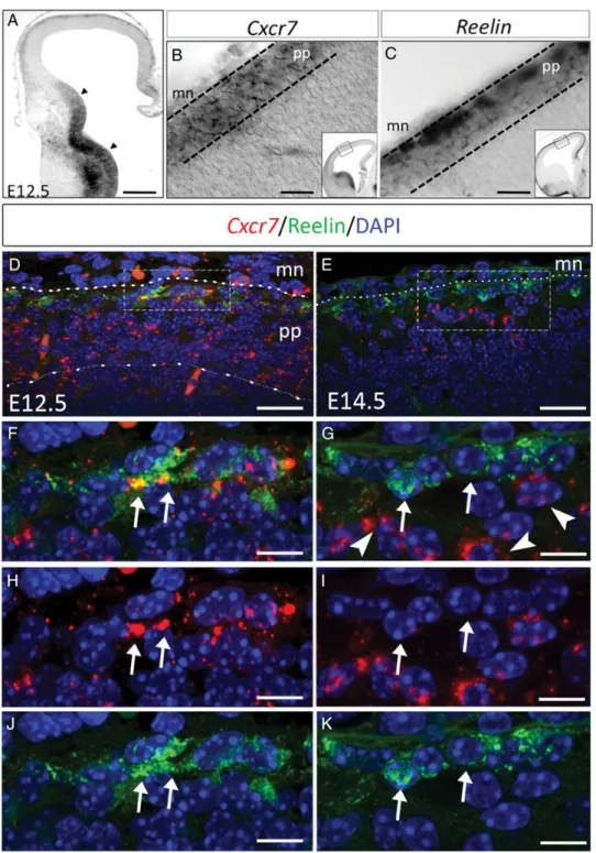
Cxcr7 expression in the developing mouse cortex was investi-gated from E11.5 to E14.5 at 3 rostro-caudal levels. At E11.5 and E12.5, Cxcr7 transcripts are localized in the subpallial ven-tricular zone (VZ) and in the emerging preplate in the pallium up to the medially located cortical hem (Fig. 1A, data not shown,Tiveron et al. 2010;Sánchez-Alcañiz et al. 2011;Wang et al. 2011). Comparison on adjacent sections of Cxcr7 and Reelin expression in the preplate at E12.5 shows that Cxcr7-positive cells are present over the whole thickness of the preplate (Fig.1B). Conversely, cells expressing Reelin, a pan-marker for CR cells (Meyer et al. 1999), are localized in the subpial area throughout the pallium (Fig. 1C). Combined
Cxcr7 in situ hybridization and Reelin immunolabeling show that Cxcr7 is expressed in Reelin-positive cells all over the pallium (Fig.1D,F,H,J, arrows). In addition, double immunola-beling for CXCR7 and Reelin confirmed in situ hybridization data (Supplementary Fig. 1A–C). Quantitative analysis at E12.5 indicates that 81 ± 7.8% of Reelin-positive cells also expressed Cxcr7.
The expression patterns of Cxcr7 and Reelin diverge at E13.5 and at E14.5 since 90 ± 9% of Reelin-positive cells are
Cxcr7-negative (Fig. 1E,G,I,K, arrows), whereas the neurons localized just below express Cxcr7 but not Reelin (arrowheads in Fig.1G and Supplementary Movie 1).
In summary, at E12.5, Cxcr7 is expressed in preplate neurons of the developing cortex, including CR cells. Then, Cxcr7 is rapidly down-regulated and at E14.5, the most superfi-cial cells are Reelin-positive and Cxcr7-negative (CR cells), whereas the immediately underlying cells are Reelin-negative and Cxcr7-positive.
Figure 1. Dynamic regulation of Cxcr7 expression in the developing pallium. (A) At E12.5, Cxcr7 expression is observed in the preplate and ganglionic eminences (arrowheads). (B and C) Within the preplate, Cxcr7 and Reelin mRNA expression profiles are overlapping. (D, F, H, and J) Double labeling with a Cxcr7 riboprobe and anti-Reelin antibody show co-expression in CR cells at E12.5 (F, H, and J, arrows). (E, G, I, and K) At E14.5, Cxcr7 expression (arrowheads) is confined in cells just below CR cells (arrows). mn, meninges (dotted lines); pp, preplate (dashed line). Scale bars: A, 160 μm; B and C, 70 μm; D, 25 μm; E, 20 μm; F–H, G–I, 8 μm.
CXCR7 Is Required for the Proper Localization of CR Cells in the Developing Cortex
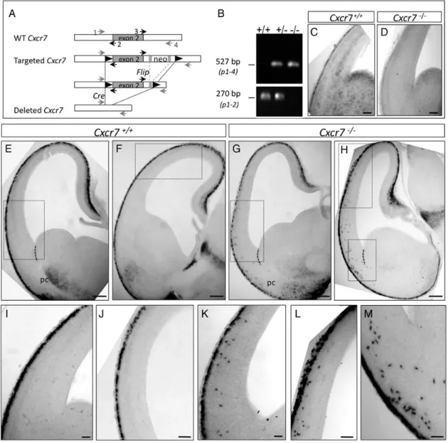
The early expression of Cxcr7 in CR cells suggests a possible role in CR migration and/or final localization. To address this question, a conditional (floxed) Cxcr7 mutant mouse line was generated (Fig.2A,B) and the entire Cxcr7 coding region was deleted by crossing Cxcr7Flox/Floxmice with an ubiquitous CMV-Cre deleter mice (Schwenk et al. 1995). Immunolabeling at E12.5 and in situ hybridization at E14.5 show that CXCR7 protein as well as Cxcr7 mRNA transcripts are absent in homo-zygous (Cxcr7−/−) embryos after Cre recombination (Supple-mentary Fig. 1D–F and Fig. 2C,D). Homozygous mice die at E17.5 as previously reported (Sierro et al. 2007;Wang et al. 2011).
Reelin mRNA expression was used to determine the localiza-tion of CR cells in Cxcr7−/−and Cxcr7+/+brains at E12.5 and
E14.5, respectively. At E12.5, Reelin-positive cells are found in the entire preplate with rare ectopic cells within the pallium (data not shown), suggesting that the initial tangential migra-tion of CR cells does not rely on Cxcr7 signaling. At E14.5, all Reelin-positive cells are confined to the subpial zone in control animals (Fig.2E,F,I,J). Conversely, in Cxcr7−/−mutants, many ectopic Reelin-positive cells are detected deep in the lateral (Fig. 2G,K) and dorsal (Fig. 2H,L) portions of the dorsal pallium, down to the prospective piriform cortex (Fig.2H,M) at different rostro-caudal levels.
The CR Cell Phenotype Is More Severe in Cxcl12−/−Than in Cxcr4−/−or Cxcr7−/−Embryos
A previous comparison of CR cell position in Cxcl12−/−and Cxcr4−/−embryos showed no major difference (Stumm et al. 2003;Borrell and Marin 2006;Paredes et al. 2006), consistent
Figure 2. CXCR7 is necessary for the localization of a subpopulation of CR cells at E14.5. (A) Schematic representation of the wild-type (WT) Cxcr7 locus, the targeted allele, and the deleted locus. The boxed region represents exon 2. The neomycin resistance cassette (neo) was excised by FRT/Flip recombination (gray rectangles). LoxP sites: black triangles; primers 1, 2, 3, and 4 used for genotyping are indicated by small arrows. (B) PCR analysis of genomic DNA from E14.5 WT (+/+), heterozygous (+/−), or homozygous (−/−) animals. (C and D) Absence of Cxcr7 mRNA expression in Cxcr7−/−(D) compared with control brains (C). (E, I, F, and J) Reelin-positive cells are all localized in the MZ in control embryos. (G, H, and K–M) In Cxcr7−/−embryos, ectopic Reelin-positive cells are observed in the ventrolateral part (G and K) and dorsal part of the dorsal pallium (H and L) as well as in the piriform cortex (H and M) at different rostro-caudal levels (G and H). pc, piriform cortex, dotted line indicates the PSB region. Scale bars: C and D, 100 μm; E–H, 200 μm; I–K, 80 μm; J–L, 100 μm; M, 60 μm.
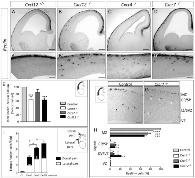
with the idea that CXCL12 acts exclusively through its canonic-al CXCR4 receptor. However, as this comparison was made at a relatively late developmental stage (E18.5,Stumm et al. 2003), we decided to analyze the Cxcl12−/−and Cxcr4−/−phenotypes at earlier stages in comparison with Cxcr7 mutant (Fig.3A–D′). We found ectopic CR cells scattered throughout the CP and SVZ/IZ at all levels along the rostro-caudal axis in Cxcl12 mutants (Fig.3B,B′) as well as in Cxcr4 and Cxcr7 mutants (Fig.3C,C′ and D,D′, respectively).
First, we quantified total Reelin-positive CR cells in the neo-cortex and found that Cxcr4−/−and Cxcl12−/− have fewer CR cells compared with control (36% and 37%, respectively, Fig.3E). No significant decrease was observed in Cxcr7 mutants.
Next, we compared the distribution of ectopic Reelin-positive cells at E14.5. We found a significant reduction in the number of Reelin-positive cells in the MZ for the 3 mutants (P < 0.001), as-sociated with a mislocalization within the CP, and the IZ/SVZ areas (Fig. 3F–H, P < 0.05). A significant higher proportion of ectopic cells in the IZ/SVZ were found in Cxcl12 mutants (22%) compared with Cxcr7 (7.1%) and Cxcr4 (5.8%) mutants (Fig.3H). Thus, both receptors might be involved in CR cells po-sitioning in the MZ. The proportion of ectopic cells represented, respectively, 15%, 25%, and 32% of total Reelin-positive cells in Cxcr4−/−, Cxcr7−/−, and Cxcl12−/−brains (Fig.3H).
Then, we analyzed the repartition of ectopic cells along the dorso-ventral axis of the neocortex to determine the preferential
Figure 3. Comparison of abnormal localization of CR cells within Cxcl12−/−, Cxcr4−/−, and Cxcr7−/−mutants. (A–C) Reelin mRNA expression in E14.5 embryos. Reelin-positive cells are observed in the deep cortical layers of the dorsal and lateral part of neocortex in Cxcl12−/−(B), Cxcr4−/−(C), and Cxcr7−/−(D) brains. (A′–D′) Higher magnifications of the lateral part of dorsal pallium from (A–D) showing the distribution of ectopic cells. (E) Quantitative analysis of the total number of Reelin-positive cells in the mutant cortices. We found that both Cxcl12–/–and Cxcr4–/–mutants had∼36% fewer Reelin-positive cells in the dorsal pallium when compared with controls and Cxcr7−/−(n = 3–4 brains per condition, 10 slices per brain; ***P < 0.001, one-way ANOVA). (F and G) Coronal cryostat sections of E14.5 Control (F) and Cxcr7−/−brains (G) from dorsal pallium (black area) after Reelin in situ hybridization. Asterisks: blood cells; arrowheads: ectopic cells. (H) Quantification of the distribution of Reelin-expressing cells in control and mutants in the thickness of the pallium, from the MZ to the ventricular zone VZ. The laminar distribution of ectopic cells shows localization in deeper layers: CP and IZ/SVZ with a large proportion for the Cxcl12 mutant. No ectopic cells were observed in the VZ. Histograms show average ± SEM (Cxcl12−/−: n = 4, Cxcr4−/−: n = 3, and Cxcr7−/−: n = 8). Non-parametric Kruskal–Wallis and Dunn’s multiple comparison tests were performed with P-values denoted as follows: *P < 0.05, ***P < 0.001. (I) Quantification of Reelin-ectopic cells in the lateral and dorsal part of the dorsal pallium in wild-type (control) and Cxcl12−/−, Cxcr4−/−, and Cxcr7−/−brains at E14.5. Significantly more ectopic Reelin-positive cells are observed in the lateral part of the dorsal pallium of Cxcr7−/−compared with Cxcr4−/−embryos (#
P < 0.05). The total number (dorsolateral + ventrolateral parts) of ectopic Reelin-positive cells is significantly higher in Cxcl12−/−and Cxcr7−/−embryos compared with Cxcr4−/−animals. Non-parametric Kruskal–Wallis and Dunn’s multiple comparison tests were performed with P-value, *P < 0.05 (10 slices per brain). MZ, marginal zone; CP/SP: cortical plate/subplate; IZ/SVZ: intermediate zone/subventricular zone; VZ: ventricular zone. Scale bars: A–D, 150 μm; A′ and B′, 70 μm; C′ and D′, 90 μm; F and G, 120 μm.
localization site. Quantification analysis indicates that the chemokine ablation has a stronger effect on CR positioning than Cxcr4 knockout (respectively, Cxcl12: 37.7 ± 3.2 ectopic Reelin-positive cells/field, n = 4; Cxcr4: 19.4 ± 1.6, n = 3, P < 0.05) (Fig.3I). Conversely, comparison between Cxcl12−/− and Cxcr7−/− embryos reveals comparable distribution of ectopic Reelin-positive cells in the dorsal or the lateral regions (Cxcl12: 37.7 ± 3.2 ectopic Reelin-positive cells/field, n = 4; Cxcr7−/−: 33.1 ± 1.4, n = 8, Fig. 3I). In addition, Cxcr7 −/− displays a higher number of ectopic Reelin-positive cells com-pared with Cxcr4−/− (respectively, 25 ± 1.6, n = 3 and 33.1 ± 1.4, n = 14, P < 0.05, Fig.3I): this difference was mainly due to an accumulation of ectopic Reelin-positive cells within the lateral part (L) (Cxcr4−/−: 9.5 ± 1.1, n = 3 vs. Cxcr7−/−: 18.2 ± 1.2, n = 8,#P < 0.001, Fig.3I). Our data not only suggest that CXCR7 plays an important role (together with Cxcr4−/−) in maintaining CR cells in the MZ, but also might acts particu-larly in the lateral region of the developing neocortex.
At E12.5, ectopic Reelin-positive cells are scattered deep within the parenchyma of Cxcl12−/−brains, but only in the lateral portion of the dorsal pallium (Supplementary Fig. 2). These cells are unlikely to be hem-derived CR cells, because at this developmental stage few cells have reached the lateral part of the dorsal pallium (Takiguchi-Hayashi et al. 2004;Borrell and Marin 2006; Yoshida et al. 2006; Tiveron et al. 2010), which is mostly populated by PSB-derived CR cells (Bielle et al. 2005;Griveau et al. 2010).
In conclusion, the phenotypes of Cxcl12, Cxcr4, and Cxcr7 mutants indicate that both receptors are involved in CR cell po-sitioning originated from the hem. In addition, preferentially mislocalized CR cells in the lateral part of the dorsal pallium in Cxcr7 mutants is consistent with the hypothesis that CXCR7 plays a specific role in the superficial localization of CR cells in this region.
Characterization of Ectopic Reelin-Positive Cells
The difference in the phenotypes observed between Cxcr7 and Cxcr4 mutants is intriguing and raises the question of the nature and origin of ectopic cells in Cxcr7 mutants.
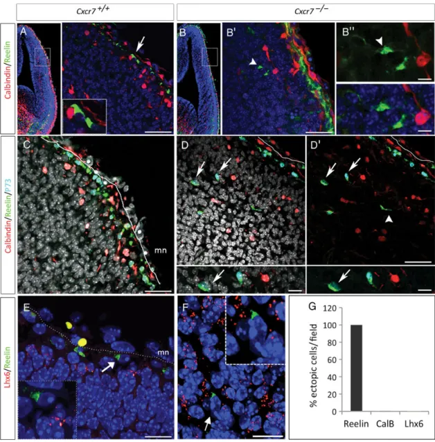
The ectopic localization of Reelin-positive cells in Cxcr7−/− telencephalon could be the result of a premature Reelin ex-pression in interneurons. During normal development, Reelin is expressed only after E15.5 in interneurons that migrate tan-gentially in the MZ and the IZ/SVZ (Alcántara et al. 1998). We combined double and triple immunolabeling for Reelin with specific interneuron markers (Calbindin, GABA, Lhx6, or Gad1) and we did not detect co-labeling with Reelin-positive ectopic cells in Cxcr7−/−embryos at E14.5 (Fig.4B–G and Sup-plementary Fig. 3), thus excluding premature expression of Reelin in interneurons.
Moreover, co-expression of Reelin and Tbr1, a transcription factor expressed in glutamatergic pallial cells (Hevner et al. 2001), demonstrates that the ectopic Reelin-positive cells ori-ginate from the pallial region (88.3 ± 7.2%, Fig.5B,C —arrows and P).
To further characterize the phenotype and origin of the ectopic Reelin-positive cells in Cxcr7−/−embryos, we explored the expression of Calretinin (normally detected in CR cells derived from the cortical hem and PSB) and p73 (a marker of CR cells derived from the cortical hem and the septum, but not from the PSB) (Takiguchi-Hayashi et al. 2004; Bielle et al. 2005;Hanashima et al. 2007;Griveau et al. 2010). At E14.5,
p73 is co-expressed in most of Reelin-positive cells in the MZ both in control and Cxcr7−/− mice (Fig. 5D–F, arrows). However, only 56.5 ± 4.7% of ectopic Reelin-positive cells
express p73 in Cxcr7−/− embryos (Fig. 5P). Note that
p73-negative ectopic Reelin cells are also negative for the inter-neuron marker Calbindin (Fig.4D–D′, arrowhead). In addition, 69.4 ± 9.1% of ectopic Reelin-positive cells expressed also Cal-retinin in Cxcr7−/−embryos (Fig.5G–H′—arrows and P).
Next, we analyzed Cxcr4, as a hem-derived CR cell marker, and found its expression in CR cells located in the MZ, compar-able in Cxcr7−/− and control embryos (Fig. 5I—O, arrows). However, in ectopic positions (CP and IZ/SVZ), both Cxcr4-positive and Cxcr4-negative/Reelin-positive cells are de-tected in the dorsal and lateral territories of the developing cortex (Fig. 5M–O). Indeed, 59.9 ± 5.6% of ectopic Reelin-positive cells co-expressed Cxcr4 (Fig.5P), which is consistent with the percentage of p73-positive ectopic cells (56.5%) and likely reflects a cortical hem origin.
Thesefindings clearly show that ectopic Reelin-positive cells are not interneurons, but CR cells as they express Tbr1 and Cal-retinin but do not express interneuron markers. We show that more than half (∼60%) of Reelin-ectopic cells originate from the cortical hem ( p73+ Cxcr4+), while the remaining (∼40%) p73- and Cxcr4-negative Reelin-ectopic cells show a PSB mo-lecular identity.
CXCR7 in the Dbx1 Lineage Is Involved in the Positioning of a Subpopulation of CR Cells
PSB-derived CR cells are produced by progenitors that express the Dbx1 gene and predominately populate the ventrolateral part of the pallium (Bielle et al. 2005). We analyzed Cxcr7 ex-pression in Dbx1-derived cells using Dbx1cre/Rosa26tdTomato embryos. At E12.5 and E14.5, Cxcr7-tdTomato-positive cells are observed predominantly in the prospective piriform cortex and the MZ of the lateral part of the dorsal pallium (Fig.6A–C, n = 3). At E14.5, they represent 63 ± 5.7% of all tdTomato labeled cells in the subpial position, consistent with a preferen-tial localization of Dbx1-derived CR cells within the lateral pallium at earlier embryonic stages (Bielle et al. 2005;Griveau et al. 2010). To investigate the specific function of CXCR7 in the Dbx1 lineage, Dbx1cremice were crossed with Cxcr7lox/lox mice to inactivate Cxcr7 specifically in Dbx1-derived cells. In Dbx1cre/Cxcr7lox/lox mutant embryos, ectopic mRNA Reelin-positive cells are specifically observed in the lateral portion of the dorsal pallium, although they are less numerous (4.5 ± 0.5 ectopic cells/field, n = 4, P < 0.05, Fig. 6D–F) compared with Cxcr7−/−embryos (18.2 ± 1.2, n = 14, Fig.3I). In addition, the nature of the ectopic cells was assessed using co-detection of Reelin/p73 and Reelin/Calbindin (Fig.6G,H). None of ectopic cells expressed Calbindin, confirming that these cells were not interneurons. The absence of p73 expression demonstrates their PSB origin.
We conclude that CXCR7 activity in Dbx1-positive progeni-tors regulates the positioning of a subpopulation of CR cells derived from the PSB. Such regulation is likely independent of CXCR4, since no Cxcr4 expression is detected in these cells (Tiveron et al. 2010).
CXCR4 Function in Cxcr7−/−Embryos
To further confirm CXCR7 role in CR positioning, CCX771 (a specific pharmacological inhibitor of CXCR7) or CCX704
(a control compound unable to bind CXCR7) was injected in the lateral ventricle of E12.5 wild-type C57BL/6J embryos in utero and then the location of Reelin-positive cells was assessed and quantified in a defined area corresponding to the injection site (dextran labeled) after 2 days, by immunohistochemistry.
CCX771 injection is associated with the presence of ectopic Reelin-positive cells specifically in the lateral part of the dorsal pallium compared with CCX704 (6.5 ± 1.2 and 2.1 ± 0.3 ectopic cells/field, respectively; P < 0.004, n = 3/each group; Fig.7A,B,G). We demonstrate that pharmacological inhibition of Cxcr7 signaling results in a mislocalization of CR cells as observed in Cxcr7−/− embryos (7.5 ± 0.5 ectopic cells/field, Fig.7G).
To assess whether CXCR4 are functional in Cxcr7 mutants, CXCR4 function was blocked by injecting the specific inhibitor
AMD3100 in the lateral ventricle of E12.5 Cxcr7−/−and wild-type littermates. At E14.5, AMD3100-treated wild-type embryos show a disorganized MZ and ectopic Reelin-positive cells in the MZ and CP of the dorsal pallium (Fig. 7C,E), compared with Cxcr7+/+
vehicle injected (data not shown). Quantification of Reelin-ectopic cells is significantly higher in AMD3100-injected Cxcr7−/− embryos compared with wild-type littermates (Cxcr7−/−/ AMD3100: 15.5 ± 1.3 and Cxcr7+/+/AMD3100: 11.4 ± 1.1 of ectopic cells/field, n = 3/group, *P < 0.05, Mann–Whitney test; Fig.7D,F, G). The inhibition of CXCR4 in the Cxcr7 mutant causes an increase of Reelin-ectopic cells when compared with Cxcr7 mutant (Cxcr7−/−: 7.65 ± 0.5, ***P < 0.001), reflecting that CXCR4 receptors remain functional in the absence of Cxcr7. Altogether, these data indicate that both receptors are required for proper CR cell localization.
Figure 4. Characterization of ectopic Reelin-positive cells in the cortex of Cxcr7−/−E14.5 embryos. Immunohistochemistry for Reelin/Calbindin (A and B″) or Reelin/Calbindin/p73 (C and D′) on coronal sections of Cxcr7−/−mutant. (A and B″) Immunostaining for Calbindin, a marker of immature interneurons. (A′ and B′) High magnifications of the dashed white boxes in A and B. In control, the CR cells are negative for Calbindin in the MZ (arrow and dashed box). (B and B″) In Cxcr7−/−embryos, the ectopic Reelin-positive cells do not express Calbindin (arrowhead). (C and D′) Immunostaining for Calbindin, p73, and Reelin shows that double-positive Reelin/p73 cells are not labeled with Calbindin (arrows, G). In addition, example of Reelin-positive/p73-negative cell that is not Calbindin positive (D′, arrowhead). (E and F) Lhx6 and Reelin expression in Cxcr7+/+and Cxcr7−/−mutant. The absence of co-expression of Lhx6 and Reelin demonstrates that ectopic cells are not interneurons. (G) Quantification of ectopic cells that co-express Reelin with Calbindin (CalB) or Lhx6 in Cxcr7−/−mutant: neither Lhx6 or CalB were co-expressed with Reelin. Scale bars: A′, 90 μm; B′, C, D, and D′, 60 μm; B″, 15 μm; E, 30 μm; F, 15 μm.
Discussion
CXCL12/CXCR4 signaling controls the position of hem-derived CR cells in the MZ. In the present study, we demonstrate
that loss of Cxcr7 is associated with ectopic location of Reelin-positive cells in the pallium. Comparison of Cxcl12−/−, Cxcr4−/−, Cxcr7−/−embryos indicates that both receptors are Figure 5. Pallial origin of ectopic Reelin-positive cells. Immunostaining for Tbr1 (A–C), p73 (D–F), Calretinin (G and H′), and Cxcr4 (I–O). (A′, B′, D′, and E′) High magnifications of the dashed white boxes in A, B, D, and E. (A and A′) Reelin-positive cells express the pallial marker Tbr1 (arrow) in the MZ in control. (B and C) Ectopic Reelin-positive cells express Tbr1 in Cxcr7−/−mutant (arrows). (B″) High magnification of B′ illustrating double Reelin/Tbr1-positive cells. (C) Another example showing Tbr1 immunoreactivity in 2 ectopic Reelin-positive cells. (D and D′) In E14.5 Cxcr7+/+
embryos, all Reelin-positive cells express p73 (marker of cortical hem- and septum-derived CR cells) in the MZ. (E and E″) In the dorsal pallium of E14.5 Cxcr7−/−embryos, ectopic Reelin-positive and p73-positive cells are observed (arrows, magnification in E″). (F) Another example showing an ectopic Reelin-positive/p73-negative (arrowhead). (G) In E14.5 Cxcr7+/+embryos, Reelin- and Calretinin-positive cells were observed in the MZ. (H and H′) In Cxcr7−/−embryos, ectopic
Reelin-positive cells in the CP express Calretinin. (H′) Higher magnification of the boxed area in H shows a Reelin-positive/Calretinin-negative cell (arrowhead) among cells that co-express Reelin and Calretinin (arrows). (I and O) Coronal sections of control (I and J) and Cxcr7−/−mutant mice (K and O) at E14.5 analyzed by ISH Cxcr4 combined to immunostaining for Reelin. (I and J) Reelin-positive cells that co-express Cxcr4 mRNA (arrows) are intermingled with Reelin-positive/Cxcr4-negative cells (arrowhead) in the CR layer. (M–O) Analysis of Cxcr4 and Reelin expression in the depth of the CP (M). Higher magnification of the boxed area in M showing an ectopic cell that co-expresses Cxcr4 and Reelin (N) next to an ectopic Reelin-positive and Cxcr4-negative cell (O). (P) Quantification of ectopic cells that co-express Reelin and Tbr1 or p73 or Calretinin (CalR) or Cxcr4 in Cxcr7−/− E14.5 embryos. mn, meninges (dotted lines); asterisks, blood cells. Scale bars: A′, B′, D′, and E′, 50 μm; B″, C, and E″, 10 μm; F–H, 30 μm; H′, 25 μm; I–M, 30 μm; N and O, 8 μm.
involved in CXCL12 signaling with a major contribution by CXCR7. Moreover a decrease of total CR cells is retrieved in Cxcl12 and Cxcr4 mutants but not in Cxcr7, indicating a dis-tinct function for both receptors.
Finally, the specific Cxcr7 ablation in cells derived from Dbx1-positive progenitors shows that CXCR7 is required in the Dbx1 lineage to position PSB-derived CR cells. We conclude that CXCR7 regulates the positioning of both hem- and
PSB-derived CR cells while CXCR4 controls only the position-ing of hem-derived CR cells.
CXCR7 Is Involved in the Positioning of Hem-Derived CR Cells
Cxcr4 and Cxcr7 receptor knockout mice display mislocalized Reelin-positive cells. Interestingly, in Cxcl12 mutants, CR cell Figure 6. Distinct function of CXCR7 in CR cells derived from Dbx1-positive progenitors. (A–C) Expression of Cxcr7 mRNA in Dbx1cre/Rosa26tdTomatoembryos shows Cxcr7/
tdTomato co-localization (arrows) in the lateral part of the dorsal pallium at E12.5 (A and B) and E14.5 (C). Asterisk indicates blood cells in the meninges (D–F) Reelin mRNA expression at E14.5 in Dbx1Cre/Cxcr7lox/lox
embryos shows ectopic Reelin-positive cells in the lateral pallium in different sections (E and F, arrowheads) compared with wild-type embryos (D). In (F), the inset represents a high magnification of an ectopic Reelin-positive cell. (G and H) Reelin-positive cells in Dbx1Cre/Crcr7lox/loxdo not express p73 or Calbindin in the lateral part of the dorsal pallium (arrowhead). mn, meninges (dotted lines). Scale bars: A, 40 μm; B and C, 15 μm; D, 30 μm; E and F, 50 μm; G, 30 μm; H, 25 μm.
mispositioning is (1) more pronounced compared with Cxcr4 mutants and (2) comparable to Cxcr7 mutants. This finding suggests a redundancy in the function of both receptors in CR cell positioning. Cxcr7 is expressed in 90% of CR cells at E12.5 when most CR cells have migrated tangentially from different sources, including hem, PSB, and septum, to cover the cortical surface. This present work shows that, in addition to CXCR4 signaling, CXCR7 is also required for proper location of hem-derived CR cells. First, in Cxcr7−/−embryos, the high propor-tion of ectopic CR cells that express the p73 marker (56%) or Cxcr4 (59%) confirms that >50% of ectopic Reelin-positive cells originate from the hem.
The functional relationship between CXCR7 and CXCR4 in hem-derived CR cells is not completely elucidated, but our data strongly suggest that CXCR7 might have a non-cell autono-mous effect through CXCR4.
Indeed, a down-regulation of CXCR4 expression has been shown after Cxcr7 inactivation in interneurons that express both receptors (Schönemeier et al. 2008;Tiveron et al. 2010; Sánchez-Alcañiz et al. 2011; Abe et al. 2014). Indeed, in Cxcr7−/−mice, CXCR4 can still bind to an excess of CXCL12, thereby inducing excessive CXCR4 activation and internaliza-tion from the neuronal surface at E14.5 and E16.5 stages (Sánchez-Alcañiz et al. 2011;Abe et al. 2014). These data are consistent with the fact that deficiency of one chemokine re-ceptor can lead to increase in the expression of its ligand, which would then promote internalization of the other recep-tor(s) for that ligand (Cardona et al. 2008). Ourfindings, in Cxcr7−/−embryos after CXCR4 antagonist, intraventricular in-jections at early developmental stages demonstrate an aggrava-tion of Cxcr7−/−phenotype and indicate that CXCR4 might still be present in Cxcr7−/− embryos. In CR cells, CXCR4 down-regulation might not occurred at this early stage (E12.5), con-versely as it is demonstrated at E14.5 and even more pro-nounced at E16.5 in interneurons (Sánchez-Alcañiz et al. 2011; Abe et al. 2014).
Many attempts to investigate the CXCR4 protein levels have not yielded conclusive results owing to nonspecific staining of the antibodies tested ( personal observations andFischer et al. 2008). Therefore, we cannot exclude non-cell autonomous effect of CXCR7 in hem-derived CR cells.
CXCR7 Plays a Role in a Subpopulation of CR Cells Originating from Dbx1 Progenitors
Defects in CR cells positioning in Cxcr7−/− and Cxcl12−/− mutants are more severe compared with Cxcr4−/− mutants, suggesting that the hem-derived CR cell subpopulation is not the only one affected.
Mapping studies indicate that CR cells originating from the PSB populate the lateral pallium and the piriform cortex, whereas hem- and pallial septum-derived CR cells invade pref-erentially the dorsal and rostral pallium, respectively (Bielle et al. 2005;Yoshida et al. 2006;Griveau et al. 2010).
The presence of ectopic Reelin-positive cells in the lateral part of the dorsal pallium of Cxcr7−/− embryos is therefore consistent with a role for CXCR7 in the subpial location of CR cells originating from the PSB, as also suggested by the fact that 40% of ectopic cells do not express p73 and Cxcr4. More-over, conditional ablation of Cxcr7 only in the Dbx1 cell lineage impaired the localization of a small population of CR cells, preferentially in the lateral part of the pallium and almost Figure 7. Pharmacological inhibition of CXCR7 or CXCR4. (A–F) Reelin
immunolabeling at E14.5 after intraventricular injection of the CXCR7 or CXCR4 antagonist at E12.5: (A) control (C57BL/6J) embryos treated with CCX704, (B) C57BL/ 6J animals treated with the CXCR7 antagonist CCX771, (C and E) Cxcr7+/+animals treated with the CXCR4 antagonist AMD3100 (AMD), and (D and F) Cxcr7−/−animals treated with AMD3100. CR cells were confined to the MZ in control embryos, but after pharmacological blocking of CXCR7 with CCX771, some CR cells were scattered within the CP (compare A with B). (C) AMD3100-treated Cxcr7+/+embryos show ectopic Reelin-positive cells in the dorsal pallium (arrows). (D) The effect of AMD3100 treatment in Cxcr7−/−embryos is stronger in the lateral part of the dorsal pallium (arrows). (E and F) High magnification showing ectopic Reelin-positive cells (arrowheads). (G) Quantification of ectopic cells in a defined field (500 × 500 μm lateral area). One-way ANOVA test, with post hoc Bonferroni’s multiple comparison test, was performed with P-values denoted as follows: *P < 0.05, ***P < 0.001 (n = 4–5 brains per condition). Asterisks indicate blood vessels. Scale bars: A–D, 100 μm; E and F, 30 μm.
all ectopic Reelin cells do not express p73, a signature of PSB-derived CR cells (Hanashima et al. 2007;Griveau et al. 2010). The smaller number of ectopic CR cells in this mouse strain compared with Cxcr7−/−embryos in the same brain area might be explained by the fact that Cre recombination does not occur in the earliest cells derived from the Dbx1 progenitors due to delayed Cre activity (Bielle et al. 2005). In addition, lineage analysis demonstrates that a large proportion of neurons derived from the Dbx1 progenitors express Cxcr7 at E12.5 and E14.5, and transcriptome analysis of CR cells derived from the Dbx1 progenitors revealed strong expression of Cxcr7 at early stages (Griveau et al. 2010; Ugo Borello and Alessandra Pierani, personal data). Taken together, these data strongly argue in favor of a cell autonomous effect of CXCR7 in positioning of PSB-derived CR cells.
The question of which cellular mechanisms are affected remains to be answered: in Cxcr7−/−mice, do CR cells fail to migrate or do they migrate, but are then unable to maintain their pial position (as in Cxcr4−/−mice)? The absence (or very low incidence) of ectopic cells in the dorsal pallium at E12.5, when migration is highly active and CR cells from different sources are invading the entire surface of the cortex, argues against the hypothesis of migration failure. However, Cxcr7 deficiency might partially affect CR cell motility as reported for Cxcr7−/− interneurons, which develop shorter leading pro-cesses and are less motile, resulting in abnormal positions within the CP (Wang et al. 2011). On the other hand, receptor antagonist injection at E12.5 has an effect on CR cell position-ing, suggesting that even after migration completion, CXCR7– CXCL12 interaction is required to retain CR cells in their subpial location. Based on our results, we propose that Cxcr7 signaling is not necessary for CR cell migration but is essential for maintaining CR cells in the MZ.
In addition, we propose that CXCR7 may also have a function of its own, independent of regulating CXCR4 signaling, by main-taining the subpial location of PSB-derived CR cells. CXCR7 and CXCR4 would then play complementary roles in, respectively, the lateral and dorsal portions of the dorsal pallium. CR cell streams originating from the hem and the PSB overlap only in the dorsolat-eral regions (Bielle et al. 2005;Yoshida et al. 2006) and appropri-ate cell density is obtained through cell interactions (Villar-Cerviño et al. 2013). Thus, a defect in either population may be partly com-pensated by the other one (Griveau et al. 2010).
Up to now, CXCL12 has been considered to influence only the migration of hem-derived CR cells (Borrell and Marin 2006; Paredes et al. 2006) and other CR populations were thought to be insensitive to CXCL12 signaling. Here, we demonstrate a novel role of CXCR7 in CR cells and complete the scheme of regulation that involved both receptors and include several CR populations.
Supplementary Material
Supplementary material can be found at: http://www.cercor.oxford journals.org/.
Funding
This work was supported by the Institut National pour la Sante et la Recherche Medicale; the Agence Nationale de la Recher-che (ANR-07-NEURO-046-01), Ville de Paris (2006 ASES 102) to A.P., and the US Department of Defense (R0705E) to S.P. H. H.B. is supported by the DFG.
Notes
We thank Eric Jouffre and Ana Bella Imap for expert animal care. We thank Vicky Diakou and Julien Cau for confocal microscopy assistance at the MRI facility; Lisa Vigier for providing the Dbx1-Cre/ Rosa-tdTomato embryos; Vassili Pachnis and André Goffinet for the gift of the riboprobe plasmids Lhx6 and Reelin, respectively; Catherine McGrath for the Cxcr4 and Cxcl12 probes; Mark E.T. Penfold of Che-moCentryx Inc. for supplying the CCX771 and CCX704 compounds; G. Naert and S. Layalle for helpful comments; Michelle Silhol for helpful technical advice. Conflict of Interest: The authors declare no competingfinancial interests.
References
Abe P, Mueller W, Schütz D, Mackay F, Thelen M, Zhang P, Stumm R. 2014. CXCR7 prevents excessive CXCL12-mediated downregulation of CXCR4 in migrating cortical interneurons. Development. 141:1857–1863.
Alcántara S, Ruiz M, D’Arcangelo G, Ezan F, de Lecea L, Curran T, Sotelo C, Soriano E. 1998. Regional and cellular patterns of Reelin mRNA expression in the forebrain of the developing and adult mouse. J Neurosci. 18:7779–7799.
Balabanian K, Lagane B, Infantino S, Chow KY, Harriague J, Moepps B, Arenzana-Seisdedos F, Thelen M, Bachelerie F. 2005. The chemokine SDF-1/CXCL12 binds to and signals through the orphan receptor RDC1 in T lymphocytes. J Biol Chem. 280: 35760–35766.
Bielle F, Griveau A, Narboux-Nême N, Vigneau S, Sigrist M, Arber S, Wassef M, Pierani A. 2005. Multiple origins of Cajal-Retzius cells at the borders of the developing pallium. Nat Neurosci. 8:1002–1012. Boldajipour B, Mahabaleshwar H, Kardash E, Reichman-Fried M,
Blaser H, Minina S, Wilson D, Xu Q, Raz E. 2008. Control of chemokine-guided cell migration by ligand sequestration. Cell. 132:463–473.
Borrell V, Marin O. 2006. Meninges control tangential migration of hem-derived Cajal-Retzius cells via CXCL12/CXCR4 signaling. Nat Neurosci. 9:1284–1293.
Burns JM, Summers BC, Wang Y, Melikian A, Berahovich R, Miao Z, Penfold ME, Sunshine MJ, Littman DR, Kuo CJ et al.2006. A novel chemokine receptor for SDF-1 and I-TAC involved in cell survival, cell adhesion, and tumor development. J Exp Med. 203:2201–2213. Cardona AE, Sasse ME, Liu L, Cardona SM, Mizutani M, Savarin C, Hu T, Ransohoff RM. 2008. Scavenging roles of chemokine receptors: chemokine receptor deficiency is associated with increased levels of ligand in circulation and tissues. Blood. 112:256–263.
Ceci ML, López-Mascaraque L, de Carlos JA. 2010. The influence of the environment on Cajal-Retzius cell migration. Cereb Cortex. 20:2348–2360.
Cubedo N, Cerdan E, Sapede D, Rossel M. 2009. CXCR4 and CXCR7 co-operate during tangential migration of facial motoneurons. Mol Cell Neurosci. 40:474–484.
Dambly-Chaudiere C, Cubedo N, Ghysen A. 2007. Control of cell mi-gration in the development of the posterior lateral line: antagonistic interactions between the chemokine receptors CXCR4 and CXCR7/ RDC1. BMC Dev Biol. 7:23.
Daniel D, Rossel M, Seki T, Konig N. 2005. Stromal cell-derived factor-1 (SDF-1) expression in embryonic mouse cerebral cortex starts in the intermediate zone close to the pallial-subpallial bound-ary and extends progressively towards the cortical hem. Gene Expr Patterns. 5:317–322.
D’Arcangelo G, Miao GG, Chen SC, Soares HD, Morgan JI, Curran T. 1995. A protein related to extracellular matrix proteins deleted in the mouse mutant reeler. Nature. 374:719–723.
Donà E, Barry JD, Valentin G, Quirin C, Khmelinskii A, Kunze A, Durdu S, Newton LR, Fernandez-Minan A, Huber W et al. 2013. Dir-ectional tissue migration through a self-generated chemokine gradi-ent. Nature. 503:285–289.
Espinosa A, Gil-Sanz C, Yanagawa Y, Fairén A. 2009. Two separate sub-types of early non-subplate projection neurons in the developing cerebral cortex of rodents. Front Neuroanat. 3:27.
Fischer T, Nagel F, Jacobs S, Stumm R, Schulz S. 2008. Reassessment of CXCR4 chemokine receptor expression in human normal and neo-plastic tissues using the novel rabbit monoclonal antibody UMB-2. PLoS ONE. 3:e4069.
Griveau A, Borello U, Causeret F, Tissir F, Boggetto N, Karaz S, Pierani A. 2010. A novel role for Dbx1-derived Cajal-Retzius cells in early regionalization of the cerebral cortical neuroepithelium. PLoS Biol. 8:e1000440.
Hanashima C, Fernandes M, Hebert JM, Fishell G. 2007. The role of Foxg1 and dorsal midline signaling in the generation of Cajal-Retzius subtypes. J Neurosci. 27:11103–11111.
Hattermann K, Mentlein R. 2013. An infernal trio: the chemokine CXCL12 and its receptors CXCR4 and CXCR7 in tumor biology. Ann Anat. 195:103–110.
Hernandez L, Magalhaes MAO, Coniglio SJ, Condeelis JS, Segall JE. 2011. Opposing roles of CXCR4 and CXCR7 in breast cancer metas-tasis. Breast Cancer Res. 13:R128.
Hevner RF, Shi L, Justice N, Hsueh Y, Sheng M, Smiga S, Bulfone A, Goffinet AM, Campagnoni AT, Rubenstein JL. 2001. Tbr1 regulates differentiation of the preplate and layer 6. Neuron. 29:353–366. Levoye A, Balabanian K, Baleux F, Bachelerie F, Lagane B. 2009.
CXCR7 heterodimerizes with CXCR4 and regulates CXCL12-mediated G protein signaling. Blood. 113:6085–6093.
Luker KE, Steele JM, Mihalko LA, Ray P, Luker GD. 2010. Constitutive and chemokine-dependent internalization and recycling of CXCR7 in breast cancer cells to degrade chemokine ligands. Oncogene. 29:4599–4610.
Madisen L, Zwingman TA, Sunkin SM, Oh SW, Zariwala HA, Gu H, Ng LL, Palmiter RD, Hawrylycz MJ, Jones AR et al. 2010. A robust and high-throughput Cre reporting and characterization system for the whole mouse brain. Nat Neurosci. 13:133–140.
Meyer G, Goffinet AM, Fairén A. 1999. What is a Cajal-Retzius cell? A re-assessment of a classical cell type based on recent observations in the developing neocortex. Cereb Cortex. 9:765–775.
Nagasawa T, Hirota S, Tachibana K, Takakura N, Nishikawa S, Kita-mura Y, Yoshida N, Kikutani H, Kishimoto T. 1996. Defects of B-cell lymphopoiesis and bone-marrow myelopoiesis in mice lacking the CXC chemokine PBSF/SDF-1. Nature. 382:635–638. Ogawa M, Miyata T, Nakajima K, Yagyu K, Seike M, Ikenaka K,
Yama-moto H, Mikoshiba K. 1995. The reeler gene-associated antigen on Cajal-Retzius neurons is a crucial molecule for laminar organization of cortical neurons. Neuron. 14:899–912.
Paredes MF, Li G, Berger O, Baraban SC, Pleasure SJ. 2006. Stromal-derived factor-1 (CXCL12) regulates laminar position of Cajal-Retzius cells in normal and dysplastic brains. J Neurosci. 26:9404–9412. Rice DS, Curran T. 2001. Role of the Reelin signaling pathway in
central nervous system development. Annu Rev Neurosci. 24: 1005–1039.
Sánchez-Alcañiz JA, Haege S, Mueller W, Pla R, Mackay F, Schulz S, López-Bendito G, Stumm R, Marín O. 2011. Cxcr7 controls neuron-al migration by regulating chemokine responsiveness. Neuron. 69:77–90.
Schönemeier B, Kolodziej A, Schulz S, Jacobs S, Hoellt V, Stumm R. 2008. Regional and cellular localization of the CXCl12/SDF-1 che-mokine receptor CXCR7 in the developing and adult rat brain. J Comp Neurol. 510:207–220.
Schwenk F, Baron U, Rajewsky K. 1995. A cre-transgenic mouse strain for the ubiquitous deletion of loxP-flanked gene segments includ-ing deletion in germ cells. Nucleic Acids Res. 23:5080–5081. Sierro F, Biben C, Martinez-Munoz L, Mellado M, Ransohoff RM, Li M,
Woehl B, Leung H, Groom J, Batten M et al. 2007. Disrupted cardiac development but normal hematopoiesis in mice deficient in the second CXCL12/SDF-1 receptor, CXCR7. Proc Natl Acad Sci USA. 104:14759–14764.
Stumm RK, Zhou C, Ara T, Lazarini F, Dubois-Dalcq M, Nagasawa T, Hollt V, Schulz S. 2003. CXCR4 regulates interneuron migration in the developing neocortex. J Neurosci. 23:5123–5130.
Takiguchi-Hayashi K, Sekiguchi M, Ashigaki S, Takamatsu M, Hasega-wa H, Suzuki-Migishima R, Yokoyama M, Nakanishi S, Tanabe Y. 2004. Generation of Reelin-positive marginal zone cells from the caudomedial wall of telencephalic vesicles. J Neurosci. 24: 2286–2295.
Teissier A, Griveau A, Vigier L, Piolot T, Borello U, Pierani A. 2010. A novel transient glutamatergic population migrating from the pallial-subpallial boundary contributes to neocortical development. J Neu-rosci. 30:10563–10574.
Tissir F, Goffinet AM. 2003. Reelin and brain development. Nat Rev Neurosci. 4:496–505.
Tiveron M-C, Boutin C, Daou P, Moepps B, Cremer H. 2010. Expression and function of CXCR7 in the mouse forebrain. J Neuroimmunol. 224:72–79.
Tiveron MC, Rossel M, Moepps B, Zhang YL, Seidenfaden R, Favor J, Konig N, Cremer H. 2006. Molecular interaction between projection neuron precursors and invading interneurons via stromal-derived factor 1 (CXCL12)/CXCR4 signaling in the cortical subventricular zone/intermediate zone. J Neurosci. 26:13273–13278.
Valentin G, Haas P, Gilmour D. 2007. The chemokine SDF1a coordi-nates tissue migration through the spatially restricted activation of Cxcr7 and Cxcr4b. Curr Biol. 17:1026–1031.
Venkiteswaran G, Lewellis SW, Wang J, Reynolds E, Nicholson C, Knaut H. 2013. Generation and dynamics of an endogenous, self-generated signaling gradient across a migrating tissue. Cell. 155:674–687.
Villar-Cerviño V, Molano-Mazón M, Catchpole T, Valdeolmillos M, Hen-kemeyer M, Martínez LM, Borrell V, Marín O. 2013. Contact repul-sion controls the disperrepul-sion andfinal distribution of Cajal-Retzius cells. Neuron. 77:457–471.
Wang Y, Li G, Stanco A, Long JE, Crawford D, Potter GB, Pleasure SJ, Behrens T, Rubenstein JLR. 2011. CXCR4 and CXCR7 have distinct functions in regulating interneuron migration. Neuron. 69:61–76.
Yoshida M, Assimacopoulos S, Jones KR, Grove EA. 2006. Massive loss of Cajal-Retzius cells does not disrupt neocortical layer order. De-velopment. 133:537–545.